Embed Size (px)
Citation preview
GUIDED PERCUTANEOUS BIOPSY OF RETROPERITONEAL LESIONS
Medical Imaging Departement; La Rabta Hospital
INTV11
INTRODUCTION
Percutaneous fine-needle aspiration biopsy (PFNAB) under computed tomographic (CT) guidance has proved to be a widely accepted method of documenting malignancy.
Refinements in technique, experience with the procedure, and improvements in CT scanners have permitted a high degree of accuracy.
We present our experience over a 8-year period with this technique.
OBJECTIVES
The aim of this work is to present an overview of Indication of percutanous needle biopsy which include diagnosis of primary or metastatic malignacy in a newly
discovered mass, diagnosis of tumor recurrence in patients known with malignancy diagnosis of infection and benign disease.
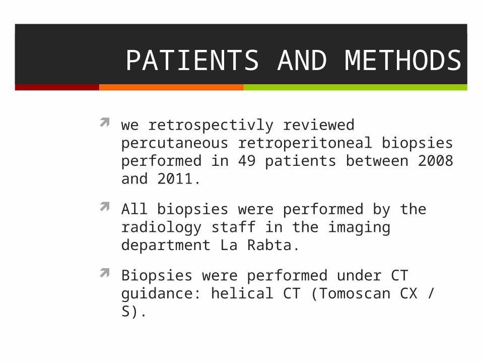
PATIENTS AND METHODS
we retrospectivly reviewed percutaneous retroperitoneal biopsies performed in 49 patients between 2008 and 2011.
All biopsies were performed by the radiology staff in the imaging department La Rabta.
Biopsies were performed under CT guidance: helical CT (Tomoscan CX / S).
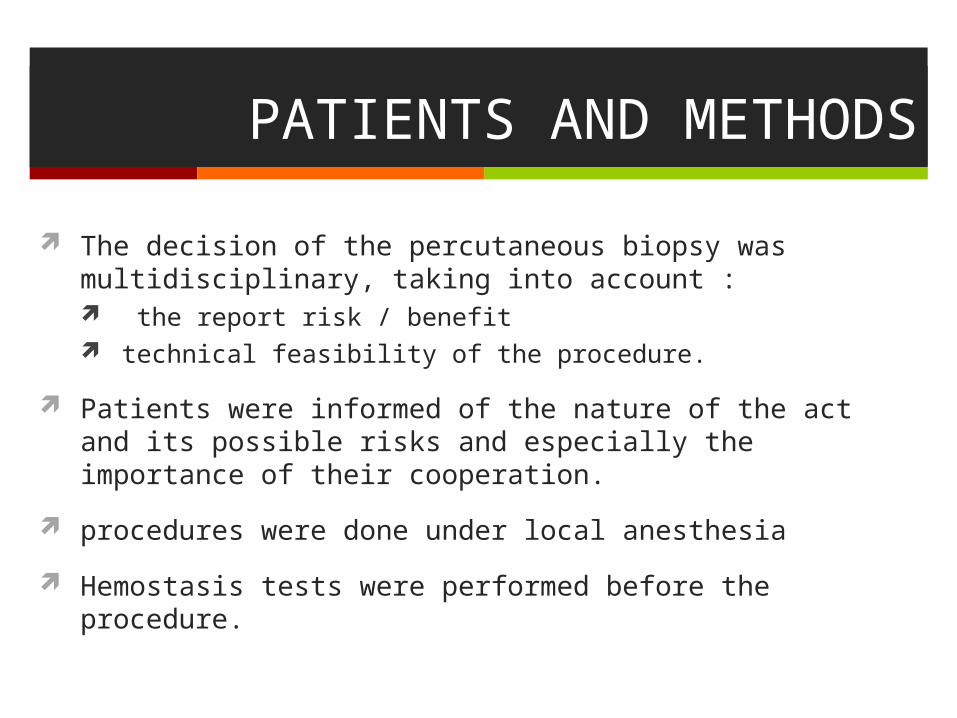
PATIENTS AND METHODS
The decision of the percutaneous biopsy was multidisciplinary, taking into account : the report risk / benefit technical feasibility of the procedure.
Patients were informed of the nature of the act and its possible risks and especially the importance of their cooperation.
procedures were done under local anesthesia
Hemostasis tests were performed before the procedure.
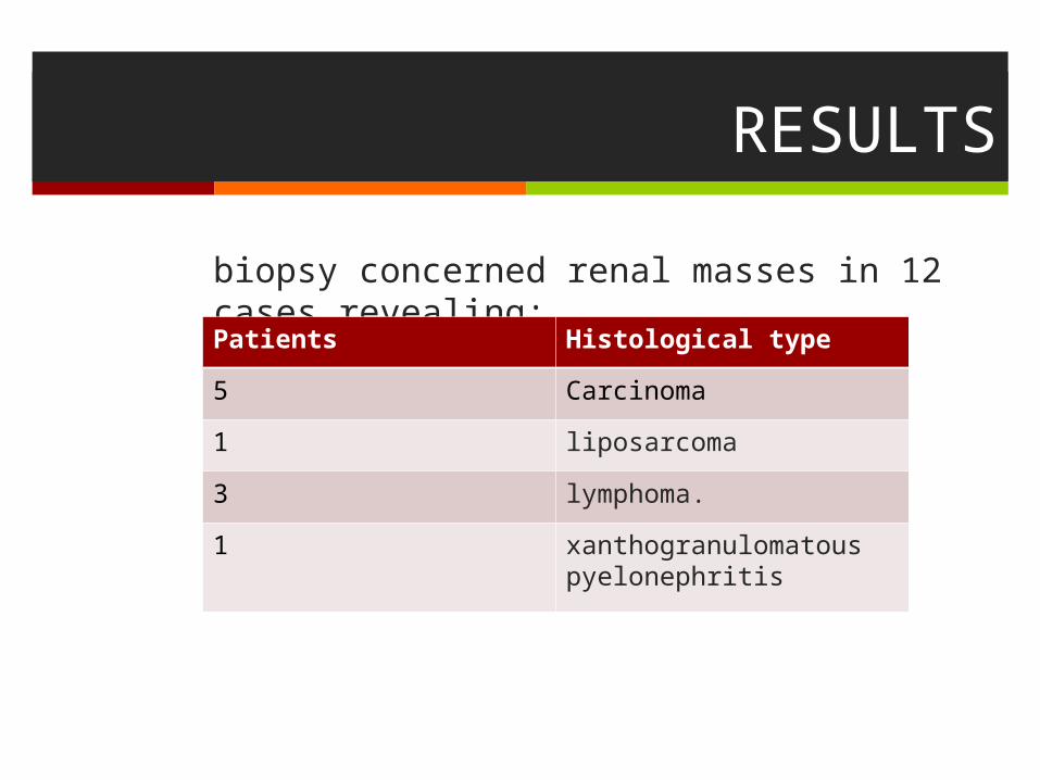
RESULTS
biopsy concerned renal masses in 12 cases revealing:
Histological typePatients
Carcinoma5
liposarcoma1
lymphoma.3
xanthogranulomatous pyelonephritis
1
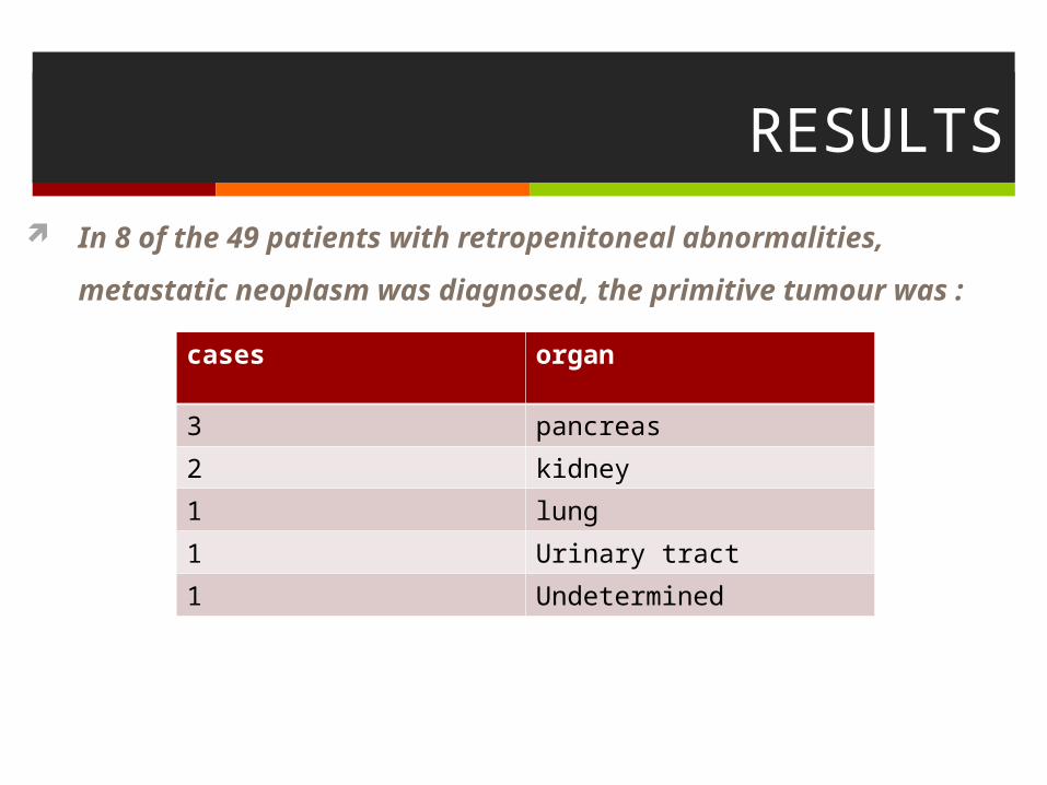
RESULTS
In 8 of the 49 patients with retropenitoneal abnormalities, metastatic
neoplasm was diagnosed, the primitive tumour was :
organcases
pancreas3
kidney2
lung1
Urinary tract1
Undetermined1
RESULTS
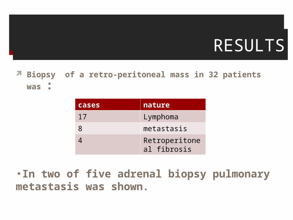
Biopsy of a retro-peritoneal mass in 32 patients was :
naturecases
Lymphoma17
metastasis8
Retroperitoneal fibrosis
4
•In two of five adrenal biopsy pulmonary metastasis was shown.
RESULTS
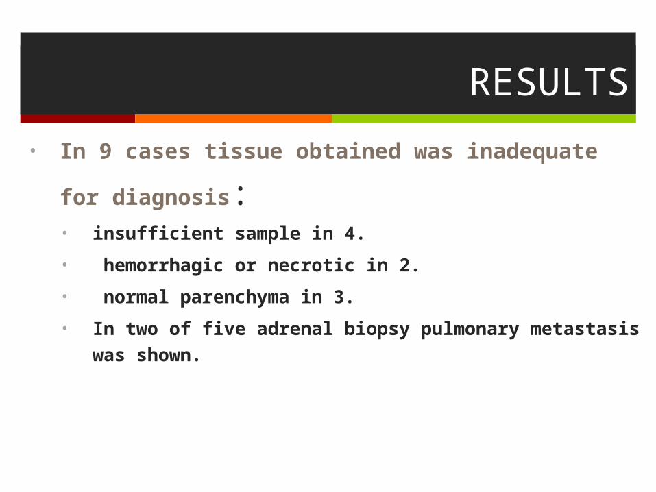
• In 9 cases tissue obtained was inadequate for diagnosis: • insufficient sample in 4.
• hemorrhagic or necrotic in 2.
• normal parenchyma in 3.
• In two of five adrenal biopsy pulmonary metastasis was shown.
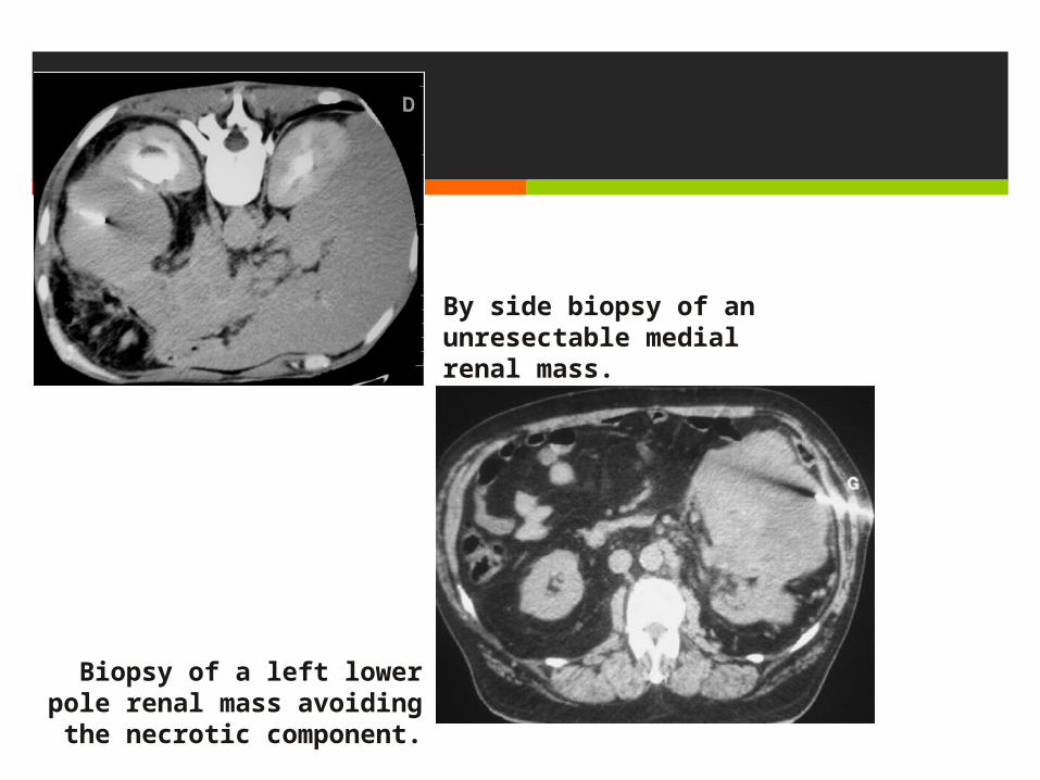
By side biopsy of an unresectable medial renal mass.
Biopsy of a left lower pole renal mass avoiding the necrotic component.
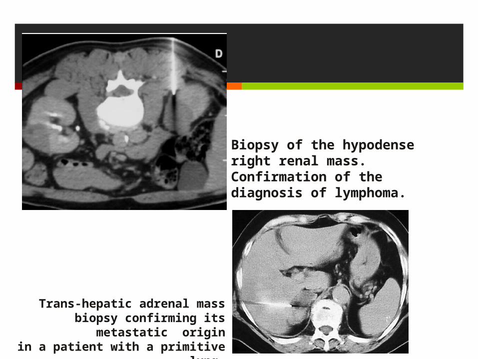
Biopsy of the hypodense right renal mass. Confirmation of the diagnosis of lymphoma.
Trans-hepatic adrenal mass biopsy confirming its metastatic origin
in a patient with a primitive lung.
Discussion In our institution, most abdominal aspiration procedures during
the past 9 years were performed under CT control.
CT permits accurate placement of a needle tip into small lesions, and its proximity to major vessels is readily ascertained.
The cross-sectional format of CT permits choice of the most appropriate needle approach to a suspected abnormality (i.e. , anterior, posterior, lateral, or oblique)
The skin puncture site, needle path, and depth can be readily determined from hard copy images and the measured depth for sampling directly transposed to the needle with a sterile rule.
DISCUSSION Problems related to patient size, bowel gas, dressings and patient
positioning, can all be accommodated by the CT guidance procedure.
As the contraindications to CT-guided biopsy the lack of patient cooperation, coagulation problems technical impossibility due to interruption by major vessels and bowel
are noted.
DISCUSSION
CT-guided biopsy was not indicated in a case that the envisaged path direction was not considered to be safe due to interruption by major vessels, bowel and vertebral bodies
enough specimens from the small lesions located at a deep site should be obtained with satisfactory sample for histological examination
DISCUSSION
There is a wide variety of needles from which to choose, with various needle gauges, tip configurations, and sampling mechanisms.
For this discussion, they will be divided into three general groups: small-gauge aspiration needles such as the Chiba: cytology study small-guage cutting-core-biopsy needles : difficult path or high hemorrhagic risk larger cutting needles such as the 18-gauge Menghini, 18-gauge Biopty, and 14-gauge TruCut.
DISCUSSION
Factors to consider when choosing a needle include location of the lesion, proximity to other structures, amount of tissue needed (pathologist’s needs), operator preference.
Aspiration needles are designed to obtain cytologic samples only. Occasionally, they obtain small pieces of tissue, which can be processed for histologic examination.
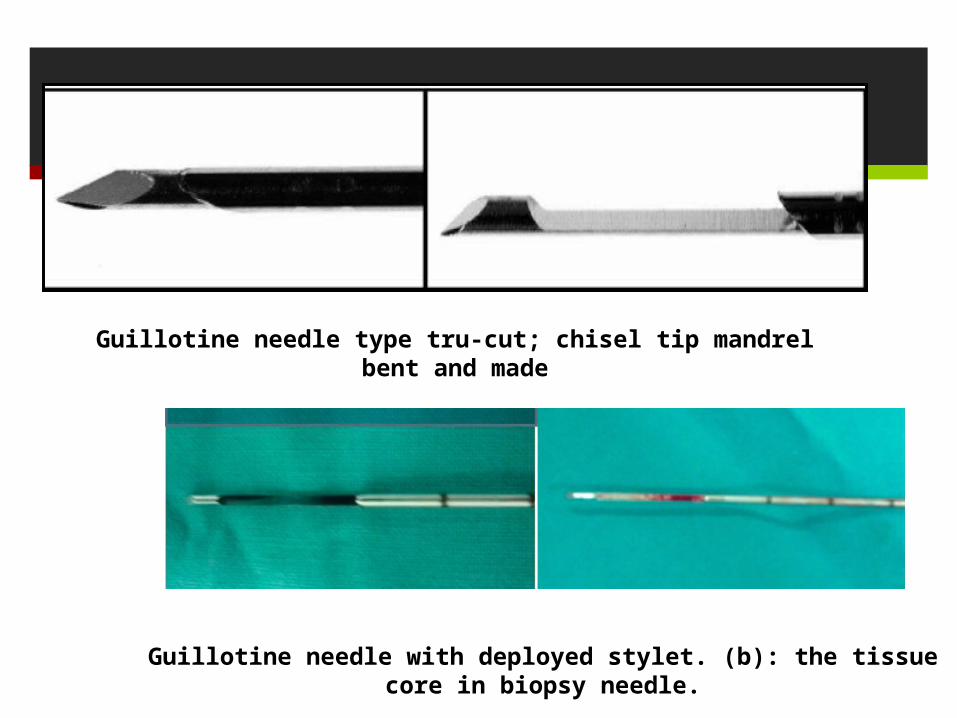
Guillotine needle with deployed stylet. (b): the tissue core in biopsy needle.
Guillotine needle type tru-cut; chisel tip mandrel bent and made
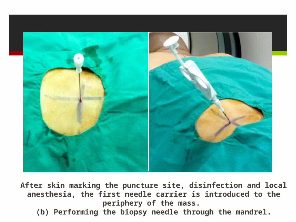
After skin marking the puncture site, disinfection and local anesthesia, the first needle carrier is introduced to the periphery of the mass.
(b) Performing the biopsy needle through the mandrel.
Discussion
Before performing any biopsy, the previous diagnostic studies should be reviewed, and the clinical findings and information sought should be discussed with the referring physician in order to plan the most appropriate procedure.
Review of the previous diagnostic studies is helpful in selecting the imaging technique, approach, and positioning of the patient for the biopsy.
Discussion
With CT guidance, most lesions are best approached by choosing a needle path that minimizes the skin-to-lesion distance.
When this involves traversing bowel or other organs, and when an alternative route is available, the alternative route is often chosen to avoid these other structures.
However, with thin-needle aspirations in the immunocompetent patient, it is possible to cross bowel, stomach, liver, or other structures without unacceptable risks
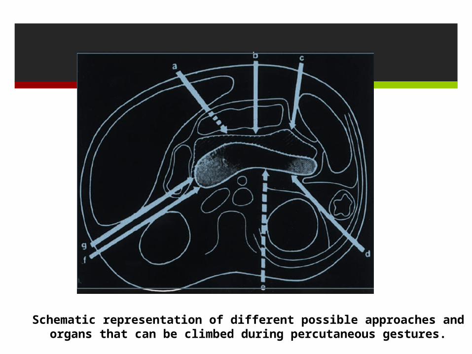
Schematic representation of different possible approaches and organs that can be climbed during percutaneous gestures.
Discussion
For ease of performance, it is best if the needle path lies in the axial plane.
This allows the entire needle to be visualized on a single image.
However, other structures often surround the lesion and preclude such an approach.
Several authors have described methods used to approach lesions that were not accessible via a direct approach.
DISCUSSION otherwise inaccessible lesions cqn be approached
either from above or below and using a geometric approach to calculate the needle angle (the so-called “triangulation method”), many ons can be sampled safely.
This is especially valuable in renal, superior retroperitoneal adrenal lesions
when avoiding the caudal extent of the pleura is important to diminish the risk of pneumothorax, pleural contamination, malignant seeding of the pleural space.
DISCUSSION
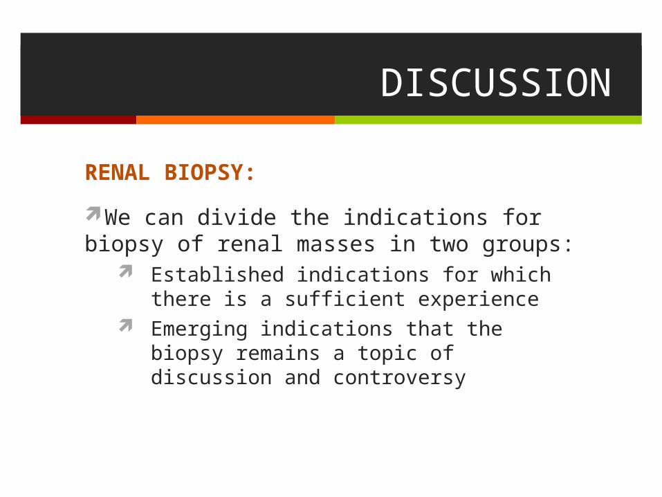
RENAL BIOPSY:
We can divide the indications for biopsy of renal masses in two groups:
Established indications for which there is a sufficient experience
Emerging indications that the biopsy remains a topic of discussion and controversy
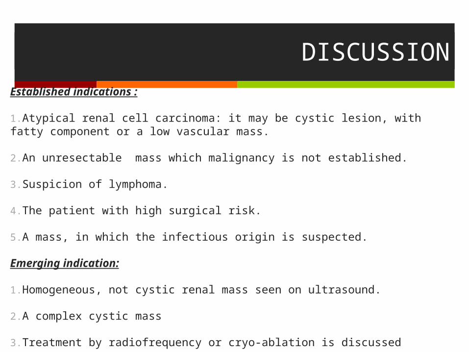
DISCUSSIONEstablished indications :
1.Atypical renal cell carcinoma: it may be cystic lesion, with fatty component or a low vascular mass.
2.An unresectable mass which malignancy is not established.
3.Suspicion of lymphoma.
4.The patient with high surgical risk.
5.A mass, in which the infectious origin is suspected.
Emerging indication:
1.Homogeneous, not cystic renal mass seen on ultrasound.
2.A complex cystic mass
3.Treatment by radiofrequency or cryo-ablation is discussed
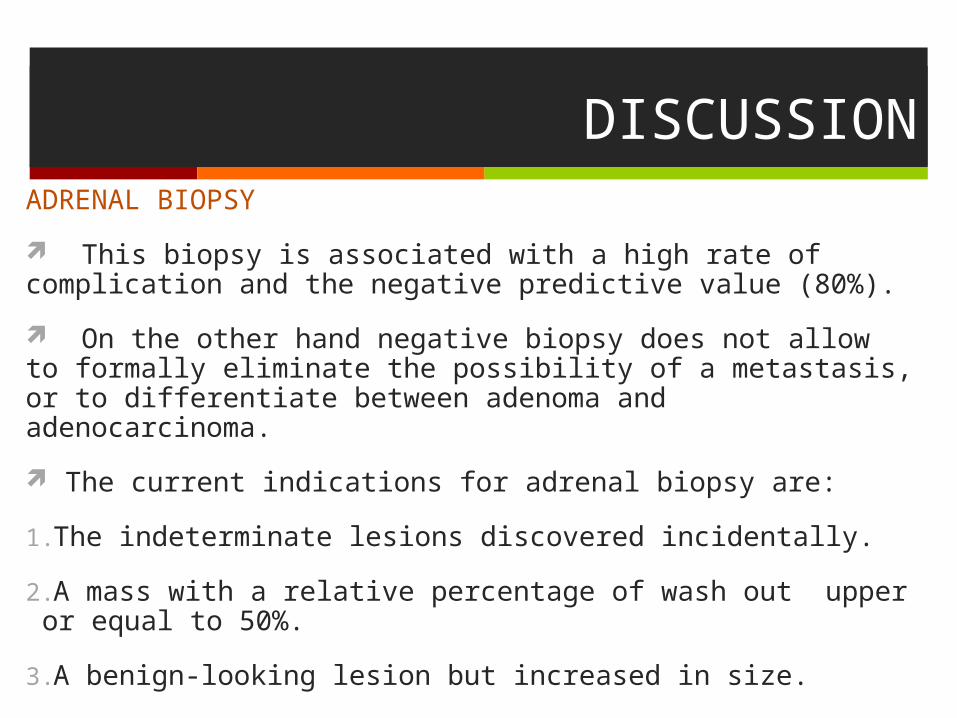
DISCUSSIONADRENAL BIOPSY
This biopsy is associated with a high rate of complication and the negative predictive value (80%).
On the other hand negative biopsy does not allow to formally eliminate the possibility of a metastasis, or to differentiate between adenoma and adenocarcinoma.
The current indications for adrenal biopsy are:
1.The indeterminate lesions discovered incidentally.
2.A mass with a relative percentage of wash out upper or equal to 50%.
3.A benign-looking lesion but increased in size.
DISCUSSION
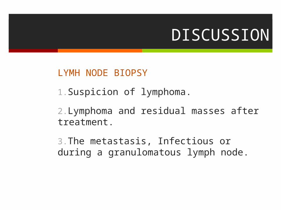
LYMH NODE BIOPSY
1.Suspicion of lymphoma.
2.Lymphoma and residual masses after treatment.
3.The metastasis, Infectious or during a granulomatous lymph node.
DISCUSSION
BIOPSY OF RETROPERITONEAL LESIONS:
Depending on the size of the lesion and its location within the retroperitoneum,
either an anterior or posterior approach can be used,
although the posterior approach is usually preferred and is most often necessary to ensure a clear path for the use of cutting needles.

CONCLUSION
Guided percutaneous biopsy of abdominal lesions
especially retroperitoneal lesions clearly has
become an important diagnostic tool.
The success of this technique lies in the accuracy
that can be achieved as well as in its relative safety
and ease of performance.