Embed Size (px)
Citation preview
SSTR2A IHC in Neuroendocrine Tumors
Am J Surg Pathol 2012;36:242–252
Introduction
• SSTR highly expressed in neuroendocrine tumors
• SSTR1, SSTR2A, SSTR3, SSTR4 and SSTR5 mostly concomitantly present
• SSTR2A shows highest expression
somatostatin analogues applied in clinical practice highest affinity for this subtype
J Tr
ansl
Med
. 20
13
Jan
15
;11
:17
Introduction
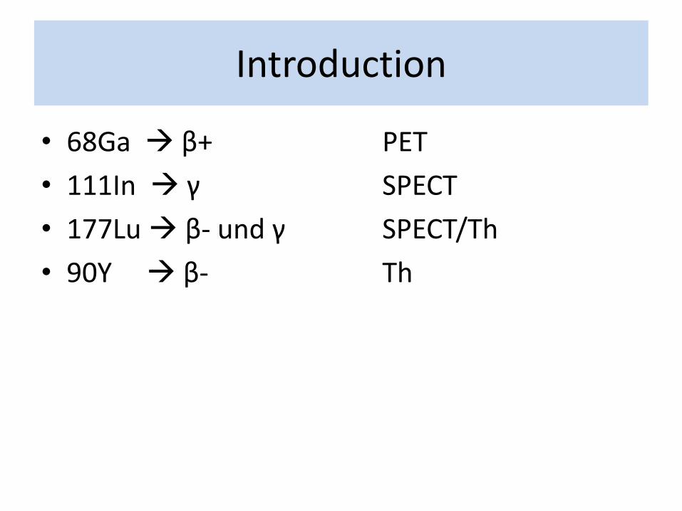
• 68Ga β+ PET
• 111In γ SPECT
• 177Lu β- und γ SPECT/Th
• 90Y β- Th
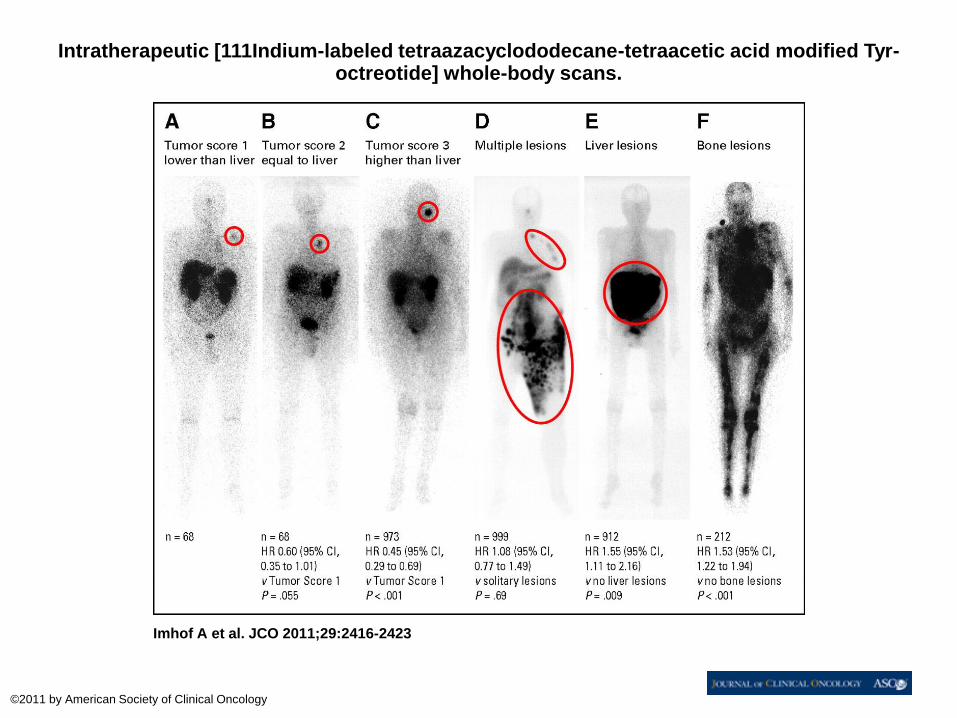
Intratherapeutic [111Indium-labeled tetraazacyclododecane-tetraacetic acid modified Tyr-octreotide] whole-body scans.
Imhof A et al. JCO 2011;29:2416-2423
©2011 by American Society of Clinical Oncology
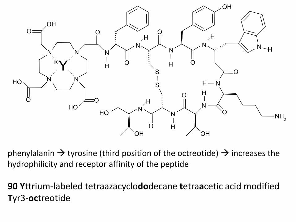
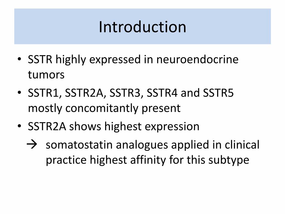
phenylalanin tyrosine (third position of the octreotide) increases the hydrophilicity and receptor affinity of the peptide
90 Yttrium-labeled tetraazacyclododecane tetraacetic acid modified Tyr3-octreotide

[90Y-DOTA]-TOC
• developed and introduced into clinical practice in 1997
Introduction
Introduction
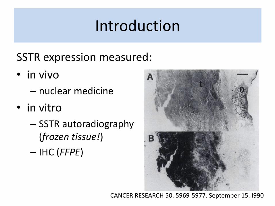
SSTR expression measured:
• in vivo
– nuclear medicine
• in vitro
– SSTR autoradiography (frozen tissue!)
– IHC (FFPE)
CANCER RESEARCH 50. 5969-5977. September 15. I990
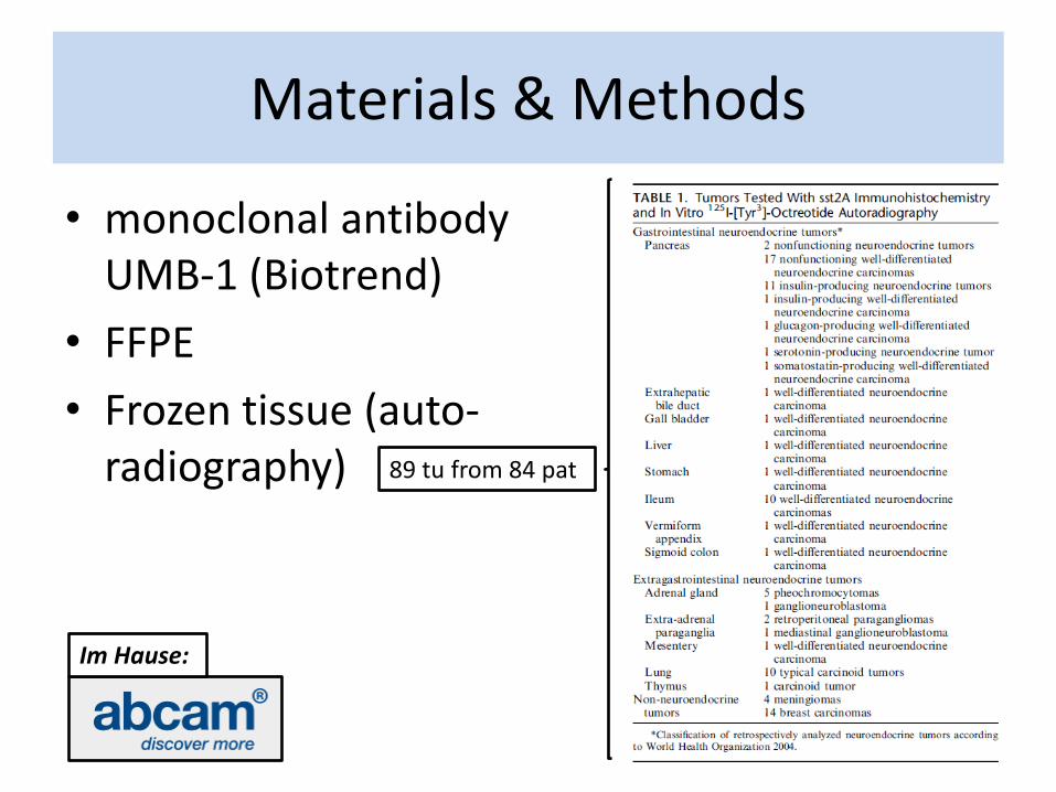
Materials & Methods
• monoclonal antibody UMB-1 (Biotrend)
• FFPE
• Frozen tissue (auto-radiography) 89 tu from 84 pat
Im Hause:
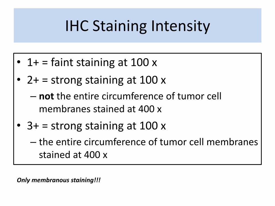
• 1+ = faint staining at 100 x
• 2+ = strong staining at 100 x
– not the entire circumference of tumor cell membranes stained at 400 x
• 3+ = strong staining at 100 x
– the entire circumference of tumor cell membranes stained at 400 x
IHC Staining Intensity
Only membranous staining!!!
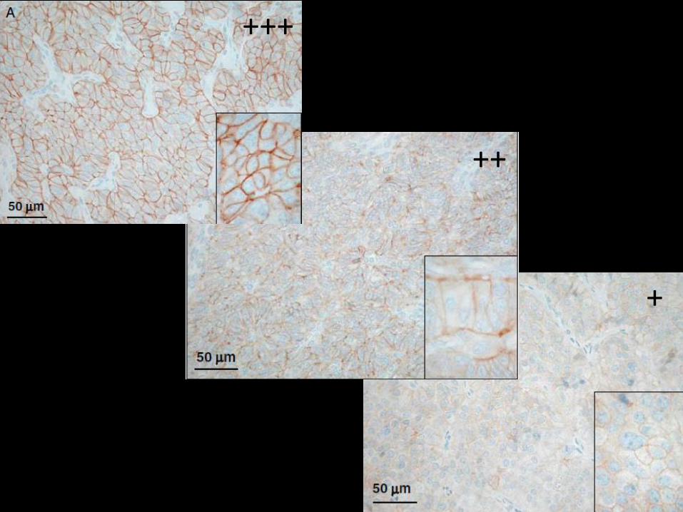
+++
++
+
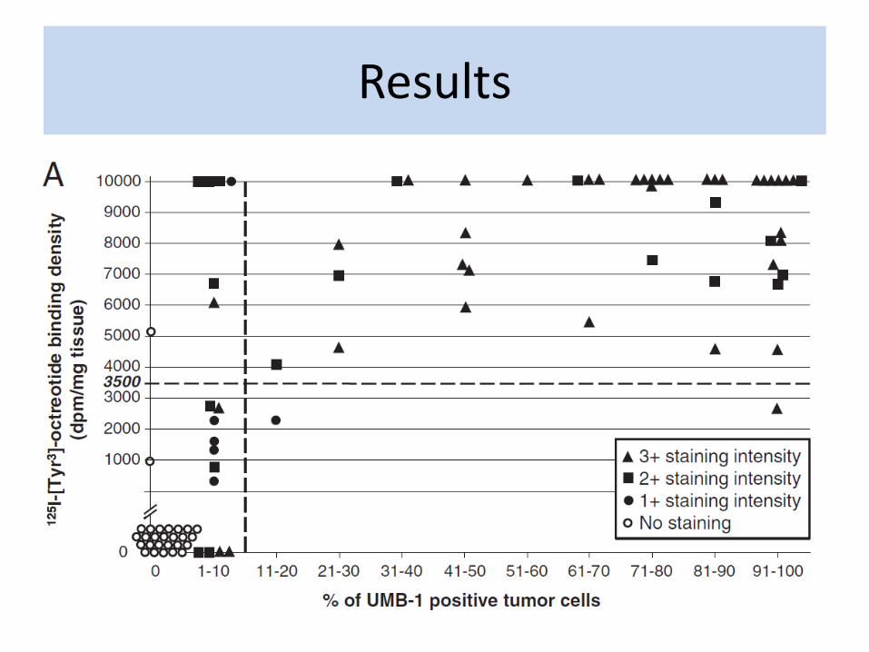
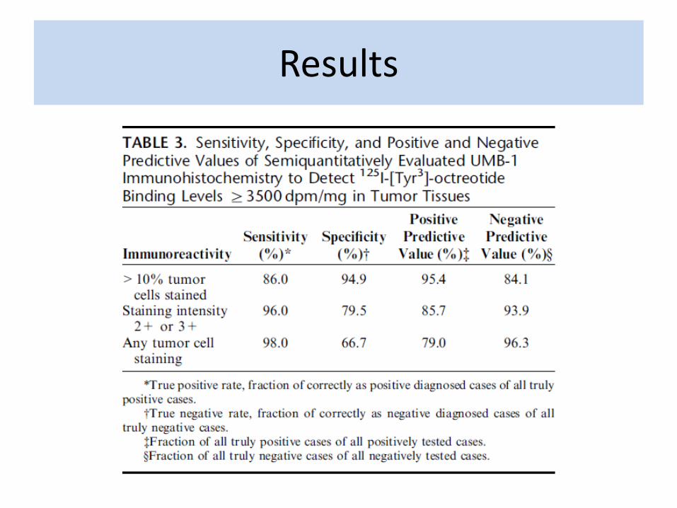
Results
Results
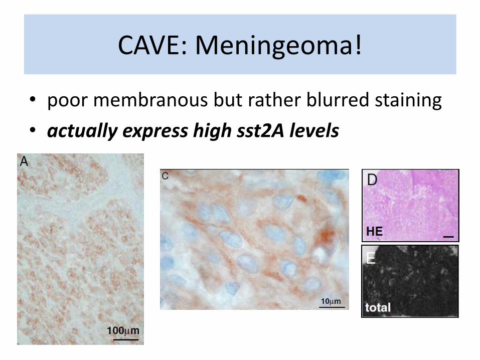
CAVE: Meningeoma!
• poor membranous but rather blurred staining
• actually express high sst2A levels
• “This particular staining pattern of meningiomas is likely explained by the prominent interdigitation of intercellular membranes typical of meningioma tumor cells, which yields an indistinct cell membrane staining at the light microscopic level.”
• “…the cytoplasmatic staining better reflects the actual somatostatin receptor levels and may well correspond to receptors located in the strongly interdigitated cell membrane. ”
CAVE: Meningeoma!
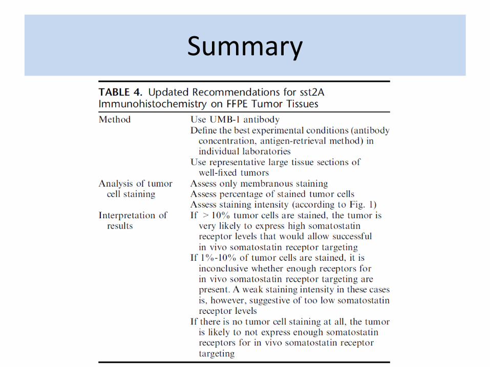
Summary
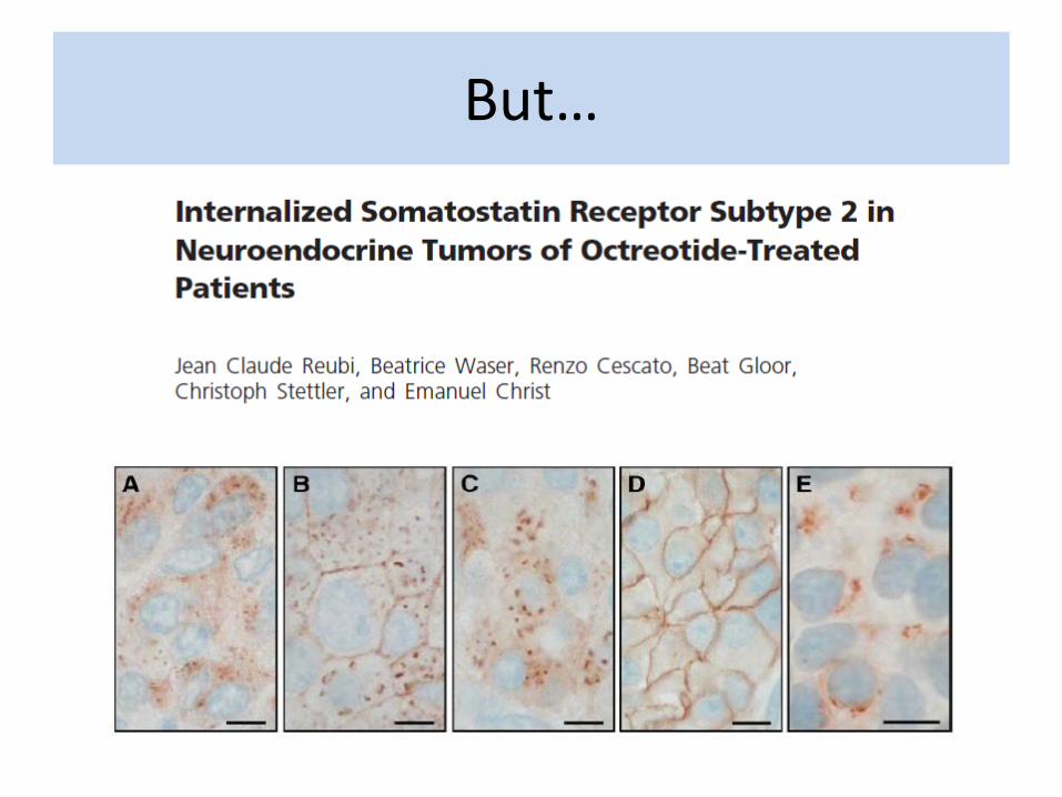
But…
So…
“The present data strongly suggest that the specific pattern of internalized sst2 receptors should also be considered as an additional reliable criterion for specific sst2 expression when it is confirmed that the patient has been on octreotide therapy immediately before or during surgery.»