Embed Size (px)
Citation preview
Appl
icAt
ion
not
E | 0
7/20
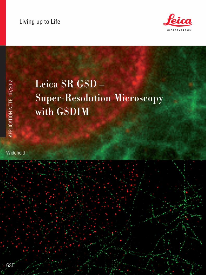
12 Leica SR GSD –Super-Resolution Microscopy with GSDIM
Widefield
GSD
brilliant
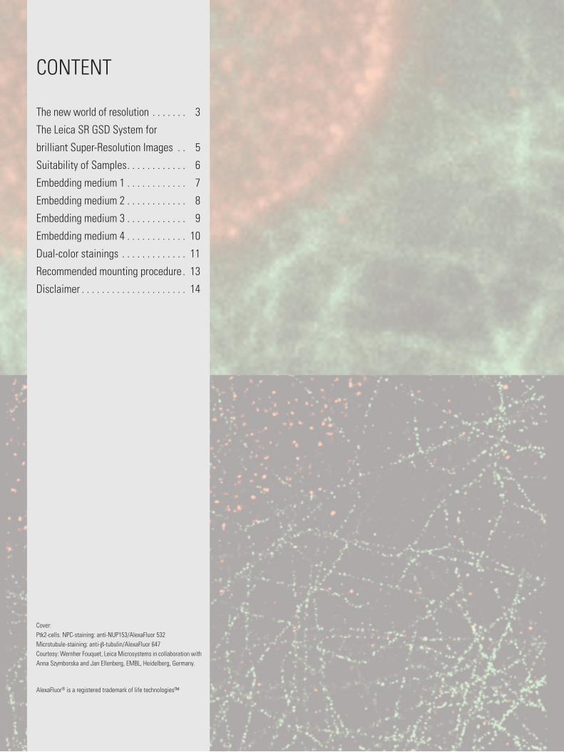
CONTENT
Cover: Ptk2-cells. NPC-staining: anti-NUP153/AlexaFluor 532Microtubule-staining: anti-β-tubulin/AlexaFluor 647Courtesy: Wernher Fouquet, Leica Microsystems in collaboration with Anna Szymborska and Jan Ellenberg, EMBL, Heidelberg, Germany.
The new world of resolution . . . . . . . 3
The Leica SR GSD System for
brilliant Super-Resolution Images . . 5
Suitability of Samples . . . . . . . . . . . . 6
Embedding medium 1 . . . . . . . . . . . . 7
Embedding medium 2 . . . . . . . . . . . . 8
Embedding medium 3 . . . . . . . . . . . . 9
Embedding medium 4 . . . . . . . . . . . . 10
Dual-color stainings . . . . . . . . . . . . . 11
Recommended mounting procedure . 13
Disclaimer . . . . . . . . . . . . . . . . . . . . . 14
AlexaFluor® is a registered trademark of life technologies™
Application Note Leica SR GSD 3
Introduction
The New World of ResolutionGreat advancements in biology have been possible by using fluorescence microscopy. So far, the resolu-tion of the images was limited due to physical con-straints. In the past couple of years, new methods evolved circumventing these limitations and bringing fluorescence microscopy to a new level of resolution, boosting the possibilities in science with fluorescence microscopes.
The resolution of a regular fluorescence microscope image is limited by diffraction to approximately half the wavelength of the emitted light. To separate fluo-rescently labeled structures that are closer together, a solution is needed to overcome Abbe’s diffraction lim-it. The most common super-resolution methods for im-aging beyond the diffraction limit are localization mi-croscopy, SIM (structured illumination) and STED (Stimulated Emission Depletion Microscope). Local-ization microscopy does not look at an ensemble of simultaneously emitting fluorophores, but at clearly separated, individual fluorophores which can be local-ized with nanometer precision. Over time, the position of each fluorophore labeling a biological structure is determined and the image is re-constructed in silico based on the position information of each fluoro-phore. The challenge is to effectively singularize an ensemble of fluorophores to allow single molecule detection.
The LeicA SR GSD
The Leica SR GSD offers the highest lateral resolution with light microscopy and is a superb tool for deter-mining the structure of fluorescently labeled speci-men in neurobiology, cell biology, virology, microbiol-ogy and physiology. The workflow is usually based on standard immunostaining techniques and integrates perfectly into existing workflows for fluorescence mi-croscopy.
The Leica SR GSD is using the localization microscopy technique Ground State Depletion followed by indi-vidual molecule return (GSDIM) to achieve superior super-resolution images. GSDIM is an innovative and reliable technology for achieving super-resolution images with a resolution of down to 20 nm.
The crucial step in GSDIM is to temporarily switch the majority of fluorophores off to allow the precise local-ization of single fluorophores. To this end, excitation light of high intensity is used in such a way that al-most all fluorophores in the sample are turned dark, leaving only single, well-separated fluorophores emit-ting fluorescence, which is a prerequisite for nanome-ter precision localization.
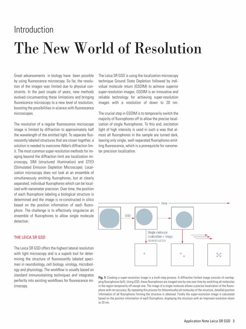
Fig. 1: Creating a super-resolution image is a multi-step process: A diffraction limited image consists of overlap-ping fluorophores (left). Using GSD, these fluorophores are imaged one-by-one over time by switching all molecules in the region temporarily off except one. The image of a single molecule allows a precise localization of the fluoro-phore with nm accuracy. By repeating this process for (theoretically) all molecules of the structure, detailed position information of all fluorophores forming the structure is obtained. Finally the super-resolution image is calculated based on the position information of each fluorophore, displaying the structure with an improved resolution down to 20 nm.
4 Application Note Leica SR GSD
The MoLecuLAR BASicS of GSDiM
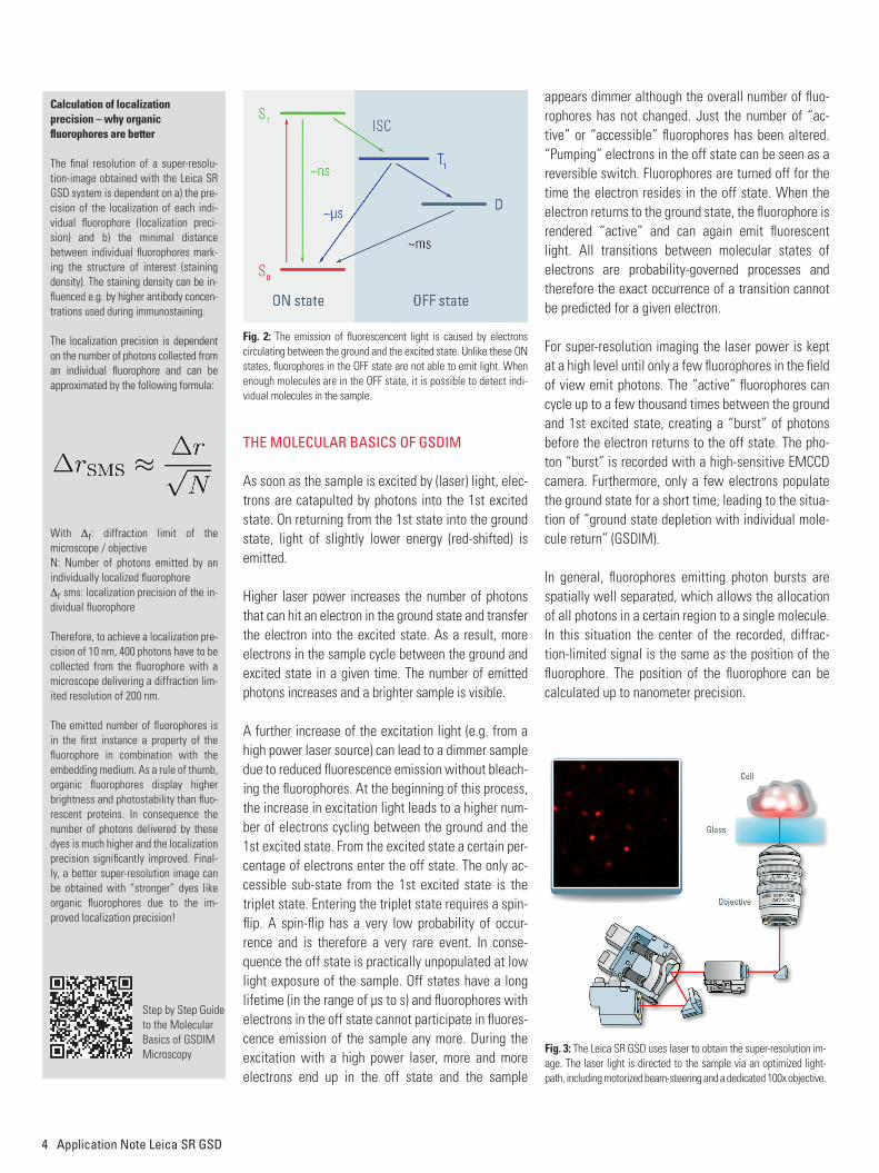
As soon as the sample is excited by (laser) light, elec-trons are catapulted by photons into the 1st excited state. On returning from the 1st state into the ground state, light of slightly lower energy (red-shifted) is emitted.
Higher laser power increases the number of photons that can hit an electron in the ground state and transfer the electron into the excited state. As a result, more electrons in the sample cycle between the ground and excited state in a given time. The number of emitted photons increases and a brighter sample is visible.
A further increase of the excitation light (e.g. from a high power laser source) can lead to a dimmer sample due to reduced fl uorescence emission without bleach-ing the fl uorophores. At the beginning of this process, the increase in excitation light leads to a higher num-ber of electrons cycling between the ground and the 1st excited state. From the excited state a certain per-centage of electrons enter the off state. The only ac-cessible sub-state from the 1st excited state is the triplet state. Entering the triplet state requires a spin-fl ip. A spin-fl ip has a very low probability of occur-rence and is therefore a very rare event. In conse-quence the off state is practically unpopulated at low light exposure of the sample. Off states have a long lifetime (in the range of µs to s) and fl uorophores with electrons in the off state cannot participate in fl uores-cence emission of the sample any more. During the excitation with a high power laser, more and more electrons end up in the off state and the sample
appears dimmer although the overall number of fl uo-rophores has not changed. Just the number of “ac-tive” or “accessible” fl uorophores has been altered. “Pumping” electrons in the off state can be seen as a reversible switch. Fluorophores are turned off for the time the electron resides in the off state. When the electron returns to the ground state, the fl uorophore is rendered “active” and can again emit fl uorescent light. All transitions between molecular states of electrons are probability-governed processes and therefore the exact occurrence of a transition cannot be predicted for a given electron.
For super-resolution imaging the laser power is kept at a high level until only a few fl uorophores in the fi eld of view emit photons. The “active” fl uorophores can cycle up to a few thousand times between the ground and 1st excited state, creating a “burst” of photons before the electron returns to the off state. The pho-ton “burst” is recorded with a high-sensitive EMCCD camera. Furthermore, only a few electrons populate the ground state for a short time, leading to the situa-tion of “ground state depletion with individual mole-cule return” (GSDIM).
In general, fl uorophores emitting photon bursts are spatially well separated, which allows the allocation of all photons in a certain region to a single molecule. In this situation the center of the recorded, diffrac-tion-limited signal is the same as the position of the fl uorophore. The position of the fl uorophore can be calculated up to nanometer precision.
Calculation of localizationprecision – why organic fl uorophores are better
The fi nal resolution of a super-resolu-tion-image obtained with the Leica SR GSD system is dependent on a) the pre-cision of the localization of each indi-vidual fl uorophore (localization preci-sion) and b) the minimal distance between individual fl uorophores mark-ing the structure of interest (staining density). The staining density can be in-fl uenced e.g. by higher antibody concen-trations used during immunostaining.
The localization precision is dependent on the number of photons collected from an individual fl uorophore and can be approximated by the following formula:
With Δr: diffraction limit of themicroscope / objectiveN: Number of photons emitted by an individually localized fl uorophoreΔr sms: localization precision of the in-dividual fl uorophore
Therefore, to achieve a localization pre-cision of 10 nm, 400 photons have to be collected from the fl uorophore with a microscope delivering a diffraction lim-ited resolution of 200 nm.
The emitted number of fl uorophores is in the fi rst instance a property of the fl uorophore in combination with the embedding medium. As a rule of thumb, organic fl uorophores display higher brightness and photostability than fl uo-rescent proteins. In consequence the number of photons delivered by these dyes is much higher and the localization precision signifi cantly improved. Final-ly, a better super-resolution image can be obtained with “stronger” dyes like organic fl uorophores due to the im-proved localization precision!
Fig. 3: The Leica SR GSD uses laser to obtain the super-resolution im-age. The laser light is directed to the sample via an optimized light-path, including motorized beam-steering and a dedicated 100x objective.
Fig. 2: The emission of fl uorescencent light is caused by electronscirculating between the ground and the excited state. Unlike these ON states, fl uorophores in the OFF state are not able to emit light. When enough molecules are in the OFF state, it is possible to detect indi-vidual molecules in the sample.
Step by Step Guide to the Molecular Basics of GSDIMMicroscopy
Application Note Leica SR GSD 5
The Leica SR GSD System for brilliant Super-Resolution Images
Key feATuReS of The LeicA SR GSD
› GSDIM-technology – Wider range of standard fluorophores
› Leica SuMo-Stage – Effective hardware drift suppression
› Super-resolution in both TIRF - and Widefield-Mode› High Power Lasers – Higher flexibility in fluorophore selection and shorter imaging time
› Multi-purpose widefield system – including TIRF / DIC / Live cell imaging
› Optimized localization algorithm – Superb image quality
› LAS AF GSD Wizard – Intuitive handling minimizes user training and makes SR imaging easier
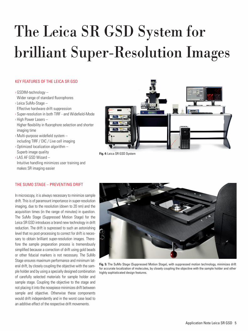
The SuMo STAGe – pReveNTiNG DRifT
In microscopy, it is always necessary to minimize sample drift. This is of paramount importance in super-resolution imaging, due to the resolution (down to 20 nm) and the acquisition times (in the range of minutes) in question. The SuMo Stage (Suppressed Motion Stage) for the Leica SR GSD introduces a brand new technology in drift reduction. The drift is supressed to such an astonishing level that no post-processing to correct for drift is neces-sary to obtain brilliant super-resolution images. There-fore the sample preparation process is tremendously simplified because a correction of drift using gold beads or other fiducial markers is not necessary. The SuMo Stage ensures maximum performance and minimum lat-eral drift, by closely coupling the objective with the sam-ple holder and by using a specially designed combination of carefully selected materials for sample holder and sample stage. Coupling the objective to the stage and not placing it into the nosepiece minimizes drift between sample and objective. Otherwise these components would drift independently and in the worst case lead to an additive effect of the respective drift movements.
Fig. 5: The SuMo Stage (Suppressed Motion Stage), with suppressed motion technology, minimizes drift for accurate localization of molecules, by closely coupling the objective with the sample holder and other highly sophisticated design features.
Fig. 4: Leica SR GSD System
6 Application Note Leica SR GSD
Suitability of SamplesGSD is suitable for a wide range of samples. Cells grown on standard glass coverslips can be used e.g. after chemical fixation. Furthermore tissue slices can be prepared for GSD and placed on a glass coverslip. The samples are normally fluorescently labeled by im-munostaining or other suitable techniques.
fLuoReSceNT pRoTeiNS
GSDIM is suitable to create super-resolution images using fluorescent proteins. E.g. the fluorescent protein YFP / Venus displayed astonishing results. Further-more, other fluorescent proteins should work with this technology (including GFP). To combine the power of organic fluorophores with the advantages of geneti-cally encoded fusion proteins (e.g. if no suitable anti-body is available), the use of tags like SNAP-, Halo- or CLIP-Tag is recommended. These tags can be geneti-cally linked to the protein of interest and later on la-belled with organic fluorophores (e.g. after fixation and permeabilization).
SupeR-ReSoLuTioN iMAGiNG of LiviNG ceLLS
Recent reports have studied the suitability of GSDIM (also known as dSTORM) to image living cells. In gen-eral, it is possible to study living cells with the Leica SR GSD, but phototoxicity - as a results of the high laser power applied - and the dynamics of structure of inter-est during the observation period should be carefully controlled.
coveRSLipS
Super-resolution imaging requires excellent optical performance throughout the whole optical system – not only within the microscope and objective. The coverslip is a major parameter with high impact on im-aging results. To obtain optimal super-resolution image quality a flat coverslip is required. Unfortunately, while being uncritical for standard widefield applications, not all chambered coverslip products can guarantee the de-gree of flatness optimal for GSD. Furthermore, cham-bered coverslip products may not provide the necessary
mechanical stability for high-resolution imaging. The re-spective products should be tested by the researcher for their suitability. A better flatness and high stability can be generally achieved by stress free mounting of a glass coverslip, e.g. in specially designed coverslip holders.
iMMuNofLuoReSceNce
Starting with samples prepared with standard immuno-fluorescence protocols optimized for diffraction-limited fluorescence should already provide stunning super-resolution images. To obtain optimal GSD images it might be necessary to increase the concentration of primary and secondary antibodies to achieve a higher labeling density. Increasing the labeling density en-sures that the structure of interest is sufficiently stained with fluorophores. A sparse labeling of structures can lead to a point-like appearance of your image. In order to reduce unspecific background signals in the GSD image, it might be necessary to increase the time for blocking your sample as well as the concentration of the blocking reagent. It is advised to wash your sam-ples thoroughly (e.g. by increasing the number and time of washing steps).
STRucTuRe pReSeRvATioN
The preservation of a specific cellular structure strongly depends on the fixation method. The exact fixation pro-tocol should be optimized for the structure of interest. For the majority of structures a Paraformaldehyde / Formaldehyde fixation is sufficient. Small amounts of Glutaraldehyde (0.05% to 0.2% (v/v)) in addition to Paraformaldehyde / Formaldehyde as a fixant can strongly improve structure preservation. Glutaralde-hyde can induce a hazy fluorescent background there-fore quenching with ammonium, NaBH4 or other suit-able reagents is recommended. Methanol fixation is common for microtubule preservation, but might not be sufficient for other structures.
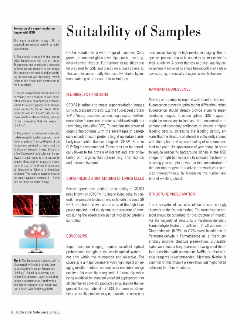
Formation of a super-resolution image with GSD
The super-resolution image (SR) is acquired and reconstructed in a multi-step process.
1. The sample is excited with a laser to drive fluorophores into the off state. This process can be seen as a decrease in fluorescence intensity in the image. The process is reversible and has noth-ing in common with bleaching, which leads to the irreversible destruction of the fluorophore.
2. As the overall fluorescence intensity decreases, the emission of well-sepa-rated individual fluorophores becomes visible for a short period until the mol-ecule returns to the off state. Other molecules will exit the off state and be-come visible at the same time, leading to the impression that the image is “blinking”.
3. The position of individual molecules is determined in each image with nano-meter precision. The coordinates of the fluorophore are used to reconstruct the final super-resolution image. Since only a few fluorescent molecules can be an-alyzed in each frame it is necessary to acquire thousands of images to detect the entire set of hundreds of thousands of fluorophores labeling a biological structure. This leads to imaging times in the range typically between 1 – 5 min min per super-resolution image.
Fig. 6: The fluorescently labeled cell is illuminated with high intensity laser light, resulting in single fluorophore “blinking”. Based on analyzing the single fluorophores a super-resolution image is reconstructed (right) with a 10x higher resolution than the diffrac-tion-limited widefield image (left).
Application Note Leica SR GSD 7
Embedding Medium 1ReAGeNTS– β-Mercaptoethylamine (MEA) (e.g. Sigma-Aldrich,
# 30070, also known as Cysteamine)– Phosphate Buffered Saline (PBS)– HCl / NaOH for pH adjustment
BuffeRPBS containing 10 mM β-Mercaptoethylamine (MEA) adjusted to pH 7.4.
Dissolve the β-Mercaptoethylamine in PBS and adjust to pH 7.4. The β-Mercaptoethylamine concentration can be varied over a wide range to optimize the GSD image – the usable range is in general between 10 and 100 mM (recommended concentrations for test-ing 10 / 30 / 100 mM).
β-Mercaptoethylamine solutions must be freshly prepared before use. Alternatively, stock solutions can be prepared and stored at -20°C and freshly thawed before use. The frozen aliquots can be stored for several weeks.
Illumination EPI ✔ TIRF ✔
Laser [nm] Dye Manufacturer
488 AlexaFluor 488 life technologies
Atto 488 Sigma
chromeo 488 Active Motif
oregon Green 488 life technologies
chromeo 505 Active Motif
532 Atto 520 Sigma
AlexaFluor 532 life technologies
AlexaFluor 568 life technologies
642 Atto 633 Sigma
AlexaFluor 647 life technologies
Atto 647n Sigma
Atto 655 Sigma
AlexaFluor 680 life technologies
AlexaFluor 700 life technologies
AlexaFluor® is a registered trademark of life technologies™
Suitability of Samples
8 Application Note Leica SR GSD
Embedding Medium 2ReAGeNTS– Glucose Oxidase (e.g. Sigma-Aldrich G2133)– Catalase (e.g. Sigma-Aldrich C1345)– Glucose – Phosphate Buffered Saline (PBS)– HCl / NaOH for pH adjustment
BuffeRPBS containing 10% (w/v) glucose, 0.5 mg/mL glucose oxidase and 40 µg/mL catalase adjusted to pH 7.4.
Note: It is recommended to prepare a glucose stock solution and mix the reagents freshly before mounting the coverslip.
Illumination EPI ✔ TIRF ✔
Laser [nm] Dye Manufacturer
532 Atto 532 Sigma
AlexaFluor 532 life technologies
Rhodamine 6G Active Motif
Atto565 Sigma
AlexaFluor 568 life technologies
642 Atto 655 Sigma
AlexaFluor 680 life technologies
Embedding Medium for YFPThe following aqueous embedding was successfully used to image YFP in fixed and permeabilized mammalian cells:
– Catalase SIGMA, C1345 40 ug/mL
– Glucose Oxidase SIGMA G2133 0.5 mg/mL
– Glucose 10 mg/mL
– β-Mercaptoethylamine (Cysteamine) 1 mM
PBS adjusted to pH 7.4.
AlexaFluor® is a registered trademark of life technologies™
Application Note Leica SR GSD 9
Embedding Medium 3ReAGeNTS– Polyvinyl alcohol (PVA) with a molecular
weight of 25,000, 88 mol% hydrolyzed, e.g. Polysciences, #02975-500
– Phosphate Buffered Saline (PBS)– HCl / NaOH for pH adjustment
hARDwAReSpincoater
BuffeR Prepare a solution of 1% PVA in PBS. Adjust the pH to 7.4.
SpiNcoATiNGPlace the coverslip on the spincoater and dry the cov-erslip with brief 5-10 sec spin at approx. 3000 rpm. Drop 50 µl of the PVA-solution onto the sample and spin the sample at approx. 3000 rpm for approximately 30 s. Let the PVA-film dry for a few minutes and mount the sample. The sample can be stored at room tem-perature and used for GSD imaging for up to three months.
Illumination EPI ✔ TIRF not possible
Laser [nm] Dye Manufacturer
488 AlexaFluor 488 life technologies
Atto 488 Sigma
chromeo 488 Active Motif
oregon Green 488 life technologies
532 Atto 520 Sigma
AlexaFluor 532 life technologies
AlexaFluor 680 life technologies
No additional mounting medium is required. The PVA-film is protecting the sample.
AlexaFluor® is a registered trademark of life technologies™
10 Application Note Leica SR GSD
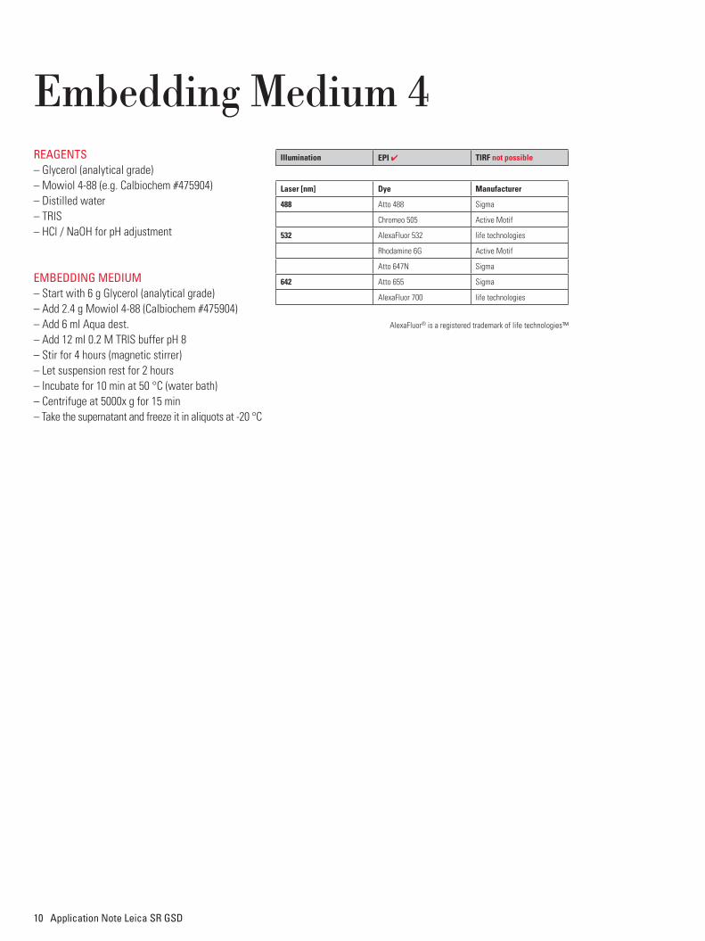
Embedding Medium 4ReAGeNTS– Glycerol (analytical grade)– Mowiol 4-88 (e.g. Calbiochem #475904)– Distilled water– TRIS– HCl / NaOH for pH adjustment
eMBeDDiNG MeDiuM– Start with 6 g Glycerol (analytical grade)– Add 2.4 g Mowiol 4-88 (Calbiochem #475904)– Add 6 ml Aqua dest.– Add 12 ml 0.2 M TRIS buffer pH 8– Stir for 4 hours (magnetic stirrer)– Let suspension rest for 2 hours– Incubate for 10 min at 50 °C (water bath)– Centrifuge at 5000x g for 15 min– Take the supernatant and freeze it in aliquots at -20 °C
Illumination EPI ✔ TIRF not possible
Laser [nm] Dye Manufacturer
488 Atto 488 Sigma
chromeo 505 Active Motif
532 AlexaFluor 532 life technologies
Rhodamine 6G Active Motif
Atto 647n Sigma
642 Atto 655 Sigma
AlexaFluor 700 life technologies
AlexaFluor® is a registered trademark of life technologies™
Application Note Leica SR GSD 11
Dual-Color StainingsLeica Microsystems recommends the combination of a) AlexaFluor 532 and AlexaFluor 647, b) AlexaFluor 488 and AlexaFluor 647 and c) Atto 488 and Alexa-Fluor 647 for sequential dual color imaging. Please image the red channel (AlexaFluor 647) first. Leica Microsystems does NOT recommend imaging the green/orange channel (e.g. AlexaFluor 488) at the
beginning to avoid bleaching of AlexaFluor 647, which can be - although at very low levels – excited by a 488 nm (or 532 nm) laser. If it is intended to use other fluorophores for double-staining of the sample, please check that both fluorophores can be imaged under the same GSD imaging conditions (embedding media) and crosstalk is negligible.
Use of 405 nm laser
The Leica SR GSD is equipped with a 405 nm laser. The lifetime of fluoro-phores in the off state can be shortened by illuminating the sample additionally with UV-light. The shorter lifetime leads to an increase of fluorophores in the on state and therefore can be used to increase the number of detected events per frame. By keeping the num-ber of events per frame in an optimal range, it is possible to
a) efficiently detect well-separated single fluorophores and
b) keep the time to acquire a super-res-olution image short, because it is pos-sible to accumulate the required number of events within fewer images.
When acquiring the longer wavelength channel of dual-color images, it is im-portant to use the 405 nm laser care-fully! The 405 nm laser can excite green dyes and therefore bleach the shorter wavelength fluorophores while acquiring the longer wavelength super-resolution image. The optimal settings should be determined for each sample type separately.
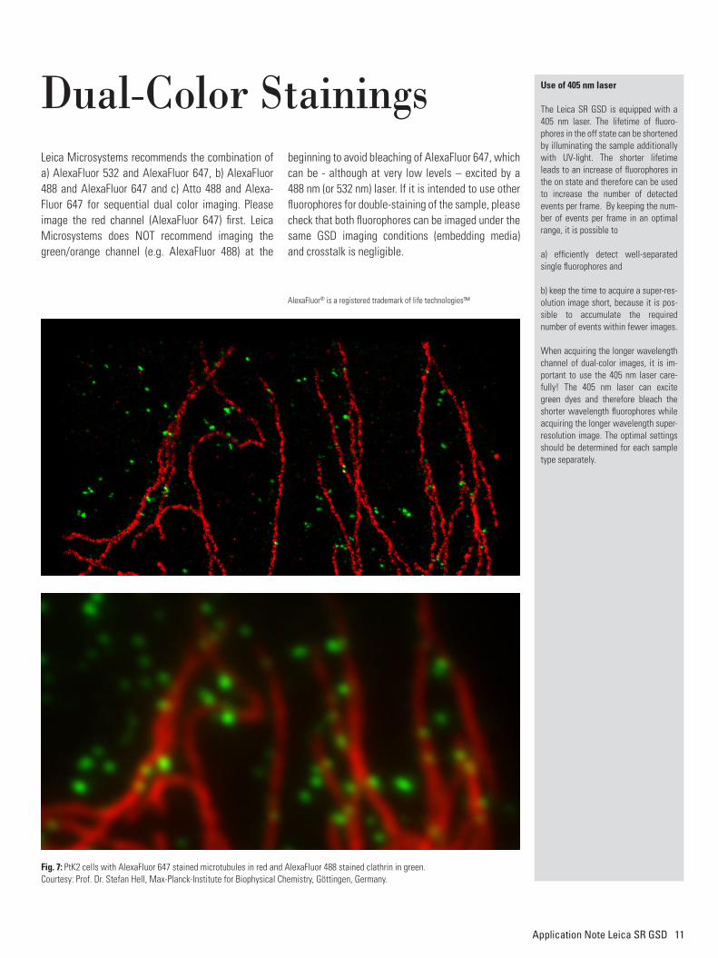
Fig. 7: ptK2 cells with AlexaFluor 647 stained microtubules in red and AlexaFluor 488 stained clathrin in green.courtesy: prof. Dr. Stefan Hell, Max-planck-institute for Biophysical chemistry, Göttingen, Germany.
AlexaFluor® is a registered trademark of life technologies™
12 Application Note Leica SR GSD
Fig. 8: The multi-step process to achieve a super-resolution image in a nutshell: The diffrac-tion limited image (top) shows all fluorophores signals overlapping. By localizing individual molecules after switching the majority of fluoro-phores temporarily off, and continuing this procedure for all fluorophores staining the structure, a super-resolution image (bottom) can be calculated with a 10x better resolution.
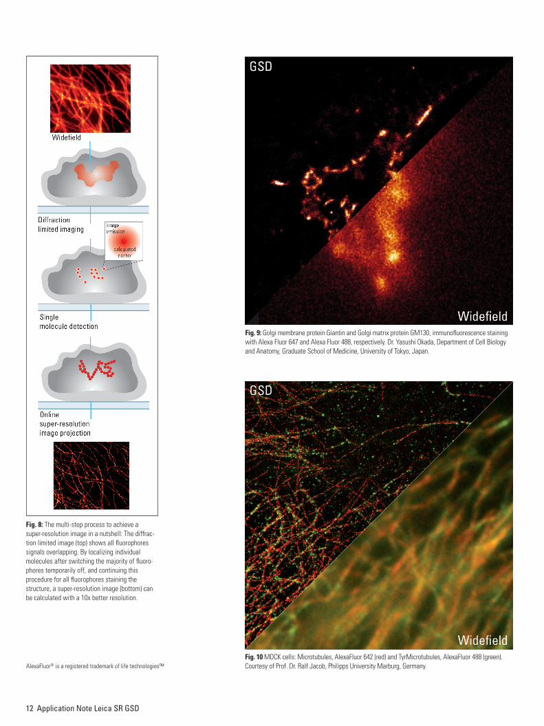
Fig. 10 MDcK cells: Microtubules, AlexaFluor 642 (red) and tyr Microtubules, AlexaFluor 488 (green). courtesy of prof. Dr. Ralf Jacob, philipps University Marburg, Germany.
Fig. 9: Golgi membrane protein Giantin and Golgi matrix protein GM130, immunofluorescence staining with Alexa Fluor 647 and Alexa Fluor 488, respectively. Dr. Yasushi okada, Department of cell Biology and Anatomy, Graduate School of Medicine, University of tokyo, Japan.
AlexaFluor® is a registered trademark of life technologies™
Application Note Leica SR GSD 13
Recommended Mounting Procedure1. Only for imaging with aqueous embedding media 1 &
2 (otherwise proceed with step two): Add 75 – 100 µl of embedding medium into the cavity of a clean de-pression slide. For embedding media 3 (PVA): leave the cavity of the depression slide empty.
2. Place the glass coverslip with your sample onto the cavity of the depression slide. The sample should face the cavity. The coverslip should cover the de-pression completely. If aqueous embedding medi-um is filled in the cavity, ensure that no air bubbles are present. Otherwise carefully remove the cover-slip and repeat the procedure.
3. For Embedding Media 1 & 2: Carefully remove excess of liquid using filter paper. Ensure that no buffer is soaked out from the reservoir.
4. Mix the yellow and blue component of Twinsil® in a ratio of 1:1 thoroughly and apply it to the edges of the glass coverslip without directly touching the glass coverslip. For Embedding Media 1 & 2: Seal the coverslip completely with Twinsil®. For Embed-ding Media 3: Seal the coverslip to approx. 75% and leave the remaining quarter open to allow ven-tilation and avoid condensation on the sample.
5. After 5 – 10 minutes the two-component glue is hardened and the sample is ready for GSD imaging.
6. The glue can be easily removed without leaving traces on the glass after hardening (e.g. to exchange the imaging buffer).
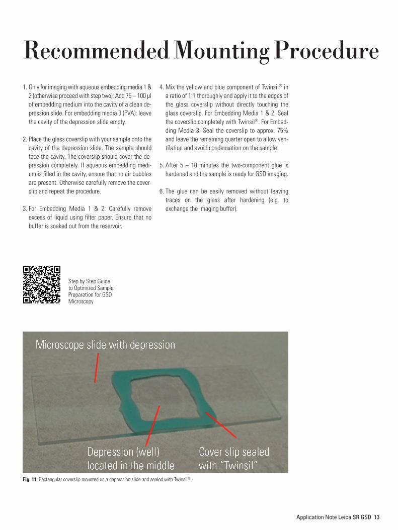
Fig. 11: Rectangular coverslip mounted on a depression slide and sealed with Twinsil®.
Step by Step Guide to Optimized Sample Preparation for GSD Microscopy
14 Application Note Leica SR GSD
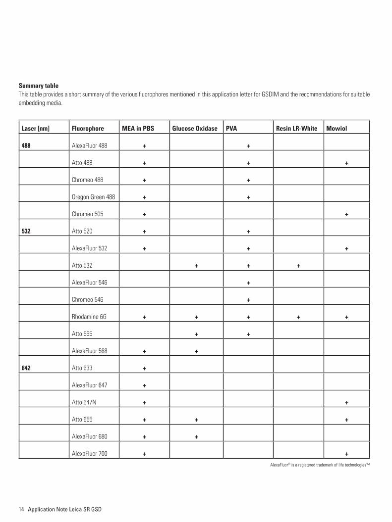
Summary tableThis table provides a short summary of the various fluorophores mentioned in this application letter for GSDIM and the recommendations for suitable embedding media.
Laser [nm] Fluorophore MEA in PBS Glucose Oxidase PVA Resin LR-White Mowiol
488 AlexaFluor 488 + +
Atto 488 + + +
Chromeo 488 + +
Oregon Green 488 + +
Chromeo 505 + +
532 Atto 520 + +
AlexaFluor 532 + + +
Atto 532 + + +
AlexaFluor 546 +
Chromeo 546 +
Rhodamine 6G + + + + +
Atto 565 + +
AlexaFluor 568 + +
642 Atto 633 +
AlexaFluor 647 +
Atto 647N + +
Atto 655 + + +
AlexaFluor 680 + +
AlexaFluor 700 + +
AlexaFluor® is a registered trademark of life technologies™
Application Note Leica SR GSD 15
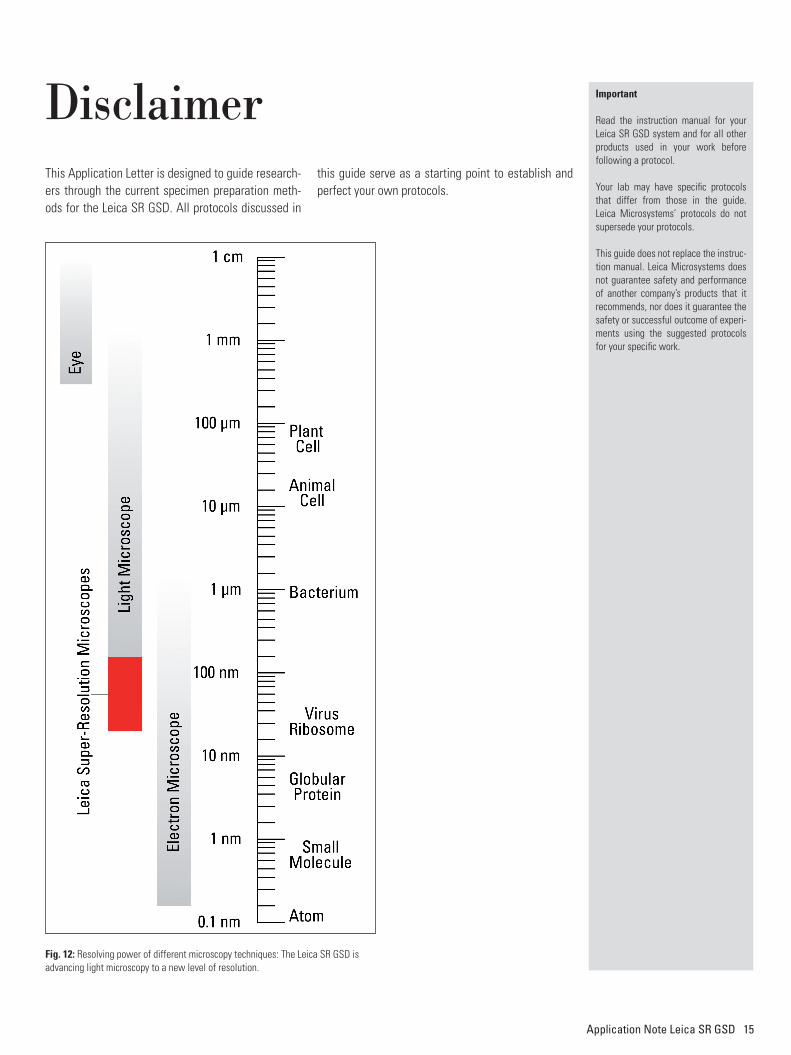
DisclaimerThis Application Letter is designed to guide research-ers through the current specimen preparation meth-ods for the Leica SR GSD. All protocols discussed in
this guide serve as a starting point to establish and perfect your own protocols.
Important
Read the instruction manual for your Leica SR GSD system and for all other products used in your work before following a protocol.
Your lab may have specific protocols that differ from those in the guide. Leica Microsystems’ protocols do not supersede your protocols.
This guide does not replace the instruc-tion manual. Leica Microsystems does not guarantee safety and performance of another company’s products that it recommends, nor does it guarantee the safety or successful outcome of experi-ments using the suggested protocols for your specific work.
Fig. 12: Resolving power of different microscopy techniques: The Leica SR GSD is advancing light microscopy to a new level of resolution.
copyright © by leica Microsystems cMS GmbH, Germany, 2012. Subject to modifications.
lEicA and the leica logo are registered trademarks of leica Microsystems iR GmbH.
leica Microsystems – an international company with a strong network of worldwide customer services:
leica Microsystems operates globally in four divisions, where we rank with the market leaders.
liFE SciEncE DiviSionthe leica Microsystems life Science Division supports the imaging needs of the scientific community with advanced innovation and technical expertise for the visualization, measurement, and analysis of microstructures. our strong focus on understanding scientific applications puts leica Microsystems’ customers at the leading edge of science.
inDUStRY DiviSionthe leica Microsystems industry Division’s focus is to support customers’ pursuit of the highest quality end result. leica Microsystems provide the best and most innovative imaging systems to see, measure, and analyze the microstructures in routine and research industrial applications, materials science, quality control, forensic science inves-tigation, and educational applications.
BioSYStEMS DiviSionthe leica Microsystems Biosystems Division brings histopathology labs and researchers the highest-quality, most comprehensive product range. From patient to pathologist, the range includes the ideal product for each histology step and high-productivity workflow solutions for the entire lab. With complete histology systems featuring innovative automation and novocastra™ reagents, leica Microsystems creates better patient care through rapid turnaround, diagnostic confidence, and close customer collaboration.
MEDicAl DiviSionthe leica Microsystems Medical Division’s focus is to partner with and support surgeons and their care of patients with the highest-quality, most innovative surgical microscope technology today and into the future.
the statement by Ernst leitz in 1907, “With the User, For the User,” describes the fruitful collaboration with end users and driving force of innovation at leica Microsystems. We have developed five brand values to live up to this tradition: pioneering, High-end Quality, team Spirit, Dedication to Science, and continuous improvement. For us, living up to these values means: living up to life.
Active worldwide Tel. Fax
Australia ∙ North Ryde +61 2 8870 3500 2 9878 1055
Austria ∙ Vienna +43 1 486 80 50 0 1 486 80 50 30
Belgium ∙ Diegem +32 2 790 98 50 2 790 98 68
Canada ∙ Concord/Ontario +1 800 248 0123 847 405 0164
Denmark ∙ Ballerup +45 4454 0101 4454 0111
France ∙ Nanterre Cedex +33 811 000 664 1 56 05 23 23
Germany ∙ Wetzlar +49 64 41 29 40 00 64 41 29 41 55
Italy ∙ Milan +39 02 574 861 02 574 03392
Japan ∙ Tokyo +81 3 5421 2800 3 5421 2896
Korea ∙ Seoul +82 2 514 65 43 2 514 65 48
Netherlands ∙ Rijswijk +31 70 4132 100 70 4132 109
People’s Rep. of China ∙ Hong Kong +852 2564 6699 2564 4163
∙ Shanghai +86 21 6387 6606 21 6387 6698
Portugal ∙ Lisbon +351 21 388 9112 21 385 4668
Singapore +65 6779 7823 6773 0628
Spain ∙ Barcelona +34 93 494 95 30 93 494 95 32
Sweden ∙ Kista +46 8 625 45 45 8 625 45 10
Switzerland ∙ Heerbrugg +41 71 726 34 34 71 726 34 44
United Kingdom ∙ Milton Keynes +44 800 298 2344 1908 246312
USA ∙ Buffalo Grove/lllinois +1 800 248 0123 847 405 0164
www.leica-microsystems.com
Leica Science Lab
Explore the knowledge portal on microscopy
Learn | Share | Contribute www.leica-microsystems.com/science-lab