Embed Size (px)
Citation preview
1
Morphogenesis is not required for Candida albicans-Staphylococcus aureus 1
intra-abdominal infection-mediated dissemination and lethal sepsis 2
3
Evelyn E. Nash1, Brian M. Peters2, Glen E. Palmer1, Paul L. Fidel1,2, and Mairi C. 4
Noverr1,2,3# 5
6
1Department of Microbiology, Immunology, and Parasitology, School of Medicine, 7
Louisiana State University Health Sciences Center, New Orleans, LA 8
2Department of Oral and Craniofacial Biology, Dental School, Louisiana State 9
University Health Sciences Center, New Orleans, LA 10
3Department of Prosthodontics, Dental School, Louisiana State University Health 11
Sciences Center, New Orleans, LA 12
13
Running title: Candida morphogenesis in polymicrobial peritonitis 14
15
# Address correspondence to: 16
Mairi C. Noverr, PhD 17
Department of Prosthodontics 18
Dental School – Rm 8405A 19
New Orleans, LA 70119 20
Phone: 504-261-9247 21
Fax: 504-941-8467 22
Email: [email protected] 23
IAI Accepts, published online ahead of print on 2 June 2014Infect. Immun. doi:10.1128/IAI.01746-14Copyright © 2014, American Society for Microbiology. All Rights Reserved.
on February 1, 2018 by guest
http://iai.asm.org/
Dow
nloaded from
2
ABSTRACT 24
Intra-abdominal polymicrobial infections cause significant morbidity and mortality. 25
An established experimental mouse model of S. aureus/C. albicans intra-26
abdominal infection results in ~60% mortality within 48 h post-inoculation 27
concomitant with amplified local inflammatory responses, while monomicrobial 28
infections are avirulent. The purpose of this study was to characterize early local 29
and systemic innate responses during co-infection, and determine the role of C. 30
albicans morphogenesis in lethality, a trait involved in virulence and physical 31
interaction with S. aureus. Local and systemic pro-inflammatory cytokines were 32
significantly elevated during co-infection at early time points (4-12 h) compared to 33
mono-infection. In contrast, microbial burden in the organs and peritoneal lavage 34
was similar between mono- and co-infected animals through 24 h, as was 35
peritoneal neutrophil infiltration. After optimizing the model for 100% mortality 36
within 48 h, using 3.5 x 10⁷ C. albicans (5x increase), co-infection with C. 37
albicans yeast-locked or hyphae-locked mutants showed similar mortality, 38
dissemination, and local and systemic inflammation as the isogenic control. 39
However, co-infection with the yeast-locked C. albicans mutant given i.v. and S. 40
aureus given i.p. failed to induce mortality. These results suggest a unique intra-41
abdominal interaction between the host and C. albicans/S. aureus that results in 42
strong inflammatory responses, dissemination and lethal sepsis, independent of 43
C. albicans morphogenesis. 44
45
on February 1, 2018 by guest
http://iai.asm.org/
Dow
nloaded from
3
INTRODUCTION 46
Intra-abdominal infections (IAIs) are a group of human infections that are 47
often polymicrobial (1, 2), and caused by invasion and replication of microbes in 48
the abdominal cavity. Severe IAIs result from a variety of insults including bowel 49
perforation, laparotomy surgery, intestinal hernias, and insertion of medical 50
devices such as peritoneal catheters [reviewed in reference (3)]. If IAIs are left 51
untreated or misdiagnosed, microorganisms can migrate into the blood stream, 52
causing sepsis or systemic inflammatory response syndrome (SIRS) (4-6). 53
In general, polymicrobial infections are most virulent when fungi are 54
implicated. During polymicrobial IAIs involving fungi, mortality rates escalate 55
between 50-75%, compared to 10-30% during polymicrobial bacterial infections 56
(7-10). Fungal/bacterial IAIs, specifically involving Candida albicans, have been 57
detected with increasing frequency (11, 12), with Staphylococcus aureus co-58
infections occurring in approximately 15 - 50% of these polymicrobial infections 59
(13-15). As nosocomial and opportunistic pathogens C. albicans and S. aureus 60
can be co-isolated from a number of anatomical sites and infections (16, 17). 61
Additionally, they are able to form polymicrobial biofilms on a variety of surfaces, 62
including peritoneal catheters, which serve as a source of contamination leading 63
to development of IAI (17, 18). Moreover, the relationship between C. albicans 64
and S. aureus is mutually beneficial; in C. albicans-S. aureus polymicrobial 65
biofilms each species displays increased growth, and S. aureus acquires 66
antimicrobial drug resistance (19), and they modulate one another’s proteomic 67
profile in vitro (20). Additionally, during in vitro and in vivo biofilm growth, S. 68
on February 1, 2018 by guest
http://iai.asm.org/
Dow
nloaded from
4
aureus preferentially adheres to C. albicans hyphae 30-fold more than yeast (19-69
21). 70
Previous studies using an experimental mouse model of IAI involving C. 71
albicans and S. aureus have demonstrated synergistic lethality, with 40-60% 72
mortality by 48 h post-inoculation (p.i) (21), compared to 0% mortality in mice 73
inoculated with either organism alone (21, 22). Co-infection is associated with 74
increased intra- and retro-peritoneal tissue microbial burdens, dramatic increases 75
in innate pro-inflammatory cytokines, prostaglandin E2 (PGE2) production, and 76
neutrophil recruitment to the peritoneal cavity at 24 h p.i. However, it is still 77
unclear whether the heightened inflammatory response, or enhanced microbial 78
burden and associated virulence factors are the major contributor to mortality in 79
co-infected mice. 80
The transition from yeast to hyphae is a well-established virulence factor 81
in C. albicans infections [reviewed in references (23), (24)]. The role of 82
morphogenesis in virulence during systemic infection was demonstrated by 83
intravenous inoculation of a yeast-locked C. albicans strain, which resulted in no 84
mortality until hyphal transition was induced in vivo (25). Morphogenesis is 85
accompanied by upregulation of hypha-specific genes, including virulence factors 86
such as adhesins, oxidative stress response genes, and secreted aspartic 87
proteases (SAPs) [reviewed in reference (26)] that mediate tissue invasion and 88
cellular damage in vitro (27-30). Furthermore, the predilection of S. aureus to 89
bind to C. albicans hyphae suggests that there may be role for this morphotype in 90
pathogenesis during polymicrobial IAIs. 91
on February 1, 2018 by guest
http://iai.asm.org/
Dow
nloaded from
5
Therefore, the purpose of this study was to characterize the early local 92
and systemic inflammatory events during C. albicans/S. aureus intra-abdominal 93
infection and determine the role of Candida morphogenesis in mediating these 94
events. 95
MATERIALS AND METHODS 96
Strains and growth conditions. The methicillin resistant S. aureus strain 97
NRS383 was obtained from the Network on Antimicrobial Resistance in S. 98
aureus (NARSA) repository, and used in all experiments. NRS383 is positive for 99
toxic shock syndrome toxin (tst) and δ-toxin genes. Frozen stocks of NRS383 100
were kept at -80°C, and streaked on to trypticase soy agar (TSA; Becton 101
Dickinson, Sparks, MD) and grown at 37°C. A single colony was transferred to 102
10 mL of trypicase soy broth (TSB; Becton Dickinson) incubated shaking at 37°C 103
overnight, diluted 1:100 in fresh TSB, and incubated similarly for 3 h until 104
reaching log-phase. The wildtype C. albicans strain used in these experiments 105
was DAY185, a prototrophic control strain that has HIS1, URA3, and ARG4 106
genes re-inserted into strain BWP17, an auxotrophic derivative of strain SC5314 107
(31). Construction of C. albicans morphogenesis mutants was described 108
previously (32-34). Briefly, mutants were constructed using the tetracycline-109
repressible tetO promoter (35) that overexpresses either the hyphal repressor 110
Nrg1p (34), or hyphal specific transcriptional activator Ume6p (36, 37). Resulting 111
mutants are “locked” in the yeast (TNRG1) or hyphal (TUME6) forms in the 112
absence of doxycycline. Isogenic control, TT21 contains tetO promoter with no 113
genes under its control, and responds normally to morphogenesis cues (data not 114
on February 1, 2018 by guest
http://iai.asm.org/
Dow
nloaded from
6
shown). Frozen stocks of Candida strains were kept at -80°C and streaked onto 115
Sabouraud dextrose agar (SAB; Becton Dickinson). TUME6 colonies were 116
plated on SAB containing 20 μg/mL of doxycycline to prevent hyphal formation. 117
A single colony was transferred to 20 mL of yeast peptone dextrose (YPD) broth 118
and incubated shaking at 30°C for 18 h. YPD broth used for culturing TUME6 119
also contained 20 μg/mL of doxycycline. Prior to inoculation, all organisms were 120
washed 3 times by centrifugation in sterile phosphate buffered saline (PBS), 121
counted on a hemocytometer, and diluted in sterile PBS to desired inocula. 122
123
Murine model of IAI. All animals were housed and handled according to 124
institutionally recommended guidelines. All animal protocols were reviewed and 125
approved by the Institutional Animal Care and Use Committee (IACUC) of the 126
LSU Health Sciences Center. Peritoneal infection was conducted as previously 127
described (21) with some modifications. Briefly, six-week old outbred Swiss 128
Webster mice, purchased from Charles Rivers at the National Cancer Institute 129
(NCI), Frederick, MD, were injected intraperitoneally with 0.2 mL of C. albicans at 130
various inocula with or without 0.2 mL of 4 x 10⁸ colony forming units (CFU) per 131
mL S. aureus. Mice intraperitoneally injected with 0.2 mL of saline served as the 132
negative control (referred to as naïve). Alternatively, mice were injected 133
intravenously with 0.2 mL of 5 x 10⁶ CFUs/mL C. albicans TNRG1 strain. After 134
inoculation, mice were observed over 5 days for morbidity (hunched posture, 135
inactivity, lethargy, ruffled fur), and mortality. In addition, separate groups of 136
mice were sacrificed at 4, 8, 12, 18, 24, and 48 h post-inoculation, and at the end 137
on February 1, 2018 by guest
http://iai.asm.org/
Dow
nloaded from
7
of the 5-day time course. At each time point several specimens were collected. 138
Whole blood was collected by retro-orbital bleeding, and serum was separated 139
by centrifugation in Microtainer serum separator tubes (BD) at 10,000 x g for 2 140
min. Peritoneal cavities were lavaged by injection of 2 mL of sterile PBS 141
containing EDTA-free protease inhibitor (Roche, Basel, Switzerland) followed by 142
gentile massaging of the peritoneal cavity. Peritoneal lavage fluid was then 143
removed using a pipette inserted into a small incision in the abdominal cavity. 144
Spleens and brains were removed, weighed, and either placed into 1 mL sterile 145
PBS with protease inhibitor, or 10% phosphate-buffered formalin for histological 146
analysis. Tissues were mechanically homogenized prior to CFU analysis. 147
148
Microbial Burden. Microbial burdens in the peritoneal lavage fluid, spleen, and 149
brain were enumerated after serial dilution and plating onto SAB agar plates 150
containing 40 μg/mL gentamicin (Invitrogen, Carlsbad, CA) and 2 μg/ml 151
vancomycin (Sigma Chemicals Co, St. Louis, MO) for C. albicans, and TSA agar 152
containing 2.5 μg/mL amphotericin B (Sigma Chemical Co, St. Louis, MO) and 40 153
μg/ml gentamicin for S. aureus. SAB plates were incubated at 35°C for 48 h, and 154
TSA plates were incubated for 37°C for 24h. Spleen and brain CFUs were 155
normalized to tissue weights (g). 156
Cytokine analysis. Concentrations of IL-6, TNF-α, and IL-1β in serum and 157
peritoneal lavage supernatant were determined by single-plex ELISAs 158
(eBioscience, San Diego, CA). Total protein levels were also measured in 159
peritoneal lavage supernatants using the BCA protein assay kit (Thermo 160
on February 1, 2018 by guest
http://iai.asm.org/
Dow
nloaded from
8
Scientific, Rockford, IL), and cytokine data from lavage fluid were expressed as 161
pg cytokine per mg total protein. 162
Neutrophil analysis. Neutrophils and mononuclear cells were identified and 163
quantified in the peritoneal lavage fluid by differential staining. Peritoneal lavage 164
fluid was centrifuged at 500 x g for 5 minutes at 4°C and resuspended in 1 mL 165
PBS. Lavage cells were stained with trypan blue and counted on a 166
hemocytometer to determine total cell number. Cells were then diluted to 1x10⁵ 167
cells in 200 μL PBS and cytospun onto Vectabond-treated (Bector Labs, 168
Burlingame, CA) glass slides (5 min at 1000 rpm). Slides (n=1/animal) were 169
stained with H&E to identify cells with single (mononuclear cells) or tri-lobed 170
(neutrophils) nuclei. Slides were visualized by light microscopy. Five random 171
fields of mono- and tri-lobed cells were counted per slide and the number of 172
neutrophils averaged per animal and ultimately for each group per time point. 173
Percentages of neutrophils were calculated from the total number of cells in each 174
field and similarly averaged. 175
176
Brain histology. Formalin-fixed tissues were sent for histological preparation 177
and were paraffin embedded, sectioned, and stained with hematoxylin-eosin 178
(H&E) or periodic acid-Schiff (PAS) (Morphology Imaging Core, LSUHSC). 179
Slides were visualized by standard light microscopy. 180
Statistics. All experiments used groups of 4-10 mice and were repeated in 181
duplicate, except where noted. All assays were repeated in duplicate, and the 182
on February 1, 2018 by guest
http://iai.asm.org/
Dow
nloaded from
9
results were averaged. Survival data were analyzed using Kaplan-Meier test. 183
The Mann-Whitney U test was used to analyze CFU data, while the unpaired 184
Student’s t test was used to analyze cytokine data and neutrophil infiltration. 185
Significant differences were defined at a confidence level where P< 0.05. All 186
statistical analyses were performed using Prism Software (Graph Pad, San 187
Diego, CA). 188
RESULTS 189
Polymicrobial infection differentially stimulates inflammation 190
Using a murine model of IAI we previously reported that co-infection with 7 191
x 106 C. albicans and 8 x 10⁷ S. aureus resulted in significantly higher mortality 192
concomitant with significantly elevated levels of organ-associated (kidney/spleen) 193
pro-inflammatory cytokines and microbial burden compared to either 194
monomicrobial infection (10-100 fold) at 24 h post-inoculation (p.i.) (21). To 195
better understand this synergistic mortality we conducted a series of kinetic 196
studies evaluating local (peritoneal) and systemic (blood/organs) cytokines and 197
microbial burden in polymicrobial versus monomicrobial-infected mice using the 198
same inocula as previously performed. In the peritoneum, C. albicans and S. 199
aureus microbial burden did not differ significantly from 4-24 h p.i. between 200
mono- and co-infections (Figure 1A). By 48 h p.i., only bacterial burden in S. 201
aureus monomicrobial infected mice was reduced. In the spleen, both C. albicans 202
and S. aureus burdens were high and did not differ significantly between mono- 203
and co-infections through 18 h p.i. (Figure 1B). By 24 h p.i. mono-infected, but 204
on February 1, 2018 by guest
http://iai.asm.org/
Dow
nloaded from
10
not co-infected mice, began to show reduced microbial burden consistent with 205
our previous data (21). Reduced microbial burden in the kidney, a retro-206
peritoneal organ, was also detected at 24 h p.i. (data not shown), thus indicating 207
similar levels of dissemination. 208
To determine whether mono-infected mice eventually clear the infection, 209
we analyzed microbial burdens at day 5 p.i (Figure 2). Results showed no 210
detectable C. albicans or S. aureus CFUs locally in the peritoneal cavity (Figure 211
2A) or the spleen (Figure 2B). In co-infected mice, recognizing that only ~60% 212
succumb to infection, those that succumbed had high levels of both C. albicans 213
and S. aureus locally and in organs, whereas the majority of those that survived 214
showed reduced local and organ burden. 215
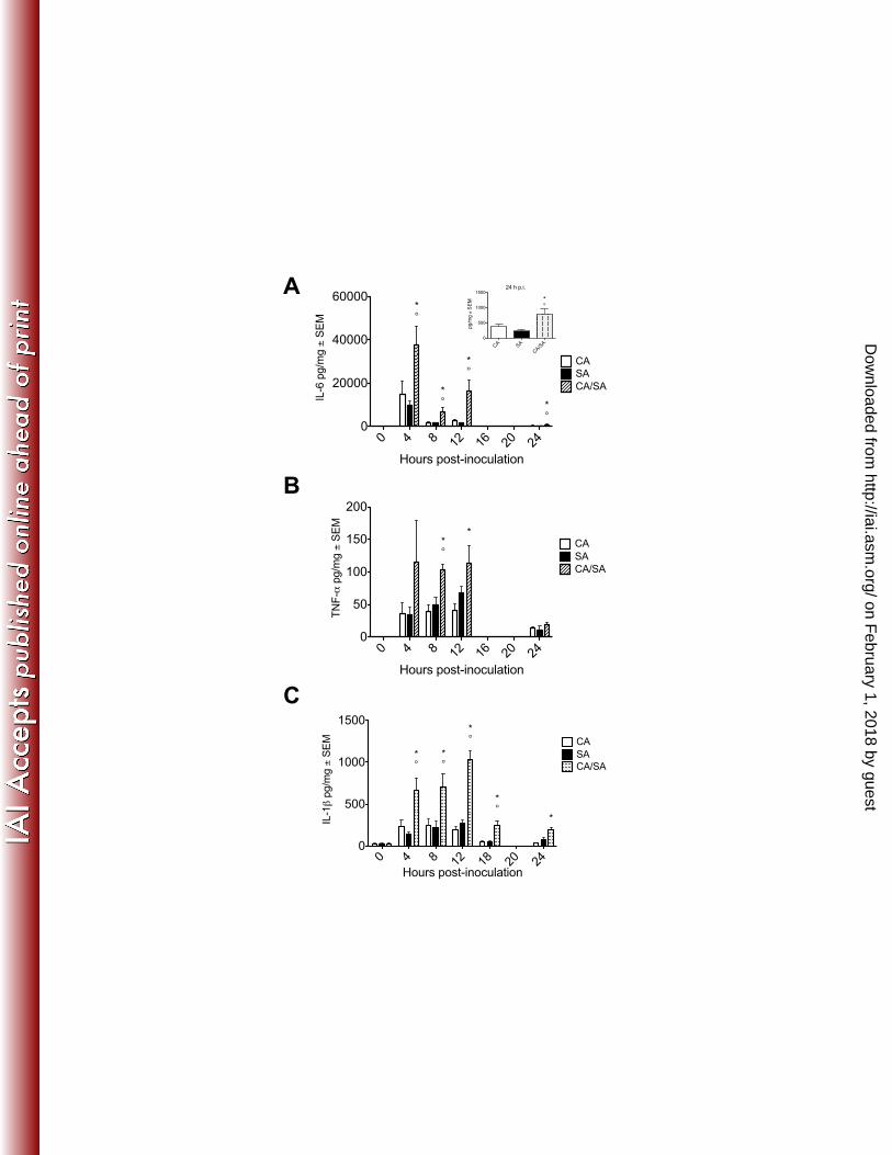
To determine whether co-infection differentially stimulates inflammation 216
despite equivalent microbial burdens, we analyzed the kinetics of inflammatory 217
cytokines produced locally in the peritoneal cavity (Figure 3). We evaluated the 218
pro-inflammatory cytokines IL-6, TNF-α and IL-1β, which have been studied in 219
other IAIs models (38). Accordingly, local production of IL-6 peaked at 4 h p.i, 220
with significantly higher levels produced during co-infection through 24 h p.i. 221
compared with either mono-infection (Figure 3A). TNF-α production peaked 222
between 4 and 12 h p.i., with significantly higher levels in co-infected mice at 223
these time points (Figure 3B). IL-1β production peaked at 12 h p.i. with dramatic 224
increases in co-infected mice through 24 h p.i. (Figure 3C). 225
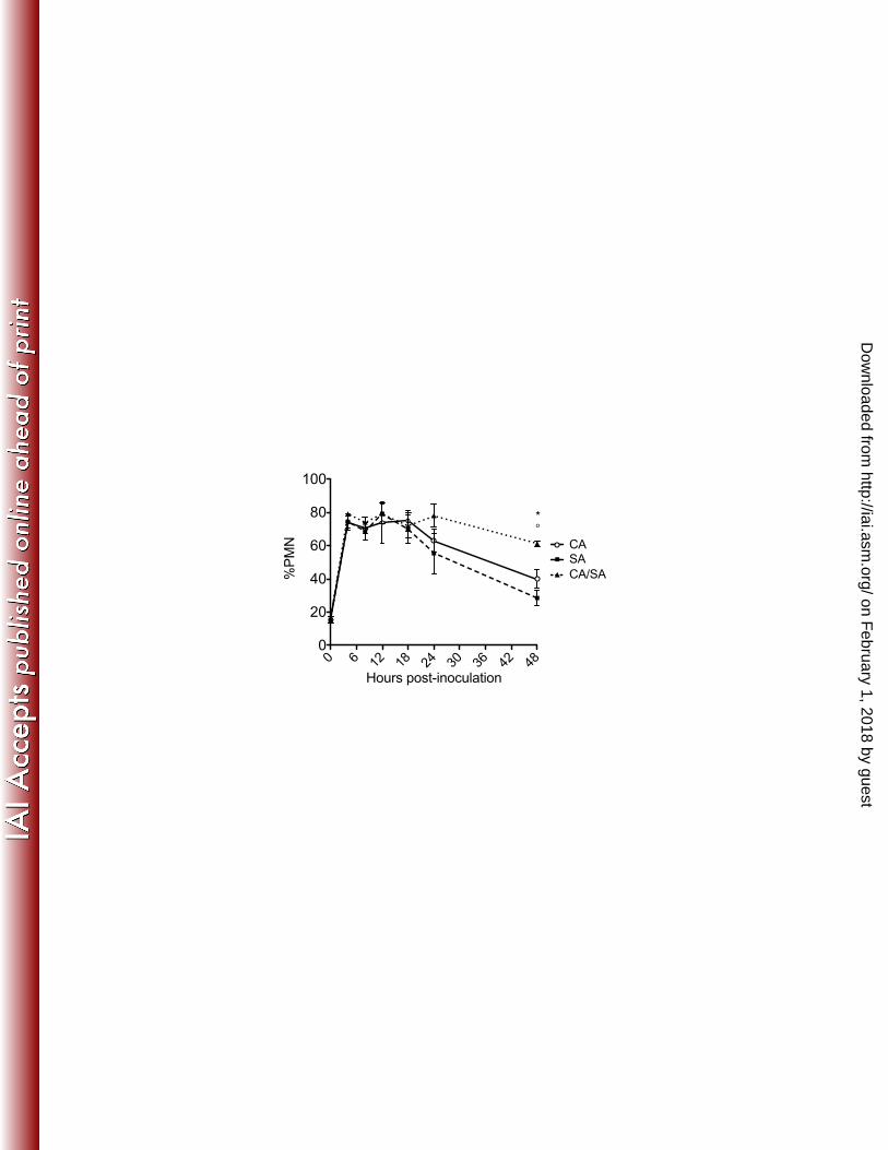
Based on our previous report showing that neutrophils are recruited to the 226
peritoneal cavity with increasing numbers during co-infection (21), it was possible 227
on February 1, 2018 by guest
http://iai.asm.org/
Dow
nloaded from
11
that the increase in pro-inflammatory cytokines observed early during co-infection 228
was due to increases in neutrophil recruitment and stimulation. However, when 229
local neutrophil infiltration was analyzed, similar percentages of neutrophils were 230
observed by 4 h p.i. in all infected groups (Figure 4). The neutrophil levels began 231
to decrease in mono-infected mice by 24 h p.i. and by 48 h in co-infected mice. 232
Absolute number of peritoneal neutrophils reflected these data (data not shown). 233
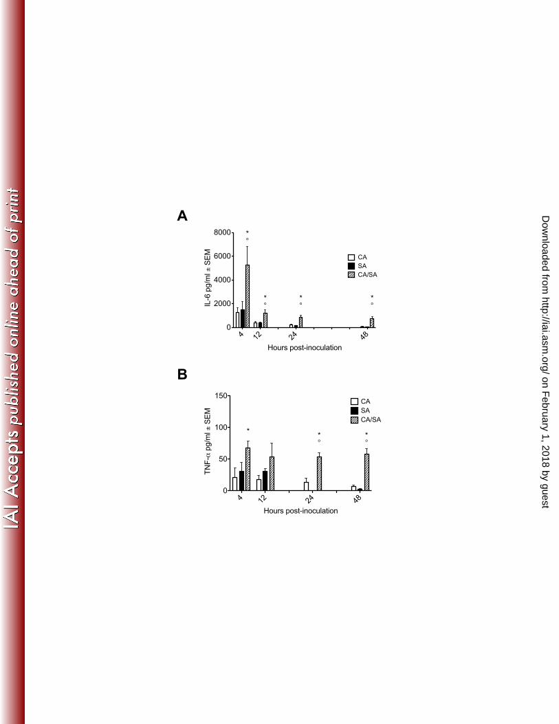
Polymicrobial infection leads to dissemination and sepsis 234
The pro-inflammatory cytokines TNF- -6, which were elevated 235
locally during co-infection, are also hallmark cytokines involved in sepsis (39, 40). 236
Therefore we analyzed whether there were also elevated levels of these 237
cytokines systemically. Serum levels of IL-6 peaked at 4 h p.i. and remained 238
elevated through 48 h with significantly higher levels produced during co-infection 239
at most time points (Figure 5A). Serum TNF-α production peaked 4 h p.i., and 240
was significantly elevated in co-infected mice from 4 - 48 h p.i. (Figure 5B). 241
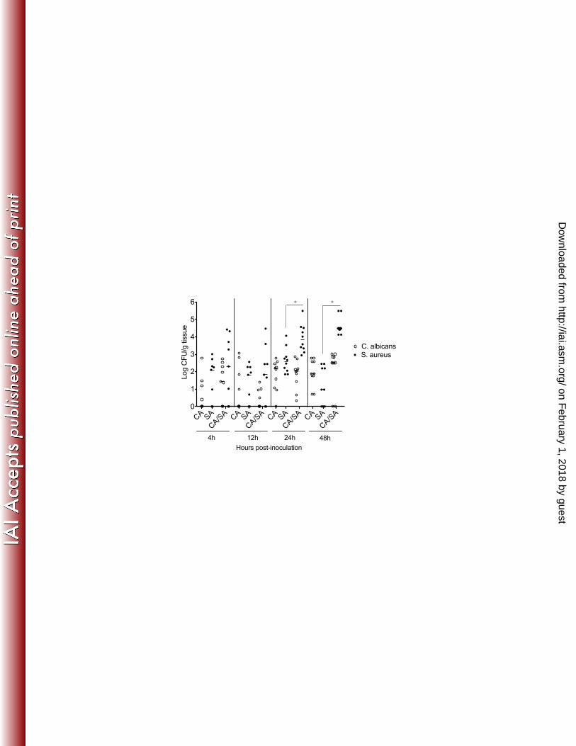
To determine the kinetics and the extent of bloodstream dissemination, we 242
examined microbial burden in the brain, a distal target organ of Candida and 243
Staphylococcal disease. Surprisingly, dissemination to the brain occurred in both 244
mono- and co-infected mice as early as 4 h p.i. (Figure 6). No differences in 245
dissemination were observed at early time points between mono- and co-infected 246
mice, similar to what was observed in the kidney and spleen. However by 24 h 247
p.i. significant increases in S. aureus CFUs were observed in co-infected mice. 248
These increases were not associated with pathology, as brain histology showed 249
on February 1, 2018 by guest
http://iai.asm.org/
Dow
nloaded from
12
no abscesses, signs of encephalitis, meningitis, or increases in immune cell 250
infiltration in mono-infected or co-infected mice (data not shown). 251
Morphogenesis does not play a role in dissemination and lethal sepsis 252
Conversion from the yeast to the hyphal form of C. albicans has been 253
shown to be required for virulence during bloodstream infection via intravenous 254
inoculation (25). In addition, the hyphal and yeast forms of C. albicans elicit 255
different responses from innate immune cells (41-43). These differing stimuli 256
may be contributing factors to the early inflammatory events and synergistic 257
pathology observed during co-infection, especially since inflammatory cytokine 258
production was independent of microbial burden. Furthermore, because S. 259
aureus primarily adheres to C. albicans hyphae, virulence factors from both 260
organisms may be modulated (20). Therefore, we sought to determine whether 261
the ability of C. albicans to form hyphae was required for virulence during intra-262
abdominal co-infection. Because the model results in only 40-60% mortality 263
during co-infection, with survivors eventually clearing the infection, data from 264
early time points include animals that will succumb to infection as well as those 265
that survive. This contributes to inherent variability in results and limits the ability 266
to determine the role of specific virulence factor during infection. Therefore, we 267
sought to optimize the model by increasing the Candida inocula to achieve 100% 268
mortality in co-infected mice by day 5, without affecting morbidity or mortality in 269
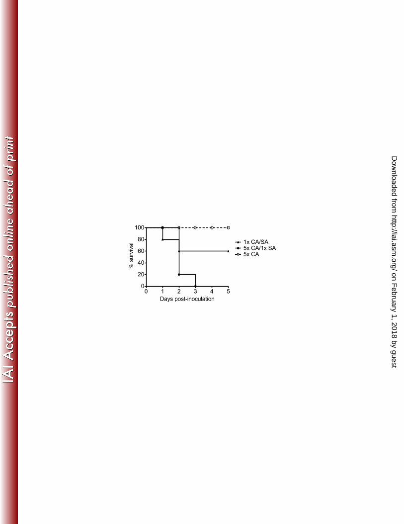
the monomicrobial-infected animals. Results in Figure 7 show that increasing the 270
C. albicans inocula 5-fold (3.5 x 10⁷ CFU), resulted in 100% mortality by day 3 271
on February 1, 2018 by guest
http://iai.asm.org/
Dow
nloaded from
13
during co-infection, and no mortality in the mono-infected animals. Therefore this 272
increased Candida inocula was utilized for subsequent studies. 273
Morphogenesis is controlled by several different transcriptional regulators 274
with overlapping function in C. albicans [reviewed in references (44), (45)]. 275
Therefore knockouts in single regulators often do not produce a uniformly strong 276
phenotype in vivo. We therefore chose to use tet-regulatable strains that remain 277
locked in either the yeast form (TNRG1) or hyphal form (TUME6) in vivo in the 278
absence of doxycycline, and the associated parental control strain (TT21) (33). 279
We visually confirmed that the TNRG1 and TUME6 strains were locked in the 280
yeast form or hyphal form respectively in vivo, and that the TT21 wildtype strain 281
formed both yeast and hyphae in vivo (data not shown) (33). As expected, no 282
mortality was observed with the wildtype strain TT21 during the monomicrobial 283
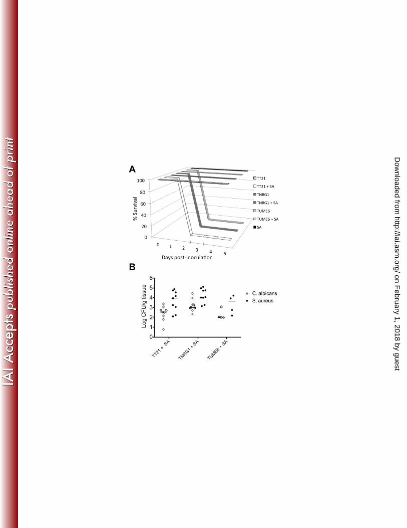
infection using the higher inocula. Surprisingly, co-infection with either the yeast-284
locked C. albicans strain TNRG1 or the hypha-locked C. albicans strain TUME6 285
at the higher inocula resulted in virtually 100% mortality (range of 80-100% 286
mortality in separate experiments) by day 2 p.i. Most mono-infected mice 287
showed no mortality, the exception was 20% mortality in mice inoculated with 288
TUME6 (Figure 8A). To confirm that this morphogenesis-independent mortality 289
was due to synergistic effects, mice were inoculated with the yeast-locked 290
TNRG1 or S. aureus alone at the total microbial burden, 1.15 x 10⁸ CFU. No 291
mortality was observed with either monomicrobial infection, confirming a 292
synergistic effect in the absence of hyphal formation (data not shown). Regarding 293
dissemination, no differences were observed in brain CFUs between wildtype 294
on February 1, 2018 by guest
http://iai.asm.org/
Dow
nloaded from
14
and the yeast-locked or hypha-locked strains during co-infection (Figure 8B). 295
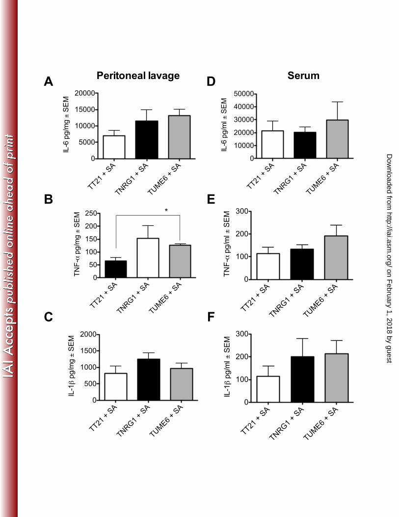
Locally in the peritoneal cavity, co-infection with TNRG1 or TUME6 resulted in 296
similar levels of pro-inflammatory cytokines as the wildtype strain (Figure 9A-C). 297
The lone exception was increased TNF-α in the TUME6 co-infected mice 298
compared to wildtype. Serum cytokines showed a similar pattern, with equivalent 299
IL-6, TNF-α, and IL-1β levels during co-infection regardless of C. albicans strain 300
(Figure 9 D-F). 301
Co-origination of disseminated infection from the abdominal cavity is 302
required to induce mortality 303
While our results indicate that co-infection with a fungal and bacterial 304
pathogen results in exacerbated stimulation of innate immune responses, leading 305
to sepsis and mortality, it remained unclear whether local vs. systemic responses 306
are more important for lethality and whether the presence of both pathogens are 307
required in the same anatomical space. To address this we asked whether 308
synergistic lethality could occur if the pathogens originated from different routes 309
of inoculation (intravenous (i.v) vs. i.p.). The LD50 for either C. albicans or S. 310
aureus is much lower for i.v. infection than i.p., limiting the ability to compare 311
effects during co-infection in our model of IAI. However, previous studies have 312
demonstrated that mice survive high dose i.v. inoculations with C. albicans yeast-313
locked strain similar to TNRG1 (34). This is not due to quick clearance of 314
infection or lack of dissemination, as similar organ burdens were observed in 315
control and yeast-locked strains. Because TNRG1 was capable of causing 316
synergistic mortality when co-infected with S. aureus, we inoculated 1 x 10⁶ 317
on February 1, 2018 by guest
http://iai.asm.org/
Dow
nloaded from
15
CFUs C. albicans TNRG1 i.v. immediately followed by 8 x 10⁷ CFUs of S. aureus 318
i.p. Surprisingly, mice appeared healthy with no morbidity or mortality observed 319
through day 10 p.i. (data not shown). S. aureus burden was confirmed in the 320
peritoneal cavity, spleen and brain of co-infected mice, but without the 321
characteristic increases shown when both organisms originated from the 322
peritoneum (Supplemental Figure 1). C. albicans burden was present in the 323
spleen and brain, but not in the peritoneal cavity (Supplemental Figure 1). 324
Finally, local and systemic cytokine production was present, but similarly not 325
enhanced during co-infection (Supplemental Figure 2). 326
DISCUSSION 327
The synergistic mortality induced during the experimental model of fungal 328
polymicrobial IAI is a striking phenotype that required further dissection to begin 329
to understand the mechanisms involved. Through previous studies, we had 330
determined that mortality induced during polymicrobial infection with C. albicans 331
and S. aureus is associated with host and microbial factors that could both 332
potentially cause damage. Thus the main objective of this study was to 333
characterize the early local and systemic inflammatory events during 334
polymicrobial IAI to further characterize and identify the host and microbial 335
factors that may contribute to mortality. 336
Overall our data support the hypothesis that the inflammatory host 337
response is primarily responsible for mortality during polymicrobial infection. This 338
is supported by equivalent fungal and bacterial burdens observed in the 339
peritoneal lavage and spleen of mono- and co-infected mice at early time points 340
on February 1, 2018 by guest
http://iai.asm.org/
Dow
nloaded from
16
prior to the onset of any morbidity and/or mortality in co-infected mice, together 341
with elevated local cytokines (IL-6, TNF-α, IL-1β) during co-infection. This is 342
further supported by previously published data (21) demonstrating that 343
inoculation of either organism alone at doses equivalent to the polymicrobial 344
inocula used does not result in mortality. 345
The cytokines detected locally during polymicrobial infection (IL-6, TNF-α, 346
IL-1β) are considered major contributors to sepsis (39, 40). Accordingly, kinetic 347
analysis of inflammatory cytokines in serum resulted in the elevated detection of 348
these septic markers early during co-infection. Heightened inflammation in the 349
periphery is also a sign of dissemination, and C. albicans and S. aureus were 350
equally present in the brains of mice during polymicrobial and monomicrobial 351
infections, surprisingly as early as 4 h p.i. Interestingly, co-infected animals 352
always developed elevated levels of S. aureus in the brain by 24 – 48 h p.i. The 353
significance is not known but may contribute to the lethal sepsis. Elevated S. 354
aureus dissemination during co-infection may be a consequence of heightened 355
peritoneal inflammation leading to peritoneal membrane damage and enhanced 356
leakage of microbes. Previous studies demonstrate that inhibiting this 357
inflammation through the use of a non-steroidal anti-inflammatory drug (NSAID) 358
during co-infection counteracts the pathological inflammation resulting in little to 359
no mortality (21, 46). This supports the concept that the exacerbated 360
inflammatory responses, which are initiated early after infection both locally and 361
systemically, are associated with mortality. This together with the present data 362
on February 1, 2018 by guest
http://iai.asm.org/
Dow
nloaded from
17
suggests that treatment for IAI should potentially include both anti-inflammatory 363
agents in conjunction with antimicrobial therapy. 364
Our polymicrobial IAI model has many similarities to other established IAI 365
models. Cecal ligation and puncture (CLP) and colon ascendens stent peritonitis 366
(CASP) are well-accepted IAI models that both involve a surgical procedure in 367
which the cecum is perforated to release the cecal contents into the normally 368
sterile peritoneal cavity. Studies using these models reported TNF-α, IL-6, and 369
IL-1β in the peritoneal lavage fluid and serum after surgery albeit with different 370
kinetics compared to our model (38, 47-51). Differences are likely attributed to 371
the continuous leakage of diverse bacterial species from the cecum of mice that 372
have undergone CLP or CASP, as compared to the bolus injection of organisms 373
used in our model. While these models are commonly used, they are limited by 374
their inability to distinguish the contributions of individual microbes to disease, do 375
not utilize a standardized inocula, and are dominated by gram-negative bacteria, 376
which predominate in the cecum (52). 377
Data from another model of fungal polymicrobial IAI was recently 378
published which involved injecting mice i.p. with sterilized mouse feces and live 379
Candida (53). This resulted in early local fungal burden and PMN infiltration to 380
the peritoneal cavity, but no mortality. This is likely because the bacterial 381
component of infection is not alive, and the sterilization process disrupts 382
pathogen associated molecular patters (PAMPs) that are necessary for 383
recognition by pattern recognition receptors (PRRs) on innate immune cells. 384
on February 1, 2018 by guest
http://iai.asm.org/
Dow
nloaded from
18
Both fungal IAI models will likely be important in dissecting mechanisms of 385
polymicrobial IAI involving fungi via alternate but complementary approaches. 386
The ability of C. albicans to switch between yeast and hypha is a well-387
established virulence factor [reviewed in references (23), (24)], as demonstrated 388
in several animal models of candidiasis (34, 54, 55). Additionally, this 389
morphological transition can modulate the host immune response, as exemplified 390
in a murine model of vulvovaginal candidiasis (33), as well as in vitro models of 391
reconstituted human or murine epithelium (56). Furthermore, S. aureus is known 392
to preferentially bind to the hyphae of C. albicans, which could affect the 393
virulence of either organism. Based on results from these models, we expected 394
C. albicans morphogenesis to be required for lethality and the amplified 395
inflammation observed during C. albicans/S. aureus polymicrobial IAI model. 396
However, surprisingly the putative avirulent yeast-locked C. albicans strain 397
resulted in equivalent mortality, inflammation, and microbial burden compared to 398
wildtype. Importantly we confirmed that the mortality using the higher inocula 399
(5x) in these experiments was due to the synergistic effect of C. albicans and S. 400
aureus during co-infection. The inoculation of either C. albicans TNRG1 or S. 401
aureus alone at the combined co-infection inoculum concentration was not lethal 402
similar to what we previously reported with the lower inocula (21). Cumulatively, 403
these results suggest that morphological transition is not required for IAI 404
sepsis/mortality. This is supported as well from results with the hypha-locked C. 405
albicans mutant that was not inherently more pathogenic in vivo. 406
on February 1, 2018 by guest
http://iai.asm.org/
Dow
nloaded from
19
The results from the morphogenesis experiments prompted other 407
important questions: 1) whether the local and systemic inflammatory events 408
induced by both organisms are sufficient to induce mortality and 2) whether 409
mortality requires the organisms to originate in the same anatomical space. We 410
reasoned that both questions could be addressed by introducing the organisms 411
via different routes (i.p./i.v.). The avirulent property of the yeast-locked C. 412
albicans given i.v. together with the virulent property of the same strain co-413
infected i.p. with S. aureus made this experimental design feasible and 414
informative. Results showing that both organisms must originate from the 415
peritoneal cavity to cause lethality suggest that local inflammation induced by 416
both organisms together in close proximity (synergistic) greatly contributes to the 417
pathogenic response. Microbial burden in the spleen supports this conclusion. 418
Although both organisms are present in the spleen during co-infection, they are 419
likely not occupying the same anatomical space in the spleen (C. albicans 420
internal, S. aureus external), and thus are not co-stimulating innate immune cells 421
to induce pathological inflammation. This leads to yet another question, whether 422
a strict physical interaction between C. albicans and S. aureus is required. 423
Previous studies from our laboratory and others have demonstrated the presence 424
of polymicrobial biofilm-like structures on target organs using this IAI model (21), 425
and several staphylococcal and fungal virulence factors are modulated during co-426
culture in vitro (20). Therefore, physical interactions between C. albicans and S. 427
aureus may modulate the degree of host immune activation. Given the fact that 428
S. aureus preferentially binds to C. albicans hyphae (20), together with the yeast-429
on February 1, 2018 by guest
http://iai.asm.org/
Dow
nloaded from
20
locked strain resulting in similar lethality during co-infection, it will be interesting 430
to determine if and what type of interactions (physical and/or chemical) occur 431
between the two C. albicans morphotypes and S. aureus during the co-infection. 432
While we have clearly demonstrated that C. albicans and S. aureus co-433
infections in the peritoneal cavity lead to synergistic mortality, we have not 434
considered other gastrointestinal bacteria or fungi that could co-infect with 435
Candida during clinical infections which also may result in mortality. Peritoneal 436
infections with C. albicans and Escherichia coli have been shown to be lethal in 437
mice (57), thus to broaden the use of this model, studies are also underway to 438
identify combinations of enteric bacteria and pathogenic Candida species that 439
result in synergistic lethality. Overall, the ability of C. albicans and S. aureus to 440
amplify the host inflammatory response to a point of sepsis and death without 441
requiring morphological transition, stresses the need to fully dissect the 442
pathogenesis during polymicrobial IAI involving fungi, which will lead to better 443
diagnosis and targeted therapeutics. Accordingly, the model will also be used to 444
further determine the role of fungal and bacterial PAMPs using killed organisms, 445
define inflammatory response through inhibition of PRR signaling on innate 446
immune cells, and to evaluate the role of the inflammasome as well as other 447
inflammatory mechanisms via genetic or pharmacologic inhibition/manipulation. 448
449
ACKNOWLEDGEMENTS 450
We kindly thank Dr. Aaron Mitchell (Carnegie Melon University) for providing C. 451
albicans strain DAY185 and the NARSA repository for S. aureus strain NRS383 452
on February 1, 2018 by guest
http://iai.asm.org/
Dow
nloaded from
21
used in this study. We also thank H. Nakayama and M. Arisawa (Nippon Roche) 453
for providing the materials required for the C. albicans tetracycline-regulatable 454
system. This study was funded by the National Institutes of Health, National 455
Institute of Allergy ad Infectious Disease grant R01-A172406. 456
457
REFERENCES 458
1. Santos SG, Serufo JC, Silva RA, Marra BA, Reis CM, Hamdan JS, Nicoli JR, 459
Carvalho MA, Farias LM. 2003. Microbiologic profile of intra-abdominal 460
infections at Belo Horizonte, Brazil. American journal of infection control 461
31:135-143. 462
2. Saini S, Gupta N, Aparna, Lokveer, Griwan MS. 2004. Surgical infections: a 463
microbiological study. The Brazilian journal of infectious diseases : an official 464
publication of the Brazilian Society of Infectious Diseases 8:118-125. 465
3. Heemken R, Gandawidjaja L, Hau T. 1997. Peritonitis: pathophysiology and 466
local defense mechanisms. Hepato-gastroenterology 44:927-936. 467
4. Cahill RA, Wang JH, Redmond HP. 2007. Enteric bacteria and their antigens 468
may stimulate postoperative peritoneal adhesion formation. Surgery 469
141:403-410. 470
5. Karantonis FF, Nikiteas N, Perrea D, Vlachou A, Giamarellos-Bourboulis 471
EJ, Tsigris C, Kostakis A. 2008. Evaluation of the effects of laparotomy and 472
laparoscopy on the immune system in intra-abdominal sepsis--A Review. 473
Journal of investigative surgery : the official journal of the Academy of 474
Surgical Research 21:330-339. 475
on February 1, 2018 by guest
http://iai.asm.org/
Dow
nloaded from
22
6. Ozmen MM, Col C, Aksoy AM, Tekeli FA, Berberoglu M. 1999. Effect of 476
CO(2) insufflation on bacteremia and bacterial translocation in an animal 477
model of peritonitis. Surgical endoscopy 13:801-803. 478
7. Dupont H, Paugam-Burtz C, Muller-Serieys C, Fierobe L, Chosidow D, 479
Marmuse JP, Mantz J, Desmonts JM. 2002. Predictive factors of mortality 480
due to polymicrobial peritonitis with Candida isolation in peritoneal fluid in 481
critically ill patients. Arch Surg 137:1341-1346; discussion 1347. 482
8. Montravers P, Gauzit R, Muller C, Marmuse JP, Fichelle A, Desmonts JM. 483
1996. Emergence of antibiotic-resistant bacteria in cases of peritonitis after 484
intraabdominal surgery affects the efficacy of empirical antimicrobial 485
therapy. Clinical infectious diseases : an official publication of the Infectious 486
Diseases Society of America 23:486-494. 487
9. Calandra T, Bille J, Schneider R, Mosimann F, Francioli P. 1989. Clinical 488
significance of Candida isolated from peritoneum in surgical patients. Lancet 489
2:1437-1440. 490
10. Blot SI, Vandewoude KH, De Waele JJ. 2007. Candida Peritonitis. Current 491
opinion in critical care 13:195-199. 492
11. de Ruiter J, Weel J, Manusama E, Kingma WP, van der Voort PH. 2009. 493
The epidemiology of intra-abdominal flora in critically ill patients with 494
secondary and tertiary abdominal sepsis. Infection 37:522-527. 495
12. Hasibeder W, Halabi M. 2014. Candida peritonitis. Minerva anestesiologica 496
80:470-481. 497
on February 1, 2018 by guest
http://iai.asm.org/
Dow
nloaded from
23
13. Miles R, Hawley CM, McDonald SP, Brown FG, Rosman JB, Wiggins KJ, 498
Bannister KM, Johnson DW. 2009. Predictors and outcomes of fungal 499
peritonitis in peritoneal dialysis patients. Kidney international 76:622-628. 500
14. Benedetti E, Gruessner AC, Troppmann C, Papalois BE, Sutherland DE, 501
Dunn DL, Gruessner RW. 1996. Intra-abdominal fungal infections after 502
pancreatic transplantation: incidence, treatment, and outcome. Journal of the 503
American College of Surgeons 183:307-316. 504
15. Govindarajulu S, Hawley CM, McDonald SP, Brown FG, Rosman JB, 505
Wiggins KJ, Bannister KM, Johnson DW. 2010. Staphylococcus aureus 506
peritonitis in Australian peritoneal dialysis patients: predictors, treatment, 507
and outcomes in 503 cases. Peritoneal dialysis international : journal of the 508
International Society for Peritoneal Dialysis 30:311-319. 509
16. Peters BM, Jabra-Rizk MA, O'May GA, Costerton JW, Shirtliff ME. 2012. 510
Polymicrobial interactions: impact on pathogenesis and human disease. 511
Clinical microbiology reviews 25:193-213. 512
17. Shirtliff ME, Peters BM, Jabra-Rizk MA. 2009. Cross-kingdom interactions: 513
Candida albicans and bacteria. FEMS microbiology letters 299:1-8. 514
18. Bender FH, Bernardini J, Piraino B. 2006. Prevention of infectious 515
complications in peritoneal dialysis: best demonstrated practices. Kidney 516
international. Supplement:S44-54. 517
19. Harriott MM, Noverr MC. 2009. Candida albicans and Staphylococcus aureus 518
form polymicrobial biofilms: effects on antimicrobial resistance. 519
Antimicrobial agents and chemotherapy 53:3914-3922. 520
on February 1, 2018 by guest
http://iai.asm.org/
Dow
nloaded from
24
20. Peters BM, Jabra-Rizk MA, Scheper MA, Leid JG, Costerton JW, Shirtliff 521
ME. 2010. Microbial interactions and differential protein expression in 522
Staphylococcus aureus -Candida albicans dual-species biofilms. FEMS 523
immunology and medical microbiology 59:493-503. 524
21. Peters BM, Noverr MC. 2013. Candida albicans-Staphylococcus aureus 525
polymicrobial peritonitis modulates host innate immunity. Infection and 526
immunity 81:2178-2189. 527
22. Carlson E. 1982. Synergistic effect of Candida albicans and Staphylococcus 528
aureus on mouse mortality. Infection and immunity 38:921-924. 529
23. Mayer FL, Wilson D, Hube B. 2013. Candida albicans pathogenicity 530
mechanisms. Virulence 4:119-128. 531
24. Kumamoto CA, Vinces MD. 2005. Contributions of hyphae and hypha-co-532
regulated genes to Candida albicans virulence. Cellular microbiology 7:1546-533
1554. 534
25. Saville SP, Lazzell AL, Bryant AP, Fretzen A, Monreal A, Solberg EO, 535
Monteagudo C, Lopez-Ribot JL, Milne GT. 2006. Inhibition of filamentation 536
can be used to treat disseminated candidiasis. Antimicrobial agents and 537
chemotherapy 50:3312-3316. 538
26. Jacobsen ID, Wilson D, Wachtler B, Brunke S, Naglik JR, Hube B. 2012. 539
Candida albicans dimorphism as a therapeutic target. Expert review of anti-540
infective therapy 10:85-93. 541
27. Zakikhany K, Naglik JR, Schmidt-Westhausen A, Holland G, Schaller M, 542
Hube B. 2007. In vivo transcript profiling of Candida albicans identifies a 543
on February 1, 2018 by guest
http://iai.asm.org/
Dow
nloaded from
25
gene essential for interepithelial dissemination. Cellular microbiology 544
9:2938-2954. 545
28. Dalle F, Wachtler B, L'Ollivier C, Holland G, Bannert N, Wilson D, 546
Labruere C, Bonnin A, Hube B. 2010. Cellular interactions of Candida 547
albicans with human oral epithelial cells and enterocytes. Cellular 548
microbiology 12:248-271. 549
29. Wachtler B, Wilson D, Haedicke K, Dalle F, Hube B. 2011. From 550
attachment to damage: defined genes of Candida albicans mediate adhesion, 551
invasion and damage during interaction with oral epithelial cells. PloS one 552
6:e17046. 553
30. Peters BM, Ovchinnikova ES, Krom BP, Schlecht LM, Zhou H, Hoyer LL, 554
Busscher HJ, van der Mei HC, Jabra-Rizk MA, Shirtliff ME. 2012. 555
Staphylococcus aureus adherence to Candida albicans hyphae is mediated by 556
the hyphal adhesin Als3p. Microbiology (Reading, England) 158:2975-2986. 557
31. Davis D, Edwards JE, Jr., Mitchell AP, Ibrahim AS. 2000. Candida albicans 558
RIM101 pH response pathway is required for host-pathogen interactions. 559
Infection and immunity 68:5953-5959. 560
32. Johnston DA, Tapia AL, Eberle KE, Palmer GE. 2013. Three prevacuolar 561
compartment Rab GTPases impact Candida albicans hyphal growth. 562
Eukaryotic cell 12:1039-1050. 563
33. Peters BM, Palmer GE, Nash AK, Lilly EA, Fidel PL, Jr., Noverr MC. 2014. 564
Fungal Morphogenetic Pathways Are Required for the Hallmark 565
on February 1, 2018 by guest
http://iai.asm.org/
Dow
nloaded from
26
Inflammatory Response during Candida albicans Vaginitis. Infection and 566
immunity 82:532-543. 567
34. Saville SP, Lazzell AL, Monteagudo C, Lopez-Ribot JL. 2003. Engineered 568
control of cell morphology in vivo reveals distinct roles for yeast and 569
filamentous forms of Candida albicans during infection. Eukaryotic cell 570
2:1053-1060. 571
35. Nakayama H, Mio T, Nagahashi S, Kokado M, Arisawa M, Aoki Y. 2000. 572
Tetracycline-regulatable system to tightly control gene expression in the 573
pathogenic fungus Candida albicans. Infection and immunity 68:6712-6719. 574
36. Banerjee M, Thompson DS, Lazzell A, Carlisle PL, Pierce C, Monteagudo 575
C, Lopez-Ribot JL, Kadosh D. 2008. UME6, a novel filament-specific 576
regulator of Candida albicans hyphal extension and virulence. Molecular 577
biology of the cell 19:1354-1365. 578
37. Carlisle PL, Kadosh D. 2010. Candida albicans Ume6, a filament-specific 579
transcriptional regulator, directs hyphal growth via a pathway involving 580
Hgc1 cyclin-related protein. Eukaryotic cell 9:1320-1328. 581
38. Ebong S, Call D, Nemzek J, Bolgos G, Newcomb D, Remick D. 1999. 582
Immunopathologic alterations in murine models of sepsis of increasing 583
severity. Infection and immunity 67:6603-6610. 584
39. Faix JD. 2013. Biomarkers of sepsis. Critical reviews in clinical laboratory 585
sciences 50:23-36. 586
on February 1, 2018 by guest
http://iai.asm.org/
Dow
nloaded from
27
40. Bozza FA, Salluh JI, Japiassu AM, Soares M, Assis EF, Gomes RN, Bozza 587
MT, Castro-Faria-Neto HC, Bozza PT. 2007. Cytokine profiles as markers of 588
disease severity in sepsis: a multiplex analysis. Crit Care 11:R49. 589
41. Blasi E, Pitzurra L, Puliti M, Lanfrancone L, Bistoni F. 1992. Early 590
differential molecular response of a macrophage cell line to yeast and hyphal 591
forms of Candida albicans. Infection and immunity 60:832-837. 592
42. Moyes DL, Runglall M, Murciano C, Shen C, Nayar D, Thavaraj S, Kohli A, 593
Islam A, Mora-Montes H, Challacombe SJ, Naglik JR. 2010. A biphasic 594
innate immune MAPK response discriminates between the yeast and hyphal 595
forms of Candida albicans in epithelial cells. Cell host & microbe 8:225-235. 596
43. d'Ostiani CF, Del Sero G, Bacci A, Montagnoli C, Spreca A, Mencacci A, 597
Ricciardi-Castagnoli P, Romani L. 2000. Dendritic cells discriminate 598
between yeasts and hyphae of the fungus Candida albicans. Implications for 599
initiation of T helper cell immunity in vitro and in vivo. The Journal of 600
experimental medicine 191:1661-1674. 601
44. Ernst JF. 2000. Transcription factors in Candida albicans - environmental 602
control of morphogenesis. Microbiology (Reading, England) 146 ( Pt 603
8):1763-1774. 604
45. Dhillon NK, Sharma S, Khuller GK. 2003. Signaling through protein kinases 605
and transcriptional regulators in Candida albicans. Critical reviews in 606
microbiology 29:259-275. 607
46. Carlson EC. 1988. Synergism of Candida albicans and delta toxin producing 608
Staphylococcus aureus on mouse mortality and morbidity: protection by 609
on February 1, 2018 by guest
http://iai.asm.org/
Dow
nloaded from
28
indomethacin. Zentralblatt fur Bakteriologie, Mikrobiologie, und Hygiene. 610
Series A, Medical microbiology, infectious diseases, virology, parasitology 611
269:377-386. 612
47. Lundblad R, Sandven P, Giercksky KE. 1995. The physical nature of a large 613
bowel perforation predicts severity of the subsequent inflammatory 614
response. Shock 3:455-461. 615
48. Villa P, Sartor G, Angelini M, Sironi M, Conni M, Gnocchi P, Isetta AM, 616
Grau G, Buurman W, van Tits LJ, et al. 1995. Pattern of cytokines and 617
pharmacomodulation in sepsis induced by cecal ligation and puncture 618
compared with that induced by endotoxin. Clinical and diagnostic laboratory 619
immunology 2:549-553. 620
49. Lustig MK, Bac VH, Pavlovic D, Maier S, Grundling M, Grisk O, Wendt M, 621
Heidecke CD, Lehmann C. 2007. Colon ascendens stent peritonitis--a model 622
of sepsis adopted to the rat: physiological, microcirculatory and laboratory 623
changes. Shock 28:59-64. 624
50. Maier S, Traeger T, Entleutner M, Westerholt A, Kleist B, Huser N, 625
Holzmann B, Stier A, Pfeffer K, Heidecke CD. 2004. Cecal ligation and 626
puncture versus colon ascendens stent peritonitis: two distinct animal 627
models for polymicrobial sepsis. Shock 21:505-511. 628
51. Busse M, Traeger T, Potschke C, Billing A, Dummer A, Friebe E, Kiank C, 629
Grunwald U, Jack RS, Schutt C, Heidecke CD, Maier S, Broker BM. 2008. 630
Detrimental role for CD4+ T lymphocytes in murine diffuse peritonitis due to 631
inhibition of local bacterial elimination. Gut 57:188-195. 632
on February 1, 2018 by guest
http://iai.asm.org/
Dow
nloaded from
29
52. Hyde SR, Stith RD, McCallum RE. 1990. Mortality and bacteriology of sepsis 633
following cecal ligation and puncture in aged mice. Infection and immunity 634
58:619-624. 635
53. Cheng S, Clancy CJ, Xu W, Schneider F, Hao B, Mitchell AP, Nguyen MH. 636
2013. Profiling of Candida albicans gene expression during intra-abdominal 637
candidiasis identifies biologic processes involved in pathogenesis. The 638
Journal of infectious diseases 208:1529-1537. 639
54. Martin MV, Craig GT, Lamb DJ. 1984. An investigation of the role of true 640
hypha production in the pathogenesis of experimental oral candidosis. 641
Sabouraudia 22:471-476. 642
55. Ray TL, Payne CD. 1988. Scanning electron microscopy of epidermal 643
adherence and cavitation in murine candidiasis: a role for Candida acid 644
proteinase. Infection and immunity 56:1942-1949. 645
56. Moyes DL, Shen C, Murciano C, Runglall M, Richardson JP, Arno M, 646
Aldecoa-Otalora E, Naglik JR. 2014. Protection Against Epithelial Damage 647
During Candida albicans Infection Is Mediated by PI3K/Akt and Mammalian 648
Target of Rapamycin Signaling. The Journal of infectious diseases. 649
57. Klaerner HG, Uknis ME, Acton RD, Dahlberg PS, Carlone-Jambor C, Dunn 650
DL. 1997. Candida albicans and Escherichia coli are synergistic pathogens 651
during experimental microbial peritonitis. The Journal of surgical research 652
70:161-165. 653
654
on February 1, 2018 by guest
http://iai.asm.org/
Dow
nloaded from
30
FIGURE LEGENDS 655
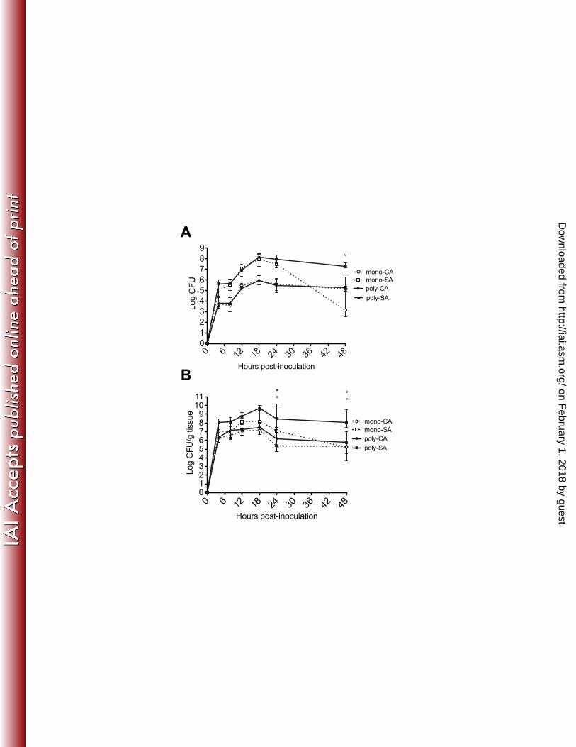
Figure 1. Microbial burden in monomicrobial and polymicrobial peritoneal 656
infection with C. albicans and S. aureus 657
Mice (n=5 mice/group) were injected i.p. with 0.2 mL of 3.5 x 10⁷ CFU/mL C. 658
albicans alone (7 x 10⁶ CFU), 0.2 mL of 4 x 10⁸ CFU/mL S. aureus alone (8 x 10⁷ 659
CFU), or 0.2 mL containing 3.5 x 10⁷ CFU/mL C. albicans and 4 x 10⁸ CFU/mL S. 660
aureus (8.7 x 10⁷ total organism). At various time points post-inoculation, mice 661
were sacrificed to quantitate C. albicans (mono, open circles vs. poly, closed 662
circles) and S. aureus (mono, open square vs. poly, closed square) in the (A) 663
peritoneal lavage fluid and (B) spleen. Results are expressed as the median CFU 664
± interquartile range. Data shown are cumulative of two repeat experiments. 665
Mono and poly infection groups were analyzed using the Mann-Whitney U test; * 666
p < 0.05 for CA vs. CA/SA, ° p < 0.05 for SA vs. CA/SA. 667
668
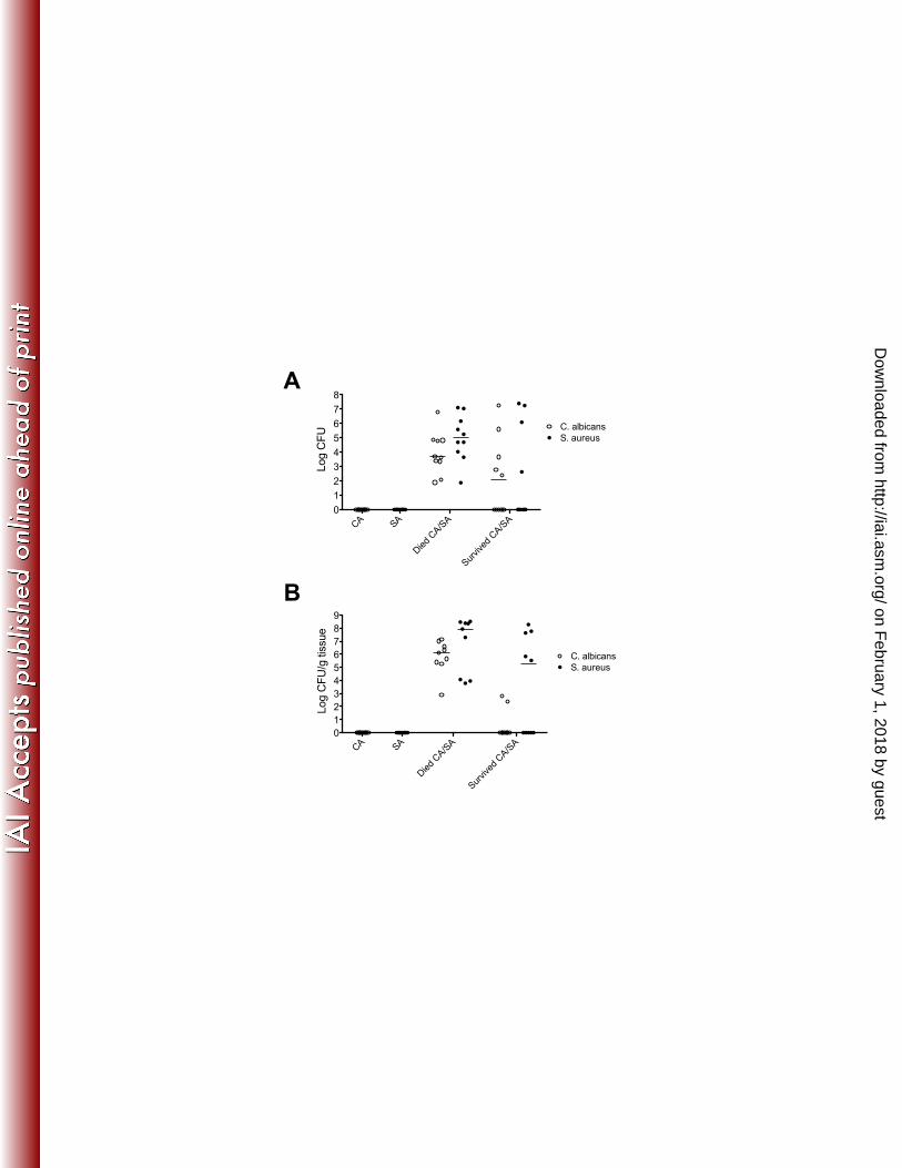
Figure 2. Microbial burden in dead vs. surviving co-infected mice 669
Mice were inoculated i.p. with C. albicans and S. aureus as described in Figure 670
1. Mice (n=5-10 mice/group) were sacrificed when moribund or after 5 days 671
post-inoculation (p.i.) to assessed fungal and bacterial CFUs in (A) peritoneal 672
lavage and (B) spleen (C. albicans, open circles; S. aureus, closed circles). 673
Results are expressed as the median CFU. Data shown are cumulative of three 674
repeat experiments. 675
676
on February 1, 2018 by guest
http://iai.asm.org/
Dow
nloaded from
31
Figure 3. Local pro-inflammatory cytokines during polymicrobial peritoneal 677
infection with C. albicans and S. aureus 678
Mice (n=5 mice/group) were inoculated i.p. with C. albicans and S. aureus as 679
described in Figure 1. At various time points post-inoculation mice were 680
sacrificed and the peritoneal cavity was lavaged. (A) IL-6, with 24 h p.i. insert, (B) 681
TNF-α, and (C) IL-1β concentrations were quantified in peritoneal lavage fluid by 682
ELISA from C. albicans infected mice (white bar), S. aureus infected mice (black 683
bar), and co-infected mice (diagonal striped bar). Results are expressed as the 684
mean cytokine level ± SEM. Data shown are cumulative of two repeat 685
experiments. Data were analyzed using the unpaired Student’s t test; * p< 0.05 686
for CA vs. CA/SA, ° p< 0.05 for SA vs. CA/SA. 687
688
Figure 4. PMN infiltration in monomicrobial and polymicrobial peritoneal 689
infection with C. albicans and S. aureus 690
Mice (n=5 mice/group) were inoculated i.p. with C. albicans and S. aureus as 691
described in Figure 1. At various time points post-inoculation, peritoneal lavage 692
fluid from mice infected with C. albicans alone (circle), S. aureus alone (square), 693
or both pathogens (triangle) was cytospun and stained with H&E. PMNs and 694
total cells from each infection group were counted in 5 nonadjacent fields and the 695
percentage of PMNs was calculated. Results are expressed as the mean %PMN 696
± SEM. Data shown are cumulative of two repeat experiments. Data were 697
analyzed using the unpaired Student’s t test; * p< 0.05 for CA vs. CA/SA; ° p< 698
0.05 for SA vs. CA/SA. 699
on February 1, 2018 by guest
http://iai.asm.org/
Dow
nloaded from
32
700
Figure 5. Systemic pro-inflammatory cytokines in polymicrobial peritoneal 701
infection with C. albicans and S. aureus 702
Mice (n=5 mice/group) were inoculated i.p. with C. albicans and S. aureus as 703
described in Figure 1. At various time points post-inoculation mice were 704
sacrificed, and serum was analyzed for (A) IL-6 and (B) TNF-α by ELISA. 705
Cytokine concentrations were analyzed in mice infected with C. albicans alone 706
(white bar), S. aureus alone (black bar), and both pathogens combined (striped 707
bar). Results are expressed as the mean cytokine level ± SEM. Data shown are 708
cumulative of two repeat experiments. Data were analyzed using the unpaired 709
Student’s t test; * p< 0.05 for CA vs. CA/SA, ° p< 0.05 for SA vs. CA/SA. 710
711
Figure 6. Dissemination of microbial burden in monomicrobial and 712
polymicrobial peritoneal infections with C. albicans and S. aureus 713
Mice (n=5 mice/group) were inoculated i.p. with C. albicans and S. aureus as 714
described in Figure 1. At various time points post-inoculation mice were 715
sacrificed, and the microbial burden in the brain was assessed in mice with 716
monomicrobial and polymicrobial infections (C. albicans, open circles; S. aureus, 717
closed circles). Results are expressed as the median CFU. Data shown are 718
cumulative of two repeat experiments. Data were analyzed using the Mann-719
Whitney U test; * p < 0.05 for CA vs. CA/SA, ° p < 0.05 for SA vs. CA/SA. 720
721
on February 1, 2018 by guest
http://iai.asm.org/
Dow
nloaded from
33
Figure 7. Effects of increased C. albicans inocula on mortality during co-722
infection 723
Mice (n=5 mice/group) were inoculated i.p. with the standard inocula of 7 x 10⁶ C. 724
albicans and 8 x 10⁷ S. aureus (1x CA/SA, triangle), 3.5 x 10⁷ C. albicans and 8 x 725
107 S. aureus (5x CA/1x SA, closed circle), or with C. albicans alone (5x CA, 726
open circle). Mice were monitored for morbidity/mortality through 5 days p.i. 727
Results are from one experiment with all groups inclusive. 728
729
Figure 8. C. albicans morphogenesis is not required for dissemination or 730
synergistic mortality. 731
Mice (n=4 or 5 mice/group) were inoculated with S. aureus and either C. albicans 732
strain TNRG1, TUME6, or the isogenic control strain TT21 using 5x inocula as 733
described in Figure 7. (A) Survival of mice co-infected with S. aureus and 734
TNRG1 using 5x inocula (dark gray line) or S. aureus and TUME6 using 5x 735
inocula (light gray line) or S. aureus and isogenic control using 5x inocula (white 736
line). Analysis was performed using Kaplan-Meier test (p <0.05). (B) At 24 h 737
post-inoculation, brains from co-infected mice were assessed for fungal and 738
bacterial burden (C. albicans, open circle; S. aureus, closed circle). Results are 739
expressed as the median CFU. Data shown are cumulative of two repeat 740
experiments, except TUME6 and TUME6 + SA inoculation, which was performed 741
once. Data were analyzed using the Mann-Whitney U test; * p < 0.05 for CA vs. 742
CA/SA, ° p < 0.05 for SA vs. CA/SA. 743
744
on February 1, 2018 by guest
http://iai.asm.org/
Dow
nloaded from
34
Figure 9. C. albicans morphogenesis is not required for inducing local and 745
systemic inflammation. 746
Mice (n=5 mice/group) were inoculated with S. aureus and either C. albicans 747
strain TNRG1, TUME6, or isogenic control strain TT21 at the 5x inocula as 748
described in Figure 7. At 24h p.i. (A-C) peritoneal lavage fluid and (D-F) serum 749
was collected and analyze for (A, D) IL-6, (B, E) TNF-α, and (C, F) IL-1β. 750
Results are expressed as the mean cytokine level ± SEM. Data shown are 751
cumulative of two repeat experiments, except TUME6 and TUME6 + SA 752
inoculation, which was performed once. Data were analyzed using the unpaired 753
Student’s t test; * p< 0.05. 754
on February 1, 2018 by guest
http://iai.asm.org/
Dow
nloaded from
0 612
18
24
30
36
42
48
0
1
2
3
4
5
6
7
8
9
Hours post-inoculation
Log CFU
mono-CA
mono-SA
poly-CA
poly-SA
°
0 612182430364248
01234567891011
Hours post-inoculation
Log CFU/g tissue
mono-CA
mono-SA
poly-CA
poly-SA
*°
*°
A
B on February 1, 2018 by guest
http://iai.asm.org/
Dow
nloaded from
CA SA
Died CA/SA
Survived CA/SA
0
1
2
3
4
5
6
7
8
Log CFU
C. albicans
S. aureus
CA SA
Died CA/SA
Survived CA/SA
0
1
2
3
4
5
6
7
8
9
Log CFU/g tissue
C. albicans
S. aureus
A
B on February 1, 2018 by guest
http://iai.asm.org/
Dow
nloaded from
0 4 8 12 16 20 240
20000
40000
60000
Hours post-inoculation
IL-6 pg/mg ± SEM
CA
SA
CA/SA
*°
*°
*°
*°
0 4 8 12 16 20 240
50
100
150
200
Hours post-inoculation
TNF-α pg/mg ± SEM
CA
SA
CA/SA
**°
0 4 8 12 18 20 240
500
1000
1500
Hours post-inoculation
IL-1β pg/mg ± SEM CA
SA
CA/SA
*°
*°
*°
*
*°
24 h p.i.
pg/mg ± SEM
CA
SA
CA/SA
0
500
1000
1500
*°
A
B
C
on February 1, 2018 by guest
http://iai.asm.org/
Dow
nloaded from
0 612
18
24
30
36
42
48
0
20
40
60
80
100
Hours post-inoculation
%PMN CA
SA
CA/SA
*
°
on February 1, 2018 by guest
http://iai.asm.org/
Dow
nloaded from
4 12 24 480
2000
4000
6000
8000
IL-6 pg/ml ± SEM
Hours post-inoculation
CA
SA
CA/SA
*°
*°
*°
*°
4 12 24 480
50
100
150
Hours post-inoculation
TNF-α pg/ml ± SEM
CA
SA
CA/SA
**°
*°
A
B on February 1, 2018 by guest
http://iai.asm.org/
Dow
nloaded from
CASA
CA/SACASA
CA/SACASA
CA/SACASA
CA/SA
0
1
2
3
4
5
6
Log CFU/g tissue
C. albicans
S. aureus
4h 12h 24h 48h
°
Hours post-inoculation
°
on February 1, 2018 by guest
http://iai.asm.org/
Dow
nloaded from
0 1 2 3 4 50
20
40
60
80
100
Days post-inoculation
% survival
5x CA/1x SA5x CA
1x CA/SA
on February 1, 2018 by guest
http://iai.asm.org/
Dow
nloaded from
!"
#!"
$!"
%!"
&!"
'!!"
!" '" #" (" $" )"
*"+,-./.01"
2034"56478/96:,10;69"
<<#'"
<<#'"="+>"
<?@A'"
<?@A'"="+>"
<BCD%"
<BCD%"="+>"
+>"
TT21 + SA
TNRG1 + SA
TUME6 + SA
0
1
2
3
4
5
6
Log CFU/g tissue
C. albicans
S. aureus
A
B on February 1, 2018 by guest
http://iai.asm.org/
Dow
nloaded from
TT21 + SA
TNRG1 + SA
TUME6 + SA
0
5000
10000
15000
20000
IL-6 pg/mg ± SEM
TT21 + SA
TNRG1 + SA
TUME6 + SA
0
50
100
150
200
250
TNF-α pg/mg ± SEM *
TT21 + SA
TNRG1 + SA
TUME6 + SA
0
500
1000
1500
2000
IL-1β pg/mg ± SEM
TT21 + SA
TNRG1 + SA
TUME6 + SA
0
10000
20000
30000
40000
50000
IL-6 pg/ml ± SEM
TT21 + SA
TNRG1 + SA
TUME6 + SA
0
100
200
300
TNF-α pg/ml ± SEM
TT21 + SA
TNRG1 + SA
TUME6 + SA
0
100
200
300
IL-1β pg/ml ± SEM
A
B
C
D
E
F
Peritoneal lavage Serum
on February 1, 2018 by guest
http://iai.asm.org/
Dow
nloaded from