Embed Size (px)
Citation preview
Evidence-Based Review of Moderate to Severe Acquired Brain Injury 2017
1 Module 11-Heterotopic Ossification and Venous Thromboembolism post ABI- V11 http://www.abiebr.com Updated August 2016
11. Heterotopic Ossification and Venous
Thromboembolism
Robert Teasell MD FRCPC, Shannon Janzen MSc, Rachel Anderson BSc, Caitlin Cassidy MD, Shawn Marshall MD FRCPC, Nora Cullen MD FRCPC
ERABI
Parkwood Institute
550 Wellington Road, London ON
Evidence-Based Review of Moderate to Severe Acquired Brain Injury 2017
2 Module 11-Heterotopic Ossification and Venous Thromboembolism post ABI- V11 http://www.abiebr.com Updated August 2016
Table of Contents
11.1 Formation of Heterotopic Ossification Post-Head Injury ................................................................. 8
11.1.1 Pathophysiology of Heterotopic Ossification Post-Head Injury ............................................... 8
11.1.2 Stimulating Factors Related to Head Injury ............................................................................. 8
11.2 Clinical Presentation of Heterotopic Ossification ............................................................................ 8
11.2.1 Location of Lesion ................................................................................................................... 8
11.2.2 Clinical Features ...................................................................................................................... 9
11.2.3 Investigations for Heterotopic Ossification ............................................................................. 9
11.3 Intervention of Heterotopic Ossification Post Head Injury ............................................................ 10
11.3.1 Physiotherapy and Range of Motion Exercises...................................................................... 10
11.3.2 Continuous Passive Motion ................................................................................................... 11
11.3.3 Nonsteroidal Anti-Inflammatory Drugs ................................................................................. 11
11.3.4 Disodium Etidronate ............................................................................................................. 11
11.4 Surgical Excision ............................................................................................................................ 12
11.5 Venous Thromboembolism ........................................................................................................... 16
11.5.1 Incidence of Venous Thromboembolism Post-Head Injury ................................................... 16
11.6 Risk Factors for DVT ...................................................................................................................... 17
11.7 Clinical Presentation of VTE .......................................................................................................... 17
11.8 Diagnostic Testing for DVT ............................................................................................................ 17
11.8.1 Diagnosis of DVT ................................................................................................................... 17
11.8.2 Venous Ultrasound ............................................................................................................... 18
11.8.3 Venography ........................................................................................................................... 18
11.8.4 D-dimer Assay ....................................................................................................................... 18
11.9 Diagnostic Testing for PE ............................................................................................................... 18
11.9.1 Ventilation/Perfusion Scanning ............................................................................................ 18
11.9.2 Pulmonary Angiography ........................................................................................................ 19
11.9.3 Spiral Computed Tomography (CT) Scan ............................................................................... 19
Evidence-Based Review of Moderate to Severe Acquired Brain Injury 2017
3 Module 11-Heterotopic Ossification and Venous Thromboembolism post ABI- V11 http://www.abiebr.com Updated August 2016
11.10 Prevention of DVTs Post ABI ....................................................................................................... 19
11.10.1 Mechanical Interventions to Prevent DVTs Post ABI ........................................................... 19
11.10.2 Pharmaceutical Therapies ................................................................................................... 21
11.10.3 Low Molecular Weight Heparin vs Low-Dose Unfractionated Heparin................................ 22
11.10.4 Combination Interventions ................................................................................................. 27
11.10 Summary ..................................................................................................................................... 29
11.11 Reference List .............................................................................................................................. 30
Evidence-Based Review of Moderate to Severe Acquired Brain Injury 2017
4 Module 11-Heterotopic Ossification and Venous Thromboembolism post ABI- V11 http://www.abiebr.com Updated August 2016
Table Directory Table 11.1 Physiotherapy and Range of Motion Exercises as Intervention for Heterotopic
Ossification
Table 11.2 Prophylactic Intervention of Heterotopic Ossification with Etidronate Disodium Table 11.3 Surgical Excision of Heterotopic Ossification Table 11.4 Probability of Pulmonary Embolism Based on Ventilation-Perfusion Scan Results and
Clinical Suspicion in PIOPED Study Table 11.5 Mechanical Interventions used to Prevent Deep Venous Thrombosis post ABI Table 11.6 Unfractionated Heparin or Low Molecular Weight Heparin versus Placebo for
Prevention Table 11.7 Physical and Pharmaceutical Methods to Prevent Deep Venous Thrombosis post ABI
Evidence-Based Review of Moderate to Severe Acquired Brain Injury 2017
5 Module 11-Heterotopic Ossification and Venous Thromboembolism post ABI- V11 http://www.abiebr.com Updated August 2016
Diagram Directory Diagram 11.1 Virchow’s Triad
Evidence-Based Review of Moderate to Severe Acquired Brain Injury 2017
6 Module 11-Heterotopic Ossification and Venous Thromboembolism post ABI- V11 http://www.abiebr.com Updated August 2016
Key Points
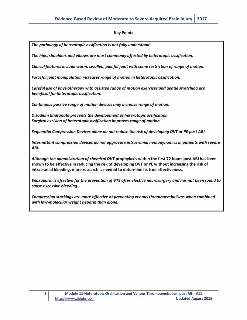
The pathology of heterotopic ossification is not fully understood. The hips, shoulders and elbows are most commonly affected by heterotopic ossification. Clinical features include warm, swollen, painful joint with some restriction of range of motion. Forceful joint manipulation increases range of motion in heterotopic ossification. Careful use of physiotherapy with assisted range of motion exercises and gentle stretching are beneficial for heterotopic ossification. Continuous passive range of motion devices may increase range of motion. Disodium Etidronate prevents the development of heterotopic ossification Surgical excision of heterotopic ossification improves range of motion. Sequential Compression Devices alone do not reduce the risk of developing DVT or PE post ABI. Intermittent compression devices do not aggravate intracranial hemodynamics in patients with severe ABI. Although the administration of chemical DVT prophylaxis within the first 72 hours post ABI has been shown to be effective in reducing the risk of developing DVT or PE without increasing the risk of intracranial bleeding, more research is needed to determine its true effectiveness. Enoxaparin is effective for the prevention of VTE after elective neurosurgery and has not been found to cause excessive bleeding. Compression stockings are more effective at preventing venous thromboembolisms when combined with low-molecular weight heparin than alone.
Evidence-Based Review of Moderate to Severe Acquired Brain Injury 2017
7 Module 11-Heterotopic Ossification and Venous Thromboembolism post ABI- V11 http://www.abiebr.com Updated August 2016
Abbreviations ABI Acquired Brain Injury
DVT Deep Venous Thrombosis
EHDP Etidronate Disodium (ethane-1-hydroxy-1, 1-diphosphonate)
GCS Glasgow Coma Scale
HO Heterotopic Ossification
LMWH Low-Molecular Weight Heparin
PE Pulmonary Embolism
PIOPED Prospective Investigation of Pulmonary Embolism Diagnosis
SCD Sequential Compression Devices
TBI Traumatic Brain Injury
VTE Venous Thromboembolism
Evidence-Based Review of Moderate to Severe Acquired Brain Injury 2017
8 Module 11-Heterotopic Ossification and Venous Thromboembolism post ABI- V11 http://www.abiebr.com Updated August 2016
11. Heterotopic Ossification and Venous Thromboembolism Post ABI Heterotopic ossification (HO) is the formation of pathologic bone within soft tissues, often muscle tissues, where bone formation does not usually occur (Watanabe & Sant 2001). The incidence of HO in patients with traumatic brain injury (TBI) has been reported to range from 11% to 77% but is clinically relevant in 10-20% (Dizdar et al. 2013; Garland et al. 1980; Rogers 1988; Sarafis et al. 1999; Sazbon et al. 1981; Zychowicz 2013). Risk factors include skeletal trauma, spasticity, diffuse axonal injury, mechanical ventilation, prolonged immobilization, and injury severity (Moreta & De los Mozos 2014). HO is often quite painful and limits joint mobility; the restricted joint range of motion may exacerbate disability and impede progress towards desired rehabilitation goals. 11.1 Formation of Heterotopic Ossification Post-Head Injury 11.1.1 Pathophysiology of Heterotopic Ossification Post-Head Injury The pathophysiology of HO is not fully understood. Mesenchymal stem cells (osteoblastic cells) can generate cartilage, bone, muscle, tendons, ligaments, or fat (Pape et al. 2004). It is thought that they play a pivotal role in the development of HO (Williams et al. 1999). HO development begins with the formation of osteoid periarticularly and intramuscularly, and progresses to full calcification within a matter of weeks (Pape et al. 2001). Over the next few months, the calcified osteoid remodels into well-organized trabecular bone at which point it is considered to have matured (Pape et al. 2001). Several months after the initial trauma, patients with HO begin to experience restricted range of motion, pain and ankylosis (Banovac & Gonzalez 1997; Garland et al. 1980). The bony lesion has been found to have a high metabolic rate, with a rate of bone formation more than three times greater than that of normal bone, and an osteoclastic density of more than twice the density found in normal bone (Puzas et al. 1987). 11.1.2 Stimulating Factors Related to Head Injury It is believed that there is likely a neurogenic factor contributing to HO, although this mechanism is not yet understood ( Antiplatelet Trialists’ Collaboration 1994; Hurvitz et al. 1992; Pape et al. 2001; Pape et al. 2004). It has also been noted that circulating factors promoting HO may be present in patients with head injuries (Pape et al. 2004). Many studies have shown enhanced osteogenesis in patients sustaining TBI (Trentz et al. 2005). The presence of certain hormonal factors early post-injury influences the stimulation of osteoprogenitors within skeletal muscles (Ivanhoe et al. 2012). Further, tissue hypoxia, sympathetic changes, immobilization, remobilization, and spasticity are additional risk factors (Ivanhoe et al. 2012). Accelerated fracture healing and HO are well documented phenomena in these patients (Bidner et al. 1990; Keret et al. 1990).
The pathophysiology of heterotopic ossification is not fully understood.
11.2 Clinical Presentation of Heterotopic Ossification 11.2.1 Location of Lesion Among individuals with TBI the most common sites of HO are the soft tissues around the hip, elbow, shoulder and knee (Garland 1991; Garland et al. 1980; Van Kampen et al. 2011; Vanden Bossche & Vanderstraeten 2005). The hip is the most frequent site of ossification (Dizdar et al. 2013; Vanden
Evidence-Based Review of Moderate to Severe Acquired Brain Injury 2017
9 Module 11-Heterotopic Ossification and Venous Thromboembolism post ABI- V11 http://www.abiebr.com Updated August 2016
Bossche & Vanderstraeten 2005); with total ankylosis of the joint occurring in 5-16% of affected hips (Stover et al. 1991). HO of the shoulder has been found to affect 5% of individuals with a brain injury (Cipriano et al. 2009), while the knee is a less common site for HO following a head injury (Sarafis et al. 1999). When HO is present in the knee, it is usually seen medially (Hosalkar et al. 2013). The distribution of HO around the elbow occurs most commonly either anteriorly in the flexor muscles or posteriorly in the extensors (Sarafis et al. 1999). Of the joints affected by HO after head injury, ankylosis is most likely to occur in the posterior elbow (Garland et al. 1980). 11.2.2 Clinical Features The onset of HO has been reported to vary between two and three weeks post injury (Watanabe & Sant 2001). More recently, clinical signs and symptoms have been said to develop 3-12 weeks post injury (Vanden Bossche & Vanderstraeten 2005; Zychowicz 2013). Pape and colleagues (2004) noted that clinical examination in the setting of HO may reveal a swollen, warm, painful joint which is often associated with a decreased range of motion. The earliest sign is typically a loss of range of motion in the involved joint (Watanabe & Sant 2001). Other findings then include erythema, palpation of a periarticular mass and fever (Varghese 1992). Because of the association with fever, it is sometimes difficult to differentiate HO from infection (Citta-Pietrolungo et al. 1992; Garland 1991; Garland et al. 1980). Moreover, the clinical picture may be confused with deep venous thrombosis (DVT), local trauma or fracture (Buschbacher 1992; Jensen et al. 1987). HO will then progress from these initial symptoms into a mass with stiffness and induration (Zychowicz 2013). Potential complications involved with HO include compression of blood vessels and nerves, breakdown of associated tissue, restricted motion and loss of function (Zychowicz 2013). 11.2.3 Investigations for Heterotopic Ossification During the initial presentation, plain radiographs may be negative and will usually remain normal until ossification begins at approximately 4-6 weeks post injury. Serum levels of alkaline phosphatase, a glycoprotein in the plasma membrane of osteoblasts, and the erythrocyte sedimentation rate may become elevated early on but are non-specific. The triple phase technicium-99 bone scan remains the diagnostic gold standard. The test is positive if there is an increased uptake during the first and second phases of the study. It typically becomes positive when clinical features appear (i.e., before an x-ray would be positive). In the HO literature there are several classification systems, with the Brooker classification system being one of the most widely used. The Brooker Classification system is typically used to classify ectopic-bone formation after total hip replacement. The system is based on anteroposterior radiograph of the pelvis and the categorization of the progression of HO into classes (Brooker et al. 1973). Brooker et al. (1973) define Class I as islands of bone within soft tissues about the hip; Class II: bone spurs from the pelvis or proximal end of the femur with at least 1cm between opposing bone surfaces; Class III: bone spurs from pelvis or proximal end of the femur, reducing space between opposing bone surfaces to less than 1cm; and Class IV: apparent bone ankylosis of hip. The classification system has been criticized and consequently the system has been modified (Della Valle et al. 2002; Mavrogenis et al. 2012; Toom et al. 2005; Wright et al. 1994).
The hips, shoulders and elbows are most commonly affected by heterotopic ossification. Clinical features include warm, swollen, painful joint with some restriction of range of motion.
Evidence-Based Review of Moderate to Severe Acquired Brain Injury 2017
10 Module 11-Heterotopic Ossification and Venous Thromboembolism post ABI- V11 http://www.abiebr.com Updated August 2016
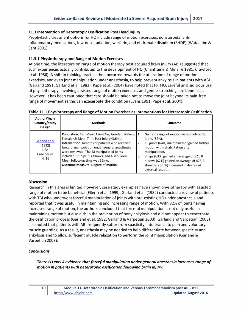
11.3 Intervention of Heterotopic Ossification Post Head Injury Prophylactic treatment options for HO include range of motion exercises, nonsteroidal anti-inflammatory medications, low-dose radiation, warfarin, and etidronate disodium (EHDP) (Watanabe & Sant 2001). 11.3.1 Physiotherapy and Range of Motion Exercises At one time, the literature on range of motion therapy post acquired brain injury (ABI) suggested that such experiences actually contributed to the development of HO (Chantraine & Minaire 1981; Crawford et al. 1986). A shift in thinking practice then occurred towards the utilization of range of motion exercises, and even joint manipulation under anesthesia, to help prevent ankylosis in patients with ABI (Garland 1991; Garland et al. 1982). Pape et al. (2004) have noted that for HO, careful and judicious use of physiotherapy, involving assisted range of motion exercises and gentle stretching, are beneficial. However, it has been cautioned that care should be taken not to move the joint beyond its pain-free range of movement as this can exacerbate the condition (Evans 1991; Pape et al. 2004). Table 11.1 Physiotherapy and Range of Motion Exercises as Interventions for Heterotopic Ossification
Author/Year/ Country/Study
Design Methods Outcome
Garland et al. (1982)
USA Case Series
N=16
Population: TBI; Mean Age=24yr; Gender: Male=8, Female=8; Mean Time Post Injury=3.6mo. Intervention: Records of patients who received forceful manipulation under general anesthesia were reviewed. The 28 manipulated joints included: 11 hips, 13 elbows, and 4 shoulders. Mean follow-up time was 15mo. Outcome Measure: Degree of motion.
1. Gains in range of motion were made in 23 joints (82%).
2. 18 joints (64%) maintained or gained further motion with rehabilitation after manipulation.
3. 7 hips (63%) gained an average of 52, 8
elbows (62%) gained an average of 47, 3 shoulders (75%) increased in degree of external rotation.
Discussion Research in this area is limited; however, case study examples have shown physiotherapy with assisted range of motion to be beneficial (Ellerin et al. 1999). Garland et al. (1982) conducted a review of patients with TBI who underwent forceful manipulation of joints with pre-existing HO under anesthesia and reported that it was useful in maintaining and increasing range of motion. With 82% of joints having increased range of motion, the authors concluded that forceful manipulation is not only useful in maintaining motion but also aids in the prevention of bony ankylosis and did not appear to exacerbate the ossification process (Garland et al. 1982; Garland & Varpetian 2003). Garland and Varpetian (2003) also noted that patients with ABI frequently suffer from spasticity, intolerance to pain and voluntary muscle guarding. As a result, anesthesia may be needed to help differentiate between spasticity and ankylosis and to allow sufficient muscle relaxation to perform the joint manipulation (Garland & Varpetian 2003). Conclusions
There is Level 4 evidence that forceful manipulation under general anesthesia increases range of motion in patients with heterotopic ossification following brain injury.
Evidence-Based Review of Moderate to Severe Acquired Brain Injury 2017
11 Module 11-Heterotopic Ossification and Venous Thromboembolism post ABI- V11 http://www.abiebr.com Updated August 2016
Forceful joint manipulation increases range of motion in heterotopic ossification.
Careful use of physiotherapy with assisted range of motion exercises and gentle stretching are
beneficial for heterotopic ossification.
11.3.2 Continuous Passive Motion Continuous passive motion devices have shown promising results in maintaining range of motion following total knee replacement (Nadler et al. 1993; Salter 1996). Animal data shows that continuous passive motion does not increase the progression of HO (Van Susante et al. 1996). Moreover, there is little human research evidence that HO is worsened by passive range of motion (Linan et al. 2001). Several studies have examined continuous passive motion in combination with surgical excision. These studies are explained further in section 11.4. Further research is needed in the ABI population.
Continuous passive range of motion devices may increase range of motion.
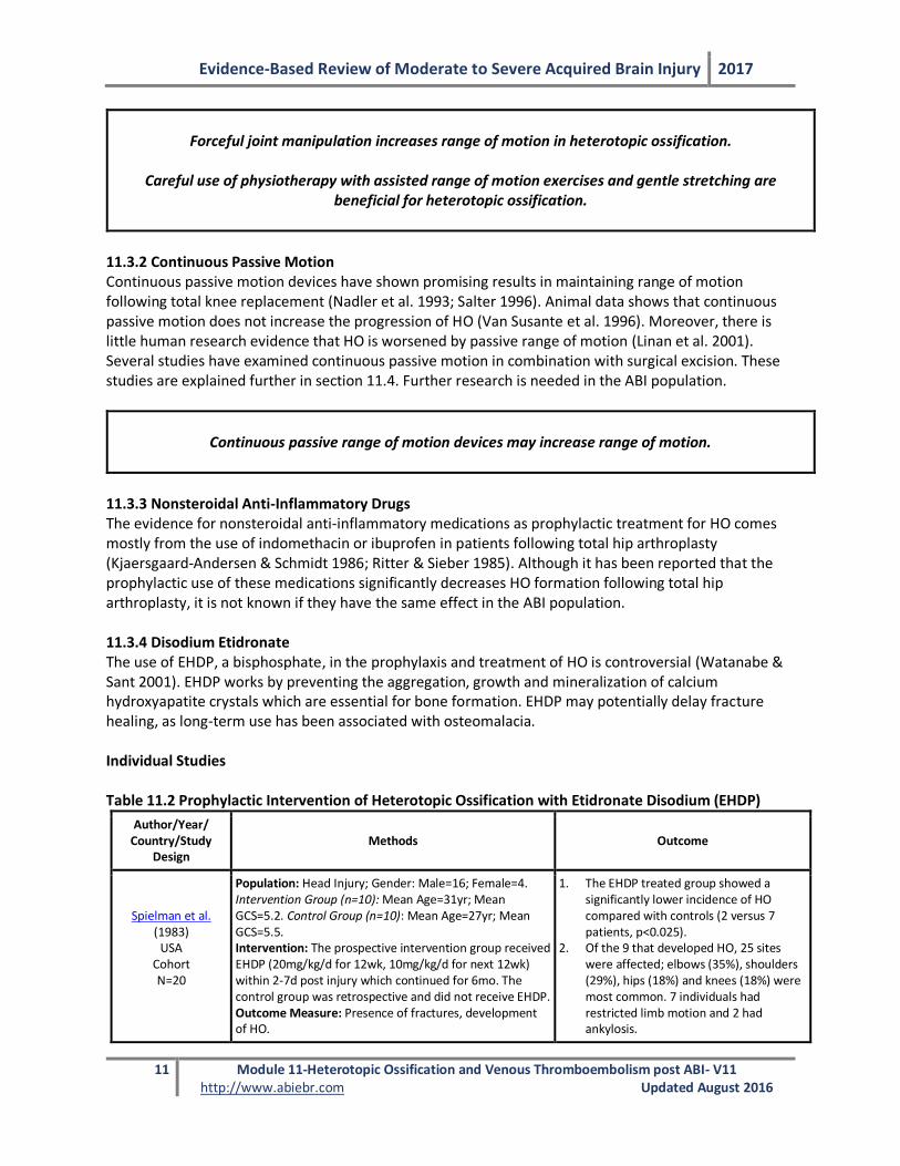
11.3.3 Nonsteroidal Anti-Inflammatory Drugs The evidence for nonsteroidal anti-inflammatory medications as prophylactic treatment for HO comes mostly from the use of indomethacin or ibuprofen in patients following total hip arthroplasty (Kjaersgaard-Andersen & Schmidt 1986; Ritter & Sieber 1985). Although it has been reported that the prophylactic use of these medications significantly decreases HO formation following total hip arthroplasty, it is not known if they have the same effect in the ABI population. 11.3.4 Disodium Etidronate The use of EHDP, a bisphosphate, in the prophylaxis and treatment of HO is controversial (Watanabe & Sant 2001). EHDP works by preventing the aggregation, growth and mineralization of calcium hydroxyapatite crystals which are essential for bone formation. EHDP may potentially delay fracture healing, as long-term use has been associated with osteomalacia. Individual Studies Table 11.2 Prophylactic Intervention of Heterotopic Ossification with Etidronate Disodium (EHDP)
Author/Year/ Country/Study
Design Methods Outcome
Spielman et al. (1983)
USA Cohort N=20
Population: Head Injury; Gender: Male=16; Female=4. Intervention Group (n=10): Mean Age=31yr; Mean GCS=5.2. Control Group (n=10): Mean Age=27yr; Mean GCS=5.5. Intervention: The prospective intervention group received EHDP (20mg/kg/d for 12wk, 10mg/kg/d for next 12wk) within 2-7d post injury which continued for 6mo. The control group was retrospective and did not receive EHDP. Outcome Measure: Presence of fractures, development of HO.
1. The EHDP treated group showed a significantly lower incidence of HO compared with controls (2 versus 7 patients, p<0.025).
2. Of the 9 that developed HO, 25 sites were affected; elbows (35%), shoulders (29%), hips (18%) and knees (18%) were most common. 7 individuals had restricted limb motion and 2 had ankylosis.
Evidence-Based Review of Moderate to Severe Acquired Brain Injury 2017
12 Module 11-Heterotopic Ossification and Venous Thromboembolism post ABI- V11 http://www.abiebr.com Updated August 2016
Discussion Although EHDP has been shown to be effective in reducing HO in other populations, such as spinal cord injury, its effectiveness among individuals with brain injury is less studied. In an ABI population, Spielman et al. (1983) found that patients treated with EHDP showed a significantly lower incidence of HO than the control group. However, due to the small sample size of the study and the research design, additional research assessing the benefit of EHDP for the intervention of HO following brain injury is needed. Conclusions
There is Level 2 evidence that Disodium Etidronate (EHDP) reduces the development of heterotopic ossification in patients with severe head injury.
Disodium Etidronate prevents the development of heterotopic ossification.
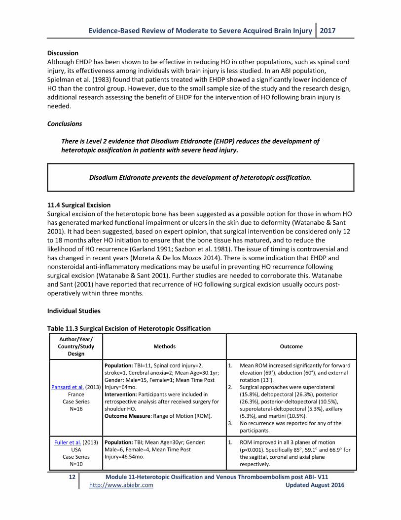
11.4 Surgical Excision Surgical excision of the heterotopic bone has been suggested as a possible option for those in whom HO has generated marked functional impairment or ulcers in the skin due to deformity (Watanabe & Sant 2001). It had been suggested, based on expert opinion, that surgical intervention be considered only 12 to 18 months after HO initiation to ensure that the bone tissue has matured, and to reduce the likelihood of HO recurrence (Garland 1991; Sazbon et al. 1981). The issue of timing is controversial and has changed in recent years (Moreta & De los Mozos 2014). There is some indication that EHDP and nonsteroidal anti-inflammatory medications may be useful in preventing HO recurrence following surgical excision (Watanabe & Sant 2001). Further studies are needed to corroborate this. Watanabe and Sant (2001) have reported that recurrence of HO following surgical excision usually occurs post-operatively within three months. Individual Studies Table 11.3 Surgical Excision of Heterotopic Ossification
Author/Year/ Country/Study
Design Methods Outcome
Pansard et al. (2013) France
Case Series N=16
Population: TBI=11, Spinal cord injury=2, stroke=1, Cerebral anoxia=2; Mean Age=30.1yr; Gender: Male=15, Female=1; Mean Time Post Injury=64mo. Intervention: Participants were included in retrospective analysis after received surgery for shoulder HO. Outcome Measure: Range of Motion (ROM).
1. Mean ROM increased significantly for forward elevation (69°), abduction (60°), and external rotation (13°).
2. Surgical approaches were superolateral (15.8%), deltopectoral (26.3%), posterior (26.3%), posterior-deltopectoral (10.5%), superolateral-deltopectoral (5.3%), axillary (5.3%), and martini (10.5%).
3. No recurrence was reported for any of the participants.
Fuller et al. (2013) USA
Case Series N=10
Population: TBI; Mean Age=30yr; Gender: Male=6, Female=4, Mean Time Post Injury=46.54mo.
1. ROM improved in all 3 planes of motion
(p<0.001). Specifically 85, 59.1 and 66.9 for the sagittal, coronal and axial plane respectively.
Evidence-Based Review of Moderate to Severe Acquired Brain Injury 2017
13 Module 11-Heterotopic Ossification and Venous Thromboembolism post ABI- V11 http://www.abiebr.com Updated August 2016
Author/Year/ Country/Study
Design Methods Outcome
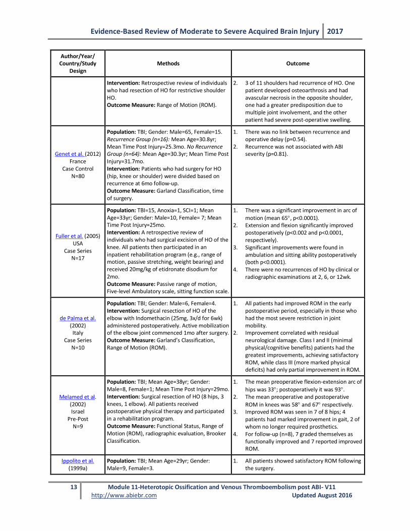
Intervention: Retrospective review of individuals who had resection of HO for restrictive shoulder HO. Outcome Measure: Range of Motion (ROM).
2. 3 of 11 shoulders had recurrence of HO. One patient developed osteoarthrosis and had avascular necrosis in the opposite shoulder, one had a greater predisposition due to multiple joint involvement, and the other patient had severe post-operative swelling.
Genet et al. (2012) France
Case Control N=80
Population: TBI; Gender: Male=65, Female=15. Recurrence Group (n=16): Mean Age=30.8yr; Mean Time Post Injury=25.3mo. No Recurrence Group (n=64): Mean Age=30.3yr; Mean Time Post Injury=31.7mo. Intervention: Patients who had surgery for HO (hip, knee or shoulder) were divided based on recurrence at 6mo follow-up. Outcome Measure: Garland Classification, time of surgery.
1. There was no link between recurrence and operative delay (p=0.54).
2. Recurrence was not associated with ABI severity (p=0.81).
Fuller et al. (2005) USA
Case Series N=17
Population: TBI=15, Anoxia=1, SCI=1; Mean Age=33yr; Gender: Male=10, Female= 7; Mean Time Post Injury=25mo. Intervention: A retrospective review of individuals who had surgical excision of HO of the knee. All patients then participated in an inpatient rehabilitation program (e.g., range of motion, passive stretching, weight bearing) and received 20mg/kg of etidronate disodium for 2mo. Outcome Measure: Passive range of motion, Five-level Ambulatory scale, sitting function scale.
1. There was a significant improvement in arc of
motion (mean 65, p<0.0001). 2. Extension and flexion significantly improved
postoperatively (p<0.002 and p<0.0001, respectively).
3. Significant improvements were found in ambulation and sitting ability postoperatively (both p<0.0001).
4. There were no recurrences of HO by clinical or radiographic examinations at 2, 6, or 12wk.
de Palma et al. (2002) Italy
Case Series N=10
Population: TBI; Gender: Male=6, Female=4. Intervention: Surgical resection of HO of the elbow with Indomethacin (25mg, 3x/d for 6wk) administered postoperatively. Active mobilization of the elbow joint commenced 1mo after surgery. Outcome Measure: Garland’s Classification, Range of Motion (ROM).
1. All patients had improved ROM in the early postoperative period, especially in those who had the most severe restriction in joint mobility.
2. Improvement correlated with residual neurological damage. Class I and II (minimal physical/cognitive benefits) patients had the greatest improvements, achieving satisfactory ROM, while class III (more marked physical deficits) had only partial improvement in ROM.
Melamed et al. (2002) Israel
Pre-Post N=9
Population: TBI; Mean Age=38yr; Gender: Male=8, Female=1; Mean Time Post Injury=29mo. Intervention: Surgical resection of HO (8 hips, 3 knees, 1 elbow). All patients received postoperative physical therapy and participated in a rehabilitation program. Outcome Measure: Functional Status, Range of Motion (ROM), radiographic evaluation, Brooker Classification.
1. The mean preoperative flexion-extension arc of
hips was 33; postoperatively it was 93. 2. The mean preoperative and postoperative
ROM in knees was 58 and 67 respectively. 3. Improved ROM was seen in 7 of 8 hips; 4
patients had marked improvement in gait, 2 of whom no longer required prosthetics.
4. For follow-up (n=8), 7 graded themselves as functionally improved and 7 reported improved ROM.
Ippolito et al. (1999a)
Population: TBI; Mean Age=29yr; Gender: Male=9, Female=3.
1. All patients showed satisfactory ROM following the surgery.
Evidence-Based Review of Moderate to Severe Acquired Brain Injury 2017
14 Module 11-Heterotopic Ossification and Venous Thromboembolism post ABI- V11 http://www.abiebr.com Updated August 2016
Author/Year/ Country/Study
Design Methods Outcome
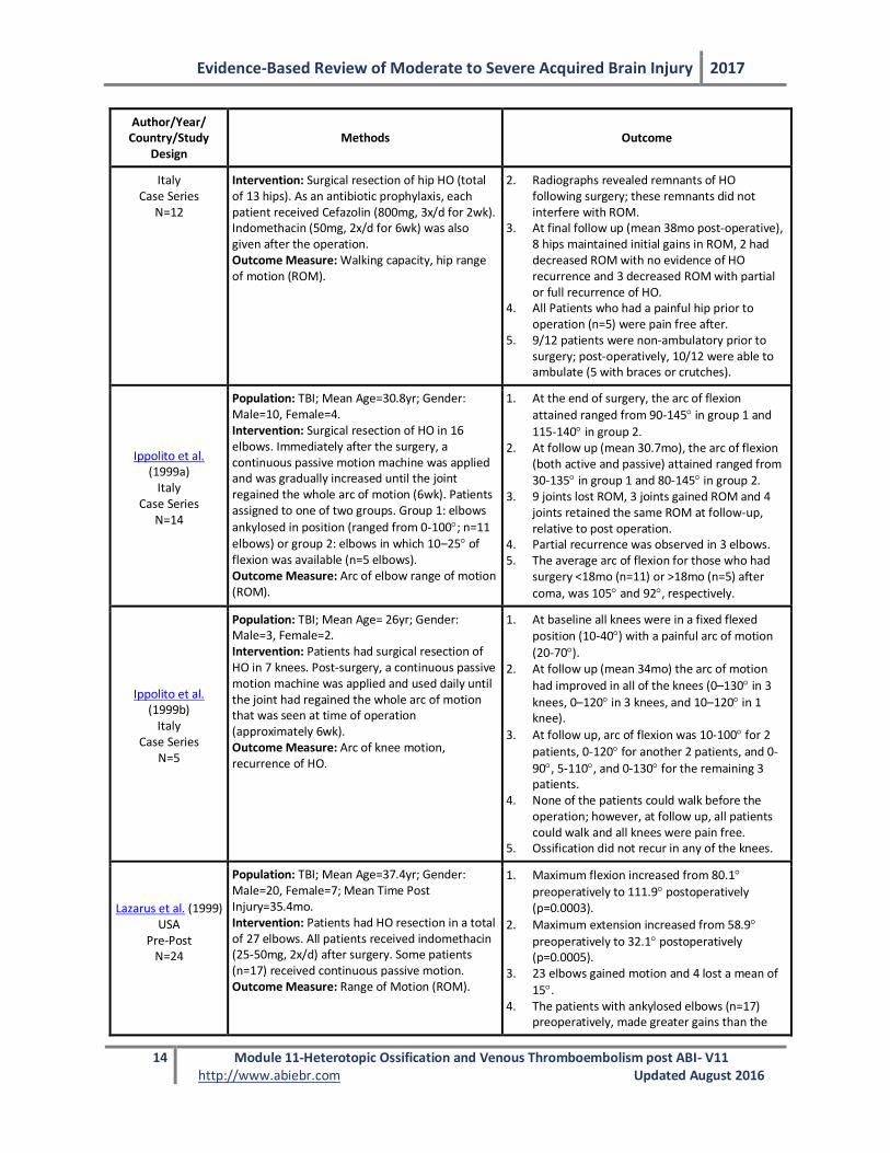
Italy Case Series
N=12
Intervention: Surgical resection of hip HO (total of 13 hips). As an antibiotic prophylaxis, each patient received Cefazolin (800mg, 3x/d for 2wk). Indomethacin (50mg, 2x/d for 6wk) was also given after the operation. Outcome Measure: Walking capacity, hip range of motion (ROM).
2. Radiographs revealed remnants of HO following surgery; these remnants did not interfere with ROM.
3. At final follow up (mean 38mo post-operative), 8 hips maintained initial gains in ROM, 2 had decreased ROM with no evidence of HO recurrence and 3 decreased ROM with partial or full recurrence of HO.
4. All Patients who had a painful hip prior to operation (n=5) were pain free after.
5. 9/12 patients were non-ambulatory prior to surgery; post-operatively, 10/12 were able to ambulate (5 with braces or crutches).
Ippolito et al. (1999a)
Italy Case Series
N=14
Population: TBI; Mean Age=30.8yr; Gender: Male=10, Female=4. Intervention: Surgical resection of HO in 16 elbows. Immediately after the surgery, a continuous passive motion machine was applied and was gradually increased until the joint regained the whole arc of motion (6wk). Patients assigned to one of two groups. Group 1: elbows
ankylosed in position (ranged from 0-100; n=11 elbows) or group 2: elbows in which 10–25 of flexion was available (n=5 elbows). Outcome Measure: Arc of elbow range of motion (ROM).
1. At the end of surgery, the arc of flexion
attained ranged from 90-145 in group 1 and
115-140 in group 2. 2. At follow up (mean 30.7mo), the arc of flexion
(both active and passive) attained ranged from 30-135 in group 1 and 80-145 in group 2.
3. 9 joints lost ROM, 3 joints gained ROM and 4 joints retained the same ROM at follow-up, relative to post operation.
4. Partial recurrence was observed in 3 elbows. 5. The average arc of flexion for those who had
surgery <18mo (n=11) or >18mo (n=5) after
coma, was 105 and 92, respectively.
Ippolito et al. (1999b)
Italy Case Series
N=5
Population: TBI; Mean Age= 26yr; Gender: Male=3, Female=2. Intervention: Patients had surgical resection of HO in 7 knees. Post-surgery, a continuous passive motion machine was applied and used daily until the joint had regained the whole arc of motion that was seen at time of operation (approximately 6wk). Outcome Measure: Arc of knee motion, recurrence of HO.
1. At baseline all knees were in a fixed flexed position (10-40) with a painful arc of motion
(20-70). 2. At follow up (mean 34mo) the arc of motion
had improved in all of the knees (0–130 in 3
knees, 0–120 in 3 knees, and 10–120 in 1 knee).
3. At follow up, arc of flexion was 10-100 for 2
patients, 0-120 for another 2 patients, and 0-
90, 5-110, and 0-130 for the remaining 3 patients.
4. None of the patients could walk before the operation; however, at follow up, all patients could walk and all knees were pain free.
5. Ossification did not recur in any of the knees.
Lazarus et al. (1999) USA
Pre-Post N=24
Population: TBI; Mean Age=37.4yr; Gender: Male=20, Female=7; Mean Time Post Injury=35.4mo. Intervention: Patients had HO resection in a total of 27 elbows. All patients received indomethacin (25-50mg, 2x/d) after surgery. Some patients (n=17) received continuous passive motion. Outcome Measure: Range of Motion (ROM).
1. Maximum flexion increased from 80.1
preoperatively to 111.9 postoperatively (p=0.0003).
2. Maximum extension increased from 58.9
preoperatively to 32.1 postoperatively (p=0.0005).
3. 23 elbows gained motion and 4 lost a mean of
15. 4. The patients with ankylosed elbows (n=17)
preoperatively, made greater gains than the
Evidence-Based Review of Moderate to Severe Acquired Brain Injury 2017
15 Module 11-Heterotopic Ossification and Venous Thromboembolism post ABI- V11 http://www.abiebr.com Updated August 2016
Author/Year/ Country/Study
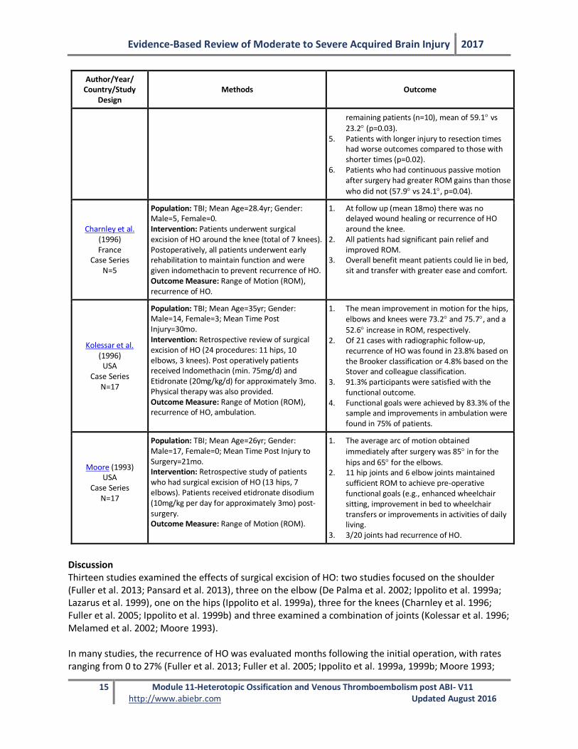
Design Methods Outcome
remaining patients (n=10), mean of 59.1 vs
23.2 (p=0.03). 5. Patients with longer injury to resection times
had worse outcomes compared to those with shorter times (p=0.02).
6. Patients who had continuous passive motion after surgery had greater ROM gains than those
who did not (57.9 vs 24.1, p=0.04).
Charnley et al. (1996) France
Case Series N=5
Population: TBI; Mean Age=28.4yr; Gender: Male=5, Female=0. Intervention: Patients underwent surgical excision of HO around the knee (total of 7 knees). Postoperatively, all patients underwent early rehabilitation to maintain function and were given indomethacin to prevent recurrence of HO. Outcome Measure: Range of Motion (ROM), recurrence of HO.
1. At follow up (mean 18mo) there was no delayed wound healing or recurrence of HO around the knee.
2. All patients had significant pain relief and improved ROM.
3. Overall benefit meant patients could lie in bed, sit and transfer with greater ease and comfort.
Kolessar et al. (1996)
USA Case Series
N=17
Population: TBI; Mean Age=35yr; Gender: Male=14, Female=3; Mean Time Post Injury=30mo. Intervention: Retrospective review of surgical excision of HO (24 procedures: 11 hips, 10 elbows, 3 knees). Post operatively patients received Indomethacin (min. 75mg/d) and Etidronate (20mg/kg/d) for approximately 3mo. Physical therapy was also provided. Outcome Measure: Range of Motion (ROM), recurrence of HO, ambulation.
1. The mean improvement in motion for the hips, elbows and knees were 73.2 and 75.7, and a
52.6 increase in ROM, respectively. 2. Of 21 cases with radiographic follow-up,
recurrence of HO was found in 23.8% based on the Brooker classification or 4.8% based on the Stover and colleague classification.
3. 91.3% participants were satisfied with the functional outcome.
4. Functional goals were achieved by 83.3% of the sample and improvements in ambulation were found in 75% of patients.
Moore (1993) USA
Case Series N=17
Population: TBI; Mean Age=26yr; Gender: Male=17, Female=0; Mean Time Post Injury to Surgery=21mo. Intervention: Retrospective study of patients who had surgical excision of HO (13 hips, 7 elbows). Patients received etidronate disodium (10mg/kg per day for approximately 3mo) post-surgery. Outcome Measure: Range of Motion (ROM).
1. The average arc of motion obtained
immediately after surgery was 85 in for the
hips and 65 for the elbows. 2. 11 hip joints and 6 elbow joints maintained
sufficient ROM to achieve pre-operative functional goals (e.g., enhanced wheelchair sitting, improvement in bed to wheelchair transfers or improvements in activities of daily living.
3. 3/20 joints had recurrence of HO.
Discussion Thirteen studies examined the effects of surgical excision of HO: two studies focused on the shoulder (Fuller et al. 2013; Pansard et al. 2013), three on the elbow (De Palma et al. 2002; Ippolito et al. 1999a; Lazarus et al. 1999), one on the hips (Ippolito et al. 1999a), three for the knees (Charnley et al. 1996; Fuller et al. 2005; Ippolito et al. 1999b) and three examined a combination of joints (Kolessar et al. 1996; Melamed et al. 2002; Moore 1993). In many studies, the recurrence of HO was evaluated months following the initial operation, with rates ranging from 0 to 27% (Fuller et al. 2013; Fuller et al. 2005; Ippolito et al. 1999a, 1999b; Moore 1993;
Evidence-Based Review of Moderate to Severe Acquired Brain Injury 2017
16 Module 11-Heterotopic Ossification and Venous Thromboembolism post ABI- V11 http://www.abiebr.com Updated August 2016
Pansard et al. 2013). The majority of the studies did not specify what qualified as recurrence; however a study by Kolessar et al. (1996) found recurrence rates differed based on the classification system utilized (23.8% versus 4.8% using the Brooker classification and the Stover and colleagues classification, respectively). A systematic review conducted by Lee et al. (2013) focused specifically on the surgical excision of HO in the elbow and found improvements in motion, with low levels of recurrence (14.3%). However, complications such as fracture, infection, nerve palsies, wound complications and loss of motion without recurrence were found in 27.5% of cases (Lee et al. 2013). Overall, the surgical excision of HO resulted in improved range of motion. One study did note a decrease in range of motion for a small portion of participants (Ippolito et al. 1999a). Improvements in activities of daily living and ambulation were also found (Fuller et al. 2005; Ippolito et al. 1999a, 1999b; Melamed et al. 2002). It is worth noting that length of time between injury and surgical resection was found to be a significant predictor of outcome, as longer times were associated with less improvement (Lazarus et al. 1999). Although therapy was provided after the surgery in many of the studies, only one study formally evaluated its effectiveness. The study conducted by Lazarus et al. (1999) found that patients who had continuous passive motion exercises post operatively made significantly greater gains those individuals
who did not (57.9 versus 24.1, p=0.04). Conclusions
There is Level 4 evidence that surgical excision of heterotopic ossification improves clinical outcomes.
Surgical excision of heterotopic ossification improves range of motion.
11.5 Venous Thromboembolism Venous thromboembolism (VTE) occurs when blood clots form within the venous system. DVT may occur if the clot forms in a deep vein (i.e., in the leg). If the clot broke off and travelled to the lungs, causing partial or full occlusion, it can become a pulmonary embolism (PE) (Office of the Surgeon General et al. 2008). VTE remains a common complication in patients who have sustained an ABI (Raslan et al. 2010; Scudday et al. 2011); however the scientific literature specific to ABI is quite limited. 11.5.1 Incidence of Venous Thromboembolism Post-Head Injury In a large sample study, 38,984 individuals with TBI, the incidence of VTE at the time of admission was 1.31% (Olufajo et al. 2016). At one month post injury, the incidence for VTE increased to 1.87% and by one year it was 2.83% (Olufajo et al. 2016). The reported incidence of DVT among patients with TBI ranges from 11% to 54% (Carlile et al. 2010; Cifu et al. 1996; Denson et al. 2007; Geerts et al. 1994). The risk of developing a DVT or PE, in the absence of prophylaxis, is estimated to be approximately 20% post-TBI (Haddad & Arabi 2012) and severity of injury is found to be associated with incidence of VTE in isolated patients with TBI (Van Gent et al. 2014). Decisions on how to treat, and when, are often made on a case by case basis (Tang & Lobel 2009). Experts recommend beginning pharmacological prophylaxis as early as 48 to 72 hours post injury (Norwood et al. 2001). Unless contraindicated, mechanical thromboprophlaxis and low-molecular weight heparin (LMWH) is recommened in the acute phase of recovery (Haddad & Arabi 2012).
Evidence-Based Review of Moderate to Severe Acquired Brain Injury 2017
17 Module 11-Heterotopic Ossification and Venous Thromboembolism post ABI- V11 http://www.abiebr.com Updated August 2016
Cifu et al. (1996) screened 153 patients admitted to a tertiary care brain injury unit within 24 hours of admission for a lower extremity DVT with colour flow duplex Doppler ultrasonography. All patients had received prophylactic intervention with either subcutaneous heparin anticoagulation or intermittent compression devices. The overall incidence of DVTs in those patients with ABI was 13%, while individuals with TBI had an incidence rate of 20%. Most of the DVTs were asymptomatic. Another study found that DVT was present in 31.6% of individuals who sustained a head injury (Ekeh et al. 2010). 11.6 Risk Factors for DVT The most recognized risk factors for VTE are venostasis, intimal damage of the vessel wall, and a hypercoagulable state (Virchow’s triad - see Diagram 11.1) (Watanabe & Sant 2001). Patients with a severe brain injury are commonly immobilized for periods of time as a result of extremity or spine fractures they experienced at the time of their injury (Vergouwen et al. 2008). The incidence of DVT appears to be impacted by length of stay in the intensive care unit and the number of days a patient is on a ventilator. There does not appear to be a correlation between DVT incidence and initial Glasgow Coma Scale (GCS) scores, Injury Severity Scale scores, or the Abbreviated Injury Scale score (Denson et al. 2007). Those at highest risk post injury are those who remain on a ventilator longer than 3 days (Olufajo et al. 2016; Raslan et al. 2010). At 1 year post-injury, risk of VTE is greatest for those discharged to extended care facilities compared to home, and for individuals who undergo an operation (Olufajo et al. 2016). Patients involved in trauma that does not specifically involve vessel injury are still at increased risk of thromboembolism, suggesting a trauma-induced hypercoagulable state (Geerts et al. 1994; Geerts et al. 1996). Therefore persons who have sustained a TBI appear to be at increased risk of developing VTE. 11.7 Clinical Presentation of VTE The clinical presentation of PE is challenging. Many cases are clinically silent (66%) with only 30% having the clinical features of a DVT (Garcia-Fuster et al. 2014). Asymptomatic PE is discovered in 70% of patients with confirmed clinically symptomatic DVT (Browse 1974; Corrigan et al. 1974; Hull & Hirsh 1983). Clinically, pulmonary embolus presents with tachycardia, tachypnea and signs of pulmonary infarction with consolidation, hemoptysis, pleuritic chest pain, pleural friction rub, pleural effusion and fever (Worku et al. 2014). Massive PE may cause right heart failure, which can progress to cardiovascular collapse, coma and death.
11.8 Diagnostic Testing for DVT
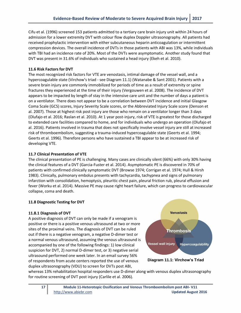
11.8.1 Diagnosis of DVT A positive diagnosis of DVT can only be made if a venogram is positive or there is a positive venous ultrasound at two or more sites of the proximal veins. The diagnosis of DVT can be ruled out if there is a negative venogram, a negative D-dimer test or a normal venous ultrasound, assuming the venous ultrasound is accompanied by one of the following findings: 1) low clinical suspicion for DVT, 2) normal D-dimer test, or 3) negative serial ultrasound performed one week later. In an email survey 56% of respondents from acute centers reported the use of venous duplex ultrasonography (VDU) to screen for DVTs post ABI, whereas 13% rehabilitation hospital responders use D-dimer along with venous duplex ultrasonography for routine screening of DVT post injury (Carlile et al. 2006).
Diagram 11.1: Virchow’s Triad
Evidence-Based Review of Moderate to Severe Acquired Brain Injury 2017
18 Module 11-Heterotopic Ossification and Venous Thromboembolism post ABI- V11 http://www.abiebr.com Updated August 2016
11.8.2 Venous Ultrasound Venous ultrasound is often used to diagnose a DVT. There are several types of venous ultrasonography. They include compression ultrasound, duplex ultrasound and color Doppler imaging. Although these types of venous ultrasonography are sometimes used interchangeably, their sensitivities and specificities for detecting acute DVT vary (Zierler 2004). The sensitivity and specificity of compression ultrasonography for detecting DVTs is 43% and 85%, respectively (Girard et al. 2005). The weighted mean sensitivity and specificity of venous ultrasonography for the diagnosis of symptomatic proximal DVT are 97% and 94%, respectively; the sensitivity falls to 73% for distal DVT (Kearon et al. 1998; Zierler 2004). Importantly, distal DVT do not confer the same risk of extension to PE as do proximal DVT. Typically, if a distal clot is going to extend proximally, this occurs within one week of its development. Consequently, serial ultrasound could be used in symptomatic patients in whom the test is initially negative as the test would become positive with the clot extension. 11.8.3 Venography Venography is considered a definitive test for DVT but it is an invasive test whereby contrast dye is injected into the leg veins. Diagnosis of DVT is made if an intraluminal-filling defect is noted. 11.8.4 D-dimer Assay D-dimer assay is a rapid, non-invasive and inexpensive test. Fibrin is the main component of thrombus formation and fibrin degradation products include D-dimers (Gill & Nahum 2000). A positive D-dimer test is highly sensitive for the presence of a thrombus but lacks specificity since D-dimers are found in other disease states, including cancer, congestive heart failure and inflammatory conditions (Raimondi et al. 1993). As a result, D-dimer assays have a high negative predictive value but a poor positive predictive value. To illustrate, Akman et al. (2004) reported that the sensitivity and negative predictive values of the D-dimer test were high, at 95.2% and 96.2% respectively, in a group of 68 rehabilitation patients (stroke, spinal cord injury, TBI, hip arthroplasty). The specificity and positive predictive values were low at 55.3% and 48.7%, respectively. 11.9 Diagnostic Testing for PE 11.9.1 Ventilation/Perfusion Scanning Nuclear ventilation/perfusion scans are often used to investigate possible PE. Palmowski et al. (2014) reported the sensitivity and specificity of ventilation/perfusion scanning as 95.8% and 82.6%, respectively, with false negative rates of 4.2% and false positive rate of 17.3%. Hence, a normal scan virtually excludes a PE (high negative predictive value). Identified perfusion defects are non-specific and only represent true PE in about one-third of cases. The probability that a perfusion defect represents a PE increases with the size, shape and number of defects as well as the presence of a normal ventilation scan. Mismatched defects, normal ventilation scan reveals poor perfusion, but are large or segmental in size are “high probability” defects and are associated with an approximately 80% prevalence of PE (Kearon et al. 1998).
Evidence-Based Review of Moderate to Severe Acquired Brain Injury 2017
19 Module 11-Heterotopic Ossification and Venous Thromboembolism post ABI-V11 http://www.abiebr.com Updated August 2016
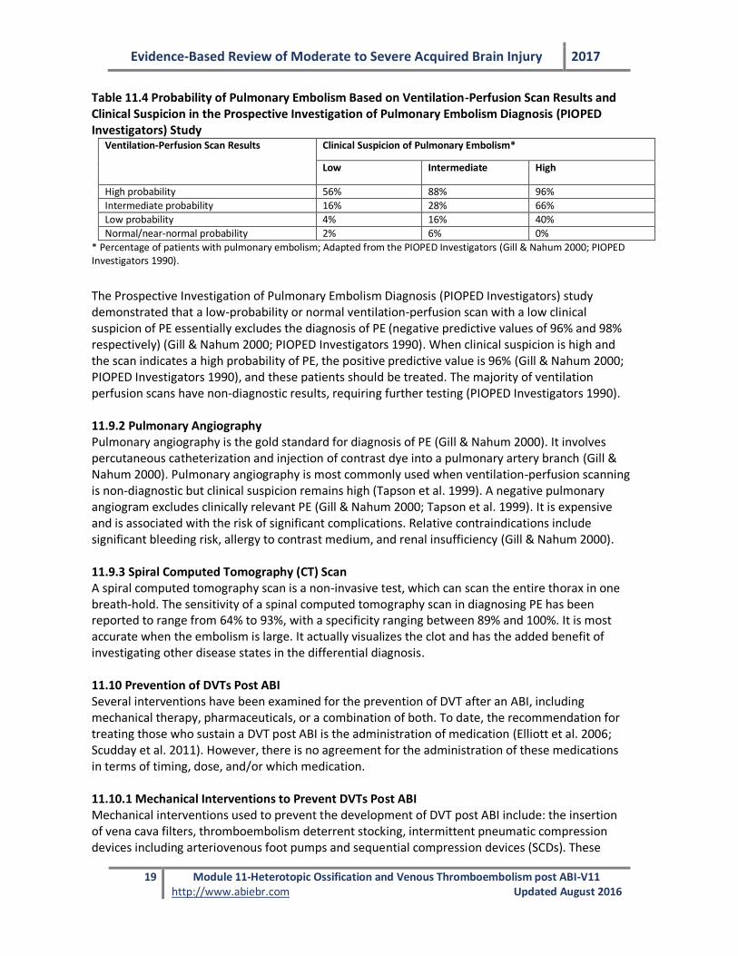
Table 11.4 Probability of Pulmonary Embolism Based on Ventilation-Perfusion Scan Results and Clinical Suspicion in the Prospective Investigation of Pulmonary Embolism Diagnosis (PIOPED Investigators) Study
Ventilation-Perfusion Scan Results Clinical Suspicion of Pulmonary Embolism*
Low Intermediate High
High probability 56% 88% 96%
Intermediate probability 16% 28% 66%
Low probability 4% 16% 40%
Normal/near-normal probability 2% 6% 0%
* Percentage of patients with pulmonary embolism; Adapted from the PIOPED Investigators (Gill & Nahum 2000; PIOPED Investigators 1990).
The Prospective Investigation of Pulmonary Embolism Diagnosis (PIOPED Investigators) study demonstrated that a low-probability or normal ventilation-perfusion scan with a low clinical suspicion of PE essentially excludes the diagnosis of PE (negative predictive values of 96% and 98% respectively) (Gill & Nahum 2000; PIOPED Investigators 1990). When clinical suspicion is high and the scan indicates a high probability of PE, the positive predictive value is 96% (Gill & Nahum 2000; PIOPED Investigators 1990), and these patients should be treated. The majority of ventilation perfusion scans have non-diagnostic results, requiring further testing (PIOPED Investigators 1990). 11.9.2 Pulmonary Angiography Pulmonary angiography is the gold standard for diagnosis of PE (Gill & Nahum 2000). It involves percutaneous catheterization and injection of contrast dye into a pulmonary artery branch (Gill & Nahum 2000). Pulmonary angiography is most commonly used when ventilation-perfusion scanning is non-diagnostic but clinical suspicion remains high (Tapson et al. 1999). A negative pulmonary angiogram excludes clinically relevant PE (Gill & Nahum 2000; Tapson et al. 1999). It is expensive and is associated with the risk of significant complications. Relative contraindications include significant bleeding risk, allergy to contrast medium, and renal insufficiency (Gill & Nahum 2000). 11.9.3 Spiral Computed Tomography (CT) Scan A spiral computed tomography scan is a non-invasive test, which can scan the entire thorax in one breath-hold. The sensitivity of a spinal computed tomography scan in diagnosing PE has been reported to range from 64% to 93%, with a specificity ranging between 89% and 100%. It is most accurate when the embolism is large. It actually visualizes the clot and has the added benefit of investigating other disease states in the differential diagnosis. 11.10 Prevention of DVTs Post ABI Several interventions have been examined for the prevention of DVT after an ABI, including mechanical therapy, pharmaceuticals, or a combination of both. To date, the recommendation for treating those who sustain a DVT post ABI is the administration of medication (Elliott et al. 2006; Scudday et al. 2011). However, there is no agreement for the administration of these medications in terms of timing, dose, and/or which medication. 11.10.1 Mechanical Interventions to Prevent DVTs Post ABI Mechanical interventions used to prevent the development of DVT post ABI include: the insertion of vena cava filters, thromboembolism deterrent stocking, intermittent pneumatic compression devices including arteriovenous foot pumps and sequential compression devices (SCDs). These
Evidence-Based Review of Moderate to Severe Acquired Brain Injury 2017
20 Module 11-Heterotopic Ossification and Venous Thromboembolism post ABI-V11 http://www.abiebr.com Updated August 2016
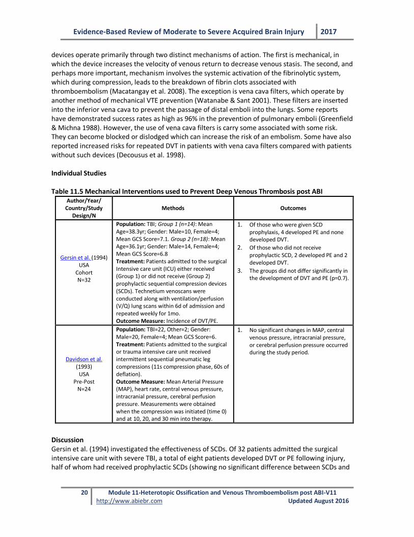
devices operate primarily through two distinct mechanisms of action. The first is mechanical, in which the device increases the velocity of venous return to decrease venous stasis. The second, and perhaps more important, mechanism involves the systemic activation of the fibrinolytic system, which during compression, leads to the breakdown of fibrin clots associated with thromboembolism (Macatangay et al. 2008). The exception is vena cava filters, which operate by another method of mechanical VTE prevention (Watanabe & Sant 2001). These filters are inserted into the inferior vena cava to prevent the passage of distal emboli into the lungs. Some reports have demonstrated success rates as high as 96% in the prevention of pulmonary emboli (Greenfield & Michna 1988). However, the use of vena cava filters is carry some associated with some risk. They can become blocked or dislodged which can increase the risk of an embolism. Some have also reported increased risks for repeated DVT in patients with vena cava filters compared with patients without such devices (Decousus et al. 1998). Individual Studies Table 11.5 Mechanical Interventions used to Prevent Deep Venous Thrombosis post ABI
Author/Year/ Country/Study
Design/N Methods Outcomes
Gersin et al. (1994) USA
Cohort N=32
Population: TBI; Group 1 (n=14): Mean Age=38.3yr; Gender: Male=10, Female=4; Mean GCS Score=7.1. Group 2 (n=18): Mean Age=36.1yr; Gender: Male=14, Female=4; Mean GCS Score=6.8 Treatment: Patients admitted to the surgical Intensive care unit (ICU) either received (Group 1) or did not receive (Group 2) prophylactic sequential compression devices (SCDs). Technetium venoscans were conducted along with ventilation/perfusion (V/Q) lung scans within 6d of admission and repeated weekly for 1mo. Outcome Measure: Incidence of DVT/PE.
1. Of those who were given SCD prophylaxis, 4 developed PE and none developed DVT.
2. Of those who did not receive prophylactic SCD, 2 developed PE and 2 developed DVT.
3. The groups did not differ significantly in the development of DVT and PE (p=0.7).
Davidson et al. (1993)
USA Pre-Post
N=24
Population: TBI=22, Other=2; Gender: Male=20, Female=4; Mean GCS Score=6. Treatment: Patients admitted to the surgical or trauma intensive care unit received intermittent sequential pneumatic leg compressions (11s compression phase, 60s of deflation). Outcome Measure: Mean Arterial Pressure (MAP), heart rate, central venous pressure, intracranial pressure, cerebral perfusion pressure. Measurements were obtained when the compression was initiated (time 0) and at 10, 20, and 30 min into therapy.
1. No significant changes in MAP, central venous pressure, intracranial pressure, or cerebral perfusion pressure occurred during the study period.
Discussion Gersin et al. (1994) investigated the effectiveness of SCDs. Of 32 patients admitted the surgical intensive care unit with severe TBI, a total of eight patients developed DVT or PE following injury, half of whom had received prophylactic SCDs (showing no significant difference between SCDs and
Evidence-Based Review of Moderate to Severe Acquired Brain Injury 2017
21 Module 11-Heterotopic Ossification and Venous Thromboembolism post ABI-V11 http://www.abiebr.com Updated August 2016
no intervention). The effectiveness of prophylactic SCDs in the prevention of post-TBI DVT or PE thus remains questionable. Davidson et al. (1993) conducted a study to evaluate the possibility that intermittent pneumatic compression could aggravate intracranial hemodynamics in severe brain injury patients. The authors reported that the use of intermittent compression devices to prevent the occurrence of DVT was not associated with any significant changes in intracranial pressure or cerebral perfusion pressure in stable patients in whom intracranial pressure was controlled by conventional measures (Davidson et al. 1993). These findings suggest that there is no contraindication to the use of pneumatic compression for the prevention of DVT in severe acute patients with brain injury who are responsive to conventional intracranial management measures. Conclusions
There is Level 2 evidence from one small study to suggest that SCDs are not entirely effective in reducing the risk of developing DVT or PE post ABI.
There is Level 4 evidence that intermittent compression devices do not cause acute elevations in intracranial pressure in patients with severe ABI.
Sequential Compression Devices alone do not reduce the risk of developing DVT or PE post
ABI.
Intermittent compression devices do not aggravate intracranial hemodynamics in patients with severe ABI.
11.10.2 Pharmaceutical Therapies Oral agents have been investigated for their prophylactic potential against DVT. Warfarin (Coumadin), a well-established anticoagulant with a predictable duration of action, is sometimes avoided as a prophylactic alternative for DVT due to its elevated bleeding side effects (Watanabe & Sant 2001). To illustrate, Albrecht and colleagues (2014) report that warfarin use is associated with lower rates of DVT and PE, but comes at the cost of the risk of increased hemorrhagic bleeding. However, some experts felt the use of warfarin was advisable, especially for high risk patients due to its benefit in treating undetected thrombosis; the therapeutic concentration for prophylaxis and treatment of thromboembolism are the same (Hirsh et al. 1992; Hyers et al. 1992; Landefeld & Goldman 1989). In a recent study with a sample of 932 patients, 71% were given LMWH, 23% heparin, 1% Coumadin, and 3% were given both LMWH and Low-dose unfractionated heparin, none of which were associated with increased intracranial or systemic hemorrhage (Carlile et al. 2010). The most recent guidelines on DVT prophylaxis recommend using LMWH or Low-dose unfractionated heparin in addition to mechanical prophylaxis (Elliott et al. 2006; Reiff et al. 2009). There is also evidence from a meta-analysis, that aspirin has positive effects in the reduction of both DVT and PE, by 40% and 60% respectively (Antiplatelet Trialists’ Collaboration 1994). Clinically there remains concern that chemical DVT prophylaxis may result in increased intracranial bleeding post ABI.
Evidence-Based Review of Moderate to Severe Acquired Brain Injury 2017
22 Module 11-Heterotopic Ossification and Venous Thromboembolism post ABI-V11 http://www.abiebr.com Updated August 2016
Overall, there is a lack of persuasive evidence to guide decisions about when to administer anticoagulant prophylaxis in those who sustain traumatic intracranial hemorrhage. Clinicians often make decisions based on their own assessments of the risks and benefits (Scales et al. 2010). To date no national standard of care exists for the administration of the pharmacological prophylaxis treatment of DVT post TBI (Phelan et al. 2012). 11.10.3 Low Molecular Weight Heparin versus Low-Dose Unfractionated Heparin Subcutaneous heparin in low doses has been reported to be both safe and effective as prophylaxis against DVT development post ABI (Watanabe & Sant 2001). The route of delivery may also affect the efficacy of anticoagulant prophylaxis (Watanabe & Sant 2001). For this reason, intravenously delivered heparin may be more effective in the prevention of thromboembolism compared with subcutaneous administration, although this method of delivery might increase the risk of bleeding (Green et al. 1988). Low-molecular weight heparins, which are injected subcutaneously, have gained popularity due to the ease of administration and dosage adjustment. Of note, low-molecular weight variants of unfractionated heparin are significantly more expansive, and thus the risks, benefits, and costs need to be balanced out on an individual basis (Watanabe & Sant 2001). Carlile et al. (2006) found that 15 of the 16 rehabilitation centers surveyed reported routinely initiating treatment with either LMWH or Low-dose unfractionated heparin. In a study with a mixed trauma population, low-dose heparin was compared to enoxaparin (LMWH) for the treatment of DVT (Geerts et al. 1996). Of those receiving low-dose heparin 44% suffered a DVT compared to 31% of patients receiving enoxaparin (p=0.014) (Geerts et al. 1996).
Individual Studies
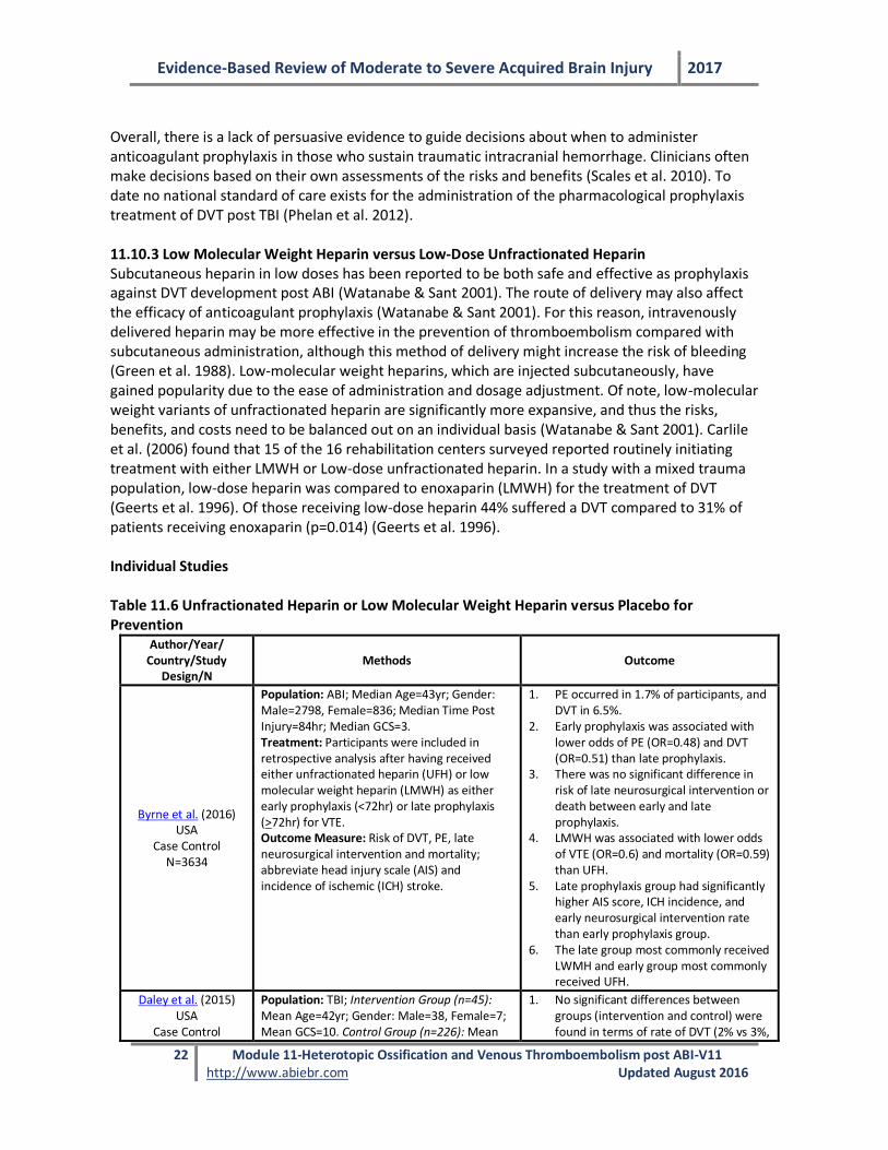
Table 11.6 Unfractionated Heparin or Low Molecular Weight Heparin versus Placebo for Prevention
Author/Year/ Country/Study
Design/N
Methods
Outcome
Byrne et al. (2016) USA
Case Control N=3634
Population: ABI; Median Age=43yr; Gender: Male=2798, Female=836; Median Time Post Injury=84hr; Median GCS=3. Treatment: Participants were included in retrospective analysis after having received either unfractionated heparin (UFH) or low molecular weight heparin (LMWH) as either early prophylaxis (<72hr) or late prophylaxis (>72hr) for VTE. Outcome Measure: Risk of DVT, PE, late neurosurgical intervention and mortality; abbreviate head injury scale (AIS) and incidence of ischemic (ICH) stroke.
1. PE occurred in 1.7% of participants, and DVT in 6.5%.
2. Early prophylaxis was associated with lower odds of PE (OR=0.48) and DVT (OR=0.51) than late prophylaxis.
3. There was no significant difference in risk of late neurosurgical intervention or death between early and late prophylaxis.
4. LMWH was associated with lower odds of VTE (OR=0.6) and mortality (OR=0.59) than UFH.
5. Late prophylaxis group had significantly higher AIS score, ICH incidence, and early neurosurgical intervention rate than early prophylaxis group.
6. The late group most commonly received LWMH and early group most commonly received UFH.
Daley et al. (2015) USA
Case Control
Population: TBI; Intervention Group (n=45): Mean Age=42yr; Gender: Male=38, Female=7; Mean GCS=10. Control Group (n=226): Mean
1. No significant differences between groups (intervention and control) were found in terms of rate of DVT (2% vs 3%,
Evidence-Based Review of Moderate to Severe Acquired Brain Injury 2017
23 Module 11-Heterotopic Ossification and Venous Thromboembolism post ABI-V11 http://www.abiebr.com Updated August 2016
N=271
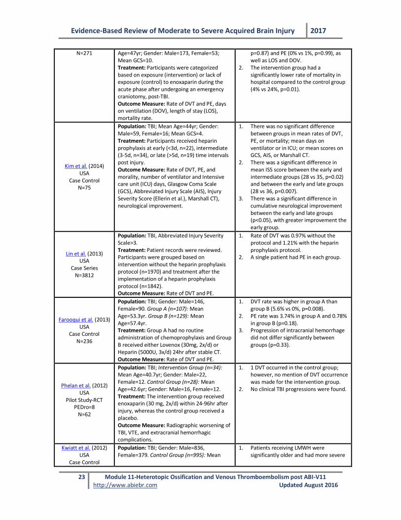
Age=47yr; Gender: Male=173, Female=53; Mean GCS=10. Treatment: Participants were categorized based on exposure (intervention) or lack of exposure (control) to enoxaparin during the acute phase after undergoing an emergency craniotomy, post-TBI. Outcome Measure: Rate of DVT and PE, days on ventilation (DOV), length of stay (LOS), mortality rate.
p=0.87) and PE (0% vs 1%, p=0.99), as well as LOS and DOV.
2. The intervention group had a significantly lower rate of mortality in hospital compared to the control group (4% vs 24%, p=0.01).
Kim et al. (2014) USA
Case Control N=75
Population: TBI; Mean Age=44yr; Gender: Male=59, Female=16; Mean GCS=4. Treatment: Participants received heparin prophylaxis at early (<3d, n=22), intermediate (3-5d, n=34), or late (>5d, n=19) time intervals post injury. Outcome Measure: Rate of DVT, PE, and morality, number of ventilator and Intensive care unit (ICU) days, Glasgow Coma Scale (GCS), Abbreviated Injury Scale (AIS), Injury Severity Score (Ellerin et al.), Marshall CT), neurological improvement.
1. There was no significant difference between groups in mean rates of DVT, PE, or mortality; mean days on ventilator or in ICU; or mean scores on GCS, AIS, or Marshall CT.
2. There was a significant difference in mean ISS score between the early and intermediate groups (28 vs 35, p=0.02) and between the early and late groups (28 vs 36, p=0.007).
3. There was a significant difference in cumulative neurological improvement between the early and late groups (p<0.05), with greater improvement the early group.
Lin et al. (2013) USA
Case Series N=3812
Population: TBI, Abbreviated Injury Severity Scale>3. Treatment: Patient records were reviewed. Participants were grouped based on intervention without the heparin prophylaxis protocol (n=1970) and treatment after the implementation of a heparin prophylaxis protocol (n=1842). Outcome Measure: Rate of DVT and PE.
1. Rate of DVT was 0.97% without the protocol and 1.21% with the heparin prophylaxis protocol.
2. A single patient had PE in each group.
Farooqui et al. (2013) USA
Case Control N=236
Population: TBI; Gender: Male=146, Female=90. Group A (n=107): Mean Age=53.3yr. Group B (n=129): Mean Age=57.4yr. Treatment: Group A had no routine administration of chemoprophylaxis and Group B received either Lovenox (30mg, 2x/d) or Heparin (5000U, 3x/d) 24hr after stable CT. Outcome Measure: Rate of DVT and PE.
1. DVT rate was higher in group A than group B (5.6% vs 0%, p=0.008).
2. PE rate was 3.74% in group A and 0.78% in group B (p=0.18).
3. Progression of intracranial hemorrhage did not differ significantly between groups (p=0.33).
Phelan et al. (2012) USA
Pilot Study-RCT PEDro=8
N=62
Population: TBI; Intervention Group (n=34): Mean Age=40.7yr; Gender: Male=22, Female=12. Control Group (n=28): Mean Age=42.6yr; Gender: Male=16, Female=12. Treatment: The intervention group received enoxaparin (30 mg, 2x/d) within 24-96hr after injury, whereas the control group received a placebo. Outcome Measure: Radiographic worsening of TBI, VTE, and extracranial hemorrhagic complications.
1. 1 DVT occurred in the control group; however, no mention of DVT occurrence was made for the intervention group.
2. No clinical TBI progressions were found.
Kwiatt et al. (2012) USA
Case Control
Population: TBI; Gender: Male=836, Female=379. Control Group (n=995): Mean
1. Patients receiving LMWH were significantly older and had more severe
Evidence-Based Review of Moderate to Severe Acquired Brain Injury 2017
24 Module 11-Heterotopic Ossification and Venous Thromboembolism post ABI-V11 http://www.abiebr.com Updated August 2016
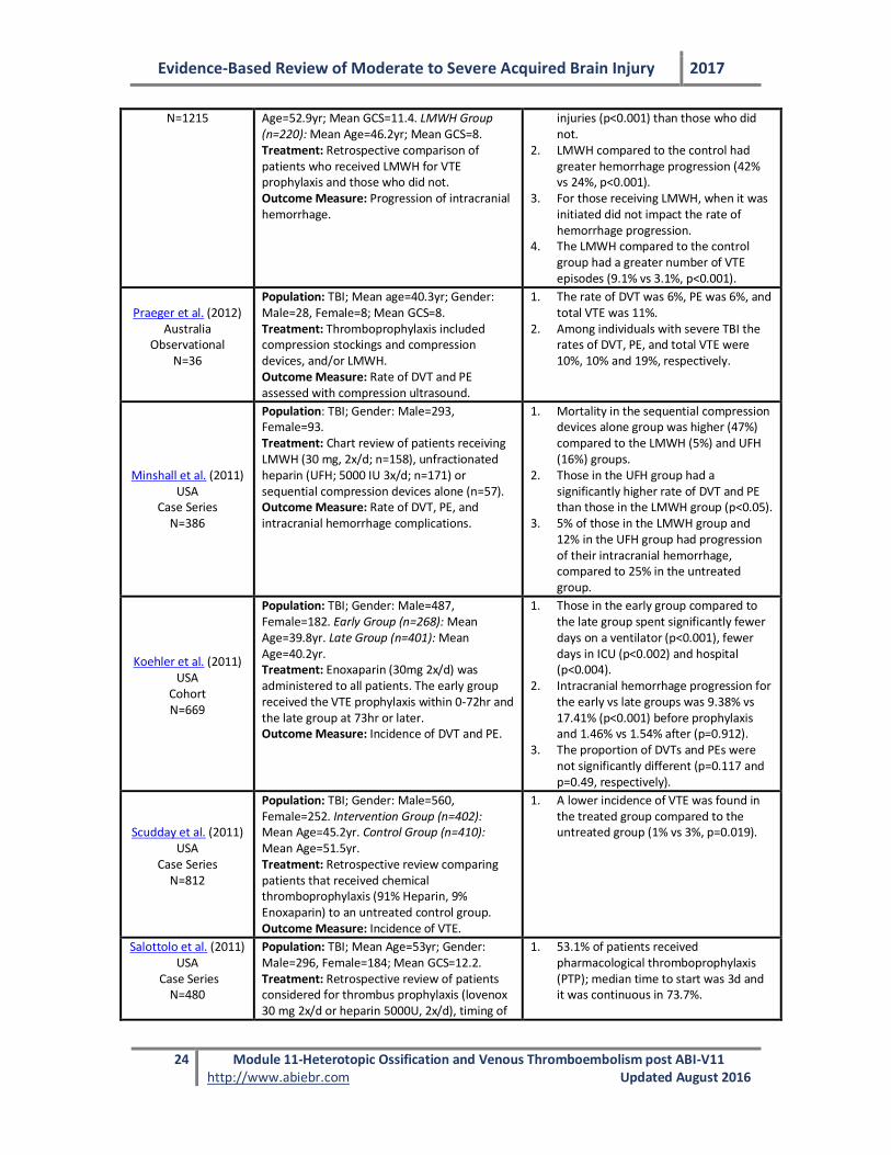
N=1215 Age=52.9yr; Mean GCS=11.4. LMWH Group (n=220): Mean Age=46.2yr; Mean GCS=8. Treatment: Retrospective comparison of patients who received LMWH for VTE prophylaxis and those who did not. Outcome Measure: Progression of intracranial hemorrhage.
injuries (p<0.001) than those who did not.
2. LMWH compared to the control had greater hemorrhage progression (42% vs 24%, p<0.001).
3. For those receiving LMWH, when it was initiated did not impact the rate of hemorrhage progression.
4. The LMWH compared to the control group had a greater number of VTE episodes (9.1% vs 3.1%, p<0.001).
Praeger et al. (2012) Australia
Observational N=36
Population: TBI; Mean age=40.3yr; Gender: Male=28, Female=8; Mean GCS=8. Treatment: Thromboprophylaxis included compression stockings and compression devices, and/or LMWH. Outcome Measure: Rate of DVT and PE assessed with compression ultrasound.
1. The rate of DVT was 6%, PE was 6%, and total VTE was 11%.
2. Among individuals with severe TBI the rates of DVT, PE, and total VTE were 10%, 10% and 19%, respectively.
Minshall et al. (2011) USA
Case Series N=386
Population: TBI; Gender: Male=293, Female=93. Treatment: Chart review of patients receiving LMWH (30 mg, 2x/d; n=158), unfractionated heparin (UFH; 5000 IU 3x/d; n=171) or sequential compression devices alone (n=57). Outcome Measure: Rate of DVT, PE, and intracranial hemorrhage complications.
1. Mortality in the sequential compression devices alone group was higher (47%) compared to the LMWH (5%) and UFH (16%) groups.
2. Those in the UFH group had a significantly higher rate of DVT and PE than those in the LMWH group (p<0.05).
3. 5% of those in the LMWH group and 12% in the UFH group had progression of their intracranial hemorrhage, compared to 25% in the untreated group.
Koehler et al. (2011) USA
Cohort N=669
Population: TBI; Gender: Male=487, Female=182. Early Group (n=268): Mean Age=39.8yr. Late Group (n=401): Mean Age=40.2yr. Treatment: Enoxaparin (30mg 2x/d) was administered to all patients. The early group received the VTE prophylaxis within 0-72hr and the late group at 73hr or later. Outcome Measure: Incidence of DVT and PE.
1. Those in the early group compared to the late group spent significantly fewer days on a ventilator (p<0.001), fewer days in ICU (p<0.002) and hospital (p<0.004).
2. Intracranial hemorrhage progression for the early vs late groups was 9.38% vs 17.41% (p<0.001) before prophylaxis and 1.46% vs 1.54% after (p=0.912).
3. The proportion of DVTs and PEs were not significantly different (p=0.117 and p=0.49, respectively).
Scudday et al. (2011) USA
Case Series N=812
Population: TBI; Gender: Male=560, Female=252. Intervention Group (n=402): Mean Age=45.2yr. Control Group (n=410): Mean Age=51.5yr. Treatment: Retrospective review comparing patients that received chemical thromboprophylaxis (91% Heparin, 9% Enoxaparin) to an untreated control group. Outcome Measure: Incidence of VTE.
1. A lower incidence of VTE was found in the treated group compared to the untreated group (1% vs 3%, p=0.019).
Salottolo et al. (2011) USA
Case Series N=480
Population: TBI; Mean Age=53yr; Gender: Male=296, Female=184; Mean GCS=12.2. Treatment: Retrospective review of patients considered for thrombus prophylaxis (lovenox 30 mg 2x/d or heparin 5000U, 2x/d), timing of
1. 53.1% of patients received pharmacological thromboprophylaxis (PTP); median time to start was 3d and it was continuous in 73.7%.
Evidence-Based Review of Moderate to Severe Acquired Brain Injury 2017
25 Module 11-Heterotopic Ossification and Venous Thromboembolism post ABI-V11 http://www.abiebr.com Updated August 2016
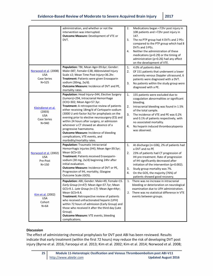
administration, and whether or not the intervention was interrupted. Outcome Measure: Development of VTE or DVT.
2. Medications began <72hr post injury in 108 patients and >72hr post injury in 147.
3. The no PTP group had 4 DVTs and 2 PEs compared to the PTP group which had 8 DVTs and 3 PEs.
4. Neither the administration of these medications (p=0.29) or the timing of administration (p=0.26) had any effect on the development of VTE.
Norwood et al. (2008) USA
Case Series N=525
Population: TBI; Mean Age=39.6yr; Gender: Male=387, Female=138; Abbreviated Injury Scale ≥2; Mean Time Post-Injury=36.2hr. Treatment: Patients were given Enoxaparin sodium (30mg, 2x/d). Outcome Measure: Incidence of DVT and PE, mortality rates.
1. 4.0% of patients died.
2. Of 151 patients that underwent a lower extremity venous Doppler ultrasound, 6 patients were diagnosed with a DVT.
3. No patients within the study group were diagnosed with a PE.
Kleindienst et al. (2003)
USA Case Series
N=940
Population: Head Injury=344, Elective Surgery (tumors)=294, Intracranial Hemorrhage (ICH)=302; Mean Age=57.3yr. Treatment: A retrospective review of patients either receiving 18mg/d of Certoparin-sodium (3000 U anti-factor Xa) for prophylaxis on the evening prior to elective neurosurgery (ES) and within 24 hours after surgery, or admission whenever a CT showed an absence of a progressive haematoma. Outcome Measure: Incidence of bleeding complications, VTE events, and morbidity/mortality rates.
1. 155 patients were excluded due to coagulation abnormalities or significant bleeding.
2. Intracranial bleeding was found in 1.5% of the total sample.
3. The incidence of VTE and PE was 0.2% and 0.1% of patients respectively, with no associated mortality.
4. No heparin induced thrombocytopenia was observed.
Norwood et al. (2002) USA
Pre-Post N=150
Population: Traumatic Intracranial Hemorrhagic injuries (IHI); Mean Age=39.5yr; Mean GCS=10. Treatment: Patients received Enaxoparin-sodium (30 mg, 2x/d) beginning 24hr after initial evaluation. Outcome Measure: Incidence of DVT or PE, Progression of IHI, mortality, Glasgow Outcome Scale (GOS).
1. At discharge (n=106), 2% of patients had a DVT and no PE
2. 23% of patients had CT progression of IHI pre-treatment. Rate of progression of IHI significantly decreased after initiation of the intervention (p=0.002).
3. Study group mortality was 7%.
4. On the GOS, the majority (76%) of patients showed good recovery.
Kim et al. (2002) USA
Cohort N=64
Population: ABI; Gender: Male=49, Female=15. Early Group (n=47): Mean Age=37.7yr; Mean GCS=9.1. Late Group (n=17): Mean Age=44yr; Mean GCS=9.4. Treatment: Retrospective review of patients who received unfractionated heparin (UFH) within 72 hours of admission (Early Group) and those who received it after the third day (Late Group). Outcome Measure: VTE events, bleeding complications.
1. There was no increase in intracranial bleeding or deterioration on neurological examination due to UFH administration.
2. There was no statistical difference in VTE events between groups.
Discussion The effect of administering chemical prophylaxis for DVT post ABI has been reviewed. Results indicate that early treatment (within the first 72 hours) may reduce the risk of developing DVT post injury (Byrne et al. 2016; Farooqui et al. 2013; Kim et al. 2002; Kim et al. 2014; Norwood et al. 2008;
Evidence-Based Review of Moderate to Severe Acquired Brain Injury 2017
26 Module 11-Heterotopic Ossification and Venous Thromboembolism post ABI-V11 http://www.abiebr.com Updated August 2016
Salottolo et al. 2011; Scudday et al. 2011) without increasing the risk of intracranial hemorrhagic injury (Byrne et al. 2016; Koehler et al. 2011; Scudday et al. 2011) or deterioration on neurological examination (Kim et al. 2002). Patients with ABI who were started on unfractionated heparin within three days of injury onset, compared to those who started after this time period, did not differ significantly in terms of the number of thromboembolic events (Kim et al. 2002; Kim et al. 2014). However, individuals who were administered heparin within three days of injury had slower progression of neurological impairments on computed tomography scans compared to late administration (Kim et al. 2014). Norwood and colleagues conducted two studies examining the benefits of administering enoxaparin (LMWH) prophylaxis to those who sustain a severe ABI within the first 48 hours post injury (Norwood et al. 2008; Norwood et al. 2002). Results from both studies indicate that administering enoxaparin post ABI reduces the risk of developing DVT and PE, without increasing the risk of bleeding post injury. Scudday et al. (2011) also found that patients who received chemical prophylaxis within 72 hours of injury had a significantly lower incidence of developing VTE post ABI (p<0.019) compared to those not receiving chemical prophylaxis (Kim et al. 2014) Overall, a meta-analysis by Jamjoom and colleagues (2013) conclude that individuals who begin pharmacological thromboprophylaxis within 72 hours of injury have half the risk of VTE without significant risk of intracranial hemorrhage progression, than those who start after 72 hours. On the contrary, few studies have demonstrated these medications may not be beneficial or superior treatments. In one study with individuals who underwent a craniotomy post-ABI, no significant differences were reported for rate of DVT and PE when comparing those administered enoxaparin prophylaxis compared to those without (Daley et al. 2015). Further, Kwiatt et al. (2012) reported patients’ receiving LMWH were at higher risk for hemorrhage progression and the risk of using LMWH may exceed its benefit. Similarly for heparin, Lin et al. (2013) did not find a reduction in DVT or PE once individuals with a severe TBI were administered a heparin prophylaxis protocol. In conclusion, a systematic review of twelve studies report that evidence is insufficient to determine effectiveness of these medications for VTE prevention; however despite the aforementioned studies without significant findings, overall evidence supports the use of enoxaparin for reduction of DVT and UFH for decreased mortality rates compared to no chemoprophylaxis (Chelladurai et al. 2013). Conclusions
There is Level 2 evidence supporting the administration of LMWH within the first 72 hours post ABI to reduce the risk of developing DVTs and PEs post injury. There is Level 2 evidence that administering LMWH (enoxaparin) or heparin post ABI does not increase the risk of intracranial bleeding, compared to no treatment. There is Level 4 evidence that the use of chemoprophylaxis 24 hours after stable head computed tomography scan decreases the rate of DVT formation post ABI.
Evidence-Based Review of Moderate to Severe Acquired Brain Injury 2017
27 Module 11-Heterotopic Ossification and Venous Thromboembolism post ABI-V11 http://www.abiebr.com Updated August 2016
Although the administration of chemical DVT prophylaxis within the first 72 hours post ABI
has been shown to be effective in reducing the risk of developing DVT or PE without increasing the risk of intracranial bleeding, more research is needed to determine its true
effectiveness.
Enoxaparin is effective for the prevention of VTE after elective neurosurgery and has not been found to cause excessive bleeding.
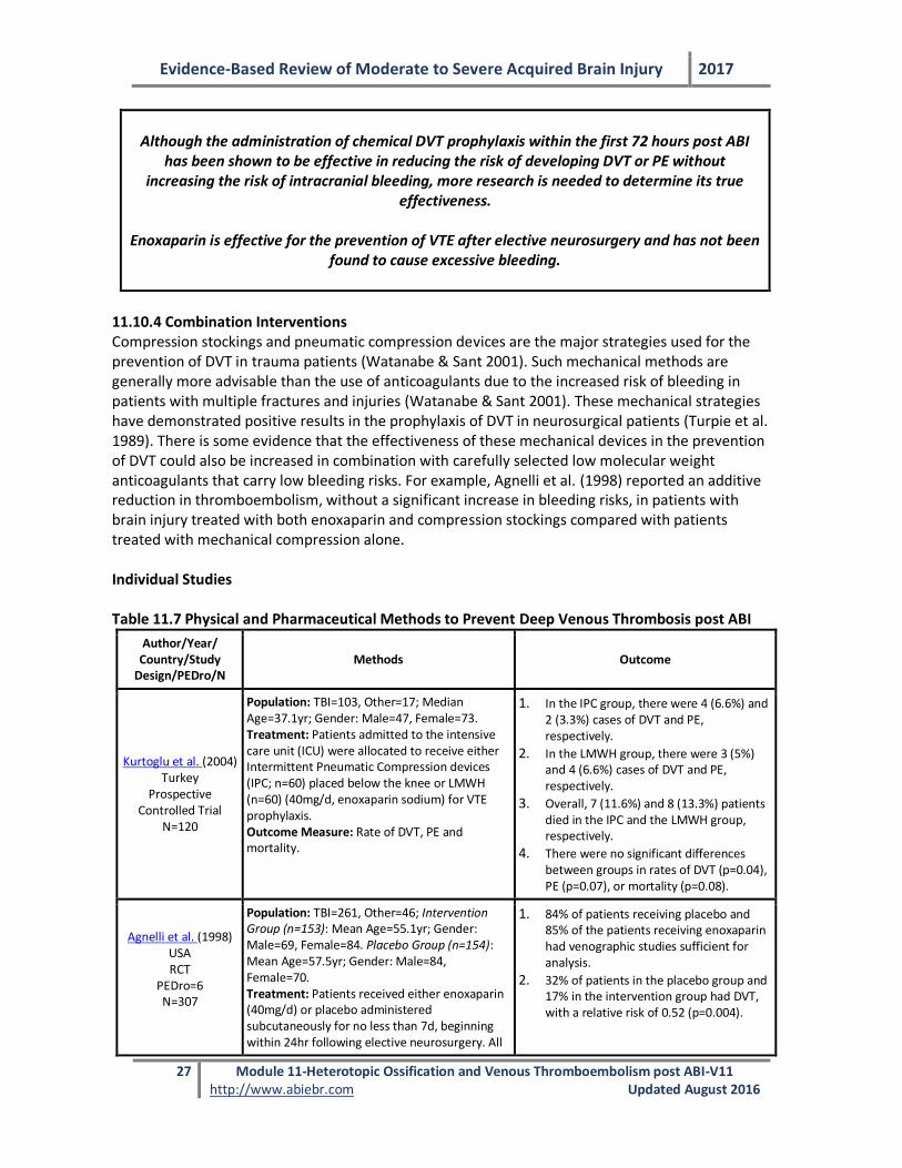
11.10.4 Combination Interventions Compression stockings and pneumatic compression devices are the major strategies used for the prevention of DVT in trauma patients (Watanabe & Sant 2001). Such mechanical methods are generally more advisable than the use of anticoagulants due to the increased risk of bleeding in patients with multiple fractures and injuries (Watanabe & Sant 2001). These mechanical strategies have demonstrated positive results in the prophylaxis of DVT in neurosurgical patients (Turpie et al. 1989). There is some evidence that the effectiveness of these mechanical devices in the prevention of DVT could also be increased in combination with carefully selected low molecular weight anticoagulants that carry low bleeding risks. For example, Agnelli et al. (1998) reported an additive reduction in thromboembolism, without a significant increase in bleeding risks, in patients with brain injury treated with both enoxaparin and compression stockings compared with patients treated with mechanical compression alone. Individual Studies Table 11.7 Physical and Pharmaceutical Methods to Prevent Deep Venous Thrombosis post ABI
Author/Year/ Country/Study
Design/PEDro/N Methods Outcome
Kurtoglu et al. (2004) Turkey
Prospective Controlled Trial
N=120
Population: TBI=103, Other=17; Median Age=37.1yr; Gender: Male=47, Female=73. Treatment: Patients admitted to the intensive care unit (ICU) were allocated to receive either Intermittent Pneumatic Compression devices (IPC; n=60) placed below the knee or LMWH (n=60) (40mg/d, enoxaparin sodium) for VTE prophylaxis. Outcome Measure: Rate of DVT, PE and mortality.
1. In the IPC group, there were 4 (6.6%) and 2 (3.3%) cases of DVT and PE, respectively.
2. In the LMWH group, there were 3 (5%) and 4 (6.6%) cases of DVT and PE, respectively.
3. Overall, 7 (11.6%) and 8 (13.3%) patients died in the IPC and the LMWH group, respectively.
4. There were no significant differences between groups in rates of DVT (p=0.04), PE (p=0.07), or mortality (p=0.08).
Agnelli et al. (1998) USA RCT
PEDro=6 N=307
Population: TBI=261, Other=46; Intervention Group (n=153): Mean Age=55.1yr; Gender: Male=69, Female=84. Placebo Group (n=154): Mean Age=57.5yr; Gender: Male=84, Female=70. Treatment: Patients received either enoxaparin (40mg/d) or placebo administered subcutaneously for no less than 7d, beginning within 24hr following elective neurosurgery. All
1. 84% of patients receiving placebo and 85% of the patients receiving enoxaparin had venographic studies sufficient for analysis.
2. 32% of patients in the placebo group and 17% in the intervention group had DVT, with a relative risk of 0.52 (p=0.004).
Evidence-Based Review of Moderate to Severe Acquired Brain Injury 2017
28 Module 11-Heterotopic Ossification and Venous Thromboembolism post ABI-V11 http://www.abiebr.com Updated August 2016
Author/Year/ Country/Study
Design/PEDro/N Methods Outcome
patients were fitted with thigh-length compression stockings, which were worn from the morning of surgery until discharge. Outcome Measure: Symptomatic, objectively documented VTE (DVT or PE) or DVT detected by bilateral venography performed at the end of the treatment period.
3. 6% in the placebo group had a clinically overt thromboembolic event compared to only 1% in the enoxaparin group.
4. The rates of proximal DVT were 13% in patients taking placebo and 5% in patients taking enoxaparin (p=0.04).
5. During the study period, death occurred in 4% and 3% of the placebo and intervention groups, respectively.
PEDro = Physiotherapy Evidence Database rating scale score (Moseley et al., 2002)
Discussion Studies investigating the benefits of using compression stockings and medication to prevent or treat DVTs post ABI were reviewed. In a RCT conducted by Agnelli et al. (1998) patients due to undergo elective neurosurgery within 24 hours were placed on LMWH or a placebo, and were also fitted with combination compression stockings. DVTs were more common in the placebo group than the intervention group (p<0.004). The combination of both the compression stockings and LMWH appeared to be more beneficial than the compression stockings alone (Agnelli et al. 1998). However, when intermittent pneumatic compression devices were compared to prophylactic LMWH for the prevention of VTE, no significant differences in the development of PEs or DVTs were found between groups (Kurtoglu et al. 2004). Conclusions
There is Level 1b evidence that low-molecular-weight heparin combined with compression stockings is more effective than compression stockings alone for the prevention of venous thromboembolism following elective neurosurgery, and that the use of low-molecular-weight heparin in this setting does not cause excessive bleeding. There is Level 2 evidence that intermittent pneumatic compression devices alone are as effective as low molecular weight heparin for the prevention of DVT in patients with ABI.
Compression stockings are more effective at preventing venous thromboembolisms when combined
with low-molecular weight heparin than alone.
Overall Conclusions The evidence tends to favor prophylactic anticoagulation, which reduces the risk of suffering a DVT without increasing the risk of intracranial bleeding. LMWH used prophylactically appears to be effective in preventing DVTs without causing increased risk of intracranial bleeding. Unfortunately, due to the limited amount of research available that is directly related to the development of DVTs post ABI, the decision to begin pharmacological prophylaxis post ABI remains a subjective one (Norwood et al. 2008).
Evidence-Based Review of Moderate to Severe Acquired Brain Injury 2017
29 Module 11-Heterotopic Ossification and Venous Thromboembolism post ABI-V11 http://www.abiebr.com Updated August 2016
11.10 Summary 1. There is Level 4 evidence that forceful manipulation under general anesthesia increases
range of motion in patients with heterotopic ossification following brain injury.
2. There is Level 2 evidence that Disodium Etidronate (EHDP) reduces the development of heterotopic ossification in patients with severe head injury.
3. There is Level 4 evidence that surgical excision of heterotopic ossification improves clinical outcomes.
4. There is Level 2 evidence from one small study to suggest that SCDs are not entirely effective in reducing the risk of developing DVT or PE post ABI.
5. There is Level 4 evidence that intermittent compression devices do not cause acute elevations in intracranial pressure in patients with severe ABI.
6. There is Level 2 evidence supporting the administration of LMWH within the first 72 hours post ABI to reduce the risk of developing DVTs and PEs post injury.
7. There is Level 2 evidence that administering LMWH (enoxaparin) or heparin post ABI does not increase the risk of intracranial bleeding, compared to no treatment.
8. There is Level 4 evidence that the use of chemoprophylaxis 24 hours after stable head computed tomography scan decreases the rate of DVT formation post ABI.
9. There is Level 1b evidence that low-molecular-weight heparin combined with compression stockings is more effective than compression stockings alone for the prevention of venous thromboembolism following elective neurosurgery, and that the use of low-molecular-weight heparin in this setting does not cause excessive bleeding.
10. There is Level 2 evidence that intermittent pneumatic compression devices alone are as effective as low molecular weight heparin for the prevention of DVT in patients with ABI.
Evidence-Based Review of Moderate to Severe Acquired Brain Injury 2017
30 Module 11-Heterotopic Ossification and Venous Thromboembolism post ABI-V11 http://www.abiebr.com Updated August 2016
11.11 Reference List
Agnelli, G., Piovella, F., Buoncristiani, P., Severi, P., Pini, M., D'Angelo, A., . . . Brambilla, G.
(1998). Enoxaparin plus compression stockings compared with compression stockings alone in the prevention of venous thromboembolism after elective neurosurgery. N Engl J Med, 339(2), 80-85.
Akman, M. N., Cetin, N., Bayramoglu, M., Isiklar, I., & Kilinc, S. (2004). Value of the D-dimer test in diagnosing deep vein thrombosis in rehabilitation inpatients. Arch Phys Med Rehabil, 85(7), 1091-1094.
Antiplatelet Trialists’ Collaboration. (1994). Collaborative overview of randomised trials of antiplatelet therapy - III: Reduction in venous thrombosis and pulmonary embolism by antiplatelet prophylaxis among surgical and medical patients. BMJ, 308(6923), 235-246.
Albrecht, J. S., Liu, X., Baumgarten, M., Langenberg, P., Rattinger, G. B., Smith, G. S., . . . Zuckerman, I. H. (2014). Benefits and risks of anticoagulation resumption following traumatic brain injury. JAMA Intern Med, 174(8), 1244-1251.
Banovac, K., & Gonzalez, F. (1997). Evaluation and management of heterotopic ossification in patients with spinal cord injury. Spinal Cord, 35(3), 158-162.
Bidner, S. M., Rubins, I. M., Desjardins, J. V., Zukor, D. J., & Goltzman, D. (1990). Evidence for a humoral mechanism for enhanced osteogenesis after head injury. J Bone Joint Surg Am, 72(8), 1144-1149.
Brooker, A. F., Bowerman, J. W., Robinson, R. A., & Riley, L. H., Jr. (1973). Ectopic ossification following total hip replacement. Incidence and a method of classification. J Bone Joint Surg Am, 55(8), 1629-1632.
Browse, N. L. (1974). Current thoughts on venous thromboembolism. Surg Clin North Am, 54(1), 229-238.
Buschbacher, R. (1992). Heterotopic ossification: a review. Clin Rev Phys Med Rehabil, 4, 199-213.
Byrne, J. P., Mason, S. A., Gomez, D., Hoeft, C., Subacius, H., Xiong, W., . . . Nathens, A. B. (2016). Timing of Pharmacologic Venous Thromboembolism Prophylaxis in Severe Traumatic Brain Injury: A Propensity-Matched Cohort Study. J Am Coll Surg, 223(4), 621-631.
Carlile, M., Nicewander, D., Yablon, S. A., Brown, A., Brunner, R., Burke, D., . . . Diaz-Arrastia, R. (2010). Prophylaxis for venous thromboembolism during rehabilitation for traumatic brain injury: A multicenter observational study. J Trauma, 68(4), 916-923.
Carlile, M. C., Yablon, S. A., Mysiw, W. J., Frol, A. B., Lo, D., & Diaz-Arrastia, R. (2006). Deep venous thrombosis management following traumatic brain injury: a practice survey of the traumatic brain injury model systems. J Head Trauma Rehabil, 21(6), 483-490.
Chantraine, A., & Minaire, P. (1981). Para-osteo-arthropathies. A new theory and mode of treatment. Scand J Rehabil Med, 13(1), 31-37.
Charnley, G., Judet, T., Garreau de Loubresse, C., & Mollaret, O. (1996). Excision of heterotopic ossification around the knee following brain injury. Injury, 27(2), 125-128.
Chelladurai, Y., Stevens, K. A., Haut, E. R., Brotman, D. J., Sharma, R., Shermock, K. M., . . . Segal, J. B. (2013). Venous thromboembolism prophylaxis in patients with traumatic brain injury: A systematic review. F1000Res, 2.
Cifu, D. X., Kaelin, D. L., & Wall, B. E. (1996). Deep venous thrombosis: incidence on admission to a brain injury rehabilitation program. Arch Phys Med Rehabil, 77(11), 1182-1185.
Cipriano, C. A., Pill, S. G., & Keenan, M. A. (2009). Heterotopic ossification following traumatic brain injury and spinal cord injury. J Am Acad Orthop Surg, 17(11), 689-697.
Evidence-Based Review of Moderate to Severe Acquired Brain Injury 2017
31 Module 11-Heterotopic Ossification and Venous Thromboembolism post ABI-V11 http://www.abiebr.com Updated August 2016
Citta-Pietrolungo, T. J., Alexander, M. A., & Steg, N. L. (1992). Early detection of heterotopic ossification in young patients with traumatic brain injury. Arch Phys Med Rehabil, 73(3), 258-262.
Corrigan, T. P., Fossard, D. P., Spindler, J., Armstrong, P., Strachan, C. J., Johnston, K. W., & Kakkar, V. V. (1974). Phlebography in the management of pulmonary embolism. Br J Surg, 61(6), 484-488.
Crawford, C. M., Varghese, G., Mani, M. M., & Neff, J. R. (1986). Heterotopic ossification: Are range of motion exercises contraindicated? J Burn Care Rehabil, 7(4), 323-327.
Daley, M. J., Ali, S., & Brown, C. V. (2015). Late venous thromboembolism prophylaxis after craniotomy in acute traumatic brain injury. Am Surg, 81(2), 207-211.
Davidson, J. E., Willms, D. C., & Hoffman, M. S. (1993). Effect of intermittent pneumatic leg compression on intracranial pressure in brain-injured patients. Crit Care Med, 21(2), 224-227.
de Palma, L., Rapali, S., Paladini, P., & Ventura, A. (2002). Elbow heterotopic ossification in head-trauma patients: Diagnosis and treatment. Orthopedics, 25(6), 665-668.
Decousus, H., Leizorovicz, A., Parent, F., Page, Y., Tardy, B., Girard, P., . . . Simonneau, G. (1998). A clinical trial of vena caval filters in the prevention of pulmonary embolism in patients with proximal deep-vein thrombosis. N Engl J Med, 338(7), 409-415.
Della Valle, A. G., Ruzo, P. S., Pavone, V., Tolo, E., Mintz, D. N., & Salvati, E. A. (2002). Heterotopic ossification after total hip arthroplasty: a critical analysis of the Brooker classification and proposal of a simplified rating system. J Arthroplasty, 17(7), 870-875.
Denson, K., Morgan, D., Cunningham, R., Nigliazzo, A., Brackett, D., Lane, M., . . . Albrecht, R. (2007). Incidence of venous thromboembolism in patients with traumatic brain injury. Am J Surg, 193(3), 380-384.
Dizdar, D., Tiftik, T., Kara, M., Tunc, H., Ersoz, M., & Akkus, S. (2013). Risk factors for developing heterotopic ossification in patients with traumatic brain injury. Brain Inj, 27(7-8), 807-811.
Ekeh, A. P., Dominguez, K. M., Markert, R. J., & McCarthy, M. C. (2010). Incidence and risk factors for deep venous thrombosis after moderate and severe brain injury. J Trauma, 68(4), 912-915.
Ellerin, B. E., Helfet, D., Parikh, S., Hotchkiss, R. N., Levin, N., Nisce, L., . . . Moni, J. (1999). Current therapy in the management of heterotopic ossification of the elbow: A review with case studies. Am J Phys Med Rehabil, 78(3), 259-271.
Elliott, B., Patel, P., Matharu, M. S., Amos, R., Machin, S., Liu, C., & Greenwood, R. (2006). Prophylaxis of venous thromboembolism during early inpatient rehabilitation after acquired brain injury: How guidelines change management. Disabil Rehabil, 28(5), 333-337.
Evans, E. B. (1991). Heterotopic bone formation in thermal burns. Clin Orthop Relat Res, 263, 94-101.
Farooqui, A., Hiser, B., Barnes, S. L., & Litofsky, N. S. (2013). Safety and efficacy of early thromboembolism chemoprophylaxis after intracranial hemorrhage from traumatic brain injury. J Neurosurg, 119(6), 1576-1582.
Fuller, D. A., Mani, U. S., & Keenan, M. A. (2013). Heterotopic ossification of the shoulder in patients with traumatic brain injury. J Shoulder Elbow Surg, 22(1), 52-56.
Fuller, D. A., Mark, A., & Keenan, M. A. E. (2005). Excision of heterotopic ossification from the knee: A functional outcome study. Clin Orthop Relat Res, 438, 197-203.
Evidence-Based Review of Moderate to Severe Acquired Brain Injury 2017
32 Module 11-Heterotopic Ossification and Venous Thromboembolism post ABI-V11 http://www.abiebr.com Updated August 2016
Garcia-Fuster, M. J., Fabia, M. J., Furio, E., Pichler, G., Redon, J., Forner, M. J., & Martinez, F. (2014). Should we look for silent pulmonary embolism in patients with deep venous thrombosis? BMC Cardiovasc Disord, 14, 178.
Garland, D. E. (1991). A clinical perspective on common forms of acquired heterotopic ossification. Clin Orthop Relat Res, 263, 13-29.
Garland, D. E., Blum, C. E., & Waters, R. L. (1980). Periarticular heterotopic ossification in head-injured adults. Incidence and location. J Bone Joint Surg Am, 62(7), 1143-1146.
Garland, D. E., Razza, B. E., & Waters, R. L. (1982). Forceful joint manipulation in head-injured adults with heterotopic ossification. Clin Orthop Relat Res, 169, 133-138.
Garland, D. E., & Varpetian, A. (2003). Heterotopic Ossification in Traumatic Brain Injury. Traumatic Brain Injury: Rehabilitation, Treatment, and Case Management, 119.
Geerts, W. H., Code, K. I., Jay, R. M., Chen, E., & Szalai, J. P. (1994). A prospective study of venous thromboembolism after major trauma. N Engl J Med, 331(24), 1601-1606.
Geerts, W. H., Jay, R. M., Code, K. I., Chen, E., Szalai, J. P., Saibil, E. A., & Hamilton, P. A. (1996). A comparison of low-dose heparin with low-molecular-weight heparin as prophylaxis against venous thromboembolism after major trauma. N Engl J Med, 335(10), 701-707.
Genet, F., Chehensse, C., Jourdan, C., Lautridou, C., Denormandie, P., & Schnitzler, A. (2012). Impact of the operative delay and the degree of neurologic sequelae on recurrence of excised heterotopic ossification in patients with traumatic brain injury. J Head Trauma Rehabil, 27(6), 443-448.
Gersin, K., Grindlinger, G. A., Lee, V., Dennis, R. C., Wedel, S. K., & Cachecho, R. (1994). The efficacy of sequential compression devices in multiple trauma patients with severe head injury. J Trauma, 37(2), 205-208.
Gill, P., & Nahum, A. (2000). Improving detection of venous thromboembolism. New technology holds promise for early, precise diagnosis. Postgrad Med, 108(4), 24-26.
Girard, P., Sanchez, O., Leroyer, C., Musset, D., Meyer, G., Stern, J. B., & Parent, F. (2005). Deep venous thrombosis in patients with acute pulmonary embolism: prevalence, risk factors, and clinical significance. Chest, 128(3), 1593-1600.
Green, D., Lee, M. Y., Ito, V. Y., Cohn, T., Press, J., Filbrandt, P. R., . . . Meyer, P. R., Jr. (1988). Fixed- vs adjusted-dose heparin in the prophylaxis of thromboembolism in spinal cord injury. JAMA, 260(9), 1255-1258.
Greenfield, L. J., & Michna, B. A. (1988). Twelve-year clinical experience with the Greenfield vena caval filter. Surgery, 104(4), 706-712.
Haddad, S. H., & Arabi, Y. M. (2012). Critical care management of severe traumatic brain injury in adults. Scand J Trauma Resusc Emerg Med, 20, 12.
Hirsh, J., Dalen, J. E., Deykin, D., Poller, L., & Bussey, H. (1992). Oral anticoagulants. Mechanism of action, clinical effectiveness, and optimal therapeutic range. Chest, 108(4 Suppl), 231s-246s.
Hosalkar, H., Pandya, N. K., Baldwin, K., & Keenan, M. A. (2013). Neuro-Orthopedics. In N. D. Zasler, D. I. Katz & R. D. Zafonte (Eds.), Brain Injury Medicine. New York, NY: Demos Medical Publishing.
Hull, R., & Hirsh, J. (1983). Long-term anticoagulant therapy in patients with venous thrombosis. Arch Intern Med, 143(11), 2061-2063.
Hurvitz, E. A., Mandac, B. R., Davidoff, G., Johnson, J. H., & Nelson, V. S. (1992). Risk factors for heterotopic ossification in children and adolescents with severe traumatic brain injury. Arch Phys Med Rehabil, 73(5), 459-462.
Evidence-Based Review of Moderate to Severe Acquired Brain Injury 2017
33 Module 11-Heterotopic Ossification and Venous Thromboembolism post ABI-V11 http://www.abiebr.com Updated August 2016
Hyers, T. M., Hull, R. D., & Weg, J. G. (1992). Antithrombotic therapy for venous thromboembolic disease. Chest, 108(4 Suppl), 335s-351s.
Ippolito, E., Formisano, R., Caterini, R., Farsetti, P., & Penta, F. (1999a). Operative treatment of heterotopic hip ossification in patients with coma after brain injury. Clin Orthop Relat Res(365), 130-138.
Ippolito, E., Formisano, R., Caterini, R., Farsetti, P., & Penta, F. (1999b). Resection of elbow ossification and continuous passive motion in postcomatose patients. J Hand Surg Am, 24(3), 546-553.
Ivanhoe, C. B., Durand-Sanchez, A., & Spier, E. T. (2012). Acute Rehabilitation. In N. Zasler, D. Katz, R. Zafonte & D. Arciniegas (Eds.), Brain Injury Medicine, 2nd Edition: Principles and Practice: Demos Medical Publishing.
Jamjoom, A. A. B., & Jamjoom, A. B. (2013). Safety and efficacy of early pharmacological thromboprophylaxis in traumatic brain injury: Systematic review and meta-analysis. J Neurotrauma, 30(7), 503-511.
Jensen, L. L., Halar, E., Little, J. W., & Brooke, M. M. (1987). Neurogenic heterotopic ossification. Am J Phys Med, 66(6), 351-363.
Kearon, C., Ginsberg, J. S., & Hirsh, J. (1998). The role of venous ultrasonography in the diagnosis of suspected deep venous thrombosis and pulmonary embolism. Ann Intern Med, 129(12), 1044-1049.
Keret, D., Harcke, H. T., Mendez, A. A., & Bowen, J. R. (1990). Heterotopic ossification in central nervous system-injured patients following closed nailing of femoral fractures. Clin Orthop Relat Res(256), 254-259.
Kim, J., Gearhart, M. M., Zurick, A., Zuccarello, M., James, L., & Luchette, F. A. (2002). Preliminary report on the safety of heparin for deep venous thrombosis prophylaxis after severe head injury. J Trauma, 53(1), 38-43.
Kim, L., Schuster, J., Holena, D., Sims, C., Levine, J., & Pascual, J. (2014). Early initiation of prophylactic heparin in severe traumatic brain injury is associated with accelerated improvement on brain imaging. J Trauma, 7(3), 141-148.
Kjaersgaard-Andersen, P., & Schmidt, S. A. (1986). Indomethacin for prevention of ectopic ossification after hip arthroplasty. Acta Orthop Scand, 57(1), 12-14.
Kleindienst, A., Harvey, H. B., Mater, E., Bronst, J., Flack, J., Herenz, K., . . . Schon, R. (2003). Early antithrombotic prophylaxis with low molecular weight heparin in neurosurgery. Acta Neurochir (Wien), 145(12), 1085-1090.
Koehler, D. M., Shipman, J., Davidson, M. A., & Guillamondegui, O. (2011). Is early venous thromboembolism prophylaxis safe in trauma patients with intracranial hemorrhage. J Trauma, 70(2), 324-329.
Kolessar, D. J., Katz, S. D., & Keenan, M. A. (1996). Functional outcome following surgical resection of heterotopic ossification in patients with brain injury. J Head Trauma Rehabil, 11(4), 78-87.
Kurtoglu, M., Yanar, H., Bilsel, Y., Guloglu, R., Kizilirmak, S., Buyukkurt, D., & Granit, V. (2004). Venous thromboembolism prophylaxis after head and spinal trauma: Intermittent pneumatic compression devices versus low molecular weight heparin. World J Surg, 28(8), 807-811.
Kwiatt, M. E., Patel, M. S., Ross, S. E., Lachant, M. T., MacNew, H. G., Ochsner, M. G., . . . Haan, J. M. (2012). Is low-molecular-weight heparin safe for venous thromboembolism prophylaxis in patients with traumatic brain injury? A Western Trauma Association multicenter study. J Trauma Acute Care Surg, 73(3), 625-628.
Evidence-Based Review of Moderate to Severe Acquired Brain Injury 2017
34 Module 11-Heterotopic Ossification and Venous Thromboembolism post ABI-V11 http://www.abiebr.com Updated August 2016
Landefeld, C. S., & Goldman, L. (1989). Major bleeding in outpatients treated with warfarin: incidence and prediction by factors known at the start of outpatient therapy. Am J Med, 87(2), 144-152.
Lazarus, M. D., Guttmann, D., Rich, C. E., & Keenan, M. A. E. (1999). Heterotopic ossification resection about the elbow. NeuroRehabilitation, 12(2), 145-153.
Lee, E. K., Namdari, S., Hosalkar, H. S., Keenan, M. A., & Baldwin, K. D. (2013). Clinical results of the excision of heterotopic bone around the elbow: a systematic review. J Shoulder Elbow Surg, 22(5), 716-722.
Lin, M., Davis, J. V., & Wong, D. T. (2013). Evaluation of heparin prophylaxis protocol on deep venous thrombosis and pulmonary embolism in traumatic brain injury. American Surgeon, 79(10), 1050-1053.
Linan, E., O'Dell, M. W., & Pierce, J. M. (2001). Continuous passive motion in the management of heterotopic ossification in a brain injured patient. Am J Phys Med Rehabil, 80(8), 614-617.
Macatangay, C., Todd, S. R., & Tyroch, A. H. (2008). Thromboembolic prophylaxis with intermittent pneumatic compression devices in trauma patients: a false sense of security? J Trauma Nurs, 15(1), 12-15.
Mavrogenis, A. F., Guerra, G., Staals, E. L., Bianchi, G., & Ruggieri, P. (2012). A classification method for neurogenic heterotopic ossification of the hip. J Orthop Traumatol, 13(2), 69-78.
Melamed, E., Robinson, D., Halperin, N., Wallach, N., Keren, O., & Groswasser, Z. (2002). Brain injury-related heterotopic bone formation: treatment strategy and results. Am J Phys Med Rehabil, 81(9), 670-674.
Minshall, C. T., Eriksson, E. A., Leon, S. M., Doben, A. R., McKinzie, B. P., & Fakhry, S. M. (2011). Safety and efficacy of heparin or enoxaparin prophylaxis in blunt trauma patients with a head abbreviated injury severity score >2. J Trauma, 71(2), 396-400.
Moore, T. J. (1993). Functional outcome following surgical excision of heterotopic ossification in patients with traumatic brain injury. J Orthop Trauma, 7(1), 11-14.
Moreta, J., & de los Mozos, J. L. M. (2014). Heterotopic Ossification after Traumatic Brain Injury. In F. Sadaka & T. Quinn (Eds.), Traumatic Brain Injury: InTech.
Nadler, S. F., Malanga, G. A., & Zimmerman, J. R. (1993). Continuous passive motion in the rehabilitation setting. A retrospective study. Am J Phys Med Rehabil, 72(3), 162-165.
Norwood, S. H., Berne, J. D., Rowe, S. A., Villarreal, D. H., & Ledlie, J. T. (2008). Early venous thromboembolism prophylaxis with enoxaparin in patients with blunt traumatic brain injury. J Trauma, 65(5), 1021-1026; discussion 1026-1027.
Norwood, S. H., McAuley, C. E., Berne, J. D., Vallina, V. L., Kerns, D. B., Grahm, T. W., & McLarty, J. W. (2001). A potentially expanded role for enoxaparin in preventing venous thromboembolism in high risk blunt trauma patients. J Am Coll Surg, 192(2), 161-167.
Norwood, S. H., McAuley, C. E., Berne, J. D., Vallina, V. L., Kerns, D. B., Grahm, T. W., . . . McLarty, J. W. (2002). Prospective evaluation of the safety of enoxaparin prophylaxis for venous thromboembolism in patients with intracranial hemorrhagic injuries. Arch Surg, 137(6), 696-701.
Office of the Surgeon General (US); National Heart, Lung, and Blood Institute (US). (2008). Publications and Reports of the Surgeon General The Surgeon General's Call to Action to Prevent Deep Vein Thrombosis and Pulmonary Embolism. Rockville (MD): Office of the Surgeon General (US).
Evidence-Based Review of Moderate to Severe Acquired Brain Injury 2017
35 Module 11-Heterotopic Ossification and Venous Thromboembolism post ABI-V11 http://www.abiebr.com Updated August 2016
Olufajo, O. A., Yorkgitis, B. K., Cooper, Z., Rios-Diaz, A., Metcalfe, D., Havens, J. M., . . . Salim, A. (2016). How long should we fear? Long-term risk of venous thromboembolism in patients with traumatic brain injury. J Trauma Acute Care Surg, 81(1), 71-77.
Palmowski, K., Oltmanns, U., Kreuter, M., Mottaghy, F. M., Palmowski, M., & Behrendt, F. F. (2014). Diagnosis of pulmonary embolism: conventional ventilation/perfusion SPECT is superior to the combination of perfusion SPECT and nonenhanced CT. Respiration, 88(4), 291-297.
Pansard, E., Schnitzler, A., Lautridou, C., Judet, T., Denormandie, P., & Genet, F. (2013). Heterotopic ossification of the shoulder after central nervous system lesion: indications for surgery and results. J Shoulder Elbow Surg, 22(6), 767-774.
Pape, H. C., Lehmann, U., van Griensven, M., Gansslen, A., von Glinski, S., & Krettek, C. (2001). Heterotopic ossifications in patients after severe blunt trauma with and without head trauma: incidence and patterns of distribution. J Orthop Trauma, 15(4), 229-237.
Pape, H. C., Marsh, S., Morley, J. R., Krettek, C., & Giannoudis, P. V. (2004). Current concepts in the development of heterotopic ossification. J Bone Joint Surg Br, 86(6), 783-787.
Phelan, H. A., Eastman, A. L., Madden, C. J., Aldy, K., Berne, J. D., Norwood, S. H., . . . Minei, J. P. (2012). TBI risk stratification at presentation: a prospective study of the incidence and timing of radiographic worsening in the Parkland Protocol. J Trauma Acute Care Surg, 73(2 Suppl 1), S122-127.
Phelan, H. A., Wolf, S. E., Norwood, S. H., Aldy, K., Brakenridge, S. C., Eastman, A. L., . . . Minei, J. P. (2012). A randomized, double-blinded, placebo-controlled pilot trial of anticoagulation in low-risk traumatic brain injury: The Delayed Versus Early Enoxaparin Prophylaxis i (DEEP I) study. J Trauma Acute Care Surg, 73(6), 1434-1441.
PIOPED Investigators. (1990). Value of the ventilation/perfusion scan in acute pulmonary embolism. Results of the prospective investigation of pulmonary embolism diagnosis (PIOPED). JAMA, 263(20), 2753-2759.
Praeger, A. J., Westbrook, A. J., Nichol, A. D., Wijemunige, R., Davies, A. R., Lyon, S. M., . . . Cooper, D. J. (2012). Deep vein thrombosis and pulmonary embolus in patients with traumatic brain injury: a prospective observational study. Care Resusc, 14(1), 10-13.
Puzas, J. E., Evarts, C. M., & Brand, J. S. (1987). The stimulus for bone formation. Hip, 25-38. Raimondi, P., Bongard, O., De Moerloose, P., Reber, G., Waldvogel, F., & Bounameaux, H. (1993).
D-Dimer plasma concentration in various clinical conditions: Implication for the use of this test in the diagnostic approach of venous thromboembolism. Thromb Res, 69(1), 125-130.
Raslan, A. M., Fields, J. D., & Bhardwaj, A. (2010). Prophylaxis for venous thrombo-embolism in neurocritical care: a critical appraisal. Neurocrit Care, 12(2), 297-309.
Reiff, D. A., Haricharan, R. N., Bullington, N. M., Griffin, R. L., McGwin, G., Jr., & Rue, L. W. (2009). Traumatic brain injury is associated with the development of deep vein thrombosis independent of pharmacological prophylaxis. J Trauma, 66(5), 1436-1440.
Ritter, M. A., & Sieber, J. M. (1985). Prophylactic indomethacin for the prevention of heterotopic bone formation following total hip arthroplasty. Clin Orthop Relat Res(196), 217-225.
Rogers, R. C. (1988). Heterotopic calcification in severe head injury: a preventive programme. Brain Inj, 2(2), 169-173.
Salottolo, K., Offner, P., Levy, A. S., Mains, C. W., Slone, D. S., & Bar-Or, D. (2011). Interrupted pharmocologic thromboprophylaxis increases venous thromboembolism in traumatic brain injury. J Trauma, 70(1), 19-25.
Evidence-Based Review of Moderate to Severe Acquired Brain Injury 2017
36 Module 11-Heterotopic Ossification and Venous Thromboembolism post ABI-V11 http://www.abiebr.com Updated August 2016
Salter, R. B. (1996). History of rest and motion and the scientific basis for early continuous passive motion. Hand Clin, 12(1), 1-11.
Sarafis, K. A., Karatzas, G. D., & Yotis, C. L. (1999). Ankylosed hips caused by heterotopic ossification after traumatic brain injury: a difficult problem. J Trauma, 46(1), 104-109.
Sazbon, L., Najenson, T., Tartakovsky, M., Becker, E., & Grosswasser, Z. (1981). Widespread periarticular new-bone formation in long-term comatose patients. J Bone Joint Surg Br, 63-b(1), 120-125.
Scales, D. C., Riva-Cambrin, J., Wells, D., Athaide, V., Granton, J. T., & Detsky, A. S. (2010). Prophylactic anticoagulation to prevent venous thromboembolism in traumatic intracranial hemorrhage: a decision analysis. Crit Care, 14(2), R72.
Scudday, T., Brasel, K., Webb, T., Codner, P., Somberg, L., Weigelt, J., . . . Peppard, W. (2011). Safety and efficacy of prophylactic anticoagulation in patients with traumatic brain injury. J Am Coll Surg, 213(1), 148-153.
Spielman, G., Gennarelli, T. A., & Rogers, C. R. (1983). Disodium etidronate: its role in preventing heterotopic ossification in severe head injury. Arch Phys Med Rehabil, 64(11), 539-542.
Stover, S. L., Niemann, K. M., & Tulloss, J. R. (1991). Experience with surgical resection of heterotopic bone in spinal cord injury patients. Clin Orthop Relat Res(263), 71-77.
Tang, M. E., & Lobel, D. A. (2009). Severe traumatic brain injury: maximizing outcomes. Mt Sinai J Med, 76(2), 119-128.
Tapson, V. F., Carroll, B. A., Davidson, B. L., Elliott, C. G., Fedullo, P. F., Hales, C. A., . . . Taylor Thompson, B. (1999). The diagnostic approach to acute venous thromboembolism. Clinical practice guideline. American Thoracic Society. Am J Respir Crit Care Med, 160(3), 1043-1066.
Toom, A., Fischer, K., Martson, A., Rips, L., & Haviko, T. (2005). Inter-observer reliability in the assessment of heterotopic ossification: proposal of a combined classification. Int Orthop, 29(3), 156-159.
Trentz, O. A., Handschin, A. E., Bestmann, L., Hoerstrup, S. P., Trentz, O. L., & Platz, A. (2005). Influence of brain injury on early posttraumatic bone metabolism. Crit Care Med, 33(2), 399-406.
Turpie, A. G. G., Hirsh, J., Gent, M., Julian, D., & Johnson, J. (1989). Prevention of deep vein thrombosis in potential neurosurgical patients. A randomized trial comparing graduated compression stockings alone or graduated compression stockings plus intermittent pneumatic compression with control. Arch Intern Med, 149(3), 679-681.
Van Gent, J. M., Bandle, J., Calvo, R. Y., Zander, A. L., Olson, E. J., Shackford, S. R., . . . Sise, M. J. (2014). Isolated traumatic brain injury and venous thromboembolism. J Trauma Acute Care Surg, 77(2), 238-242.
van Kampen, P. J., Martina, J. D., Vos, P. E., Hoedemaekers, C. W., & Hendricks, H. T. (2011). Potential risk factors for developing heterotopic ossification in patients with severe traumatic brain injury. J Head Trauma Rehabil, 26(5), 384-391.
van Susante, J. L., Buma, P., Kirn, H. K., & Salter, R. B. (1996). Traumatic heterotopic bone formation in the quadriceps muscle. No progression by continuous passive motion in rabbits. Acta Orthop Scand, 67(5), 450-454.
Vanden Bossche, L., & Vanderstraeten, G. (2005). Heterotopic ossification: a review. J Rehabil Med, 37(3), 129-136.
Varghese, G. (1992). Traumatic brain injury: heterotopic ossification. Phys Med Rehabil Clin North Am, 3, 407-415.
Evidence-Based Review of Moderate to Severe Acquired Brain Injury 2017
37 Module 11-Heterotopic Ossification and Venous Thromboembolism post ABI-V11 http://www.abiebr.com Updated August 2016
Vergouwen, M. D., Roos, Y. B., & Kamphuisen, P. W. (2008). Venous thromboembolism prophylaxis and treatment in patients with acute stroke and traumatic brain injury. Curr Opin Crit Care, 14(2), 149-155.
Watanabe TK, & MO., S. (2001). Common medical complications of traumatic brain injury. Phys Medand Rehabi: state of the art reviews, 15, 283-299.
Williams, J. T., Southerland, S. S., Souza, J., Calcutt, A. F., & Cartledge, R. G. (1999). Cells isolated from adult human skeletal muscle capable of differentiating into multiple mesodermal phenotypes. Am Surg, 65(1), 22-26.
Worku, B., Gulkarov, I., Girardi, L. N., & Salemi, A. (2014). Pulmonary Embolectomy in the Treatment of Submassive and Massive Pulmonary Embolism. Cardiology, 129(2), 106-110.
Wright, J. G., Moran, E., & Bogoch, E. (1994). Reliability and validity of the grading of heterotopic ossification. J Arthroplasty, 9(5), 549-553.
Zierler, B. K. (2004). Ultrasonography and diagnosis of venous thromboembolism. Circulation, 109(12 Suppl 1), I9-14.
Zychowicz, M. E. (2013). Pathophysiology of heterotopic ossification. Orthopaedic Nursing, 32(3), 173-177.