Embed Size (px)
Citation preview
NAME PERIOD DATE
PASCO / PS-2852A 1
11. MITOSIS
Background Eukaryotes carry out mitosis to produce identical cells. In most circumstances new cells are needed for tissue repair or growth. Mitosis is a complicated process involving many significant cellular changes, such as the disappearance of the nuclear membrane to allow chromosome movement and separation. The phases of mitosis are carefully regulated to ensure that heritable information is transmitted correctly to new cells, and to limit cell division to times when it is needed. Unregulated cell division can lead to cancer.
Onion roots are commonly used as a source of cells undergoing mitosis. As roots grow, mitosis occurs in the apical meristem to add cells to the root tip. Cells from the root tip can be stained and viewed under a microscope. The stain darkens condensed chromosomes, which helps to distinguish interphase cells from cells in mitosis. Many science research studies involve determination of the mitotic index, that is, determining the proportion of cells in interphase compared to mitosis. To understand normal growth and development, as well as cancer, biologists need to identify factors that affect mitosis and cell cycle regulation in organisms.
Driving Question Under normal conditions, what proportion of cells in an onion root tip will be in mitosis?
Materials and Equipment Use the following materials to complete the initial investigation. For conducting an experiment of your own design, check with your teacher to see what materials and equipment are available.
• Dissection scissors • Spot plate • Forceps • Carbol fuchsin solution, 1 mL • Razor blade or scalpel • 1 M Warm hydrochloric acid (HCl), 1 mL • Glass test tube • Onion bulb • Glass microscope slides (3) • Paper towel • Cover slips (2) • Large toothpicks (4) • Compound microscope with 400× magnification • Pencil with eraser • Disposable pipets (2), 1-mL • Plastic wrap • Plastic cup, 16-oz • Disposable plastic gloves • Personal protective equipment: • Permanent marker
Disposable gloves and chemical apron • Distilled water
Safety Follow these important safety precautions in addition to your regular classroom procedures:
• Wear safety goggles at all times.
• Wear disposable gloves and a chemical apron while performing the staining and microscope slide preparation steps.
• Use caution when cutting with the razor blade or scalpel. Cut away from the body and away from other students, and do not use excessive force when cutting.
• Wear disposable plastic gloves when handling treated onion bulbs.
11. MITOSIS / STUDENT HANDOUT
2 PASCO / PS-2852A
Initial Investigation Complete the following investigation before designing and conducting your own experiment. Record all observations, data, explanations, and answers in your lab notebook.
Part 1 – Growing root tips
1. Obtain a plastic cup and fill it two-thirds full with water. Label the cup with today's date and your initials.
2. Obtain a small onion. Remove any dry outer skin and any green leaves.
3. Use a razor blade to carefully cut off dry roots.
NOTE: Make a shallow cut, such that the existing roots are cut back to the base of the bulb, but take care not to cut into the bottom of the bulb.
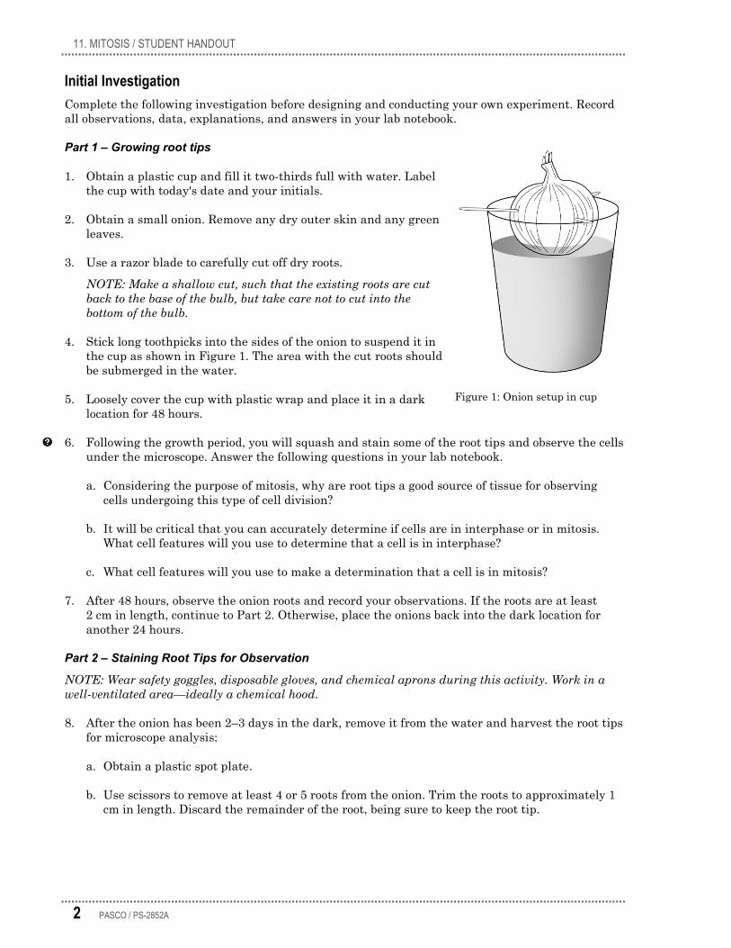
4. Stick long toothpicks into the sides of the onion to suspend it in the cup as shown in Figure 1. The area with the cut roots should be submerged in the water.
5. Loosely cover the cup with plastic wrap and place it in a dark location for 48 hours.
6. Following the growth period, you will squash and stain some of the root tips and observe the cells under the microscope. Answer the following questions in your lab notebook.
a. Considering the purpose of mitosis, why are root tips a good source of tissue for observing cells undergoing this type of cell division?
b. It will be critical that you can accurately determine if cells are in interphase or in mitosis. What cell features will you use to determine that a cell is in interphase?
c. What cell features will you use to make a determination that a cell is in mitosis?
7. After 48 hours, observe the onion roots and record your observations. If the roots are at least 2 cm in length, continue to Part 2. Otherwise, place the onions back into the dark location for another 24 hours.
Part 2 – Staining Root Tips for Observation
NOTE: Wear safety goggles, disposable gloves, and chemical aprons during this activity. Work in a well-ventilated area—ideally a chemical hood.
8. After the onion has been 2–3 days in the dark, remove it from the water and harvest the root tips for microscope analysis:
a. Obtain a plastic spot plate.
b. Use scissors to remove at least 4 or 5 roots from the onion. Trim the roots to approximately 1 cm in length. Discard the remainder of the root, being sure to keep the root tip.
Figure 1: Onion setup in cup
11. MITOSIS / STUDENT HANDOUT
PASCO / PS-2852A 3
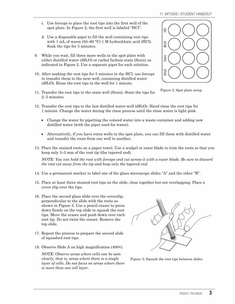
c. Use forceps to place the root tips into the first well of the spot plate. In Figure 2, the first well is labeled “HCl”.
d. Use a disposable pipet to fill the well containing root tips with 1 mL of warm (50–60 °C) 1 M hydrochloric acid (HCl). Soak the tips for 5 minutes.
9. While you wait, fill three more wells in the spot plate with either distilled water (dH2O) or carbol fuchsin stain (Stain) as indicated in Figure 2. Use a separate pipet for each solution.
10. After soaking the root tips for 5 minutes in the HCl, use forceps to transfer them to the next well, containing distilled water (dH2O). Rinse the root tips in the well for 1 minute.
11. Transfer the root tips to the stain well (Stain). Stain the tips for 2–3 minutes.
12. Transfer the root tips to the last distilled water well (dH2O). Hand rinse the root tips for 1 minute. Change the water during the rinse process until the rinse water is light pink.
• Change the water by pipetting the colored water into a waste container and adding new distilled water (with the pipet used for water).
• Alternatively, if you have extra wells in the spot plate, you can fill them with distilled water and transfer the roots from one well to another.
13. Place the stained roots on a paper towel. Use a scalpel or razor blade to trim the roots so that you keep only 3–5 mm of the root tip (the tapered end).
NOTE: You can hold the root with forceps and cut across it with a razor blade. Be sure to discard the root cut away from the tip and keep only the tapered end.
14. Use a permanent marker to label one of the glass microscope slides “A” and the other “B”.
15. Place at least three stained root tips on the slide, close together but not overlapping. Place a cover slip over the tips.
16. Place the second glass slide over the coverslip, perpendicular to the slide with the roots as shown in Figure 3. Use a pencil eraser to press down firmly on the top slide to squash the root tips. Move the eraser and push down over each root tip. Do not twist the eraser. Remove the top slide.
17. Repeat the process to prepare the second slide of squashed root tips.
18. Observe Slide A on high magnification (400×).
NOTE: Observe areas where cells can be seen clearly, that is, areas where there is a single layer of cells. Do not focus on areas where there is more than one cell layer.
Figure 3: Squash the root tips between slides
Figure 2: Spot plate setup
11. MITOSIS / STUDENT HANDOUT
4 PASCO / PS-2852A
19. Some cells will appear elongated and rectangular, other cells will appear small and square in shape. In regions with small, square cells, look for cells in metaphase or anaphase. These phases will be the easiest to recognize and can help you locate a good field of view (FOV) to count cells.
20. When you find a FOV that has at least two cells showing evidence of mitosis, determine a systematic way of viewing and counting all of the cells in that FOV. Every well-stained, distinct cell in the field of view should be counted.
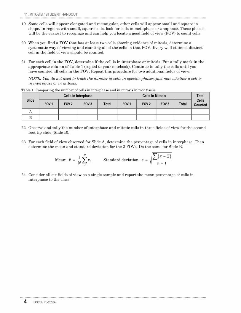
21. For each cell in the FOV, determine if the cell is in interphase or mitosis. Put a tally mark in the appropriate column of Table 1 (copied to your notebook). Continue to tally the cells until you have counted all cells in the FOV. Repeat this procedure for two additional fields of view.
NOTE: You do not need to track the number of cells in specific phases, just note whether a cell is in interphase or in mitosis.
Table 1: Comparing the number of cells in interphase and in mitosis in root tissue
Slide Cells in Interphase Cells in Mitosis Total
Cells Counted FOV 1 FOV 2 FOV 3 Total FOV 1 FOV 2 FOV 3 Total
A B
22. Observe and tally the number of interphase and mitotic cells in three fields of view for the second root tip slide (Slide B).
23. For each field of view observed for Slide A, determine the percentage of cells in interphase. Then determine the mean and standard deviation for the 3 FOVs. Do the same for Slide B.
( )1
1Mean: Standard deviation: 1
N
ii
x xx x s
N n=
−= =
−∑∑
24. Consider all six fields of view as a single sample and report the mean percentage of cells in interphase to the class.
11. MITOSIS / STUDENT HANDOUT
PASCO / PS-2852A 5
Data Analysis
1. Find the mean and standard deviation for the data provided by all groups.
2. Given the standard deviation for your group data and class data, what can you conclude regarding the amount of variation in the samples?
Chi-square analysis
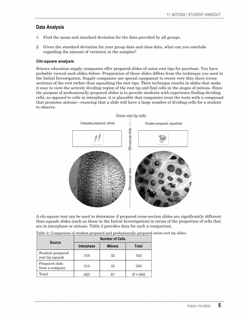
Science education supply companies offer prepared slides of onion root tips for purchase. You have probably viewed such slides before. Preparation of these slides differs from the technique you used in the Initial Investigation. Supply companies use special equipment to create very thin slices (cross-sections) of the root rather than squashing the root tips. Their technique results in slides that make it easy to view the actively dividing region of the root tip and find cells in the stages of mitosis. Since the purpose of professionally prepared slides is to provide students with experience finding dividing cells, as opposed to cells in interphase, it is plausible that companies treat the roots with a compound that promotes mitosis—ensuring that a slide will have a large number of dividing cells for a student to observe.
A chi-square test can be used to determine if prepared cross-section slides are significantly different than squash slides (such as those in the Initial Investigation) in terms of the proportion of cells that are in interphase or mitosis. Table 2 provides data for such a comparison. Table 2: Comparison of student-prepared and professionally-prepared onion root tip slides
Source Number of Cells
Interphase Mitosis Total Student-prepared root tip squash 310 32 342
Prepared slide from a company 315 35 350
Total 625 67 N = 692
11. MITOSIS / STUDENT HANDOUT
6 PASCO / PS-2852A
The formula used to calculate a chi-square value is:
( )22 o e
e−
= ∑Χ
Typically, a chi-square test is a goodness-of-fit test. This test is applicable in situations where expected results can be calculated from theory, such as a predicted phenotypic ratio in the F1 generation of fruit flies based on the principles of Mendelian inheritance. The null hypothesis for a goodness-of-fit test states there is no difference between the observed o and expected e (theoretical) values.
However, in treatment studies, a different type of chi-square test is applied. This test of independence operates under the null hypothesis that there is no association between two groups (or two variables)—the two groups are independent. For the provided data, the null hypothesis is that the probability of a cell being in interphase or mitosis is independent of its source: a prepared slide from a company or a student-prepared squashed root tip. The same chi-square formula is used for goodness-of-fit tests and tests of independence. However, in the test of independence expected frequencies are derived from observed frequencies, rather than from theory. The observations for the two groups for each category are recorded in a contingency table. Table 2 is an example of a contingency table (also known as a 2 × 2 contingency table).
NOTE: The degrees of freedom (df) for this type of chi-square test is one.
In the chi-square test of independence, to calculate the expected values from observed values, apply the following Law of Probability:
If A and B are independent, then the probability P of A and B both occurring is:
P(A and B) = P(A) × P(B)
Considering the total number of cells observed, the probability of A and B both occurring in a sample size of N would be:
[P(A) × P(B)]/N
3. Copy Table 3 into your lab notebook. Table 3: Calculation of chi square
Source Observed Expected (o – e) (o – e)2 [(o – e)2]/e Root tip squash Interphase 310
Root tip squash Mitosis 32
Prepared slide Interphase 315
Prepared slide Mitosis 35
X2 = Σ[(o – e)2]/e =
From the observed values o and the total number of cells counted N, calculate each of the following:
a. What is the probability that a cell will be in interphase and sourced from the student-prepared squash? Record the value in the “Expected” column of Table 3.
b. What is the probability that a cell will be in mitosis and sourced from the student-prepared squash? Record the value in the “Expected” column of Table 3.
RECORD ANSWERS & DATA IN YOUR NOTEBOOK.
11. MITOSIS / STUDENT HANDOUT
PASCO / PS-2852A 7
4. Calculate and record the remaining expected values to complete the Expected column of Table 3.
5. Perform the calculations necessary to complete the remainder of Table 3. Find the sum of the values in the last column to determine the chi-square value.
6. With regard to the null hypothesis, what can you conclude from the calculated chi square? Table 4: Chi-square distribution
Degrees of Freedom
Probability p Value
0.75 0.50 0.25 0.10 0.05 0.01
1 0.10 0.46 1.32 2.71 3.84 6.64 2 0.58 1.30 2.77 4.60 5.99 9.21 3 1.21 2.37 4.11 6.25 7.82 11.34 4 1.92 3.36 5.39 7.78 9.49 13.28
df = (number of groups – 1) × (number of categories – 1)
7. Do the chi-square results support the conjecture that supply companies treat roots with a compound to increase the rate of mitosis? Does the data provide conclusive evidence? Explain your answer.
8. Identify any new questions that have arisen as a result of your research.
Synthesis Questions
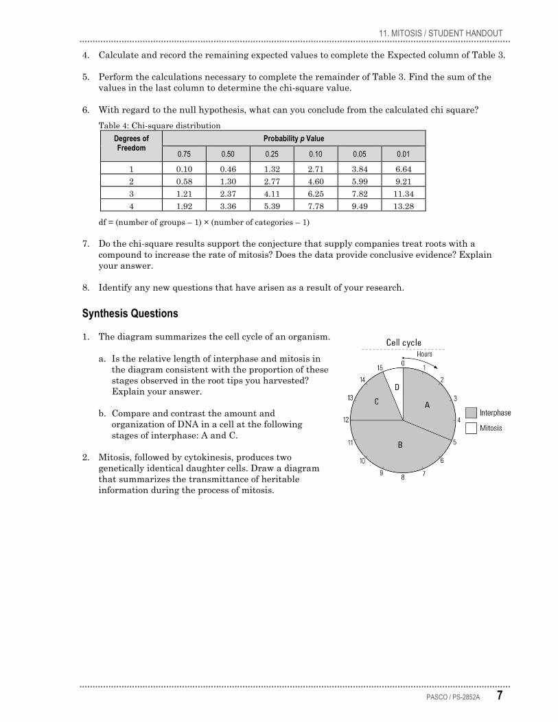
1. The diagram summarizes the cell cycle of an organism.
a. Is the relative length of interphase and mitosis in the diagram consistent with the proportion of these stages observed in the root tips you harvested? Explain your answer.
b. Compare and contrast the amount and organization of DNA in a cell at the following stages of interphase: A and C.
2. Mitosis, followed by cytokinesis, produces two genetically identical daughter cells. Draw a diagram that summarizes the transmittance of heritable information during the process of mitosis.
11. MITOSIS / STUDENT HANDOUT
8 PASCO / PS-2852A
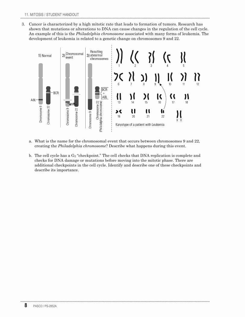
3. Cancer is characterized by a high mitotic rate that leads to formation of tumors. Research has shown that mutations or alterations to DNA can cause changes in the regulation of the cell cycle. An example of this is the Philadelphia chromosome associated with many forms of leukemia. The development of leukemia is related to a genetic change on chromosomes 9 and 22.
a. What is the name for the chromosomal event that occurs between chromosomes 9 and 22, creating the Philadelphia chromosome? Describe what happens during this event.
b. The cell cycle has a G2 “checkpoint.” The cell checks that DNA replication is complete and checks for DNA damage or mutations before moving into the mitotic phase. There are additional checkpoints in the cell cycle. Identify and describe one of these checkpoints and describe its importance.

11. MITOSIS / STUDENT HANDOUT
PASCO / PS-2852A 9
4. A study was performed to determine the effects of 24-epibrassinolide (BL), a plant steroid hormone, on the mitotic index of onion root tips.1 Investigators compared meristematic tissues of control onion bulbs to onion bulbs in experimental groups after 48 hours of root growth.
The mitotic index is the percentage of cells in mitosis relative to the total number of cells examined. The mitotic index was calculated by counting 400 cells from five root tips obtained from each group (2000 cells total from each group). Data from this study is summarized in Table 5. Table 5: Effects of BL on mitosis in Allium roots
Concentration of BL Number of Cells
Mitotic Index ± SD Interphase Mitosis Total
Control (spring water) 1907 93 2000 4.65 ± 1.34
0.5 ppm BL 1918 82 2000 4.10 ± 1.34 0.05 ppm BL 1867 133 2000 6.65 ± 0.69
a. What can be concluded from this study? Use chi-square analysis to provide evidence for your conclusions.
b. Another study showed that hormones like BL induce transcription of a cyclin gene. Explain the role of cyclins in the cell cycle.
Design and Conduct an Experiment If your teacher determines there is sufficient time and materials for you to carry out an experiment of your own design, explore factors that are likely to affect the rate of mitosis in organisms. For example, a number of studies involve testing compounds to see if they stimulate or inhibit mitosis.
Design and carry out your experiment using either the Design and Conduct an Experiment Worksheet or the Experiment Design Plan.
1 Howell, W.M.; Keller III, G.E.; Kirkpatrick, J.D.; Jenkins, R.L.; Hunsinger, R.N.; McLaughlin, E.W. Effects of the plant steroidal hormone, 24-epibrassinolide, on the mitotic index and growth of onion (Allium cepa) root tips. Genetics and Molecular Research, Online Journal. Dept. of Biology, Samford University, Birmingham, AL. 2007 Retrieved April, 2014 from http://www.funpecrp.com.br/gmr/year2007/vol1-6/gmr0259_full_text.htm
11. MITOSIS / STUDENT HANDOUT
10 PASCO / PS-2852A
Design and Conduct an Experiment Worksheet A number of factors, internal and external, are likely to affect the rate of mitosis in organisms. Identify one of these factors and design an experiment to determine how that factor stimulates or inhibits mitosis.
Develop and conduct your experiment using the following guide.
1. Based on your knowledge of mitosis, what factors (abiotic or biotic) could affect this process?
____________________________________________________________________________________________
____________________________________________________________________________________________
2. Create a driving question: choose one of the factors you've identified that can be controlled in the lab and develop a testable question for your experiment.
____________________________________________________________________________________________
____________________________________________________________________________________________
3. What is the justification for your question? That is, why is it biologically significant, relevant, or interesting?
____________________________________________________________________________________________
____________________________________________________________________________________________
4. What will be the independent variable of the experiment? Describe how this variable will be manipulated in your experiment.
____________________________________________________________________________________________
____________________________________________________________________________________________
____________________________________________________________________________________________
5. What is the dependent variable of the experiment? Describe how the data will be collected and processed in the experiment.
____________________________________________________________________________________________
____________________________________________________________________________________________
6. Write a testable hypothesis (If…then…).
____________________________________________________________________________________________
____________________________________________________________________________________________
7. What conditions will need to be held constant in the experiment? Quantify these values where possible.
____________________________________________________________________________________________
____________________________________________________________________________________________
11. MITOSIS / STUDENT HANDOUT
PASCO / PS-2852A 11
8. How many trials will be run for each experimental group? Justify your choice.
____________________________________________________________________________________________
____________________________________________________________________________________________
9. It is beneficial to estimate at the beginning of the experiment the sample size needed to make statistically valid conclusions. The formula below can be used for this purpose. In the formula, n represents an adequate sample size, that is, the number of cells that need to be counted to compare the control and experimental groups. Solve for n in the equation. How many cells should you count in each of your groups?
NOTE: The formula makes assumptions of other statistical values, such as margin of error and critical value.
0.9 0.10.03 2n×
= ×
10. What will you compare or calculate? What analysis will you perform to evaluate your results and hypothesis?
____________________________________________________________________________________________
____________________________________________________________________________________________
11. Describe at least 3 potential sources of error that could affect the accuracy or reliability of data.
____________________________________________________________________________________________
____________________________________________________________________________________________
12. Use the space below to create an outline of the experiment. In your lab notebook, write the steps for the procedure of the lab. (Another student or group should be able to repeat the procedure and obtain similar results.)
13. Have your teacher approve your answers to these questions and your plan before beginning the experiment.
![Index [ ] · PDF fileBone Holding Forceps ... Burley Disc Scoop ... Eye Enucleation Forceps](https://img.pdfslide.net/doc/110x75/5aa15ab17f8b9ac67a8baaaa/index-holding-forceps-burley-disc-scoop-eye-enucleation-forceps.jpg)