Embed Size (px)
Citation preview
251
CHAPTER
1616.1 Glucose Can Be Synthesized from
Noncarbohydrate Precursors
16.2 Gluconeogenesis and GlycolysisAre Reciprocally Regulated
16.3 Metabolism in Context:Precursors Formed by Muscle AreUsed by Other Organs
16.4 Glycolysis and GluconeogenesisAre Evolutionarily Intertwined
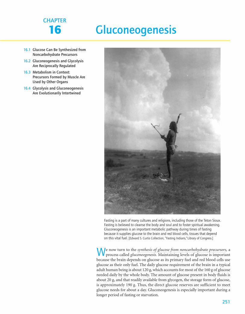
Fasting is a part of many cultures and religions, including those of the Teton Sioux.Fasting is believed to cleanse the body and soul and to foster spiritual awakening.Gluconeogenesis is an important metabolic pathway during times of fastingbecause it supplies glucose to the brain and red blood cells, tissues that dependon this vital fuel. [Edward S. Curtis Collection, ”Fasting Indians,”Library of Congress.]
Gluconeogenesis
We now turn to the synthesis of glucose from noncarbohydrate precursors, aprocess called gluconeogenesis. Maintaining levels of glucose is important
because the brain depends on glucose as its primary fuel and red blood cells useglucose as their only fuel. The daily glucose requirement of the brain in a typicaladult human being is about 120 g, which accounts for most of the 160 g of glucoseneeded daily by the whole body. The amount of glucose present in body fluids isabout 20 g, and that readily available from glycogen, the storage form of glucose,is approximately 190 g. Thus, the direct glucose reserves are sufficient to meetglucose needs for about a day. Gluconeogenesis is especially important during alonger period of fasting or starvation.
The major site of gluconeogenesis is the liver, with a small amount also tak-ing place in the kidney. Little gluconeogenesis takes place in the brain, skeletalmuscle, or heart muscle. Rather, gluconeogenesis in the liver and kidney helps tomaintain the glucose level in the blood where it can be extracted by the brain andmuscle to meet their metabolic demands.
In this chapter, we begin by examining the reactions that constitute the glu-coneogenic pathway. We then investigate the reciprocal regulation of gluconeoge-nesis and glycolysis, and end the chapter with a look at how gluconeogenesis andglycolysis are coordinated between tissues.
16.1 Glucose Can Be Synthesized from Noncarbohydrate Precursors
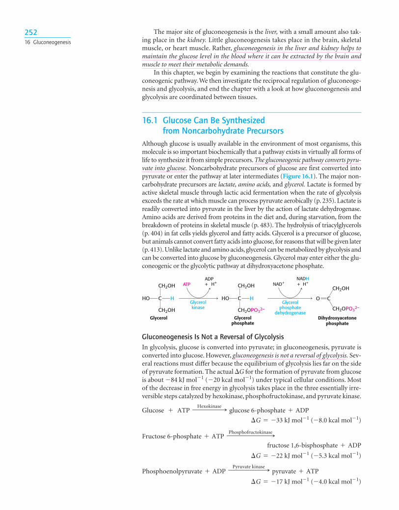
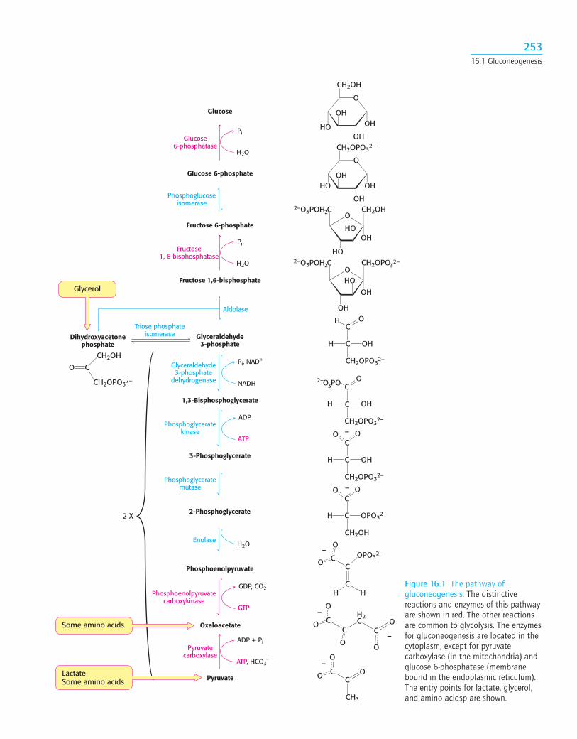
Although glucose is usually available in the environment of most organisms, thismolecule is so important biochemically that a pathway exists in virtually all forms oflife to synthesize it from simple precursors. The gluconeogenic pathway converts pyru-vate into glucose. Noncarbohydrate precursors of glucose are first converted intopyruvate or enter the pathway at later intermediates (Figure 16.1). The major non-carbohydrate precursors are lactate, amino acids, and glycerol. Lactate is formed byactive skeletal muscle through lactic acid fermentation when the rate of glycolysisexceeds the rate at which muscle can process pyruvate aerobically (p. 235). Lactate isreadily converted into pyruvate in the liver by the action of lactate dehydrogenase.Amino acids are derived from proteins in the diet and, during starvation, from thebreakdown of proteins in skeletal muscle (p. 483). The hydrolysis of triacylglycerols(p. 404) in fat cells yields glycerol and fatty acids. Glycerol is a precursor of glucose,but animals cannot convert fatty acids into glucose, for reasons that will be given later(p. 413). Unlike lactate and amino acids, glycerol can be metabolized by glycolysis andcan be converted into glucose by gluconeogenesis. Glycerol may enter either the glu-coneogenic or the glycolytic pathway at dihydroxyacetone phosphate.
25216 Gluconeogenesis
ATP NAD+NADH+ H+
Glycerolkinase
Glycerolphosphate
dehydrogenaseGlycerol Glycerol
phosphateDihydroxyacetone
phosphate
CH2OH
C
CH2OH
HHO
CH2OH
CH2OPO32–
HHO O
CH2OH
CH2OPO32–
C C
ADP+ H+
Gluconeogenesis Is Not a Reversal of GlycolysisIn glycolysis, glucose is converted into pyruvate; in gluconeogenesis, pyruvate isconverted into glucose. However, gluconeogenesis is not a reversal of glycolysis. Sev-eral reactions must differ because the equilibrium of glycolysis lies far on the sideof pyruvate formation. The actual �G for the formation of pyruvate from glucoseis about �84 kJ mol�1 (�20 kcal mol�1) under typical cellular conditions. Mostof the decrease in free energy in glycolysis takes place in the three essentially irre-versible steps catalyzed by hexokinase, phosphofructokinase, and pyruvate kinase.
¢G = -17 kJ mol-1 (-4.0 kcal mol-1)
Phosphoenolpyruvate + ADPPyruvate kinase99999: pyruvate + ATP
¢G = -22 kJ mol-1 (-5.3 kcal mol-1)
fructose 1,6-bisphosphate + ADPFructose 6-phosphate + ATP
Phosphofructokinase999999:¢G = -33 kJ mol-1 (-8.0 kcal mol-1)
Glucose + ATPHexokinase9999: glucose 6-phosphate + ADP
Glycerol
Glucose
Glucose 6-phosphate
Fructose 6-phosphate
Fructose 1,6-bisphosphate
Dihydroxyacetonephosphate
Glyceraldehyde3-phosphate
1,3-Bisphosphoglycerate
3-Phosphoglycerate
2-Phosphoglycerate
Phosphoenolpyruvate
Pyruvate
Oxaloacetate
2 X
Aldolase
O
OH
CH2OH
OH
OH
HO
O
OH
CH2OPO32–
OH
OHHO
OCH2OH
HO
HO
2–O3POH C2
OH
32–
OCH2OPO
HO
OH
2–O3POH C2
OH
C
C
CH2OPO32–
OHH
OO –
C
C OPO32–H
OO –
CH2OH
OPO32–
H H
O
O
–
–
CH3
O
CO
CH2OPO32–
CH2OH
C
C
CH2OPO32–
OHH
O PO32– O
C
C
CH2OPO32–
OHH
OH
Pi
H2O
Glucose6-phosphatase
Pi
H2O
Fructose1, 6-bisphosphatase
Phosphoglucoseisomerase
Triose phosphateisomerase
Pi, NAD+
NADH
Glyceraldehyde3-phosphate
dehydrogenase
Phosphoglyceratekinase
ADP
ATP
Phosphoenolpyruvatecarboxykinase
GDP, CO2
GTP
Phosphoglyceratemutase
H2OEnolase
Pyruvatecarboxylase
ADP + Pi
ATP, HCO3–
H2C
O
CO
O
Some amino acids
LactateSome amino acids
C
O
O
–C
O
O
–C
C
C
C
C
Figure 16.1 The pathway ofgluconeogenesis. The distinctivereactions and enzymes of this pathwayare shown in red. The other reactionsare common to glycolysis. The enzymesfor gluconeogenesis are located in thecytoplasm, except for pyruvatecarboxylase (in the mitochondria) andglucose 6-phosphatase (membranebound in the endoplasmic reticulum).The entry points for lactate, glycerol,and amino acidsp are shown.
25316.1 Gluconeogenesis
In gluconeogenesis, the following new steps bypass these virtually irreversiblereactions of glycolysis:
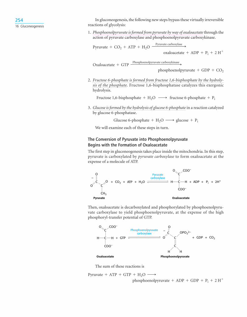
1. Phosphoenolpyruvate is formed from pyruvate by way of oxaloacetate through theaction of pyruvate carboxylase and phosphoenolpyruvate carboxykinase.
2. Fructose 6-phosphate is formed from fructose 1,6-bisphosphate by the hydroly-sis of the phosphate. Fructose 1,6-bisphosphatase catalyzes this exergonichydrolysis.
3. Glucose is formed by the hydrolysis of glucose 6-phosphate in a reaction catalyzedby glucose 6-phosphatase.
We will examine each of these steps in turn.
The Conversion of Pyruvate into Phosphoenolpyruvate Begins with the Formation of OxaloacetateThe first step in gluconeogenesis takes place inside the mitochondria. In this step,pyruvate is carboxylated by pyruvate carboxylase to form oxaloacetate at theexpense of a molecule of ATP.
Glucose 6-phosphate + H2O ¡ glucose + Pi
Fructose 1,6-bisphosphate + H2O ¡ fructose 6-phosphate + Pi
phosphoenolpyruvate + GDP + CO2
Oxaloacetate + GTPPhosphoenolpyruvate carboxykinase99999999999:
oxaloacetate + ADP + Pi + 2 H+Pyruvate + CO2 + ATP + H2O
Pyruvate carboxylase9999999:
25416 Gluconeogenesis
+ + H2O+CO2 + Pi+ +ADP 2H+
C
C
COO–
HH
O COO–
Pyruvate Oxaloacetate
Pyruvatecarboxylase
ATP
O
CC
CH3
OO
–
+ GTP + +GDPC
C
H H
C
O
OPO32–
OC
COO–
HH
Oxaloacetate Phosphoenolpyruvate
CO2
–CO COO–
Phosphoenolpyruvatecarboxylase
Then, oxaloacetate is decarboxylated and phosphorylated by phosphoenolpyru-vate carboxylase to yield phosphoenolpyruvate, at the expense of the highphosphoryl-transfer potential of GTP.
The sum of these reactions is
phosphoenolpyruvate + ADP + GDP + Pi + 2 H+Pyruvate + ATP + GTP + H2O ¡
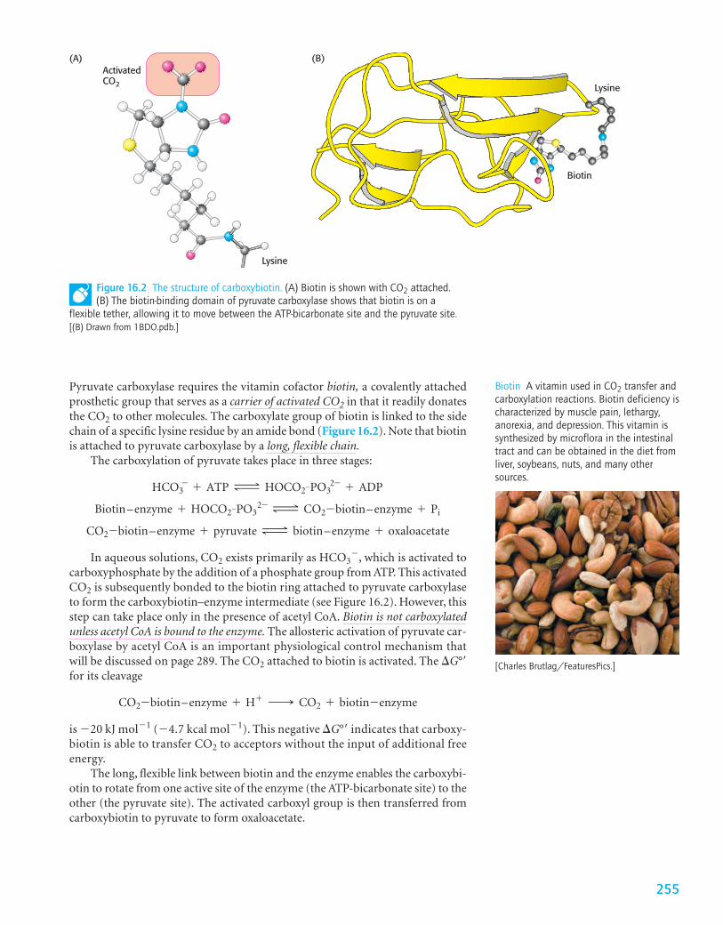
Pyruvate carboxylase requires the vitamin cofactor biotin, a covalently attachedprosthetic group that serves as a carrier of activated CO2 in that it readily donatesthe CO2 to other molecules. The carboxylate group of biotin is linked to the sidechain of a specific lysine residue by an amide bond (Figure 16.2). Note that biotinis attached to pyruvate carboxylase by a long, flexible chain.
The carboxylation of pyruvate takes place in three stages:
In aqueous solutions, CO2 exists primarily as HCO3�, which is activated to
carboxyphosphate by the addition of a phosphate group from ATP. This activatedCO2 is subsequently bonded to the biotin ring attached to pyruvate carboxylaseto form the carboxybiotin–enzyme intermediate (see Figure 16.2). However, thisstep can take place only in the presence of acetyl CoA. Biotin is not carboxylatedunless acetyl CoA is bound to the enzyme. The allosteric activation of pyruvate car-boxylase by acetyl CoA is an important physiological control mechanism thatwill be discussed on page 289. The CO2 attached to biotin is activated. The �G°�for its cleavage
is �20 kJ mol�1 (�4.7 kcal mol�1). This negative �G°� indicates that carboxy-biotin is able to transfer CO2 to acceptors without the input of additional freeenergy.
The long, flexible link between biotin and the enzyme enables the carboxybi-otin to rotate from one active site of the enzyme (the ATP-bicarbonate site) to theother (the pyruvate site). The activated carboxyl group is then transferred fromcarboxybiotin to pyruvate to form oxaloacetate.
CO2-biotin–enzyme + H+ ¡ CO2 + biotin-enzyme
CO2-biotin–enzyme + pyruvate Δ biotin–enzyme + oxaloacetate
Biotin–enzyme + HOCO2-PO32- Δ CO2-biotin–enzyme + Pi
HCO3- + ATP Δ HOCO2-PO3
2- + ADP
Biotin A vitamin used in CO2 transfer andcarboxylation reactions. Biotin deficiency ischaracterized by muscle pain, lethargy,anorexia, and depression. This vitamin issynthesized by microflora in the intestinaltract and can be obtained in the diet fromliver, soybeans, nuts, and many othersources.
(A)ActivatedCO2
Lysine
Figure 16.2 The structure of carboxybiotin. (A) Biotin is shown with CO2 attached.(B) The biotin-binding domain of pyruvate carboxylase shows that biotin is on a
flexible tether, allowing it to move between the ATP-bicarbonate site and the pyruvate site.[(B) Drawn from 1BDO.pdb.]
Biotin
Lysine
(B)
255
[Charles Brutlag/FeaturesPics.]
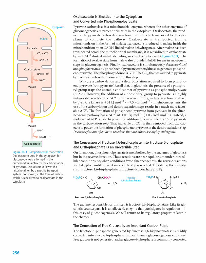
Oxaloacetate Is Shuttled into the Cytoplasm and Converted into PhosphoenolpyruvatePyruvate carboxylase is a mitochondrial enzyme, whereas the other enzymes ofgluconeogenesis are present primarily in the cytoplasm. Oxaloacetate, the prod-uct of the pyruvate carboxylase reaction, must thus be transported to the cyto-plasm to complete the pathway. Oxaloacetate is transported from amitochondrion in the form of malate: oxaloacetate is reduced to malate inside themitochondrion by an NADH-linked malate dehydrogenase. After malate has beentransported across the mitochondrial membrane, it is reoxidized to oxaloacetateby an NAD�-linked malate dehydrogenase in the cytoplasm (Figure 16.3). Theformation of oxaloacetate from malate also provides NADH for use in subsequentsteps in gluconeogenesis. Finally, oxaloacetate is simultaneously decarboxylatedand phosphorylated by phosphoenolpyruvate carboxykinase to generate phospho-enolpyruvate. The phosphoryl donor is GTP. The CO2 that was added to pyruvateby pyruvate carboxylase comes off in this step.
Why are a carboxylation and a decarboxylation required to form phospho-enolpyruvate from pyruvate? Recall that, in glycolysis, the presence of a phospho-ryl group traps the unstable enol isomer of pyruvate as phosphoenolpyruvate(p. 233). However, the addition of a phosphoryl group to pyruvate is a highlyunfavorable reaction: the �G°� of the reverse of the glycolytic reaction catalyzedby pyruvate kinase is �31 kJ mol�1 (�7.5 kcal mol�1). In gluconeogenesis, theuse of the carboxylation and decarboxylation steps results in a much more favor-able �G°�. The formation of phosphoenolpyruvate from pyruvate in the gluco-neogenic pathway has a �G°� of �0.8 kJ mol�1 (�0.2 kcal mol�1). Instead, amolecule of ATP is used to power the addition of a molecule of CO2 to pyruvatein the carboxylation step. That molecule of CO2 is then removed from oxaloac-etate to power the formation of phosphoenolpyruvate in the decarboxylation step.Decarboxylations often drive reactions that are otherwise highly endergonic.
The Conversion of Fructose 1,6-bisphosphate into Fructose 6-phosphateand Orthophosphate Is an Irreversible StepOn formation, phosphoenolpyruvate is metabolized by the enzymes of glycolysisbut in the reverse direction. These reactions are near equilibrium under intracel-lular conditions; so, when conditions favor gluconeogenesis, the reverse reactionswill take place until the next irreversible step is reached. This step is the hydroly-sis of fructose 1,6-bisphosphate to fructose 6-phosphate and Pi.
256
Cytoplasm
Matrix
Pyruvate
CO2 + ATP
ADP + Pi
NADH + H+
NAD+
NADH + H+
NAD+
Oxaloacetate
Oxaloacetate
Malate
Malate
Figure 16.3 Compartmental cooperation.Oxaloacetate used in the cytoplasm forgluconeogenesis is formed in themitochondrial matrix by the carboxylationof pyruvate. Oxaloacetate leaves themitochondrion by a specific transportsystem (not shown) in the form of malate,which is reoxidized to oxaloacetate in thecytoplasm.
+ H2O + Pi
Fructose1,6-bisphosphatase
Fructose 1,6-bisphosphate Fructose 6-phosphate
OCH2OH
HO
2–O3POH C2
OH
O32–CH2OPO
HO
OHOH
2–O3POH C2
OH
The enzyme responsible for this step is fructose 1,6-bisphosphatase. Like its gly-colytic counterpart, it is an allosteric enzyme that participates in regulation—inthis case, of gluconeogenesis. We will return to its regulatory properties later inthe chapter.
The Generation of Free Glucose Is an Important Control PointThe fructose 6-phosphate generated by fructose 1,6-bisphosphatase is readilyconverted into glucose 6-phosphate. In most tissues, gluconeogenesis ends here.Free glucose is not generated; rather glucose 6-phosphate is commonly converted
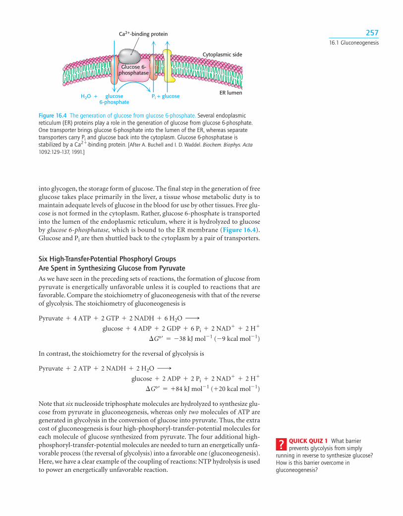
into glycogen, the storage form of glucose. The final step in the generation of freeglucose takes place primarily in the liver, a tissue whose metabolic duty is tomaintain adequate levels of glucose in the blood for use by other tissues. Free glu-cose is not formed in the cytoplasm. Rather, glucose 6-phosphate is transportedinto the lumen of the endoplasmic reticulum, where it is hydrolyzed to glucoseby glucose 6-phosphatase, which is bound to the ER membrane (Figure 16.4).Glucose and Pi are then shuttled back to the cytoplasm by a pair of transporters.
Six High-Transfer-Potential Phosphoryl Groups Are Spent in Synthesizing Glucose from PyruvateAs we have seen in the preceding sets of reactions, the formation of glucose frompyruvate is energetically unfavorable unless it is coupled to reactions that arefavorable. Compare the stoichiometry of gluconeogenesis with that of the reverseof glycolysis. The stoichiometry of gluconeogenesis is
In contrast, the stoichiometry for the reversal of glycolysis is
Note that six nucleoside triphosphate molecules are hydrolyzed to synthesize glu-cose from pyruvate in gluconeogenesis, whereas only two molecules of ATP aregenerated in glycolysis in the conversion of glucose into pyruvate. Thus, the extracost of gluconeogenesis is four high-phosphoryl-transfer-potential molecules foreach molecule of glucose synthesized from pyruvate. The four additional high-phosphoryl-transfer-potential molecules are needed to turn an energetically unfa-vorable process (the reversal of glycolysis) into a favorable one (gluconeogenesis).Here, we have a clear example of the coupling of reactions: NTP hydrolysis is usedto power an energetically unfavorable reaction.
¢G°¿ = +84 kJ mol-1 (+20 kcal mol-1)
glucose + 2 ADP + 2 Pi + 2 NAD+ + 2 H+Pyruvate + 2 ATP + 2 NADH + 2 H2O ¡
¢G°¿ = -38 kJ mol-1 (-9 kcal mol-1)
glucose + 4 ADP + 2 GDP + 6 Pi + 2 NAD+ + 2 H+Pyruvate + 4 ATP + 2 GTP + 2 NADH + 6 H2O ¡
25716.1 Gluconeogenesis
Cytoplasmic side
Glucose 6-phosphatase
Ca2+-binding protein
Pi + glucoseH2O + glucose6-phosphate
ER lumen
Figure 16.4 The generation of glucose from glucose 6-phosphate. Several endoplasmicreticulum (ER) proteins play a role in the generation of glucose from glucose 6-phosphate.One transporter brings glucose 6-phosphate into the lumen of the ER, whereas separatetransporters carry Pi and glucose back into the cytoplasm. Glucose 6-phosphatase isstabilized by a Ca2�-binding protein. [After A. Buchell and I. D. Waddel. Biochem. Biophys. Acta1092:129–137, 1991.]
QUICK QUIZ 1 What barrierprevents glycolysis from simply
running in reverse to synthesize glucose?How is this barrier overcome ingluconeogenesis?
16.2 Gluconeogenesis and Glycolysis Are Reciprocally Regulated
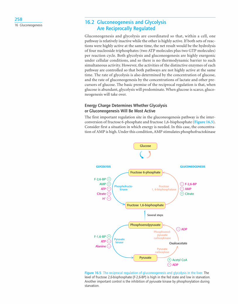
Gluconeogenesis and glycolysis are coordinated so that, within a cell, one pathway is relatively inactive while the other is highly active. If both sets of reac-tions were highly active at the same time, the net result would be the hydrolysisof four nucleoside triphosphates (two ATP molecules plus two GTP molecules)per reaction cycle. Both glycolysis and gluconeogenesis are highly exergonicunder cellular conditions, and so there is no thermodynamic barrier to suchsimultaneous activity. However, the activities of the distinctive enzymes of eachpathway are controlled so that both pathways are not highly active at the sametime. The rate of glycolysis is also determined by the concentration of glucose,and the rate of gluconeogenesis by the concentrations of lactate and other pre-cursors of glucose. The basic premise of the reciprocal regulation is that, whenglucose is abundant, glycolysis will predominate. When glucose is scarce, gluco-neogenesis will take over.
Energy Charge Determines Whether Glycolysis or Gluconeogenesis Will Be Most ActiveThe first important regulation site in the gluconeogenesis pathway is the inter-conversion of fructose 6-phosphate and fructose 1,6-bisphosphate (Figure 16.5).Consider first a situation in which energy is needed. In this case, the concentra-tion of AMP is high. Under this condition, AMP stimulates phosphofructokinase
25816 Gluconeogenesis
+−
Phosphoenolpyruvate
Pyruvatekinase
Pyruvatecarboxylase
Phosphoenol-pyruvate
carboxykinase
Several steps
Pyruvate
Glucose
Acetyl CoA
ADP
+−
F-1,6-BP
ATP−Alanine
− ADP
Oxaloacetate
Fructose 6-phosphate
Phosphofructo-kinase
Fructose1, 6-bisphosphatase
GLUCONEOGENESISGLYCOLYSIS
Fructose 1,6-bisphosphate
+−
AMP
+F-2,6-BP
ATP−Citrate−H+
−+ Citrate
AMP
− F-2,6-BP
Figure 16.5 The reciprocal regulation of gluconeogenesis and glycolysis in the liver. Thelevel of fructose 2,6-bisphosphate (F-2,6-BP) is high in the fed state and low in starvation.Another important control is the inhibition of pyruvate kinase by phosphorylation duringstarvation.
but inhibits fructose 1,6-bisphosphatase. Thus, glycolysis is turned on and gluconeogenesis is inhibited. Conversely, high levels of ATP and citrate indicatethat the energy charge is high and that biosynthetic intermediates are abundant.ATP and citrate inhibit phosphofructokinase, whereas citrate activates fructose1,6-bisphosphatase. Under these conditions, glycolysis is nearly switched off andgluconeogenesis is promoted. Why does citrate take part in this regulatoryscheme? As we will see in Chapter 18, citrate reports on the status of the citricacid cycle, the primary pathway for oxidizing fuels in the presence of oxygen.High levels of citrate indicate an energy-rich situation and the presence ofprecursors for biosynthesis.
Glycolysis and gluconeogenesis are also reciprocally regulated at the inter-conversion of phosphoenolpyruvate and pyruvate in the liver. The glycolyticenzyme pyruvate kinase is inhibited by allosteric effectors ATP and alanine,which signal that the energy charge is high and that building blocks are abun-dant. Conversely, pyruvate carboxylase, which catalyzes the first step in gluco-neogenesis from pyruvate, is inhibited by ADP. Likewise, ADP inhibitsphosphoenolpyruvate carboxykinase. Pyruvate carboxylase is activated by acetylCoA, which, like citrate, indicates that the citric acid cycle is producing energyand biosynthetic intermediates (Chapter 18). Hence, gluconeogenesis is favoredwhen the cell is rich in biosynthetic precursors and ATP.
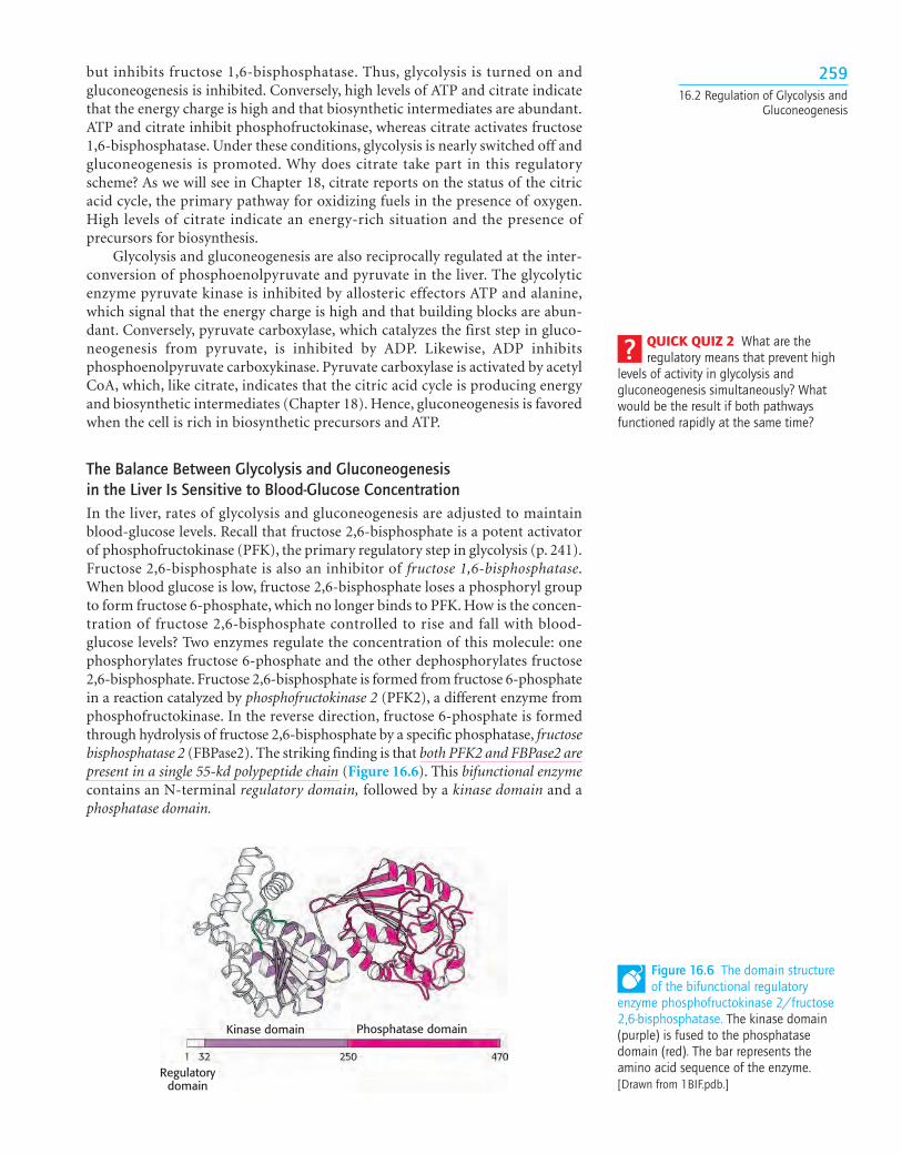
The Balance Between Glycolysis and Gluconeogenesis in the Liver Is Sensitive to Blood-Glucose ConcentrationIn the liver, rates of glycolysis and gluconeogenesis are adjusted to maintainblood-glucose levels. Recall that fructose 2,6-bisphosphate is a potent activatorof phosphofructokinase (PFK), the primary regulatory step in glycolysis (p. 241).Fructose 2,6-bisphosphate is also an inhibitor of fructose 1,6-bisphosphatase.When blood glucose is low, fructose 2,6-bisphosphate loses a phosphoryl groupto form fructose 6-phosphate, which no longer binds to PFK. How is the concen-tration of fructose 2,6-bisphosphate controlled to rise and fall with blood-glucose levels? Two enzymes regulate the concentration of this molecule: onephosphorylates fructose 6-phosphate and the other dephosphorylates fructose2,6-bisphosphate. Fructose 2,6-bisphosphate is formed from fructose 6-phosphatein a reaction catalyzed by phosphofructokinase 2 (PFK2), a different enzyme fromphosphofructokinase. In the reverse direction, fructose 6-phosphate is formedthrough hydrolysis of fructose 2,6-bisphosphate by a specific phosphatase, fructosebisphosphatase 2 (FBPase2). The striking finding is that both PFK2 and FBPase2 arepresent in a single 55-kd polypeptide chain (Figure 16.6). This bifunctional enzymecontains an N-terminal regulatory domain, followed by a kinase domain and aphosphatase domain.
25916.2 Regulation of Glycolysis and
Gluconeogenesis
Regulatorydomain
Kinase domain Phosphatase domain
Figure 16.6 The domain structureof the bifunctional regulatory
enzyme phosphofructokinase 2/fructose2,6-bisphosphatase. The kinase domain(purple) is fused to the phosphatasedomain (red). The bar represents theamino acid sequence of the enzyme.[Drawn from 1BIF.pdb.]
QUICK QUIZ 2 What are theregulatory means that prevent high
levels of activity in glycolysis andgluconeogenesis simultaneously? Whatwould be the result if both pathwaysfunctioned rapidly at the same time?
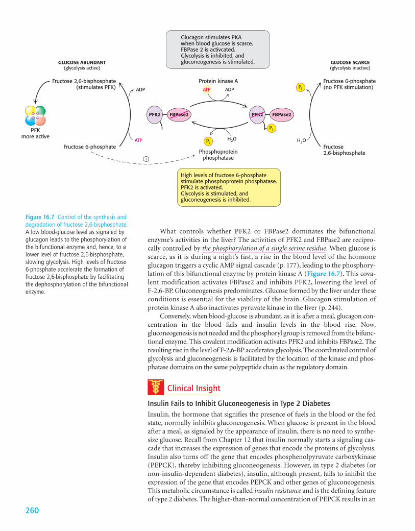
What controls whether PFK2 or FBPase2 dominates the bifunctionalenzyme’s activities in the liver? The activities of PFK2 and FBPase2 are recipro-cally controlled by the phosphorylation of a single serine residue. When glucose isscarce, as it is during a night’s fast, a rise in the blood level of the hormoneglucagon triggers a cyclic AMP signal cascade (p. 177), leading to the phosphory-lation of this bifunctional enzyme by protein kinase A (Figure 16.7). This cova-lent modification activates FBPase2 and inhibits PFK2, lowering the level ofF-2,6-BP. Gluconeogenesis predominates. Glucose formed by the liver under theseconditions is essential for the viability of the brain. Glucagon stimulation ofprotein kinase A also inactivates pyruvate kinase in the liver (p. 244).
Conversely, when blood-glucose is abundant, as it is after a meal, glucagon con-centration in the blood falls and insulin levels in the blood rise. Now,gluconeogenesis is not needed and the phosphoryl group is removed from the bifunc-tional enzyme. This covalent modification activates PFK2 and inhibits FBPase2. Theresulting rise in the level of F-2,6-BP accelerates glycolysis. The coordinated control ofglycolysis and gluconeogenesis is facilitated by the location of the kinase and phos-phatase domains on the same polypeptide chain as the regulatory domain.
Clinical Insight
Insulin Fails to Inhibit Gluconeogenesis in Type 2 DiabetesInsulin, the hormone that signifies the presence of fuels in the blood or the fedstate, normally inhibits gluconeogenesis. When glucose is present in the bloodafter a meal, as signaled by the appearance of insulin, there is no need to synthe-size glucose. Recall from Chapter 12 that insulin normally starts a signaling cas-cade that increases the expression of genes that encode the proteins of glycolysis.Insulin also turns off the gene that encodes phosphenolpyruvate carboxykinase(PEPCK), thereby inhibiting gluconeogenesis. However, in type 2 diabetes (ornon-insulin-dependent diabetes), insulin, although present, fails to inhibit theexpression of the gene that encodes PEPCK and other genes of gluconeogenesis.This metabolic circumstance is called insulin resistance and is the defining featureof type 2 diabetes. The higher-than-normal concentration of PEPCK results in an
Protein kinase A Fructose 6-phosphate(no PFK stimulation)
Fructose2,6-bisphosphate
Fructose 6-phosphate
Fructose 2,6-bisphosphate(stimulates PFK)
PFKmore active
GLUCOSE ABUNDANT(glycolysis active)
Phosphoproteinphosphatase
ADPADP
+
ATP Pi
Pi
H2OH2O
PFK2 FBPase2
GLUCOSE SCARCE(glycolysis inactive)
ATP
PFK2 FBPase2
Pi
Glucagon stimulates PKAwhen blood glucose is scarce.FBPase 2 is activcated.Glycolysis is inhibited, andgluconeogenesis is stimulated.
High levels of fructose 6-phosphatestimulate phosphoprotein phosphatase.PFK2 is activated. Glycolysis is stimulated, and gluconeogenesis is inhibited.
Figure 16.7 Control of the synthesis anddegradation of fructose 2,6-bisphosphate.A low blood-glucose level as signaled byglucagon leads to the phosphorylation ofthe bifunctional enzyme and, hence, to alower level of fructose 2,6-bisphosphate,slowing glycolysis. High levels of fructose6-phosphate accelerate the formation offructose 2,6-bisphosphate by facilitatingthe dephosphorylation of the bifunctionalenzyme.
260
increased output of glucose by the liver even when glucosefrom the diet is present. Blood glucose rises to abnormallyhigh levels (hyperglycemia). High levels of blood glucoseresult in excessive thirst, frequent urination, blurred vision,fatigue, and frequent or slow-healing infections. The cause oftype 2 diabetes, the most common metabolic disease in theworld, is unknown, although obesity may be a contributingfactor. Untreated, type 2 diabetes can progress to type 1, orinsulin-dependent, diabetes. The treatment of type 2 diabetesincludes weight loss, a healthy diet, exercise, and drug treat-ment to enhance sensitivity to insulin (Figure 16.8). ■
Substrate Cycles Amplify Metabolic SignalsA pair of reactions such as the phosphorylation of fructose 6-phosphate to fructose 1,6-bisphosphate in the glycolytic path-way and its hydrolysis back to fructose 6-phosphate in thegluconeogenic pathway is called a substrate cycle. As alreadymentioned, both reactions are not simultaneously fully activein most cells, because of reciprocal allosteric controls. However, the results of iso-tope-labeling studies have shown that some fructose 6-phosphate is phosphory-lated to fructose 1,6-bisphosphate even in gluconeogenesis. There is also a limiteddegree of cycling in other pairs of opposed irreversible reactions. This cycling wasregarded as an imperfection in metabolic control, and so substrate cycles havesometimes been called futile cycles. Indeed, there are pathological conditions, suchas malignant hyperthermia, in which control is lost and both pathways proceedrapidly. In malignant hyperthermia, there is rapid, uncontrolled hydrolysis of ATP,which generates heat and can raise body temperature to 44 °C (111 °F). Musclesmay become rigid and destroyed as well.
Despite such extraordinary circumstances, substrate cycles now seem likelyto be biologically important. One possibility is that substrate cycles amplify meta-bolic signals. Suppose that the rate of conversion of A into B is 100 and of B intoA is 90, giving an initial net flux of 10. Assume that an allosteric effectorincreases the A S B rate by 20% to 120 and reciprocally decreases the B S Arate by 20% to 72. The new net flux is 48, and so a 20% change in the rates ofthe opposing reactions has led to a 380% increase in the net flux. In the exam-ple shown in Figure 16.9, this amplification is made possible by the rapidhydrolysis of ATP. The flux of each step of the glycolytic pathway has been sug-gested to increase as much as 1000-fold at the initiation of intense exercise,when a lot of ATP is needed. Because the allosteric activation of enzymes aloneseems unlikely to explain this increased flux, the existence of substrate cyclesmay partly account for the rapid rise in the rate of glycolysis.
16.3 Metabolism in Context: Precursors Formedby Muscle Are Used by Other Organs
Lactate produced by active skeletal muscle and red blood cells is a source ofenergy for other organs. Red blood cells lack mitochondria and can never oxi-dize glucose completely. Recall that, in contracting skeletal muscle during vig-orous exercise, the rate at which glycolysis produces pyruvate exceeds the rateat which the citric acid cycle oxidizes it. In these cells, lactate dehydrogenasereduces excess pyruvate to lactate to restore redox balance (p. 235). However,lactate is a dead end in metabolism. It must be converted back into pyruvatebefore it can be metabolized. Both pyruvate and lactate diffuse out of these cellsthrough carriers into the blood. In contracting skeletal muscle, the formation and
261
Figure 16.8 Diet can help to prevent the development of type 2diabetes. A healthy diet, one rich in fruits and vegetables, is animportant step in preventing or treating type 2 diabetes.[Photodisc/Getty Images.]
A
Net flux of B = 10
ATP ADP
Pi H2O
B
100
90
A
Net flux of B = 48
ATP ADP
Pi H2O
B
120
72
Figure 16.9 A substrate cycle. This ATP-driven cycle operates at two different rates.A small change in the rates of the twoopposing reactions results in a largechange in the net flux of product B.
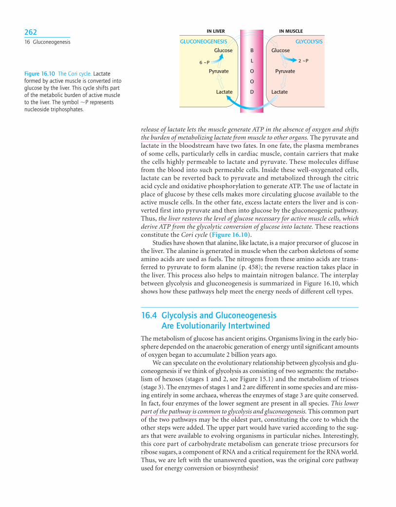
release of lactate lets the muscle generate ATP in the absence of oxygen and shiftsthe burden of metabolizing lactate from muscle to other organs. The pyruvate andlactate in the bloodstream have two fates. In one fate, the plasma membranesof some cells, particularly cells in cardiac muscle, contain carriers that makethe cells highly permeable to lactate and pyruvate. These molecules diffusefrom the blood into such permeable cells. Inside these well-oxygenated cells,lactate can be reverted back to pyruvate and metabolized through the citricacid cycle and oxidative phosphorylation to generate ATP. The use of lactate inplace of glucose by these cells makes more circulating glucose available to theactive muscle cells. In the other fate, excess lactate enters the liver and is con-verted first into pyruvate and then into glucose by the gluconeogenic pathway.Thus, the liver restores the level of glucose necessary for active muscle cells, whichderive ATP from the glycolytic conversion of glucose into lactate. These reactionsconstitute the Cori cycle (Figure 16.10).
Studies have shown that alanine, like lactate, is a major precursor of glucose inthe liver. The alanine is generated in muscle when the carbon skeletons of someamino acids are used as fuels. The nitrogens from these amino acids are trans-ferred to pyruvate to form alanine (p. 458); the reverse reaction takes place inthe liver. This process also helps to maintain nitrogen balance. The interplaybetween glycolysis and gluconeogenesis is summarized in Figure 16.10, whichshows how these pathways help meet the energy needs of different cell types.
16.4 Glycolysis and Gluconeogenesis Are Evolutionarily Intertwined
The metabolism of glucose has ancient origins. Organisms living in the early bio-sphere depended on the anaerobic generation of energy until significant amountsof oxygen began to accumulate 2 billion years ago.
We can speculate on the evolutionary relationship between glycolysis and glu-coneogenesis if we think of glycolysis as consisting of two segments: the metabo-lism of hexoses (stages 1 and 2, see Figure 15.1) and the metabolism of trioses(stage 3). The enzymes of stages 1 and 2 are different in some species and are miss-ing entirely in some archaea, whereas the enzymes of stage 3 are quite conserved.In fact, four enzymes of the lower segment are present in all species. This lowerpart of the pathway is common to glycolysis and gluconeogenesis. This common partof the two pathways may be the oldest part, constituting the core to which theother steps were added. The upper part would have varied according to the sug-ars that were available to evolving organisms in particular niches. Interestingly,this core part of carbohydrate metabolism can generate triose precursors forribose sugars, a component of RNA and a critical requirement for the RNA world.Thus, we are left with the unanswered question, was the original core pathwayused for energy conversion or biosynthesis?
26216 Gluconeogenesis
Glucose
GLUCONEOGENESIS GLYCOLYSIS
6 ~P 2 ~P
Glucose
IN MUSCLEIN LIVER
Pyruvate Pyruvate
B
L
O
O
DLactate Lactate
Figure 16.10 The Cori cycle. Lactateformed by active muscle is converted intoglucose by the liver. This cycle shifts partof the metabolic burden of active muscleto the liver. The symbol �P representsnucleoside triphosphates.
SUMMARY
16.1 Glucose Can Be Synthesized from Noncarbohydrate PrecursorsGluconeogenesis is the synthesis of glucose from noncarbohydratesources, such as lactate, amino acids, and glycerol. Several of the reactionsthat convert pyruvate into glucose are common to glycolysis. Gluconeoge-nesis, however, requires four new reactions to bypass the essential irre-versibility of three reactions in glycolysis. In two of the new reactions,pyruvate is carboxylated in mitochondria to oxaloacetate, which, in turn,is decarboxylated and phosphorylated in the cytoplasm to phospho-enolpyruvate. Two molecules having high phosphoryl-transfer potentialare consumed in these reactions, which are catalyzed by pyruvate carboxy-lase and phosphoenolpyruvate carboxykinase. Pyruvate carboxylasecontains a biotin prosthetic group. The other distinctive reactions of glu-coneogenesis are the hydrolyses of fructose 1,6-bisphosphate and glucose 6-phosphate, which are catalyzed by specific phosphatases. Themajor raw materials for gluconeogenesis by the liver are lactate and alanineproduced from pyruvate by active skeletal muscle. The formation oflactate during intense muscular activity buys time and shifts part of themetabolic burden from muscle to the liver.
16.2 Gluconeogenesis and Glycolysis Are Reciprocally RegulatedGluconeogenesis and glycolysis are reciprocally regulated so that one path-way is relatively inactive while the other is highly active. Phosphofructoki-nase and fructose 1,6-bisphosphatase are key control points. Fructose2,6-bisphosphate, an intracellular signal molecule present at higher levelswhen glucose is abundant, activates glycolysis and inhibits gluconeogenesisby regulating these enzymes. Pyruvate kinase and pyruvate carboxylase areregulated by other effectors so that both are not maximally active at thesame time. Allosteric regulation and reversible phosphorylation, which arerapid, are complemented by transcriptional control, which takes place inhours or days.
16.3 Metabolism in Context: Precursors Formed by Muscle Are Used by Other OrgansLactate that is generated by glycolysis in contracting muscle is released intothe bloodstream. This lactate is removed from the blood by the liver and isconverted into glucose by gluconeogenesis. This metabolic cooperationbetween muscle and liver is called the Cori cycle. Alanine is used to trans-port nitrogen as well as carbon skeletons from muscle to the liver.
16.4 Glycolysis and Gluconeogenesis Are Evolutionarily IntertwinedParts of glycolysis and gluconeogenesis are ancient pathways. In partic-ular, the metabolism of trioses that is common to both pathways may bethe oldest part. This common set of reactions formed the basis of carbo-hydrate metabolism from which glycolysis and gluconeogenesis evolved.
263Key Terms
Key Terms
gluconeogenesis (p. 251)pyruvate carboxylase (p. 254)biotin (p. 255)
glucose 6-phosphatase (p. 257)bifunctional enzyme (p. 259)
substrate cycle (p. 261)Cori cycle (p. 262)
Answers to QUICK QUIZZES
1. The reverse of glycolysis is highly endergonic undercellular conditions. The expenditure of 6 NTP molecules ingluconeogenesis renders gluconeogenesis exergonic.
2. Reciprocal regulation at the key allosteric enzymes in the two pathways. For instance, PFK is stimulated byfructose 2,6-bisphosphate and AMP. The effect of these sig-nals is opposite that of fructose 1,6-bisphosphatase. If bothpathways were operating simultaneously, a futile cyclewould result. ATP would be hydrolyzed, yielding only heat.
1. Road blocks. What reactions of glycolysis are not read-ily reversible under intracellular conditions? How are thesereactions bypassed in gluconeogenesis?
2. Waste not, want not. Why is it in an organism’s bestinterest to convert lactic acid from the blood into glucose inthe liver?
3. Metabolic mutant. What are the likely consequencesof a genetic disorder rendering fructose 1,6-bisphos-phatase in the liver less sensitive to regulation by fructose2,6-bisphosphate?
4. Biotin snatcher. Avidin, a 70-kd protein in egg white, hasvery high affinity for biotin. In fact, it is a highly specificinhibitor of biotin enzymes. Which of the following conver-sions would be blocked by the addition of avidin to a cellhomogenate?
(a) Glucose S pyruvate(b) Pyruvate S glucose(c) Oxaloacetate S glucose(d) Malate S oxaloacetate(e) Pyruvate S oxaloacetate(f) Glyceraldehyde 3-phosphate S fructose 1,6-bisphosphate
5. Tracing carbon atoms. If cells synthesizing glucosefrom lactate are exposed to CO2 labeled with 14C, whatwill be the distribution of label in the newly synthesizedglucose?
6. Working at cross-purposes? Gluconeogenesis takes placeduring intense exercise, which seems counterintuitive. Whywould an organism synthesize glucose and, at the sametime, use glucose to generate energy?
7. Powering pathways. Compare the stoichiometries ofglycolysis and gluconeogenesis. Recall that the input of oneATP equivalent changes the equilibrium constant of a reac-tion by a factor of about 108 (p. 207). By what factor do theadditional high-phosphoryl-transfer compounds alter theequilibrium constant of gluconeogenesis?
8. Different needs. Liver is primarily a gluconeogenic tis-sue, whereas muscle is primarily glycolytic. Why does thisdivision of labor make good physiological sense?
9. Metabolic mutants. What would be the effect on anorganism’s ability to use glucose as an energy source if amutation inactivated glucose 6-phosphatase in the liver?
10. Never let me go. Why does the lack of glucose 6-phos-phatase activity in the brain and muscle make good physio-logical sense?
11. Match ’em 1. The following sequence is a part of thesequence of reactions in gluconeogenesis.
A B C
D
Match the capital letters representing the reaction in thegluconeogenic pathway with parts a, b, c, etc.
(a) takes place in the mitochondria.(b) takes place in the cytoplasm.(c) produces CO2.(d) consumes CO2.(e) requires NADH.(f) produces NADH.(g) requires ATP.(h) requires GTP.(i) requires thiamine.(j) requires biotin.(k) is regulated by acetyl CoA.
12. Salvaging resources. In starvation, protein degradationtakes place in muscle. Explain how this degradation mightaffect gluconeogenesis in the liver.
13. Counting high-energy compounds 1. How many NTPmolecules are required for the synthesis of one molecule ofglucose from two molecules of pyruvate? How many NADHmolecules?
14. Counting high-energy compounds 2. How many NTPmolecules are required to synthesize glucose from each ofthe following compounds?
(a) Glucose 6-phosphate(b) Fructose 1,6-bisphosphate(c) Two molecules of oxaloacetate(d) Two molecules of dihydroxyacetone phosphate
oxaloacetate ¡ Phosphoenolpyruvate
Pyruvate ¡ Oxaloacetate ¡ Malate ¡
Problems
264 16 Gluconeogenesis
Enzy
me
activ
ity (
units
g−1
tho
rax)
0
B. te
rres
tris
B. a
ffini
sB.
bim
acul
atus
B. im
patie
nsB.
vag
ans
B. ru
foci
nctu
s
B. p
erpl
exus
P. c
itrin
us
B. g
riseo
colli
s
120
PFKFBPase
100
80
60
40
20
Problems 265
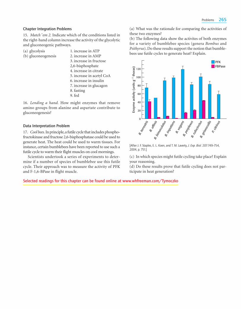
[After J. F. Staples, E. L. Koen, and T. M. Laverty, J. Exp. Biol. 207:749–754,2004, p. 751.]
(a) What was the rationale for comparing the activities ofthese two enzymes?(b) The following data show the activites of both enzymesfor a variety of bumblebee species (genera Bombus andPsithyrus). Do these results support the notion that bumble-bees use futile cycles to generate heat? Explain.
Chapter Integration Problems
15. Match ’em 2. Indicate which of the conditions listed inthe right-hand column increase the activity of the glycolyticand gluconeogenic pathways.
(a) glycolysis 1. increase in ATP(b) gluconeogenesis 2. increase in AMP
3. increase in fructose2,6-bisphosphate4. increase in citrate5. increase in acetyl CoA6. increase in insulin7. increase in glucagon8. fasting9. fed
16. Lending a hand. How might enzymes that removeamino groups from alanine and aspartate contribute togluconeogenesis?
Data Interpretation Problem
17. Cool bees. In principle,a futile cycle that includes phospho-fructokinase and fructose 2,6-bisphosphatase could be used togenerate heat. The heat could be used to warm tissues. Forinstance, certain bumblebees have been reported to use such afutile cycle to warm their flight muscles on cool mornings.
Scientists undertook a series of experiments to deter-mine if a number of species of bumblebee use this futilecycle. Their approach was to measure the activity of PFKand F-1,6-BPase in flight muscle.
(c) In which species might futile cycling take place? Explainyour reasoning.(d) Do these results prove that futile cycling does not par-ticipate in heat generation?
Selected readings for this chapter can be found online at www.whfreeman.com/Tymoczko


![Biochem [Gluconeogenesis]](https://img.pdfslide.net/doc/110x75/577c82b31a28abe054b1e4af/biochem-gluconeogenesis.jpg)