Embed Size (px)
Citation preview
Faculty Scholarship
1984
Competition between hydrocarbon andbarbiturate for spectral binding to hepaticcytochrome P ≃ 450. Inferences concerning spinstate of the enzymeW. L. Backes
M Means
W. J. Canady
Follow this and additional works at: https://researchrepository.wvu.edu/faculty_publications
This Article is brought to you for free and open access by The Research Repository @ WVU. It has been accepted for inclusion in Faculty Scholarshipby an authorized administrator of The Research Repository @ WVU. For more information, please contact [email protected].
Digital Commons CitationBackes, W. L.; Means, M; and Canady, W. J., "Competition between hydrocarbon and barbiturate for spectral binding to hepaticcytochrome P ≃ 450. Inferences concerning spin state of the enzyme" (1984). Faculty Scholarship. 93.https://researchrepository.wvu.edu/faculty_publications/93
THE JOURNAL OF BIOLOGICAL CHEMISTRY Q 1984 by The American Society of Biological Chemists, Inc.
Vol. 259, No. 16, Issue of August 25, pp. 10092-10099,19&L Printed in U.S.A.
Competition between Hydrocarbon and Barbiturate for Spectral Binding to Hepatic Cytochrome P-450 INFERENCES CONCERNING SPIN STATE OF THE ENZYME*
(Received for publication, December 5, 1983)
Wayne L. BackesS, Melissa Means, and William J. Canady From the Department of Biochemistry, West Virginia University School of Medicine, Morgantown, West Virginia 26506
The substrates hexobarbital and ethylbenzene have been shown to compete for the spectral binding site of phenobarbital-induced rat hepatic microsomal cyto- chrome P-450. The two substrates produce different AAbs,,,, values, and the presence of one substrate does not affect the AAbs,, of the other substrate and vice versa. The respective binding constants for the two substrates are similarly unaffected. The conclusion drawn from these observations is that, over the con- centration ranges studied, there is no change in the availability of the enzyme as a result of substrate ad- dition; the difference in AAbs-, apparently being due to varying abilities of different substrates to bring about a spin shift in the enzyme.
Evidence is presented to indicate that differences between enzymes from untreated male rats and phe- nobarbital-treated male rats are attributable to differ- ences in the enzyme itself and not to changes in the nature of the membrane brought about by phenobar- bital administration, at least insofar as heat entropy compensation is concerned. The enthalpy-entropy compensation observed in the binding of a homologous series of barbiturates to the microsomal membrane as determined from the membrane concentration depend- ence of their binding constants is shown to agree sur- prisingly well with the direct determination performed by Sitar and Mannering.
Liver microsomal cytochrome P-450 is known to metabolize a wide variety of both exogenous and endogenous substrates (1-3). One of the most important classes of substrates for the cytochrome P-450 system is the barbiturates. When most barbiturates (as well as many other substrates) are added to oxidized cytochrome P-450 in the absence of reducing equiv- alents (4-6) a spectral change demonstrated by difference spectroscopy is observed in the Soret region; it is known as a type I spectral change. This difference spectrum generally exhibits an absorption maximum and minimum at 385 and 420 nm, respectively, with increasing quantities of the drug producing proportionately larger spectral changes. An asso- ciation constant for enzyme-substrate complex formation may be determined by plotting the reciprocal of the substrate concentration uersus the reciprocal of the absorbance change
* This work was supported by Department of Energy/Morgantown Energy Research Center Contract D4-77-C-21-8087 and the West Virginia Medical Corporation. The costs of publication of this article were defrayed in part by the payment of page charges. This article must therefore be hereby marked “advertisement” in accordance with 18 U.S.C. Section 1734 solely to indicate this fact.
Connecticut Health Center, Farmington, CT 06032. 3 Present address, Department of Pharmacology, University of
(4,6) in a manner analogous to the Lineweaver-Burk plot (7) for kinetic data.
The interactions of a series of barbiturates with liver mi- crosomes were previously studied by Jansson et al. (8). In this work, the spectrally determined binding constants for a series of barbiturates were compared to the partition constants of the same substrates from water into an organic (corn oil) phase. Only a small correlation between these parameters was obtained. A weak correlation was also found with the metab- olism of these substrates and the partition process with a stronger relationship obtained between partitioning and in- hibition of NADH oxidation. Later, Sitar and Mannering (9) directly measured the partitioning of a series of barbiturates into the microsomal membrane and compared these values with corn oil/water partition coefficients. A series of barbi- turates were found to partition into microsomes in such a way that those with more carbon atoms (more hydrophobic) ex- hibited a larger affinity for microsomes than the smaller (less hydrophobic) ones. However, this effect was larger when the partitioning of the same series of barbiturates from water into corn oil was observed by these investigators. In other words, making a barbiturate substrate more hydrophobic increased its tendency to be transferred from water to both the micro- somal membrane and to corn oil, but a considerably larger increase was observed with the corn oil system.
Since cytochrome P-450 enzymes reside in the microsomal membrane, the presence of a lipid membrane phase would be expected to influence the apparent binding of hydrophobic drug substrates. The treatment of Parry et al. (10) assumes that the active site of a cytochrome P-450 molecule either resides in the membrane or faces the aqueous phase. Sub- strates for such an enzyme generally have hydrophobic char- acter and will partition into the microsomal lipid as well as bind to the active site of the enzyme. Therefore, if the active site faces the aqueous phase, the substrate which partitions into the microsomal lipid is unavailable for binding to the enzyme. Conversely, a similar situation is seen if the active site of the enzyme is buried within the lipid phase. In this case, substrate which remains in the aqueous phase would not be “seen” by the enzyme which is totally buried in the lipid phase. Under either of the two circumstances considered above, the reciprocal of the apparent association constant for substrate binding increases in a linear manner as the concen- tration of enzyme (microsomes) is increased. This effect has been reported by Ebel et al. (11) and by Backes and Canady (12) and Backes et al. (13).
Extensions of the treatment of Parry et al. have been put forward by this laboratory (13). The hydrophobicity of the enzyme-binding site was probed by the means of a homologous series of aromatic hydrocarbons. If one plots the reciprocal of the apparent association constant against the microsome con-
10092
Competition between Substrates in Cytochrome P-450 10093
centration, a linear relationship is obtained, the y intercept representing the binding constant corrected for the presence of the microsomal phase. It can be shown (13, 14) that the x intercept represents the reciprocal of the microsomal partition coefficient for the distribution of the substrate between aqueous phase and microsomal phase.
In the present study, the binding of a series of barbiturates to both the enzyme and to the microsomal membrane, mea- sured indirectly using the enzyme as a marker, were investi- gated and compared with the partitioning of these substrates from the aqueous phase, into an organic solvent and from the aqueous phase into the microsomal phase as determined by direct measurement. Competitive studies involving the sub- strates hexobarbital and ethylbenzene were performed with phenobarbital-induced enzyme.
EXPERIMENTAL PROCEDURES
Male Wistar rats (Hilltop Farms, Scottsdale, PA) weighing 250- 350 g were used. The animals were killed by decapitation and the livers were removed and prepared by a modification of the method of Omura and Sat0 (15) as previously described (12). The microsomal pellet was suspended in 0.15 M KCl, 50 mM Tris buffer (pH 7.4 at 25 “C). Sodium phenobarbital, where appropriate, was administered by daily intraperitoneal injections (75 mg/kg of body weight) for 3 days prior to the experiments. Cytochrome P-450 levels were mea- sured according to the method of Estabrook et al. (16). Protein concentrations were determined by the method of Lowry et al. (17).
Partition coefficients for the series of barbiturates used in these studies were determined by dissolving them in 0.1 M sodium phos- phate buffer, pH 7.4, and extracting the solution with either octanol or corn oil for 1 h at 25 “C. The barbiturate concentration was measured in the aqueous phase before and after the partitioning process, by means of the addition of 0.5 ml of 0.1 N NaOH solution to 2.5 ml of the barbiturate preparation under investigation. The absorbance due to enol formation by the barbiturate was measured at 238-242 nm (8, 18).
Spectral binding constants were determined using difference spec- troscopy as described by Schenkman et al. (5), measuring the mag- nitude of the type I spectral change as described previously (13). The absorbances were traced on an expanded scale and measured precisely with a draughtsman’s rule.
When hexobarbital and ethylbenzene were investigated in the presence of each other (for the determination of the type of interac- tion of these compounds with cytochrome P-450) ethylbenzene was added without the use of an organic solvent as carrier as described previously (12); however, after the absorbances of each of these preparations were determined, increments of an aqueous solution (8.4 mM) of hexobarbital were added to both cuvettes and the absorbance was measured after each addition. The reciprocal of the change in absorbance was plotted as a function of the reciprocal of the ethyl- benzene concentration at fixed concentrations of hexobarbital. The nature of interaction with the enzyme was determined by the use of equations presented under “Results.” It should be noted that in the studies presented here, the spectral association constants were used because of their direct relationships to the free energies of binding.
Ethylbenzene was purchased from Eastman. Monobasic sodium phosphate was obtained from Mallinkrodt. Butabarbital, barbital, pentobarbital, amobarbital, secobarbital, and Tris were obtained from Sigma. Phenobarbital, hexobarbital, and methohexital were pur- chased from J. T. Baker, Winthrop Laboratories, and Lilly, respec- tively. The absolute ethanol was obtained from U. S. Industrial Chemical Co., Tuscola, IL. All other chemicals were supplied by Fisher.
RESULTS AND DISCUSSION
The competitive binding of two substrates ( A and B ) to cytochrome P-450 ( E ) may be described by the following scheme (14):
E B % E * E A RE KA
The following equilibrium constants are involved
The equation (12) describing such interactions is:
Where \ k A and \ks represent the molar absorbances of the EA and EB complexes, respectively. The total enzyme con- centration is (E) , , and AAbhhl represents the total ab- sorbance change caused by the addition of substrates ( A and B ) .
If only substrate A is added to the enzyme preparation, then Equation 1 simplifies to:
If, making use of difference spectroscopy, both substrates are added to the sample cuvette, and an equal quantity of substrate A is added to the reference cuvette, as described under “Experimental Procedures,” then the resultant ab- sorbance becomes:
which rearranges to:
If substrate A is added to each cuvette and its concentration kept constant for that experiment, then a plot of the reciprocal of aAbhhl, against the reciprocal of [ B ] would be expected to be linear. By performing a series of experiments at different concentrations of A, a family of lines is obtained. By setting the reciprocal of AAbsmhl equal to zero, it is possible to determine the apparent association constant, i t .pp, for B. The following relationship between the apparent binding constant Kapp and the other association constants involved can be easily shown
Therefore, a concentration dependence of absorbance for substrate B may be determined at a fixed concentration of substrate A. This experiment is then repeated at a variety of fixed concentrations of A and the apparent association con- stants are determined in the usual way. A replot of the reciprocal of Kapp versus the concentration of A employed in each of the above titrations (Equation 5) permits the deter- mination of the association constants for both the EA and EB complexes (it, and &). The slope is equal to KA/& and the intercept is equal to the reciprocal of RE. In addition to obtaining the individual association constants, it can be shown that replots of the reciprocal of AAbsma, and the reciprocal of the slope uersus [ A ] will mot be linear.
If the assumption is made that all substrates that bind to the active site produce the same spectral change (all com- plexes have the same extinction coefficient), \ k A is equal to \kB and the resultant equation which is a special case of
10094 Competition between Substrates in Cytochrome P-450
Equation 4 is
1 (1 + KA[A])* + 1 + KA[A] ~- AAbhd RB*B[EIo [BI *B[EIo
- (6)
In such a situation, a plot of the reciprocal of AAbsmml against the reciprocal of [ B ] will result in a linear relationship. This equation is identical to that derived by Van den Berg et al. (19) who made this same moot assumption concerning ex- tinction coefficients in their derivation. A plot of l/KaPp uersus [ A ] has the same significance as in Equation 4. A plot of the reciprocal of the slope against [ A ] yields nonlinear results, but l/AAbs,,, uersus [ A ] results in a linear plot from which qB [ Elo can be determined from the intercept.
0 4 8 12 1 6 2 0 1 /[mM Ethylbenzenel
Another special case of Equation 4 which is of interest is that in which the EA complex can form but cannot bring about an absorbance change; therefore, \ k A is equal to zero. This is the spectral analog of competitive inhibition in enzyme kinetics. The equation is
Hence, the usual double reciprocar plot of the reciprocal of the absorbance uersus the reciprocal of the concentration of substrate B is linear provided that the concentration of sub- strate A is kept constant for that titration.
A replot of the reciprocal of the apparent binding constant KaPp uersus [ A ] has the same significance as in Equation 5 . A replot of the reciprocal of AAbs,., uersus [ A ] has zero de- pendence; a replot of the slope uersus [ A ] would be expected to be linear and to have the following significance
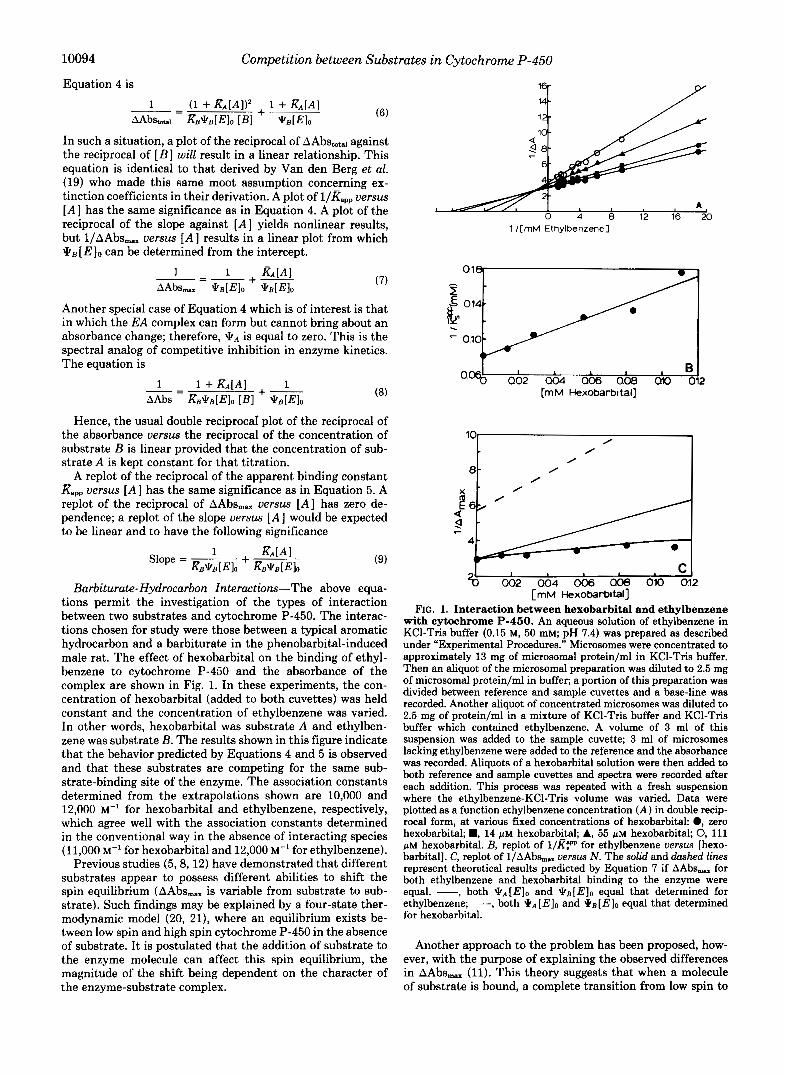
Barbiturate-Hydrocarbon Interactions-The above equa- tions permit the investigation of the types of interaction between two substrates and cytochrome P-450. The interac- tions chosen for study were those between a typical aromatic hydrocarbon and a barbiturate in the phenobarbital-induced male rat. The effect of hexobarbital on the binding of ethyl- benzene to cytochrome P-450 and the absorbance of the complex are shown in Fig. 1. In these experiments, the con- centration of hexobarbital (added to both cuvettes) was held constant and the concentration of ethylbenzene was varied. In other words, hexobarbital was substrate A and ethylben- zene was substrate B. The results shown in this figure indicate that the behavior predicted by Equations 4 and 5 is observed and that these substrates are competing for the same sub- strate-binding site of the enzyme. The association constants determined from the extrapolations shown are 10,000 and 12,000 M” for hexobarbital and ethylbenzene, respectively, which agree well with the association constants determined in the conventional way in the absence of interacting species (11,000 M” for hexobarbital and 12,000 M” for ethylbenzene).
Previous studies (5 , 8, 12) have demonstrated that different substrates appear to possess different abilities to shift the spin equilibrium (AAbs,,. is variable from substrate to sub- strate). Such findings may be explained by a four-state ther- modynamic model (20, 21), where an equilibrium exists be- tween low spin and high spin cytochrome P-450 in the absence of substrate. It is postulated that the addition of substrate to the enzyme molecule can affect this spin equilibrium, the magnitude of the shift being dependent on the character of the enzyme-substrate complex.
B 0.4 062 a b a b a b A J 2 [mM Hexobarbttal]
L / I
L 4 a62 064 066 Obe oio 0!2 [mM Hexobarb~tal]
FIG. 1. Interaction between hexobarbital and ethylbenzene with cytochrome P-460. An aqueous solution of ethylbenzene in KC1-Tris buffer (0.15 M, 50 mM; pH 7.4) was prepared as described under “Experimental Procedures.” Microsomes were concentrated to approximately 13 mg of microsomal protein/ml in KC1-Tris buffer. Then an aliquot of the microsomal preparation was diluted to 2.5 mg of microsomal protein/ml in buffer; a portion of this preparation was divided between reference and sample cuvettes and a base-line was recorded. Another aliquot of concentrated microsomes was diluted to 2.5 mg of protein/ml in a mixture of KC1-Tris buffer and KC1-Tris buffer which contained ethylbenzene. A volume of 3 ml of this suspension was added to the sample cuvette; 3 ml of microsomes lacking ethylbenzene were added to the reference and the absorbance was recorded. Aliquots of a hexobarbital solution were then added to both reference and sample cuvettes and spectra were recorded after each addition. This process was repeated with a fresh suspension where the ethylbenzene-KC1-Tris volume was varied. Data were plotted as a function ethylbenzene concentration ( A ) in double recip- rocal form, at various fixed concentrations of hexobarbitak 0, zero hexobarbital; D, 14 p~ hexobarbital; A, 55 p~ hexobarbital; 0, 111 p~ hexobarbital. B, replot of l/Rpp for ethylbenzene versus [hexo- barbital]. C, replot of l/AAbs, versus N . The solid and dashed lines represent theoretical results predicted by Equation 7 if AAbs, for both ethylbenzene and hexobarbital binding to the enzyme were equal. -, both [E l0 and qB[ Elo equal that determined for ethylbenzene; -- -, both * A [E10 and *B[E]O equal that determined for hexobarbital.
Another approach to the problem has been proposed, how- ever, with the purpose of explaining the observed differences in AAbsmex (11). This theory suggests that when a molecule of substrate is bound, a complete transition from low spin to
Competition between Substrates in Cytochrome P-450 10095
high spin state in the enzyme takes place. According to this theory, different substrates bring about different maximal absorbance changes by binding to different amounts of cyto- chrome P-450. Thus, this model is based on the assumption that the microsomal membrane has a heterogeneous compo- sition with different regions possessing different gel-fluid transition temperatures.
Therefore, only that portion of cytochrome P-450 in the fluid portion of the membrane is actually available for binding. Now, the addition of some substrates, particularly those which more readily dissolve in the membrane, can bind some of the previously unavailable cytochrome P-450 because of the abil- ity of that substrate to affect the equilibrium constant for the gel-fluid transition in favor of the fluid phase. In other words, a given substrate becomes more available to the enzyme than another, provided that it can also alter the fluidity of the membrane to a greater extent. It is important to keep in mind that, according to this model, the addition of a given amount of one substrate (which itself has an effect upon membrane fluidity) would be expected to alter the apparent affinity constant and AAbs,,, of the enzyme for another competing substrate of somewhat different character and vice versa. Indeed it should be virtually impossible to observe simple competition, especially between two unlike substrates such as these that we have used. Our substrate B is ethylbenzene, a very simple hydrophobic probe that we have used before (12, 13). Substrate A is hexobarbital, a substrate having a hydro- phobic component, but, in addition, is highly polar in nature.
Thus, we have a tool by which we may determine whether the differences in AAbs,. are due to actual differences in the ability of a substrate to perturb the spin equilibrium or to differences in the availability of enzyme to different sub- strates. If two compounds can be found that 1) possess differ- ent values for AAbs,,, 2) compete in a conventional manner for the same binding site, and 3) whose spectral parameters can be measured directly by conventional methods for com- parison to those derived from the competitive studies, then the membrane fluidity theory described above must be re- examined, and at the very least be considerably modified.
The results shown in Fig. 1C as well as the direct conven- tional experiments show that the extinction coefficients (AAbs,,.) for ethylbenzene and hexobarbital are different. This is shown by the deviation from linearity of the plot of l/AAbs,, uersus [A] in the experiment where the substrates are allowed to compete with one another. If AAbs,, for both hexobarbital and ethylbenzene were equal ( \ k B = \ k A ) , then according to Equation 7, a replot of l/AAbs,, uersus [A] would be linear with a slope equal to &\k[E]o. If \ k A were actually equal to \ kB , the theoretical lines shown in Fig. 1C should adequately describe the experimental results. Neither theoretical line assuming JTB is equal to \ k A fits the actual experiment. The curve that indeed does fit the experimental data points in Fig. 1C was drawn using the values for \ k B
(0.34) and \ k A (0.18) obtained from direct conventional titra- tions of each of the substrates in separate experiments and these then inserted into Equation 6. The results described here clearly demonstrate that the spectral change elicited by substrate-cytochrome P-450 interaction is not independent of the substrate employed. If different pools of enzyme were being affected, a competitive interaction would not have been observed. If a smaller percentage of hemoprotein bound the barbiturate than the hydrocarbon, deviations in the linearity of the double reciprocal plots (Fig. 1A) would be observed. For the above two substrates at least, and with the PB'-
The abbreviation used is: PB, phenobarbital.
induced enzyme, the results are consistent with the four-state model of Cinti et al. (21), although they do not constitute a proof of such a mechanism.
Previous studies (12) have demonstrated that the maximal type I absorbance of an enzyme-solvent (ethanol) complex differs from that of the enzyme-hydrocarbon complex (ethyl- benzene) in phenobarbital-pretreated animals; the situation was complicated by the fact that the solvent ethanol produced both a type I1 and type I spectral change, the former ordinarily overshadowing the latter. Corrections had to be introduced in order to take this fact into account. Therefore, in this present work, two simple type I compounds were studied with regard to their competitive behavior and their respective AAbs,.. values. The results unequivocally indicate that the barbiturate hexobarbital and the hydrocarbon ethylbenzene exhibit com- petitive behavior for the same site in PB-treated male rats, and produce significantly different AAbs,., values. Values obtained by the methods outlined show good agreement with those obtained by conventional direct spectral measurements in the absence of a corresponding competing substrate.
It is interesting to note that compounds as different in nature as barbiturates and aromatic hydrocarbons apparently bind to the same particular cytochrome P-450 in the PB- induced rat.
It should be kept in mind, however, that because we observe simple competitive behavior between hexobarbital and ethyl- benzene in the PB-treated rat, it does not necessarily follow from this that competitive behavior might be expected be- tween any pair of substrate types that might have been selected. The reason for this has to do with the well known fact that multiple forms of cytochrome P-450 exist in the microsome. This aspect of the problem has been discussed earlier (12, 13) insofar as experiments of the present type are concerned. The fortunate selection of the two classes of com- pounds reported here for the PB-induced enzyme may have been fortuitous, but the fact remains that this system behaves as though the two substrates were competing for a single enzyme site; our results are entirely consistent with this concept. Practical experimental constraints prohibit investi- gation over a much wider range of substrate concentrations than those the results of which are reported here; we have not been able to obtain any significant or consistent evidence of downward curvature of the double reciprocal plots that one would ordinarily expect if two or more enzymes were inter- acting with either of these substrates (24, 25).
The interactions of hexobarbital and ethylbenzene with the enzyme(s) from untreated rats follow a more complex law and will be presented and interpreted at a later time. Although analysis of the data is not yet complete, curvature of the double reciprocal plots under competitive conditions indicates that a minimum of two enzymes are involved. The data given here for the preparation from the untreated animal are valid, but some of the constants involved may be more complex than originally envisaged. The enzyme preparation from the PB-treated animal represents the simplest case insofar as competitive interactions of hexobarbital and ethylbenzene are concerned and, considering the present stage and limitations of our experimental techniques, we feel that the most likely explanation of this finding is that all or most of the absor- bance change monitored in the PB-induced microsomal prep- aration is due to a single isozyme. Experiments were under- taken to compare the substrate binding characteristics of both the microsomal membrane and the enzyme; we have previ- ously reported observations on the nature of hydrocarbon binding to both of these entities (13).
Organic Solvent Partitioning of a Series of Barbiturates-A
10096 Competition between Substrates in Cytochrome P-450
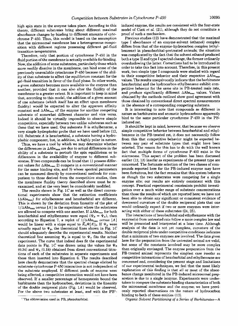
list of the barbiturates used in this study plus their partition coefficients obtained in both corn oil/phosphate buffer (8) and octanol/phosphate buffer systems are presented in Table I. From these partition coefficients, the apparent free energy of transfer from phosphate buffer (0.1 M, pH 7.4) and either corn oil or octanol were plotted as a function of the number of substituent carbon atoms. The results are to be seen in Fig. 2. The free energy of transfer, AGO, in the corn oilbuffer system is smaller than in the octanol/buffer system which indicates that octanol is more effective than corn oil insofar as barbiturate transfer is concerned. Despite the differences observed in the absolute values for the two systems, the size dependences of transfer from aqueous to organic phase were identical. In other words, when the free energy of transfer from water to organic solvent was plotted against number of carbon atoms added to the homologous series of barbiturates, a linear relationship was obtained in both cases, the slopes being, within experimental error, identical. This value was -0.66 kcal/mol/carbon atom. This is very similar to the value obtained for the size dependence of the free energy of transfer for a series of hydrocarbons from the aqueous phase to various organic solvents (13). The size dependence of transfer at pH 7.4 was very similar to those obtained in which partition coefficients were determined with barbiturates in the un- ionized form (23), where a slope of -0.66 kcal/mol/carbon atom was also obtained. This similarity of results of experi- ments done under highly acid conditions and those described here at pH 7.4 is easy to understand.
Let us consider an acid whose uncharged form is the only one to partition from the aqueous environment into the organic solvent, enzyme or membrane lipid.
KA AHA, e A & + HA,
Since only AH partitions
AHA, G KP AHL
where
This would be the case at very low pH where ionization of our organic acid is suppressed.
If KpBm, the apparent partition coefficient at a given pH, is
defined as
where [AItotAq is the sum of the ionized and un-ionized forms in the aqueous phase.
It can be shown that Kp. 10WL - PHI
K P ~ = 1 + ~ o ' P K . - pH)
If the working pH is about one unit or more lower than the pK, of the acid (barbiturate) under consideration, then 10'pK~ - pH) will be an order of a magnitude or more greater than unity. Therefore,
Kpm z Kp
The pK, values for the barbiturates used in this experiment which have been previously determined all meet this criterion. Indeed, as would be expected, introduction of the above cor- rection only alters the slope of the size dependence plot seen in Fig. 2 by less than lo%, as compared to the value obtained without correction.
Barbiturate Binding to the Enzyme in Untreated Rats- Jansson et al. (8) determined the binding of a series of barbiturates of differing lipid solubility to cytochrome P-450. In their study, only a rough correlation could be found be- tween affinity for the enzyme and lipid solubility in the phenobarbital-pretreated male rat. Recent reports (10-13) have stressed, however, that microsomal partitioning may have a marked effect on the apparent ability of a substrate to be bound by the enzyme. Therefore, this laboratory has rein- vestigated comparisons of lipid solubility with enzyme-sub- strate-binding affinities in both the untreated and phenobar- bital-treated male rat.
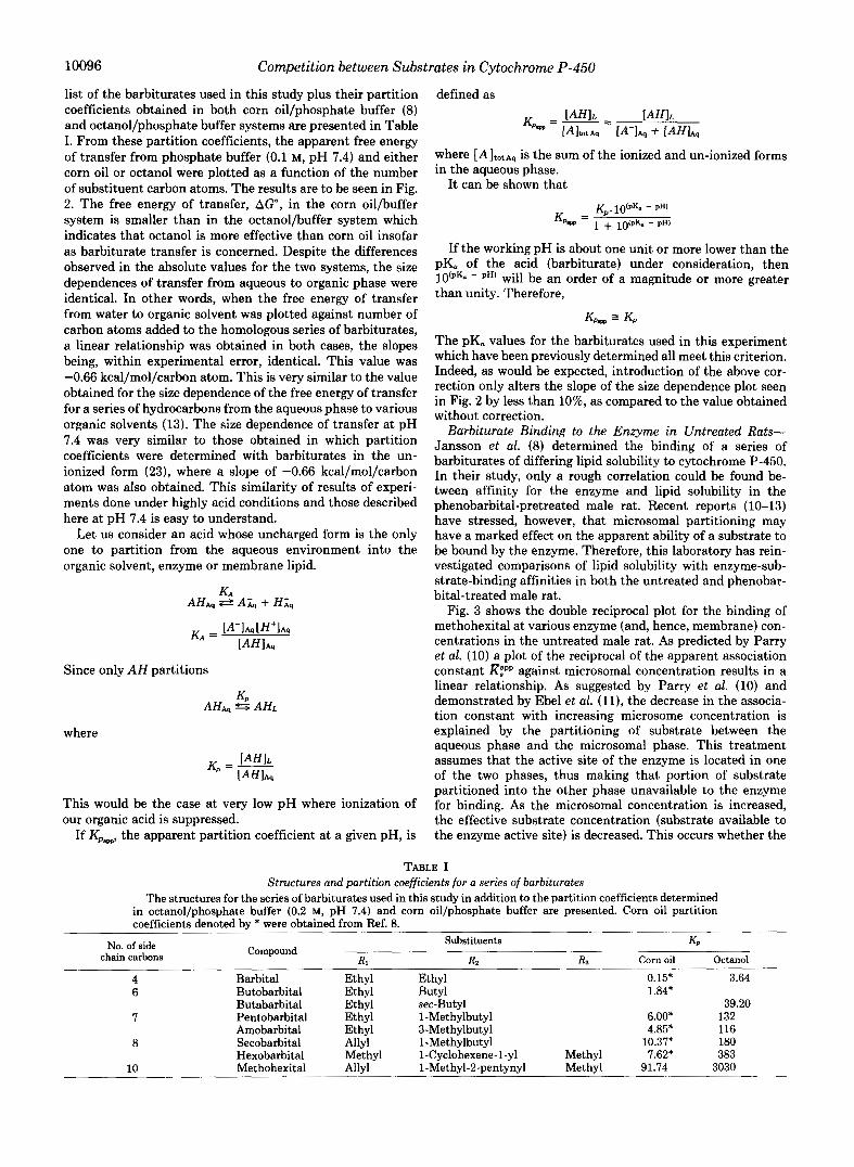
Fig. 3 shows the double reciprocal plot for the binding of methohexital at various enzyme (and, hence, membrane) con- centrations in the untreated male rat. As predicted by Parry et al. (10) a plot of the reciprocal of the apparent association constant against microsomal concentration results in a linear relationship. As suggested by Parry et al. (10) and demonstrated by Ebel et al. (ll), the decrease in the associa- tion constant with increasing microsome concentration is explained by the partitioning of substrate between the aqueous phase and the microsomal phase. This treatment assumes that the active site of the enzyme is located in one of the two phases, thus making that portion of substrate partitioned into the other phase unavailable to the enzyme for binding. As the microsomal concentration is increased, the effective substrate concentration (substrate available to the enzyme active site) is decreased. This occurs whether the
TABLE I Structures and partition coefficients for a series of barbiturates
The structures for the series of barbiturates used in this study in addition to the partition coefficients determined in octanol/phosphate buffer (0.2 M, pH 7.4) and corn oil/phosphate buffer are presented. Corn oil partition coefficients denoted by * were obtained from Ref. 8.
chain carbons No. of side Compound
Substituents
R1 R2
KP ~
R3 Corn oil Octanol
4 6
7
8
10
Barbital Butobarbital Butabarbital Pentobarbital Amobarbital Secobarbital Hexobarbital Methohexital
Ethyl Ethyl Ethyl Ethyl Ethyl Allyl Methyl Allyl
Ethyl 0.15* Butyl 1.84* sec-Butyl 1-Methylbutyl 6.00* 3-Methylbutyl 4.85* 1-Methylbutyl 10.37* 1-Cyclohexene-1-yl Methyl 7.62* 1-Methyl-2-pentynyl Methyl 91.74
3.64
39.20 132 116 180 383
3030
Competition between Substrates in Cytochrome P-450 10097 04
A 1 4t
X I 063 I
I
- 4 I 1 I I 1
Number of carbon atoms
FIG. 2. Dependence of AGO of partitioning between organic solvents and phosphate buffer, pH 7.4. Using partition coeffi- cients presented in Table I, the free energy of partitioning in both corn oil/phosphate buffer (0.1 M, pH 7.4), and octanol/phosphate buffer (0.1 M, pH 7.4) systems were plotted against the number of substituent carbon atoms. Points represent: 6, butabarbital; 7, pen- tobarbital and amobarbital (open symbols); 8, secobarbital and hex- obarbital (open symbols); and 10, methohexital. Circles represent octanol system; triangles represent corn oil system.
5 6 7 8 9 10
I J
/ I
1 /[Methohexitall
0 50 100 150 260
FIG. 3. Dependence of the apparent association of metho- hexital for the type I site on microsome concentration in untreated male rats. Double reciprocal plots for the binding of methohexital to the type I site at various microsome concentrations. @, 1.2 mg of protein/ml; A, 4.6 mg of protein/ml; and 0, 7.7 mg of protein/ml. For microsome concentrations above 4.5 mg of micro- somal proteins/ml, 1.5 ml of the microsomal suspension were added to cuvettes with a 0.45-cm path length.
mg mlcrosMnal proteln / ml
7
%J Number of carton atoms
active site of the enzyme faces the lipid phase or the aqueous phase. More complete explanations of these effects have been described previously (13, 14). Fig. 4A shows the effect of microsome concentration upon l/m for a series of barbitu- rates in the untreated male rat. As described previously (13), extrapolation to zero microsome concentration produces an association constant corrected for the effect of microsomal partitioning of the substrate.
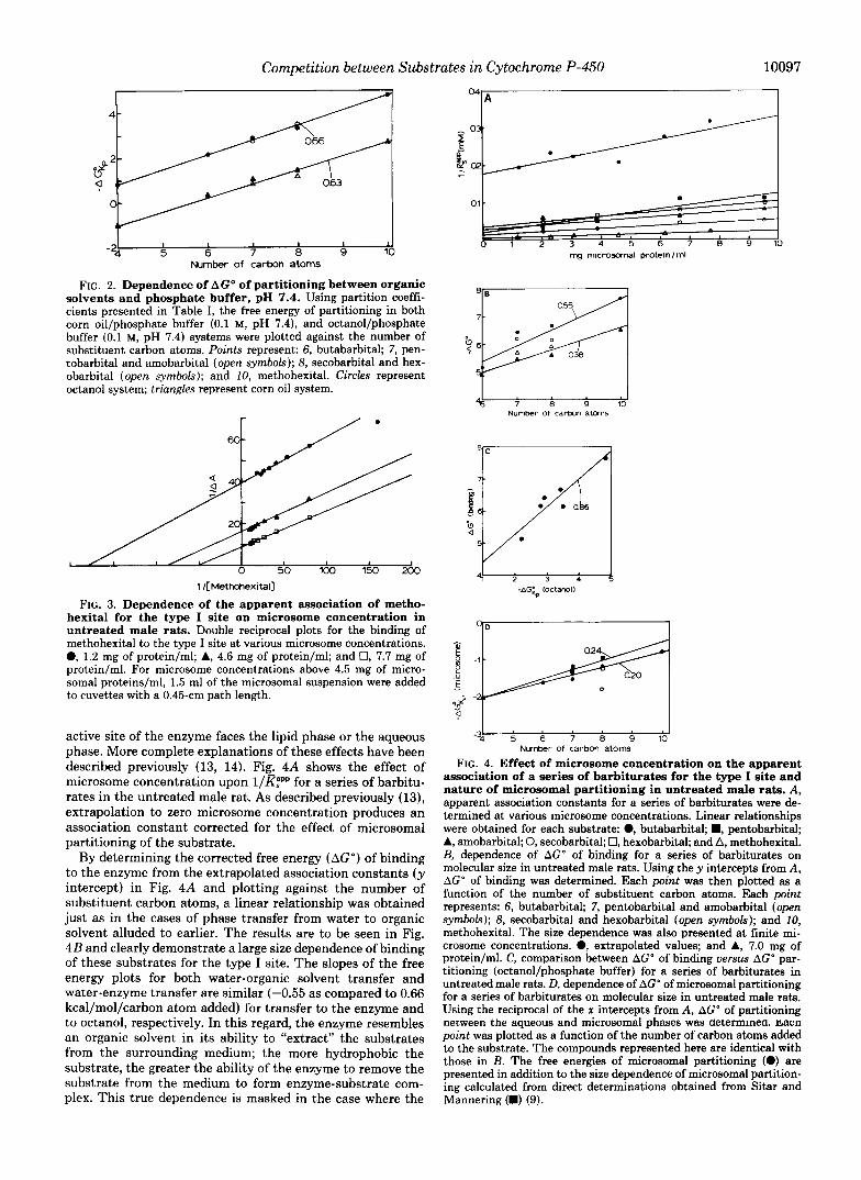
By determining the corrected free energy (AGO) of binding to the enzyme from the extrapolated association constants (y intercept) in Fig. 4A and plotting against the number of substituent carbon atoms, a linear relationship was obtained just as in the cases of phase transfer from water to organic solvent alluded to earlier. The results are to be seen in Fig. 4B and clearly demonstrate a large size dependence of binding of these substrates for the type I site. The slopes of the free energy plots for both water-organic solvent transfer and water-enzyme transfer are similar (-0.55 as compared to 0.66 kcal/mol/carbon atom added) for transfer to the enzyme and to octanol, respectively. In this regard, the enzyme resembles an organic solvent in its ability to “extract” the substrates from the surrounding medium; the more hydrophobic the substrate, the greater the ability of the enzyme to remove the substrate from the medium to form enzyme-substrate com- plex. This true dependence is masked in the case where the
Number of carbon atoms FIG. 4. Effect of microsome concentration on the apparent
association of a series of barbiturates for the type I site and nature of microsomal partitioning in untreated male rats. A, apparent association constants for a series of barbiturates were de- termined at various microsome concentrations. Linear relationships were obtained for each substrate: @, butabarbital; B, pentobarbital; A, amobarbital; 0, secobarbital; 0, hexobarbital; and A, methohexital. B, dependence of AGO of binding for a series of barbiturates on molecular size in untreated male rats. Using the y intercepts from A , AG” of binding was determined. Each point was then plotted as a function of the number of substituent carbon atoms. Each point represents: 6, butabarbital; 7, pentobarbital and amobarbital (open symbols); 8, secobarbital and hexobarbital (open symbols); and 10, methohexital. The size dependence was also presented at finite mi- crosome concentrations. 0, extrapolated values; and A, 7.0 mg of protein/ml. C, comparison between AGO of binding versus AGO par- titioning (octanol/phosphate buffer) for a series of barbiturates in untreated male rats. D, dependence of AGO of microsomal partitioning for a series of barbiturates on molecular size in untreated male rats. Using the reciprocal of the x intercepts from A, AGO of partitioning oecween the aqueous and microsomal phases was determmea. fiacn point was plotted as a function of the number of carbon atoms added to the substrate. The compounds represented here are identical with those in B. The free energies of microsomal partitioning (e) are presented in addition to the size dependence of microsomal partition- ing calculated from direct determinations obtained from Sitar and Mannering (B) (9).
10098 Competition between Substrates in Cytochrome P-450
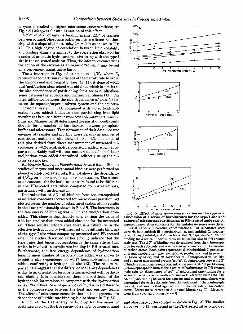
enzyme is studied at higher microsome concentrations; see Fig. 4B (triangles) for an illustration of this effect.
A plot of AGO of enzyme binding against AG" of transfer between octanol/phosphate buffer results in a linear relation- ship with a slope of almost unity ( m = 0.9) as shown in Fig. 4C. This high degree of correlation between lipid solubility and binding affinity is similar to the correlation observed for a series of aromatic hydrocarbons interacting with the type I site in the untreated male rat. Thus, the inferences concerning the action of the enzyme as an organic "solvent" may be put on a convenient quantitative basis.
The x intercept in Fig. 4A is equal to -I/Kp, where Kp represents the partition coefficient of the barbiturate between the aqueous and microsomal phases (13,14). A slope of -0.20 kcal/mol/carbon atom added was obtained which is similar to the size dependence of partitioning for a series of alkylben- zenes between the aqueous and microsomal phases (13). The large difference between the size dependence of transfer be- tween the aqueous/organic solvent system and the aqueous/ microsomal system (-0.66 compared with -0.20 kcal/mol/ carbon atom added) indicates that partitioning into lipid membranes is quite different from octanol/water partitioning. Sitar and Mannering (9) determined the partition coefficients directly for a number of barbiturates between phosphate buffer and microsomes. Transformation of their data into free energies of transfer and plotting these uersus the number of substituent carbons is also shown in Fig. 40. The slope of this plot derived from direct measurement of untreated mi- crosomes is -0.24 kcal/mol/carbon atom added, which com- pares remarkably well with our measurement of -0.20 kcal/ mol/carbon atom added determined indirectly using the en- zyme as a marker.
Barbiturate Binding in Pherwbarbital-treated Rats-Similar studies of enzyme and microsomal binding were performed on phenobarbital-pretreated rats. Fig. 5A shows the dependence of l/Kapp on microsome (enzyme) concentration. The associ- ation constants for the barbiturates were found to be different in the PB-treated rats when compared to untreated rats, particularly with methohexital.
Determination of AGO of binding from the extrapolated association constants (corrected for microsomal partitioning) plotted uersus the number of substituent carbon atoms results in the linear relationship shown in Fig. 5B. The increment in the free energy of binding was -0.31 kcal/mol/carbon atom added. This slope is significantly smaller than the value of 0.55 kcal/mol/carbon atom added obtained for the untreated rat. These results indicate that there is a difference in the effective hydrophobicity (with respect to barbiturate binding) of the type I site when comparing untreated and PB-treated rats. The studies described earlier (Fig. 1) indicate that the type I site that binds hydrocarbons is the same site as that which is involved in barbiturate binding in PB-treated rats. Furthermore, the free energy dependence of hydrocarbon binding upon number of carbon atoms added was shown to exhibit a size dependence of -0.77 kcal/mol/carbon atom added, confirming a hydrophobic site (13). The results re- ported here suggest that the difference in the size dependences is due to an orientation term or terms involved with barbitu- rate binding. It is possible that there is a restriction of the hydrophobic interactions as compared with the hydrocarbon series. The difference in slope is, no doubt, due to a difference in the compensation between the heat and entropy terms. The effect of microsome concentration on the apparent size dependence of barbiturate binding is also shown in Fig. 5B.
A plot of the free energy of binding for the series of barbiturates uersus the free energy of transfer between octanol
A I a20t
015 I
- I "k
a
1 2 3 4 5 6 7 8 9 1 0 I
mg microsomal protem Iml
46- Number d carbon atoms
! 5 6 7 8 9 1 0 ' Number of carbon a t M s
FIG. 5. Effect of microsome concentration on the apparent association of a series of barbiturates for the type I site and nature of microsomal partitioning in PB-treated male rats. A, apparent association constants for the barbiturate series were deter- mined at various microsome concentrations. The substrates used were: 0, butabarbital; B, pentobarbital; A, amobarbital; 0, secobar- bital; 0, hexobarbital; and A, methohexital. B, dependence of AGO of binding for a series of barbiturates on molecular size in PB-treated male rats. The AGO of binding was determined from the y intercepts in A for each substrate and was plotted as a function of the number of carbon atoms. Each point represents: 6, butabarbital, 7, pentobar- bital and amobarbital (open symbols); 8, secobarbital and hexobarbi- tal (open symbols); and 10, methohexital. Extrapolated values (O), and 10 mg of microsomal protein/ml (A). C, comparison between AGO of binding at zero microsome concentration versus AGO of partitioning (octanol/phosphate buffer) for a series of barbiturates in PB-treated male rats. D, dependence of AGO of microsomal partitioning for a series of barbiturates on molecular size in PB-treated male rats. The AGO of partitioning between the aqueous and microsomal phases was determined for each substrate from the reciprocal of the x intercepts from A, and was plotted against the number of side chain carbon atoms. Direct measurement of Sitar and Mannering (0). Measure- ment from spectral binding data (0).
and phosphate buffer solution is shown in Fig. 5C. The smaller slope ( m = 0.45) was found in the PB-treated rat as compared
Competition between Substrates in Cytochrome P-450 10099
to the untreated rat, which confirms results obtained by Jansson et al. (8); those workers obtained a slope of 0.23. The difference in magnitudes of these slopes is due to our extrap- olation of the association constants to zero microsome con- centration which produces “true” association constants free of membrane concentration effects; Jansson et al. (8) did not make such corrections.
Fig. 5 0 shows the size dependence of the free energy of transfer between aqueous phase and microsomal lipid for the series of barbiturates reported here. Data obtained from the direct determinations of Sitar and Mannering (9) are shown as a reference. The results show a similarity in the microsomal partitioning to those obtained in the untreated male rat (-0.31 kcal/mol/carbon atom added as compared to -0.20 kcal/mol/ carbon atom added from the PB-treated and untreated rats, respectively). These observations indicate that the major dif- ferences in barbiturate binding between the untreated and PB-treated rats are due primarily to differences in the type I binding sites of their respective cytochrome P-450 enzymes, rather than to alteration of the characteristics of the micro- somal membrane of the phenobarbital-treated rat.
To sum up, we have shown in a previous publication (12) that ethanol is a competitive substrate for the spectral binding site of cytochrome P-450 in hepatic microsomes obtained from PB-induced male rats. Ethanol does not interact in this way with the enzyme site of normal untreated male rats. It was necessary to use valid but indirect means to show this, since the type I interaction with ethanol is masked by a concurrent type I1 spectral change in the PB-induced animals. In the work described in this present communication, two very dif- ferent substrates were used, both of which may be considered to be classical type I substrates and for which the association constants and AAbs- values can be obtained separately in the conventional way. The fact that both of these substrates show simple competitive behavior for the same spectral bind- ing site indicates that the presence of hydrocarbon substrate such as ethylbenzene does not affect the binding constant or AAbs,, for hexobarbital and vice versa. The conclusion to be drawn from such studies is that, at the substrate concen- trations used, there is no change in the availability of the enzyme as a result of substrate addition; at least there is none “seen” by the enzyme embedded in that membrane. It should be emphasized again that these comments apply only to microsomal enzyme of PB-induced male rats. Any theory which suggests that the amount of effective enzyme present is a function of substrate concentration would involve a very complex set of equations to describe the effect of substrate concentration on optical density. For example, it would be necessary to predict the effect not only of one added substrate upon the liquid/gel equilibrium (only the gel in the immediate vicinity of the enzyme) but, in the case of competition, the effect of a mixture of such substrates. This would be difficult enough to do in a bulk solution but we have shown that the membrane is a different kind of “solvent” which follows different laws (13). In general, such a scheme would predict that substrate concentration terms other than to the first power would be involved in describing the dependence of absorbance on substrate concentration.
In addition, we have shown that the differences observed between enzymes from the untreated male and the PB-treated male can be attributed to differences in the enzyme itself and not to changes in the nature of the membrane brought about by PB administration at least insofar as heat entropy com- pensation is concerned. Put in another way, the major differ- ence shown here between the normal and PB-treated animals is that, as a barbiturate is made more hydrophobic by adding more carbon and hydrogen atoms, the affinity for the enzyme increases. This is true for both the treated and untreated rat, but the affinity for the normal enzyme is increased to a greater extent as a hydrophobic moiety is added to the substrate structure than is the affinity for the PB-pretreated enzyme. It should be kept in mind that the free energies quoted are, of course, logarithmic functions of the association constants, so that the differences in the effects on the association con- stants for the two kinds of enzymes are very different indeed from the arithmetical standpoint.
REFERENCES 1. Conney, A. H. (1967) Pharmacol. Reu. 19, 317-366 2. Gillette, J. R. (1967) Adu. Pharmacol. 4, 219-261 3. Kurtzman, R. (1969) Annu. Rev. Pharmacol. 9,21-36 4. Remmer, H., Schenkman, J. B., Estabrook, R. W., Sasame, H.,
Gillette, J., Cooper, D. Y., Narasimhulu, S., and Rosenthal, 0. (1966) Mol. Pharmacol. 2 , 187-190
5. Schenkman, J. B., Remmer, H., and Estabrook, R. W. (1967) MOL Pharmacol. 3,113-123
6. Schenkman, J. B. (1970) Biochemistry 9,2081-2091 7. Lineweaver, H., and Burk, D. (1934) J. Am. Chem. Soc. 56,658-
8. Jansson, I., Orrenius, S., Ernster, L., and Schenkman, J. B. (1972)
9. Sitar, D. S., and Mannering, G. J. (1977) Biochem. Pharmacol.
10. Parry, G., Palmer, D. N., and Williams, D. J. (1976) FEBS Lett.
11. Ebel, R. E., O’Keefe, D. H., and Peterson, J. A. (1978) J . Biol.
12. Backes, W. L., and Canady, W. J. (1981) J. Biol. Chem. 256 ,
13. Backes, W. L., Hogaboom, M., and Canady, W. J. (1982) J. Biol.
14. Backes, W. L., and Canady, W. J. (1981) Pharmacol. Ther. 12,
15. Omura, T., and Sato, R. (1964) J. Biol. Chem. 239,2370-2378 16. Estabrook, R. W., Peterson, J., Baron, J., and Hildebrandt, A. G.
17. Lowry, 0. H., Rosebrough, N. J., Farr, A. L., and Randall, R. J.
18. Broughton, P. M. G. (1956) Biochem. J. 6 3 , 207-213 19. Van den Berg. A.. Noordhoek. J.. and Kooaman-Kool. E. (1979)
666
Arch. Biochern. Biophys. 151,391-400
26,988-991
67,123-129
Chem. 253,3888-3897
7213-7227
Chem. 257,4063-4070
133-158
(1972) Methods Pharmacol. 2,320-321
(1951) J. Biol. Chem. 193 , 265-275
Biochem. PharmOCol. 28,37-4i 20. Sligar, S. G. (1976) Biochemistry 15, 5399-5406 21. Cinti, D. L., Sligar, S. G., Gibson, G. G., and Schenkman, J. B.
(1979) Biochemistry 1 8 , 36-42 22. Leo, A., Hansch, C., and Elkins, D. (1971) Chem. Reu. 7 1 , 525-
616 23. Bush, M. T. (1963) in Physiological Pharmacology (Root, W. S.,
and Hoffman. F. G.. eds) Vol. 1, D. 205, Academic Press Inc., New York
. -
24. Klotz. I. M., and Hunston. D. L. (1971) Biochemistrv 10.3065- 3069
. , I ,
25. Klotz, I. M., and Hunston, D. L. (1975) J. Bwl. Chem. 2 5 0 , 3001-3009