Embed Size (px)
Citation preview
1
22 Heart Transplantation
PIERRE COUTURE, MICHEL CARRIER
University of Montreal, Montreal, Canada
FRANCOIS HADDAD
Stanford University, San-Francisco, California, U.S.A.
I. Introduction 00
II. Donors and Recipients
III. Surgical Considerations 00
A. Standard Technique 00
B. Bicaval Technique 00
IV. Transesophageal Echocardiography in the Perioperative Period 00
V. Echocardiography in the Postoperative Period 00
A. Right Ventricular Remodeling, Tricuspid Regurgitation,
and Abnormal Movement of the Ventricular Septum
B. Left Ventricular Mass and Mitral Valve Function
C. Pericardial Effusion
D. Left Ventricular Diastolic Function
E. Normal Echocardiographic Profile after Heart Transplantation
F. Abnormal Echocardiographic Findings Following Heart Transplantation
G. Detection of Acute Allograft Rejection and Coronary Artery Disease
VI. Conclusion 00
References 00
I. INTRODUCTION
Orthotopic heart transplantation (OHT) is a proven surgical option for selected patients with
advanced heart failure refractory to surgical or medical management and poor short-term
prognosis but good indications for long-term survival (1). Transesophageal echocardiography
(TEE) plays an important role during the process of heart transplantation. Therefore, a clear
understanding of the surgical procedure helps to provide key diagnostic information and
2
appropriate monitoring parameters before, during and after the procedure. This chapter will
review the surgical technique and the role of TEE in the perioperative management of heart
transplant patients.
II. DONORS AND RECIPIENTS
The availability of donor organs has led to a plateau of the annual number of heart transplants
at 5,000 cases worldwide (2). Broader donor eligibility criteria, to include older donors (> 50
years old), marginal donors and resuscitated hearts, have yet to have a substantial impact on
the cardiac donor pool (2,3). Currently older donors (50-60 years old) have increased to
account for 12% of all donors (4). A marginal donor heart is defined as an organ that fails to
meet one or more of the traditional criteria for an optimal donor (3). Marginal donor hearts
may have coronary artery disease, reduced left ventricular ejection fraction (LVEF), left
ventricular (LV) hypertrophy, and successful resuscitation after asystole or exposure to high
dose inotropes. A structurally normal young heart with LV dysfunction can recover normal
function after hemodynamic and metabolic management. This form of donor heart
resuscitation involves invasive monitoring to optimize fluid status, arginine vasopressin to
maintain a normal systemic vascular resistance (SVR) and intravenous treatment of steroids,
insulin and thyroid hormone (3).
Donor ischemic time is defined as the time from aortic cross-clamp at harvest to
cross-clamp release following implantation. Donor ischemic time has increased beyond the
gold standard of four hours without a significant increase in mortality (5).
Immediately prior to transplantation recipients should have a pulmonary artery
catheter (PAC) inserted to assess pulmonary artery (PA) pressures. Elevated PA pressures,
whether fixed or reactive, are a risk factor for post-transplant right heart failure and may
preclude transplantation (6). General guidelines for unacceptable PA pressures and a relative
contraindication to heart transplantation include: a) absolute systolic PA pressure > 60mmHg,
b) calculated pulmonary vascular resistance (PVR) > 6 woods or PVR index > 6, c)
transpulmonary gradient (TPG) = mean PA pressure - mean pulmonary capillary wedge
pressure (PCWP) > 16-20 mmHg (6). A vasodilator challenge using nitroprusside,
nitroglycerin or nesiritide and diuretics may lower PA pressures though these patients are still
3
at higher risk of early mortality (7).
III. SURGICAL CONSIDERATIONS
There are two surgical techniques of OHT currently used in clinical practice: the standard
technique originally described by Lower and Shumway in 1966 (8,9) and the bicaval technique
described by Dreyfus et al. (10) in 1991.
A. Standard Technique
A standard anesthetic preparation for routine cardiac surgery is performed including the use
of a TEE probe for monitoring. The right internal jugular vein is left undisturbed for later use
for endomyocardial biopsies. The Swan–Ganz catheter is initially inserted to measure real
time RV and PA pressure but is pulled back during surgery. Both groins are prepared for
emergency cannulation and initiation of cardiopulmonary bypass (CPB) if needed.
Through a median sternotomy following full systemic heparinization, standard
cannulation of the ascending aorta (Ao) and both vena cava is performed. Cardiopulmonary
bypass is initiated when the donor heart arrives in the Operating Room (OR). Cross-clamping
of the Ao is followed by removal of the recipient’s failing heart leaving in place the posterior
aspect of the left and right atria (LA, RA), and keeping as much as possible of the recipient’s
ascending Ao and PA tissue. Mild systemic hypothermia is easily reached and maintained
throughout surgery (34oC).
While the cardiectomy is performed, the donor heart is inspected and prepared in a
cold bath saline solution. The integrity of the atrial septum is ensured and corrected if, for
instance, a foramen ovale is detected. The tricuspid and mitral valve (TV, MV) apparatus are
also carefully inspected. Proper ligation of the superior vena cava (SVC) is secured, the right
atrial wall is opened from the inferior vena cava (IVC) to the right atrial appendage (RAA) and
the LA wall is trimmed and prepared. The ascending Ao and the PA are completely dissected
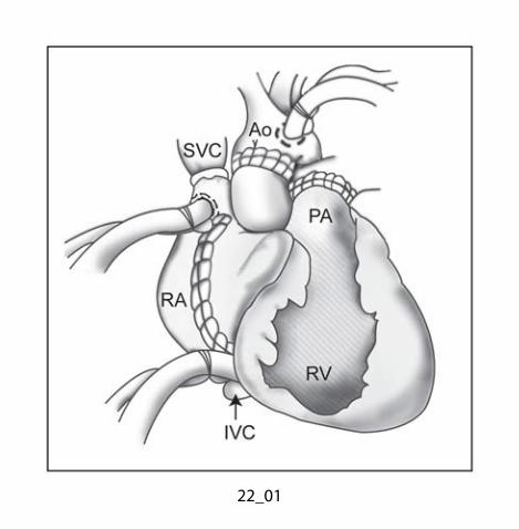
and separated to facilitate proper exposure and suturing approach (Fig. 22.1).
The donor heart is brought into the thoracic cavity and the LA anastomosis is
performed first with 3.0 polypropylene continuous sutures. To keep the heart as cold as
possible during surgical implantation standard cold blood cardioplegia, at a rate of 100–200
4
mL/min, is administered retrograde through a coronary sinus catheter in addition to
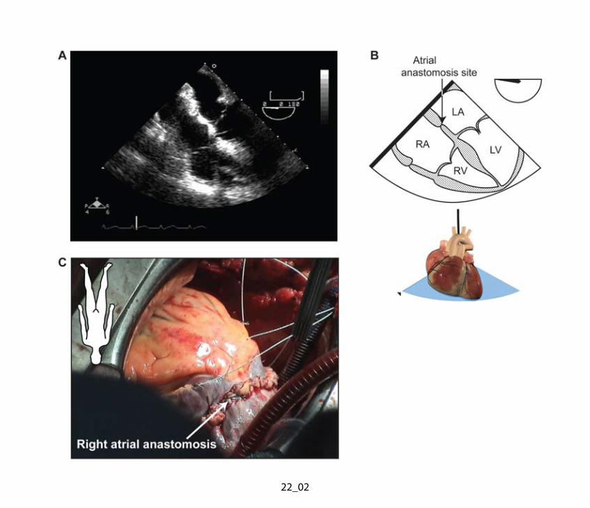
application of ice slush onto the right ventricle (RV) (11). The RA anastomosis (Fig. 22.2) is
completed with 3.0 polypropylene sutures, leaving a small gap for the coronary sinus catheter
prior to completion of both the PA and aortic anastomoses. Donor and recipient pulmonary
arteries are prepared, trimmed and anastomosed with a continuous 4.0 polypropylene suture.
Next, snares on both vena cava are released, the coronary sinus catheter is
removed, the operating table is put in the Trendelenburg position and the ascending Ao clamp
is released. With gentle massage of the heart, air is evacuated from the LV and the ascending
Ao through the suture line via the puncture used to inject the antegrade cardioplegic solution
at the donor site. The hole created in the left atrial appendage (LAA) to decompress the LV
during the harvest of the donor heart is closed with a 3.0 polypropylene suture.
The patient is weaned from the CPB with a heart rate averaging 100 beats/min
achieved by an isoproterenol infusion or atrial pacing and a good cardiac performance with
systolic blood pressure (SBP) >90–100 mmHg. Atrial and aortic cannulae are removed while
500 mg of solumedrol is administered and protamine is injected intravenously. The Swan–
Ganz catheter is refloated through the PA to monitor cardiac output (CO) and PA pressure.
Two chest tubes are inserted in the mediastinum. Temporary pacing wires are sutured onto
the RV. The sternum is then closed according to a standard technique.
B. Bicaval Technique
This alternative technique of cardiectomy and anastomoses is now more widely accepted as it
minimizes the problems arising from oversized post-transplant atria, such as stasis,
spontaneous echo contrast and embolic complications (10). The right aspect of the LA is
entered as for a routine MV surgical approach. The incision is extended superiorly under the
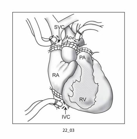
SVC and inferiorly under the IVC (Fig. 22.3). During excision of the RA, generous portions of
the SVC and IVC are left to facilitate donor and recipient anastomoses. The donor LA is
anastomosed to the recipient LA as described in the standard method. The IVC and SVC are
then anastomosed with 4-0 polypropylene suture. The rest of the procedure continues as
described above for the standard approach to OHT.
5
IV. TRANSESOPHAGEAL ECHOCARDIOGRAPHY IN THE PERIOPERATIVE PERIOD
The intraoperative use of TEE is a Society of Cardiovascular Anesthesiologists (SCA)
category 2 indication during OHT (12). The role of TEE is less valuable during the pre-CPB
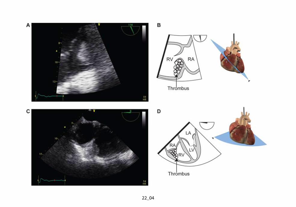
period as the recipient’s failing heart is completely removed. Rarely, thrombotic material in the
native heart can be mobilized during manipulation and lead to pulmonary embolism (Fig.
22.4). During the CPB weaning process, TEE helps to define the etiology of hemodynamic
instability, assess the efficacy of de-airing procedures and confirm the integrity of surgical
anastomosis in the post-transplantation period.
Hemodynamic instability after heart transplantation may arise from a single or
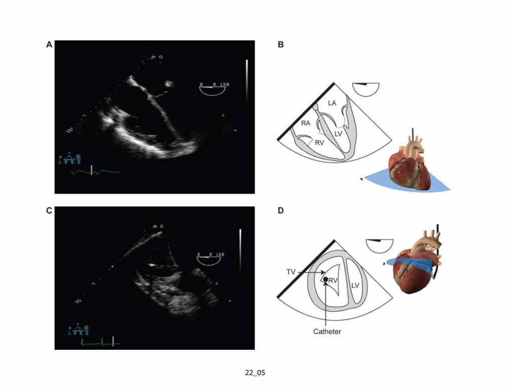
multiple causes such as primary graft failure, hyperacute rejection, RV dysfunction (Fig. 22.5),
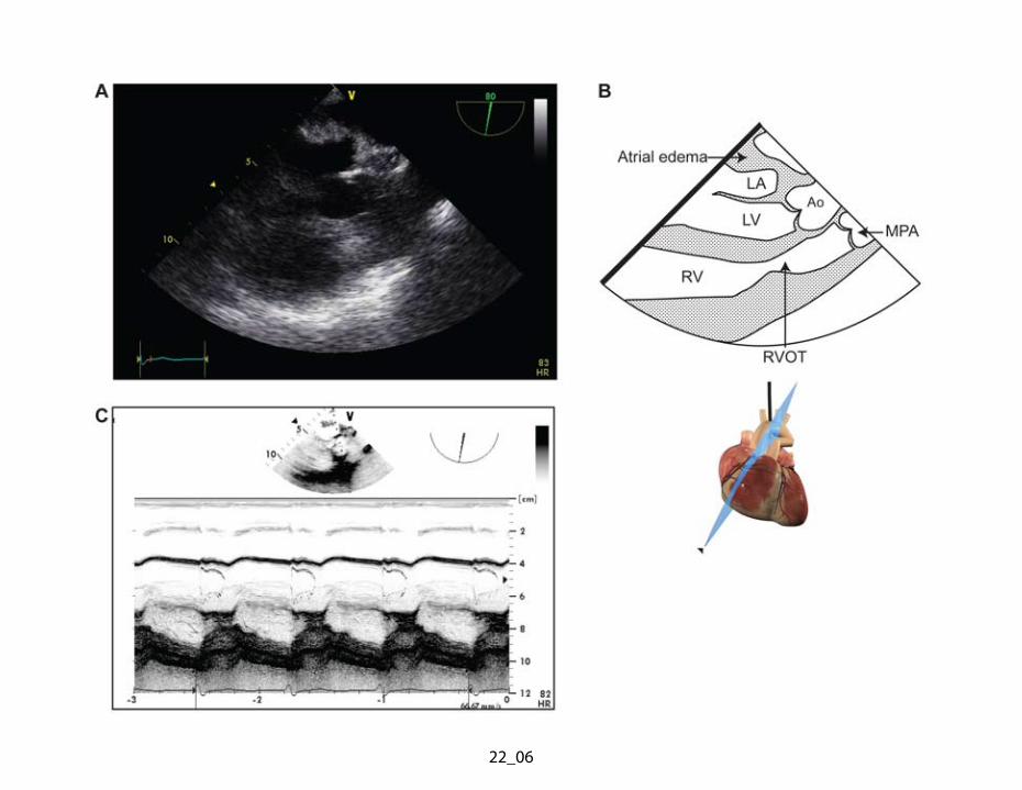
hypovolemia, vasodilatation, tamponade, right ventricular outflow tract (RVOT) obstruction
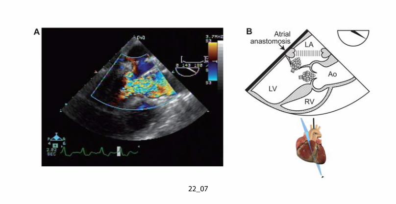
(Fig. 22.6) or left ventricular outflow tract (LVOT) obstruction (Fig. 22.7).
Nonspecific graft failure is still the most common cause of death in the early
postoperative period (13). Severe systolic dysfunction in the recipient may result from
inadequate myocardial protection during the harvesting of the donor heart (role of cardioplegic
solution, temperature, and duration of ischemia) and/or during transplantation. In rare
instances, hyperacute rejection, rather than insufficient myocardial protection, may be
responsible for the heart failure.
Right ventricular failure may occur in the recipient with pre-existing and
underestimated pulmonary hypertension (PH) (Fig. 22.5). Registry data from the International
Society of Heart and Lung Transplantation show that, despite advances in perioperative
management, RV dysfunction accounts for 50% of all cardiac complications and 19% of early
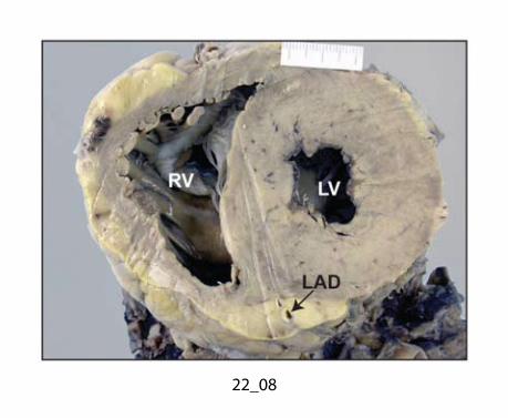
death after heart transplantation (2,13,14) (Fig. 22.8). There is approximately a fourfold higher
mortality among transplanted patients with fixed PH compared with patients without PH (13).
Pulmonary hypertension, defined as a PVR >480 dyne s/cm5 or 6 Wood units, is currently
considered a contraindication to OHT. Patients with marked PH (PVR >640 dyne s/cm5 or 8
Wood units) should be considered for heart and lung transplantation and, more recently, long
term LV assistance followed with transplantation. To date, there have been no reports in the
literature of a threshold beyond which donor RV heart failure could occur which would
constitute a clear contraindication to heart transplantation. A normal preoperative PVR does
6
provide some potential protection from acute increase in PVR causing RV failure after heart
transplantation.
Organ preservation and CPB may have deleterious effects on ventricular function.
Indeed CPB has been shown to cause an increase in PVR (13). Reviews have been
published on the management of RV failure after heart transplantation and the role for inhaled
nitric oxide (NO) treatment (13,15,16).
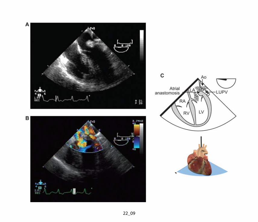
Transesophageal echocardiography is also useful to evaluate surgical anastomosis in
the post-transplantation period (Fig. 22.9). The main PA anastomosis appears as a suture
ridge within the vessels. Stenosis should be ruled out by two-dimensional (2D) color Doppler
imaging to detect turbulent flow and by continuous wave (CW) Doppler to measure the
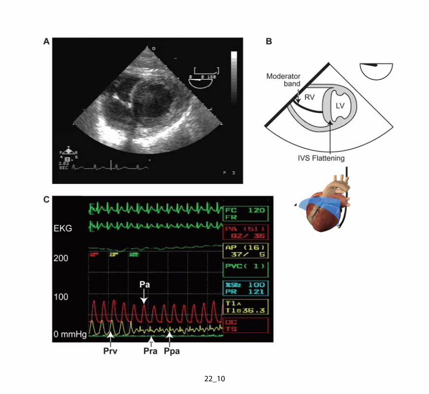
systolic gradient across the anastomosis. The PAC can also be used to document any
gradient across the RV and the PA (Fig. 22.10) (17). The mid-esophageal long-axis (LAX) or
two-chamber views show an elongated LA now composed of both donor and recipient atrial
tissue (Fig. 22.11). The suture line within the LA and RA also appears predominantly as an
echodense ridge (Fig. 22.9). Acquired cor triatriatum may develop secondary to infolding of
the redundant tissue from excessive donor atrial tissue (Fig. 22.7). Anastamoses of both
cavae are examined to rule out stenosis.
V. ECHOCARDIOGRAPHY IN THE POSTOPERATIVE PERIOD
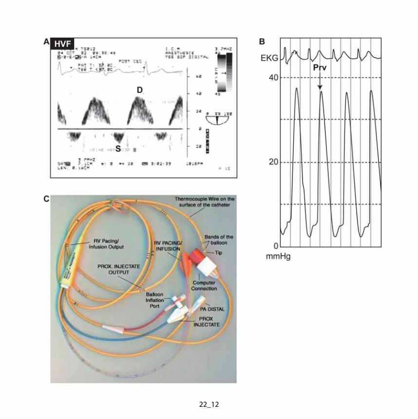
The early postoperative changes following heart transplantation include RV remodeling,
abnormal septal motion, increase in LV mass, presence of pericardial fluid and abnormal LV
and RV filling patterns (Fig. 22.12) (18).
A. Right Ventricular Remodeling, Tricuspid Regurgitation, and Abnormal Movement of
the Ventricular Septum
The time course of the resolution of PH and RV remodeling after OHT has been described by
Bhatia et al. (19). In 24 patients with moderate PH (mean PA pressure <50 mmHg) after heart
transplantation, the PH resolved rapidly, with the mean PA pressure approaching normal
values at two weeks after surgery. In parallel, the PVR had returned to normal in 80% of
patients by one year. Patients with the highest preoperative PVR had the greatest decrease
7
after the transplantation (ranging from 10% to 84%). Right and left heart filling pressure also
decreased to a nadir in the upper normal range at two weeks after surgery and remained
unchanged over one year of follow-up.
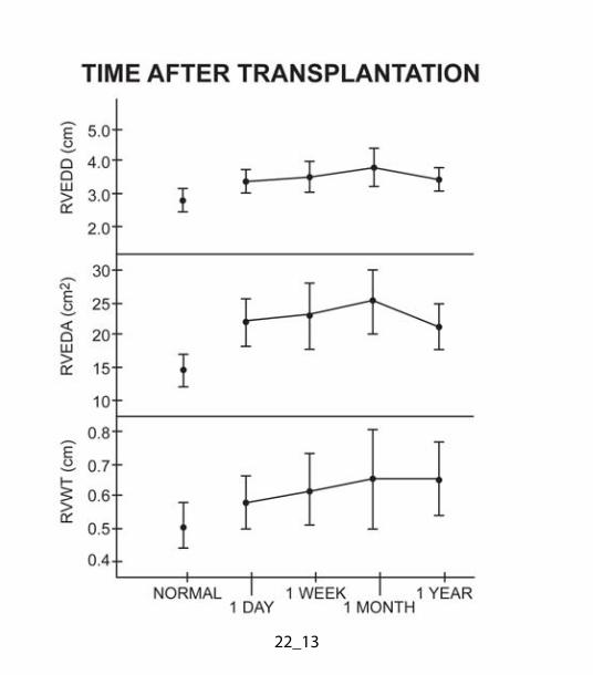
The donor RV remodels in response to recipient perioperative PH. Initially enlarged, it
dilates further during the first month and subsequently returns to its immediate perioperative
dimensions at one year after surgery (Fig. 22.13) (19). The RV wall thickness does not
increase significantly probably because the PVR declines to normal level. Thus, at one year,
while resting hemodynamics are normal, the donor RV remains enlarged, probably as an
adaptation to chronic volume overload.
Tricuspid regurgitation (TR) is common after heart transplantation due to the afterload
mismatch and RV dilatation. The incidence of TR (grades 1–3) changes from 67% on day one
after heart transplantation to 36% at one year. Nevertheless, TR is generally well tolerated
and rarely of clinical significance (19). Finally, abnormal diastolic flattening of the ventricular
septum was present in all patients immediately postoperatively (Fig. 22.10). This proportion
decreased to 75% at one month and 42% at one year. This decline parallels the decrease in
TR and RV dimensions. On the other hand, systolic paradoxical septal motion was
uncommon, probably because the mean PA pressure decreases significantly immediately
after OHT (19).
B. Left Ventricular Mass and Mitral Valve Function
In the immediate postoperative period, both LV wall thickness and mass increase, most likely
from perioperative myocardial ischemia and edema. Systemic hypertension and side effects
from cyclosporine and corticosteroid administration can further contribute to LV mass
augmentation seen at one year. Acute increase in myocardial thickness may also occur later
with allograft rejection (Fig. 22.8) (20). Left ventricular contractility and contractile reserve both
appear to be normal after heart transplantation (21).
Mild to moderate mitral regurgitation (MR) has been reported to occur with a
frequency ranging from 55% to 87% without major clinical implications (19,22). It usually
occurs in a donor heart without structural abnormality of the mitral apparatus or LV
dysfunction. The MR may arise from biatrial enlargement which exerts tension on the
8
posterior MV leaflet and causes incomplete closure of the valve and annuloventricular
disproportion (19). Moreover, the presence of both donor and recipient atria and sinus nodes
results in asynchrony and inhomogenous atrial contraction, which may also play a role in the
genesis of MR (19). Finally, MR can occur in association with LVOT obstruction.
C. Pericardial Effusion
Pericardial effusions are commonly observed in 85% of patients after heart transplantation
(23). The loss of lymphatic drainage and the discrepancy in size between the new donor heart
and the large remaining pericardial cavity are plausible explanations (20). Even though
pericardial effusions are common, rarely do they precipitate hemodynamic instability, such as
cardiac tamponade. While large but slowly accumulating pericardial effusions usually cause
little hemodynamic impairment, a loculated hematoma developing at a critical location may
conversely cause acute cardiac tamponade (see Chapter 12). Thus, the relationship between
the size of the effusion and the clinical outcome is likely to show a poor correlation (20). Most
effusions are nearly, or completely, resolved by 30 days after surgery (23).
D. Left Ventricular Diastolic Function
Diastolic function is also altered in the heart transplant recipient (in the absence of rejection)
during the early postoperative period. Initial pulsed wave (PW) Doppler examination of the
mitral inflow shows a profile comparable to previously defined restrictive parameters (see
Chapter 10); this evolves into a non-restrictive pattern over a six week period leading to
progressive improvement in postoperative diastolic function parameters and decrease in left
heart filling pressures (24). The presence of recurrent or persistent severe diastolic
dysfunction with restrictive filling within six months after transplantation is associated with a
reduced late-term actuarial survival, independent of graft rejection (25).
Evidence suggests that retaining normal LA size and shape by using the bicaval
technique promotes ventricular filling dynamics which more closely approximates normal
physiology (20). Ventricular filling is influenced by the mechanical activity of residual recipient
atrial tissue over that of the donor heart. In addition, “parasystolic” contraction of residual
recipient atrial tissue also modifies the pulmonary venous flow (PVF): recipient atrial
9
contraction occurring in late systole results in an increase in the diastolic component (D-
wave); if it happens in early systole the systolic component (S-wave) is decreased. End-
diastolic atrial contraction will increase the velocity observed during atrial reversal (AR) (18).
E. Normal Echocardiographic Profile after Heart Transplantation
One year after OHT, echocardiograms of recipients doing clinically well are characterized by
increased LV wall thickness and mass. Left ventricular dimensions, volumes and ejection
fraction are within normal limits. Right ventricular wall thickness and cavity size are increased
with preserved RV systolic function. The transplanted heart also shows an anteromedial
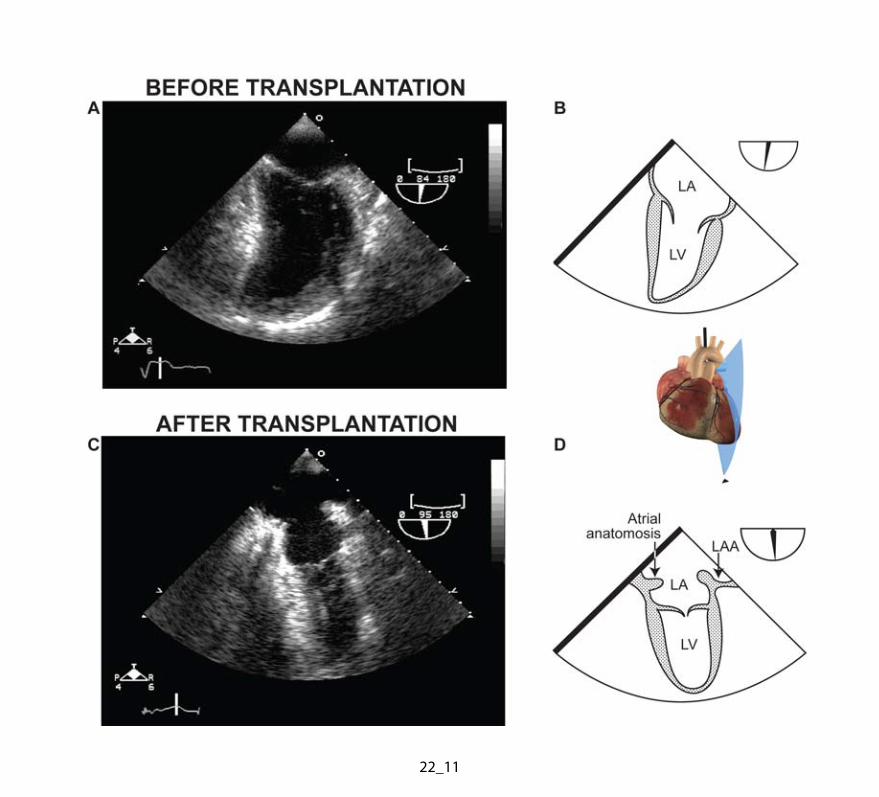
translational motion during systole. The atria of the transplanted heart have unique
echocardiographic features. The anastomotic suture line is easily identified on 2D imaging as
the waist of these hourglass-shaped atria (Fig. 22.11). This waist creates a natural point of
subdivision within the native and donor atria. The markedly enlarged atrial volume results
primarily from an increased LAX dimension (Fig. 22.11), although maximal width or short-axis
dimension is also slightly larger than in controls (26). The increase in both donor and recipient
atrial size is inversely correlated with survival (27).
F. Abnormal Echocardiographic Findings Following Heart Transplantation
The presence of the atrial suture line, increased atrial size with subcontractile portion of the
recipient atrium and asynchrony between the donor and recipient atria contraction, promotes
stasis. These factors may account for the high prevalence of atrial spontaneous echo contrast
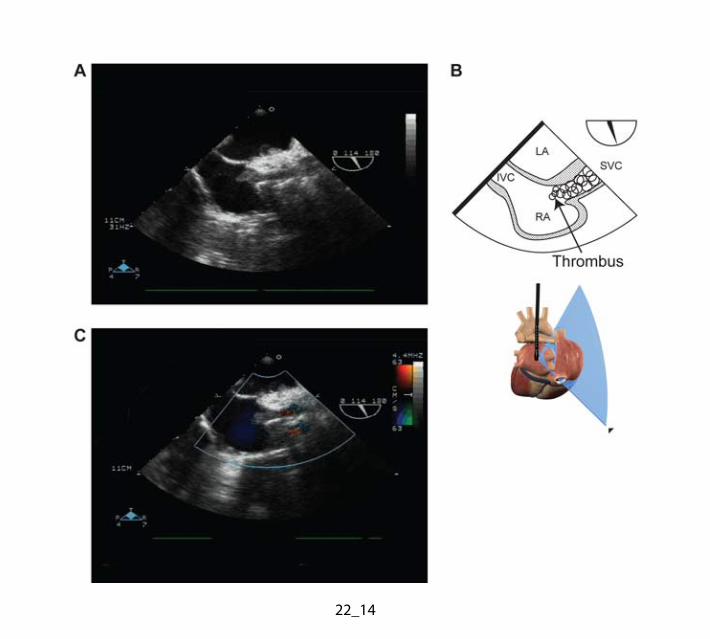
(55%) as assessed with TEE (28). Left atrial thrombi were observed in 38% by TEE and are
often missed by transthoracic echocardiography (TTE). Thrombi were located in the donor
LAA (10/18), on the posterior wall of the LA (6/18), on the donor component of the atrial
septum (1/18) and on the left atrial suture (1/18) (28). Thrombi occurred only in patients
displaying spontaneous echocardiographic contrast (Fig. 22.14). Episodes of arterial
embolism were documented in 22 % of patients with both spontaneous echocardiographic
contrast and LA thrombus (6% of heart transplantation recipients) (28). The use of the
modified bicaval OHT technique seems to decrease the incidence of this problem
considerably (29).
10
Transesophageal echocardiography has also been found to be superior to TTE in
demonstrating thickening of the atrial septum, bulging of the recipient and donor atrial
septum, and shunt at the atrial level (28). It may identify uncommon patent foramen ovale
after heart transplantation (30). Coronary fistula is another finding detected by TEE after heart
transplantation. The incidence of this iatrogenic complication has been estimated between 5%
and 15%, a 20-fold increase over the incidence of congenital coronary artery fistula (31). The
increased incidence in this group is attributed to injury from multiple routine surveillance RV
endomyocardial biopsy procedures for detection of cardiac rejection, which frequently
involves the right coronary artery (RCA). The vast majority of these fistulas communicate
directly with the RV, are usually diagnosed by routine coronary angiography and are without
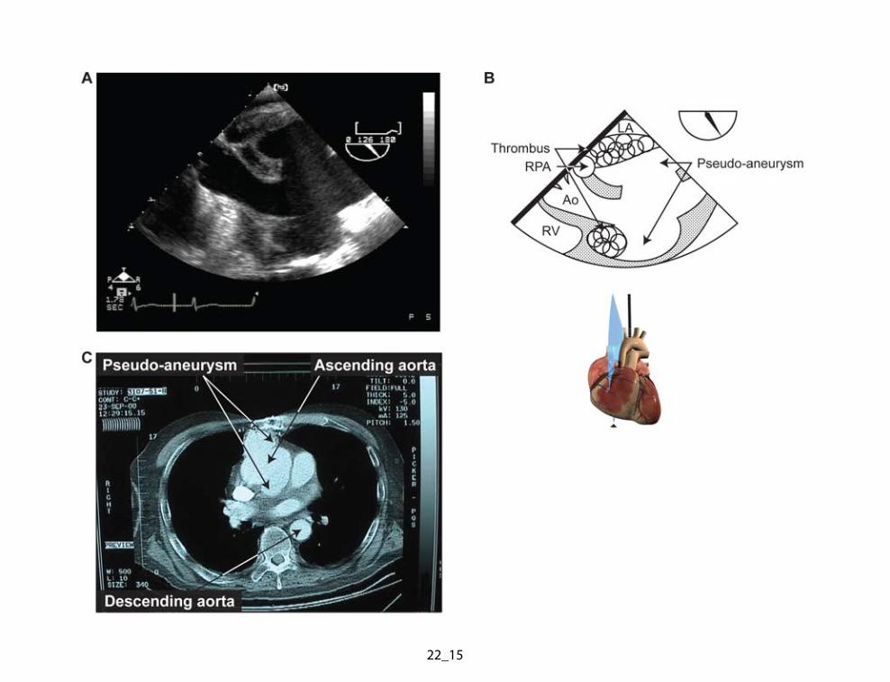
hemodynamic significance (31). Finally pseudoaneurysm can occur after cardiac
transplantation at any site of major vessel anastomosis (Fig. 22.11).
G. Detection of Acute Allograft Rejection and Coronary Artery Disease
Acute allograft rejection is common, particularly in the first year after transplantation. It
constitutes the most frequent cause of death during this period. Morphologic features
suggestive of allograft rejection include an increase in myocardial mass due to inflammatory
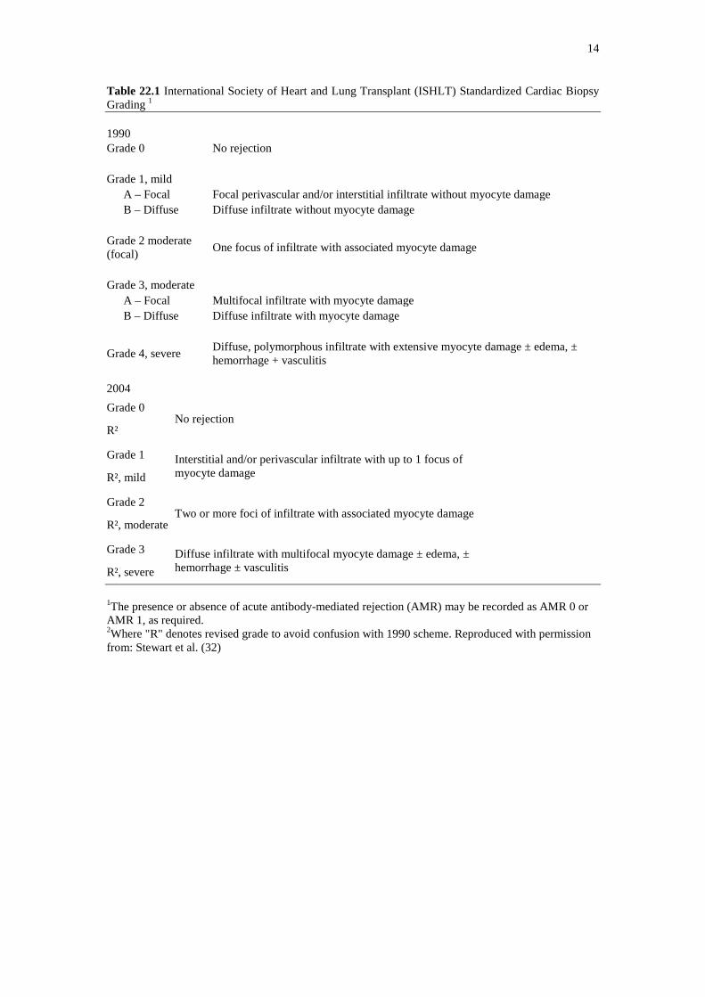
cell infiltration and myocardial edema (Fig. 22.8) (Table 22.1). However, with the addition of
cyclosporine to the immunosuppressive medical regimen, cellular rejection is associated with
less myocardial edema: the evaluation of ventricular systolic function and myocardial mass
has thus become obsolete as a means of detecting early rejection. Doppler parameters of
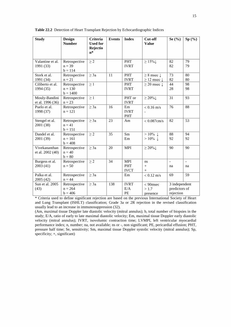
diastolic function have recently been used to detect acute rejection (Table 22.2). It appears
that moderate to severe rejection leads to a fall of at least 15% in the mitral deceleration or
the isovolemic relaxation time. However, wide inter-patient variability in Doppler variables
renders isolated measurement cut-offs less predictive. Each patient acting as her/his own
control (from baseline) provides the basis for Doppler-based surveillance of allograft rejection.
Currently, Doppler-derived parameters of diastolic function are used as an adjunct rather than
a replacement for endomyocardial biopsy. Protocols now combine routine biopsies, with
supplemental biopsy in the event of echocardiographic evidence of acutely restrictive
physiology (20).
11
In patients who survive the first two years of transplantation, coronary artery disease
(CAD) constitutes an important cause of mortality. There is increasing interest in the ability of
dobutamine stress echocardiography to predict adverse cardiac events in OHT recipients. A
normal dobutamine stress echocardiography result is a very powerful determinant of a benign
clinical course with a negative predictive value in excess of 90–95% (20).
VI. CONCLUSION
In summary, echocardiography may significantly contribute to the evaluation of patients at
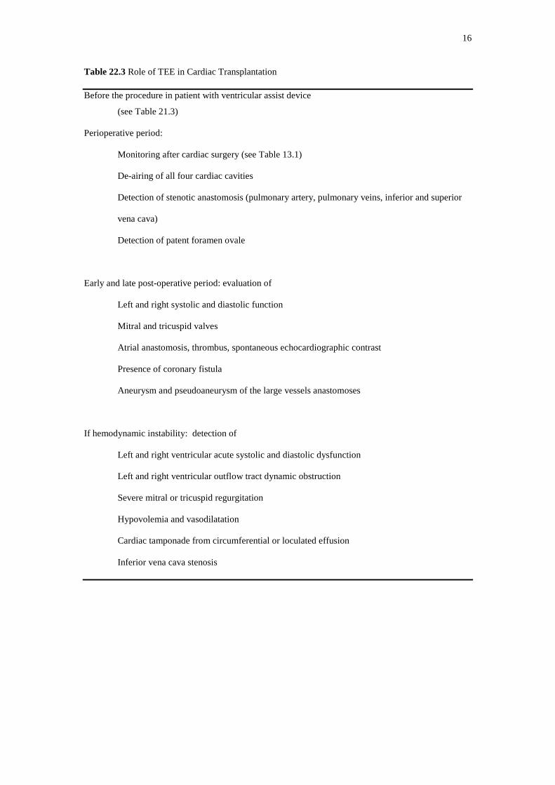
various stages of heart transplantation (Table 22.3). During the initial perioperative period, it
provides timely assessment of cardiac anatomy and physiology and bestows the opportunity
to detect specific problems. Later, detection of allograft rejection and accelerated CAD
benefits from the addition of this noninvasive diagnostic tool.
12
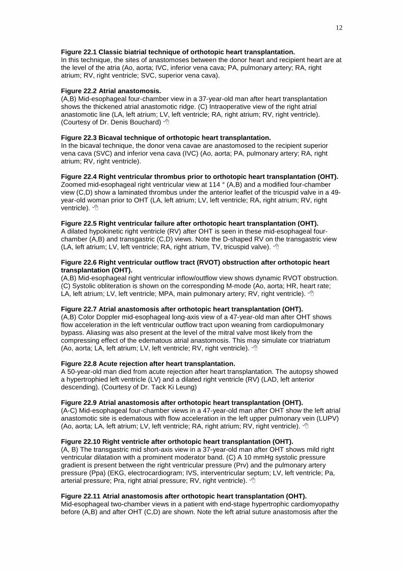
Figure 22.1 Classic biatrial technique of orthotopic heart transplantation. In this technique, the sites of anastomoses between the donor heart and recipient heart are at the level of the atria (Ao, aorta; IVC, inferior vena cava; PA, pulmonary artery; RA, right atrium; RV, right ventricle; SVC, superior vena cava). Figure 22.2 Atrial anastomosis. (A,B) Mid-esophageal four-chamber view in a 37-year-old man after heart transplantation shows the thickened atrial anastomotic ridge. (C) Intraoperative view of the right atrial anastomotic line (LA, left atrium; LV, left ventricle; RA, right atrium; RV, right ventricle). (Courtesy of Dr. Denis Bouchard) Figure 22.3 Bicaval technique of orthotopic heart transplantation. In the bicaval technique, the donor vena cavae are anastomosed to the recipient superior vena cava (SVC) and inferior vena cava (IVC) (Ao, aorta; PA, pulmonary artery; RA, right atrium; RV, right ventricle). Figure 22.4 Right ventricular thrombus prior to orthotopic heart transplantation (OHT). Zoomed mid-esophageal right ventricular view at 114 ° (A,B) and a modified four-chamber view (C,D) show a laminated thrombus under the anterior leaflet of the tricuspid valve in a 49-year-old woman prior to OHT (LA, left atrium; LV, left ventricle; RA, right atrium; RV, right ventricle). Figure 22.5 Right ventricular failure after orthotopic heart transplantation (OHT). A dilated hypokinetic right ventricle (RV) after OHT is seen in these mid-esophageal four-chamber (A,B) and transgastric (C,D) views. Note the D-shaped RV on the transgastric view (LA, left atrium; LV, left ventricle; RA, right atrium, TV, tricuspid valve). Figure 22.6 Right ventricular outflow tract (RVOT) obstruction after orthotopic heart transplantation (OHT). (A,B) Mid-esophageal right ventricular inflow/outflow view shows dynamic RVOT obstruction. (C) Systolic obliteration is shown on the corresponding M-mode (Ao, aorta; HR, heart rate; LA, left atrium; LV, left ventricle; MPA, main pulmonary artery; RV, right ventricle). Figure 22.7 Atrial anastomosis after orthotopic heart transplantation (OHT). (A,B) Color Doppler mid-esophageal long-axis view of a 47-year-old man after OHT shows flow acceleration in the left ventricular outflow tract upon weaning from cardiopulmonary bypass. Aliasing was also present at the level of the mitral valve most likely from the compressing effect of the edematous atrial anastomosis. This may simulate cor triatriatum (Ao, aorta; LA, left atrium; LV, left ventricle; RV, right ventricle). Figure 22.8 Acute rejection after heart transplantation. A 50-year-old man died from acute rejection after heart transplantation. The autopsy showed a hypertrophied left ventricle (LV) and a dilated right ventricle (RV) (LAD, left anterior descending). (Courtesy of Dr. Tack Ki Leung) Figure 22.9 Atrial anastomosis after orthotopic heart transplantation (OHT). (A-C) Mid-esophageal four-chamber views in a 47-year-old man after OHT show the left atrial anastomotic site is edematous with flow acceleration in the left upper pulmonary vein (LUPV) (Ao, aorta; LA, left atrium; LV, left ventricle; RA, right atrium; RV, right ventricle). Figure 22.10 Right ventricle after orthotopic heart transplantation (OHT). (A, B) The transgastric mid short-axis view in a 37-year-old man after OHT shows mild right ventricular dilatation with a prominent moderator band. (C) A 10 mmHg systolic pressure gradient is present between the right ventricular pressure (Prv) and the pulmonary artery pressure (Ppa) (EKG, electrocardiogram; IVS, interventricular septum; LV, left ventricle; Pa, arterial pressure; Pra, right atrial pressure; RV, right ventricle). Figure 22.11 Atrial anastomosis after orthotopic heart transplantation (OHT). Mid-esophageal two-chamber views in a patient with end-stage hypertrophic cardiomyopathy before (A,B) and after OHT (C,D) are shown. Note the left atrial suture anastomosis after the
13
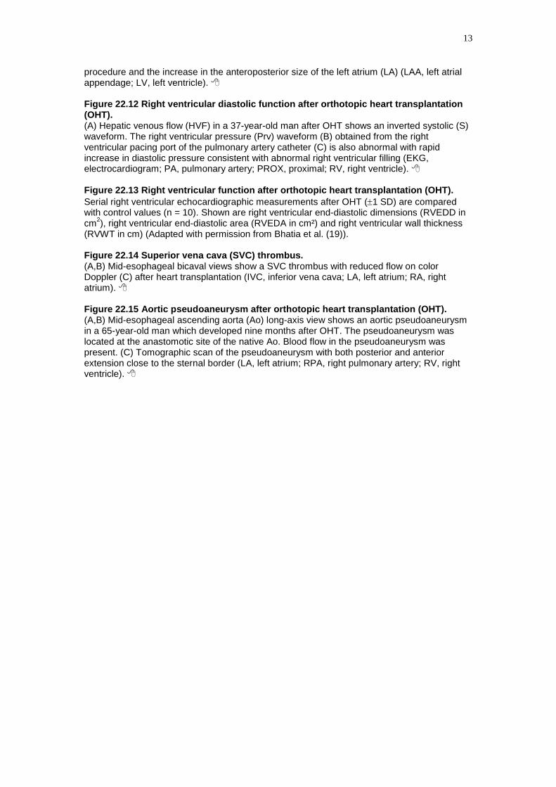
procedure and the increase in the anteroposterior size of the left atrium (LA) (LAA, left atrial appendage; LV, left ventricle). Figure 22.12 Right ventricular diastolic function after orthotopic heart transplantation (OHT). (A) Hepatic venous flow (HVF) in a 37-year-old man after OHT shows an inverted systolic (S) waveform. The right ventricular pressure (Prv) waveform (B) obtained from the right ventricular pacing port of the pulmonary artery catheter (C) is also abnormal with rapid increase in diastolic pressure consistent with abnormal right ventricular filling (EKG, electrocardiogram; PA, pulmonary artery; PROX, proximal; RV, right ventricle). Figure 22.13 Right ventricular function after orthotopic heart transplantation (OHT). Serial right ventricular echocardiographic measurements after OHT (±1 SD) are compared with control values (n = 10). Shown are right ventricular end-diastolic dimensions (RVEDD in cm2), right ventricular end-diastolic area (RVEDA in cm²) and right ventricular wall thickness (RVWT in cm) (Adapted with permission from Bhatia et al. (19)). Figure 22.14 Superior vena cava (SVC) thrombus. (A,B) Mid-esophageal bicaval views show a SVC thrombus with reduced flow on color Doppler (C) after heart transplantation (IVC, inferior vena cava; LA, left atrium; RA, right atrium). Figure 22.15 Aortic pseudoaneurysm after orthotopic heart transplantation (OHT). (A,B) Mid-esophageal ascending aorta (Ao) long-axis view shows an aortic pseudoaneurysm in a 65-year-old man which developed nine months after OHT. The pseudoaneurysm was located at the anastomotic site of the native Ao. Blood flow in the pseudoaneurysm was present. (C) Tomographic scan of the pseudoaneurysm with both posterior and anterior extension close to the sternal border (LA, left atrium; RPA, right pulmonary artery; RV, right ventricle).
14
Table 22.1 International Society of Heart and Lung Transplant (ISHLT) Standardized Cardiac Biopsy Grading 1
1990 Grade 0 No rejection Grade 1, mild
A – Focal Focal perivascular and/or interstitial infiltrate without myocyte damage B – Diffuse Diffuse infiltrate without myocyte damage
Grade 2 moderate (focal) One focus of infiltrate with associated myocyte damage
Grade 3, moderate
A – Focal Multifocal infiltrate with myocyte damage B – Diffuse Diffuse infiltrate with myocyte damage
Grade 4, severe Diffuse, polymorphous infiltrate with extensive myocyte damage ± edema, ± hemorrhage + vasculitis
2004 Grade 0
R² No rejection
Grade 1
R², mild Interstitial and/or perivascular infiltrate with up to 1 focus of myocyte damage
Grade 2
R², moderate Two or more foci of infiltrate with associated myocyte damage
Grade 3
R², severe Diffuse infiltrate with multifocal myocyte damage ± edema, ± hemorrhage ± vasculitis
1The presence or absence of acute antibody-mediated rejection (AMR) may be recorded as AMR 0 or AMR 1, as required. 2Where "R" denotes revised grade to avoid confusion with 1990 scheme. Reproduced with permission from: Stewart et al. (32)
15
Table 22.2 Detection of Heart Transplant Rejection by Echocardiographic Indices Study Design
Number Criteria Used for Rejection*
Events
Index Cut-off Value
Se (%) Sp (%)
Valantine et al. 1991 (33)
Retrospective n = 39 b = 114
≥ 2
PHT IVRT
≥ 15%↓ 82 82
79 79
Stork et al. 1991 (34)
Retrospective n = 21
≥ 3a 11 PHT IVRT
≥ 8 msec ↓ ≥ 12 msec ↓
73 82
80 80
Ciliberto et al. 1994 (35)
Retrospective n = 130 b = 1400
≥ 1 PHT IVRT
≥ 20 msec ↓
44 28
98 98
Mouly-Bandini et al. 1996 (36)
Retrospective n = 23
≥ 1 PHT or IVRT
≥ 20%↓ 31 93
Puelo et al. 1998 (37)
Retrospective n = 121
≥ 3a 16 Em IVRT PHT
< 0.16 m/s -
76 88
Stengel et al. 2001 (38)
Retrospective n = 41 b = 151
≥ 3a 23 Am < 0.087cm/s
82 53
Dandel et al. 2001 (39)
Retrospective n = 161 b = 408
≥ 2 35 Sm Em
> 10% ↓ > 10% ↓
88 92
94 92
Vivekananthan et al. 2002 (40)
Retrospective n = 40 b = 80
≥ 3a 20 MPI ≥ 20%↓
90 90
Burgess et al. 2003 (41)
Retrospective n = 50
≥ 2
34 MPI PHT IVCT
ns + +
- na
- na
Palka et al. 2005 (42)
Retrospective n = 44
≥ 3a Em < 0.12 m/s 69 59
Sun et al. 2005 (43)
Retrospective n = 264 b = 406
≥ 3a 138 IVRT E/A PE
< 90msec > 1.7 presence
3 independent predictors of rejection
* Criteria used to define significant rejection are based on the previous International Society of Heart and Lung Transplant (ISHLT) classification; Grade 3a or 2R rejection in the revised classification usually lead to an increase in immunosuppression (32). (Am, maximal tissue Doppler late diastolic velocity (mitral annulus); b, total number of biopsies in the study; E/A, ratio of early to late maximal diastolic velocity; Em, maximal tissue Doppler early diastolic velocity (mitral annulus); IVRT, isovolumic contraction time; LVMPI, left ventricular myocardial performance index; n, number; na, not available; ns or -, non significant; PE, pericardial effusion; PHT, pressure half time; Se, sensitivity; Sm, maximal tissue Doppler systolic velocity (mitral annulus); Sp, specificity; +, significant)
16
Table 22.3 Role of TEE in Cardiac Transplantation
Before the procedure in patient with ventricular assist device
(see Table 21.3)
Perioperative period:
Monitoring after cardiac surgery (see Table 13.1)
De-airing of all four cardiac cavities
Detection of stenotic anastomosis (pulmonary artery, pulmonary veins, inferior and superior
vena cava)
Detection of patent foramen ovale
Early and late post-operative period: evaluation of
Left and right systolic and diastolic function
Mitral and tricuspid valves
Atrial anastomosis, thrombus, spontaneous echocardiographic contrast
Presence of coronary fistula
Aneurysm and pseudoaneurysm of the large vessels anastomoses
If hemodynamic instability: detection of
Left and right ventricular acute systolic and diastolic dysfunction
Left and right ventricular outflow tract dynamic obstruction
Severe mitral or tricuspid regurgitation
Hypovolemia and vasodilatation
Cardiac tamponade from circumferential or loculated effusion
Inferior vena cava stenosis
17
References
1. Hunt SA, Abraham WT, Chin MH et al. ACC/AHA 2005 Guideline Update for the Diagnosis and
Management of Chronic Heart Failure in the Adult: a report of the American College of
Cardiology/American Heart Association Task Force on Practice Guidelines (Writing Committee to
Update the 2001 Guidelines for the Evaluation and Management of Heart Failure): developed in
collaboration with the American College of Chest Physicians and the International Society for
Heart and Lung Transplantation: endorsed by the Heart Rhythm Society. Circulation
2005;112:e154-e235.
2. Taylor DO, Edwards LB, Boucek MM et al. Registry of the International Society for Heart and
Lung Transplantation: twenty-fourth official adult heart transplant report--2007. J Heart Lung
Transplant 2007;26:769-781.
3. Zaroff JG, Rosengard BR, Armstrong WF et al. Consensus conference report: maximizing use of
organs recovered from the cadaver donor: cardiac recommendations, March 28-29, 2001, Crystal
City, Va. Circulation 2002;106:836-841.
4. Renlund DG, Taylor DO, Kfoury AG et al. New UNOS rules: historical background and
implications for transplantation management. United Network for Organ Sharing. J Heart Lung
Transplant 1999;18:1065-1070.
5. Russo MJ, Chen JM, Sorabella RA et al. The effect of ischemic time on survival after heart
transplantation varies by donor age: an analysis of the United Network for Organ Sharing database.
J Thorac Cardiovasc Surg 2007;133:554-559.
6. Mehra MR, Kobashigawa J, Starling R et al. Listing criteria for heart transplantation: International
Society for Heart and Lung Transplantation guidelines for the care of cardiac transplant
candidates--2006. J Heart Lung Transplant 2006;25:1024-1042.
7. Butler J, Stankewicz MA, Wu J et al. Pre-transplant reversible pulmonary hypertension predicts
higher risk for mortality after cardiac transplantation. J Heart Lung Transplant 2005;24:170-177.
18
8. Lower RR, Shumway NE. Studies on orthotopic homotransplantation of the canine heart. Surg
Forum 1960;11:18-19.
9. Shumway NE, Lower RR, Stofer RC. Transplantation of the heart. Adv Surg 1966;2:265-284.
10. Dreyfus G, Jebara V, Mihaileanu S et al. Total orthotopic heart transplantation: an alternative to
the standard technique. Ann Thorac Surg 1991;52:1181-1184.
11. Carrier M, Leung TK, Solymoss BC et al. Clinical trial of retrograde warm blood reperfusion
versus standard cold topical irrigation of transplanted hearts. Ann Thorac Surg 1996;61:1310-
1314.
12. Practice guidelines for perioperative transesophageal echocardiography. A report by the American
Society of Anesthesiologists and the Society of Cardiovascular Anesthesiologists Task Force on
Transesophageal Echocardiography. Anesthesiology 1996;84:986-1006.
13. Stobierska-Dzierzek B, Awad H, Michler RE. The evolving management of acute right-sided heart
failure in cardiac transplant recipients. J Am Coll Cardiol 2001;38:923-931.
14. Hosenpud JD, Bennett LE, Keck BM et al. The Registry of the International Society for Heart and
Lung Transplantation: seventeenth official report-2000. J Heart Lung Transplant 2000;19:909-931.
15. Carrier M, Blaise G, Belisle S et al. Nitric oxide inhalation in the treatment of primary graft failure
following heart transplantation. J Heart Lung Transplant 1999;18:664-667.
16. Haddad F, Couture P, Tousignant C et al. The right ventricle in cardiac surgery, a perioperative
perspective: II. Pathophysiology, clinical importance, and management. Anesth Analg
2009;108:422-433.
17. Suriani RJ. Transesophageal echocardiography during organ transplantation. J Cardiothorac Vasc
Anesth 1998;12:686-694.
18. Durennes ME. Échocardiographie transoesophagienne chez le transplanté cardiaque. In: Cohen A,
ed. Échocardiographie transoesophagienne en cardiologie et en anesthésie. Édition Arnette,
1992:265-279.
19
19. Bhatia SJ, Kirshenbaum JM, Shemin RJ et al. Time course of resolution of pulmonary
hypertension and right ventricular remodeling after orthotopic cardiac transplantation. Circulation
1987;76:819-826.
20. Burgess MI, Bhattacharyya A, Ray SG. Echocardiography after cardiac transplantation. J Am Soc
Echocardiogr 2002;15:917-925.
21. Borow KM, Neumann A, Arensman FW et al. Left ventricular contractility and contractile reserve
in humans after cardiac transplantation. Circulation 1985;71:866-872.
22. Angermann CE, Spes CH, Tammen A et al. Anatomic characteristics and valvular function of the
transplanted heart: transthoracic versus transesophageal echocardiographic findings. J Heart
Transplant 1990;9:331-338.
23. Weitzman LB, Tinker WP, Kronzon I et al. The incidence and natural history of pericardial
effusion after cardiac surgery--an echocardiographic study. Circulation 1984;69:506-511.
24. StGoar FG, Gibbons R, Schnittger I et al. Left ventricular diastolic function. Doppler
echocardiographic changes soon after cardiac transplantation. Circulation 1990;82:872-878.
25. Ross HJ, Gullestad L, Hunt SA et al. Early Doppler echocardiographic dysfunction is associated
with an increased mortality after orthotopic cardiac transplantation. Circulation 1996;94:II289-
II293.
26. Gorcsan J, III, Snow FR, Paulsen W et al. Echocardiographic profile of the transplanted human
heart in clinically well recipients. J Heart Lung Transplant 1992;11:80-89.
27. Gudmundsson GS, Smull DL, Pisani BA et al. Increase in atrial size in long-term survivors of
heart transplant. J Am Soc Echocardiogr 2003;16:1043-1048.
28. Derumeaux G, Mouton-Schleifer D, Soyer R et al. High incidence of left atrial thrombus detected
by transoesophageal echocardiography in heart transplant recipients. Eur Heart J 1995;16:120-125.
29. Riberi A, Ambrosi P, Habib G et al. Systemic embolism: a serious complication after cardiac
transplantation avoidable by bicaval technique. Eur J Cardiothorac Surg 2001;19:307-311.
20
30. Ouseph R, Stoddard MF, Lederer ED. Patent foramen ovale presenting as refractory hypoxemia
after heart transplantation. J Am Soc Echocardiogr 1997;10:973-976.
31. Lowry RW, Young JB, Kleiman NS et al. Transesophageal echocardiographic demonstration of
right coronary artery-to-coronary sinus fistula in a heart transplant recipient. J Am Soc
Echocardiogr 1993;6:449-452.
32. Stewart S, Winters GL, Fishbein MC et al. Revision of the 1990 working formulation for the
standardization of nomenclature in the diagnosis of heart rejection. J Heart Lung Transplant
2005;24:1710-1720.
33. Valantine HA, Yeoh TK, Gibbons R et al. Sensitivity and specificity of diastolic indexes for
rejection surveillance: temporal correlation with endomyocardial biopsy. J Heart Lung Transplant
1991;10:757-765.
34. Stork T, Mockel M, Eichstadt H et al. Noninvasive diagnosis of cardiac allograft rejection by
means of pulsed Doppler and M-mode ultrasound. J Ultrasound Med 1991;10:569-575.
35. Ciliberto GR, Mascarello M, Gronda E et al. Acute rejection after heart transplantation:
noninvasive echocardiographic evaluation. J Am Coll Cardiol 1994;23:1156-1161.
36. Mouly-Bandini A, Vion-Dury J, Viout P et al. Value of Doppler echocardiography in the detection
of low-grade rejections after cardiac transplantation. Transpl Int 1996;9:131-136.
37. Puleo JA, Aranda JM, Weston MW et al. Noninvasive detection of allograft rejection in heart
transplant recipients by use of Doppler tissue imaging. J Heart Lung Transplant 1998;17:176-184.
38. Stengel SM, Allemann Y, Zimmerli M et al. Doppler tissue imaging for assessing left ventricular
diastolic dysfunction in heart transplant rejection. Heart 2001;86:432-437.
39. Dandel M, Hummel M, Muller J et al. Reliability of tissue Doppler wall motion monitoring after
heart transplantation for replacement of invasive routine screenings by optimally timed cardiac
biopsies and catheterizations. Circulation 2001;104:I184-I191.
40. Vivekananthan K, Kalapura T, Mehra M et al. Usefulness of the combined index of systolic and
21
diastolic myocardial performance to identify cardiac allograft rejection. Am J Cardiol
2002;90:517-520.
41. Burgess MI, Bright-Thomas RJ, Yonan N et al. Can the index of myocardial performance be used
to detect acute cellular rejection after heart transplantation? Am J Cardiol 2003;92:308-311.
42. Palka P, Lange A, Galbraith A et al. The role of left and right ventricular early diastolic Doppler
tissue echocardiographic indices in the evaluation of acute rejection in orthotopic heart transplant.
J Am Soc Echocardiogr 2005;18:107-115.
43. Sun JP, Abdalla IA, Asher CR et al. Non-invasive evaluation of orthotopic heart transplant
rejection by echocardiography. J Heart Lung Transplant 2005;24:160-165.
22_01
22_02
22_03
22_04
22_05
22_06
22_07
22_08
22_09
22_10
22_11
22_12
22_13
22_14
22_15