Embed Size (px)
Citation preview
© Copyright 2013 Elsevier, Ltd. All rights reserved.
24 Disorders of the thumb
CHAPTER CONTENTS
Disorders of the inert structures 351
Rheumatoid arthritis . . . . . . . . . . . . . . . . . 351Traumatic arthritis . . . . . . . . . . . . . . . . . . 351Arthrosis . . . . . . . . . . . . . . . . . . . . . . . 351
Disorders of the contractile structures 353
Pain 353Resisted extension. . . . . . . . . . . . . . . . . . 353Resisted flexion . . . . . . . . . . . . . . . . . . . 357Weakness 358Rupture . . . . . . . . . . . . . . . . . . . . . . . 358Nerve lesions . . . . . . . . . . . . . . . . . . . . 358
Disorders affecting the base of the thumb may cause pain over the radial aspect of the wrist. This is why the trapezium–first metacarpal joint and its neighbouring tendinous structures are tested in examination of the wrist.
Disorders of the inert structures
The only relevant passive test for the first carpometacarpal joint is backward movement during extension. Pain and/or limitation indicate a capsular lesion. In the majority of cases, it is the anterior aspect of the capsule that is affected and the joint is found to be tender anteriorly.
The three following conditions are quite common.
Rheumatoid arthritis
Both first carpometacarpal joints are frequently affected in rheumatoid arthritis and respond well to intra-articular triam-cinolone. Surgery is seldom necessary but, if so, resection arthroplasty is preferred.1
Traumatic arthritis
The patient describes an injury to the thumb, usually over-stretching. Discomfort and pain are experienced over the thenar area and the radial side of the wrist. The backward movement during extension elicits pain and is limited. The radiograph is negative.1
Spontaneous recovery may take many months. The condi-tion can be treated either by intra-articular triamcinolone or by 2 weeks’ treatment with deep transverse friction. Friction is given on alternate days and directed to the anterior and anterolateral capsuloligamentous structures.
Arthrosis
Arthrosis of the trapeziometacarpal joint, or ‘rhizarthrosis’, is very common. It occurs most frequently in middle-aged or postmenopausal women2,3 and affects at least 1 in 3 women over 65 and a quarter of men over 75.4 Rhizarthrosis is often bilateral and is sometimes found in association with arthrosis at the distal interphalangeal joints of the fingers.5 The aetiology is still very unclear. There is evidence that ligament laxity6 and trapeziometacarpal subluxation are important early events in the development of thumb arthrosis.7
The main symptoms are pain in the dorsoradial and thenar area of the hand and a loss of manual ability and grip strength. In the beginning the pain is cyclic and is felt only during par-ticular activities. As the disease progresses, pain may become constant and may even be present at night. In the final stage, when there is gross joint destruction and subluxation, pain eases but weakness of grip and inability to pinch remain.
Inspection often reveals a dorsoradial prominence of the thumb metacarpal base secondary to subluxation and osteo-phyte formation. Later on, adduction and Z-deformity of the thumb develop8: the first metacarpal is displaced radially and dorsally and the metacarpophalangeal joint is in a hyperex-tended position. The thenar muscles are atrophic.
The Wrist, Thumb and Hand
352
On examination, the combined extension–abduction move-ment is limited and extremely painful. Crepitus may be felt, especially when the joint is axially compressed and then cir-cumducted – the ‘grind’ test.9,10
The radiographic changes in trapeziometacarpal osteoarthri-tis are classified into four stages, ranging from mild joint nar-rowing and subchondral sclerosis to complete joint destruction with cystic changes and bone sclerosis.11 The final stage shows clear signs of sclerosis of the bone, gross osteophytes and a metacarpal that is displaced radially and dorsally. However, care must be taken to base the diagnosis not only on radio-graphic evidence, but also on symptoms and the physical exam-ination. Most people with radiographic evidence of degeneration of the trapeziometacarpal joint remain asymptomatic and, when questioned, only 28% will admit to pain.12
TreatmentThe treatment that is chosen depends on the stage of the arthrosis and the functional disability of the joint (Table 24.1). Classically, conservative treatment of osteoarthritis of the tra-peziometacarpal joint includes analgesics, joint protection, strengthening exercises of the intrinsic and extrinsic muscles of the thumb, and splints.13 Surgical management is recom-mended to relieve intractable pain.
In early arthrosis, deep transverse friction to the anterior and lateral aspect of the capsule can cause the pain to cease but does not influence mobility.
Later in the course of the condition, intra-articular triamci-nolone can be tried. It usually has a temporary result only. An open label trial found that steroids had no benefit on car-pometacarpal pain at 26 weeks,14 while a randomized control-led trial evaluating steroids against placebo injection showed that steroids had no benefit in moderate to severe trapezio-metacarpal osteoarthritis at 24 weeks.15 However, the long-term results of triamcinolone are more effective when a traumatic arthritis has supervened.
During the last few decades, intra-articular injections with hyaluronic acid have been promoted as a valuable alternative to intra-articular injections with steroids.16 A few open label trials17,18 found that hyaluronic acid reduced pain and improved grip strength during 6-month follow-up, and two randomized controlled trials stated ‘non-inferiority’ of hyaluronic acid com-pared with steroids for pain relief at 26 weeks.19,20
Indications for surgical intervention in trapeziometacarpal arthritis are similar to those for arthroplasty of most joints: persistent pain, decreased function, instability, and failure of conservative management.21,22 Surgical options vary with the stage and nature of the disease. In the early stages,
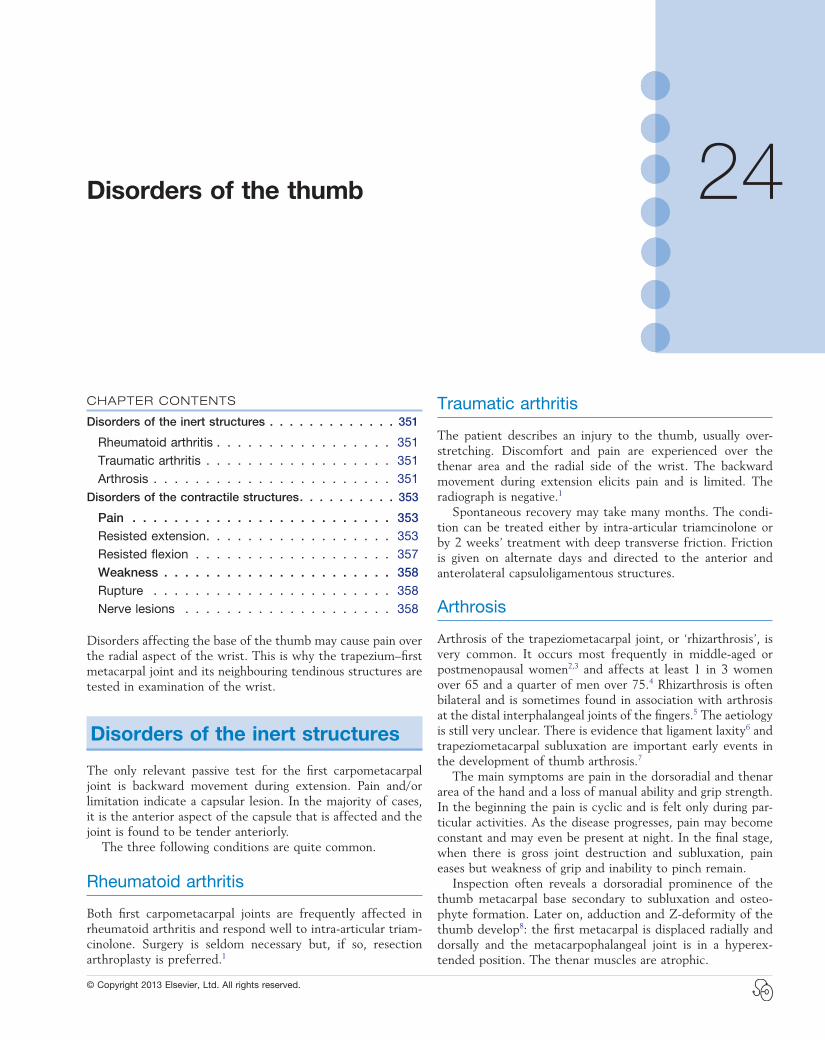
Table 24.1 Summary of treatment of capsular disorders
Deep friction Intra-articular injection Surgery
– Rheumatoid arthritis Rheumatoid arthritis
Traumatic arthritis Traumatic arthritis –
Early arthrosis Moderate arthrosis Severe arthrosis
trapezio metacarpal ligament reconstruction and tendon inter-position procedures23–27 have been shown to provide good symptomatic relief. For severe or late-stage disease, some have advocated arthrodesis of the trapeziometacarpal joint, assum-ing that there is good mobility of the joints proximal and distal.28–30 Arthroplasty techniques have ranged from simple partial or complete trapeziectomy to various implant and liga-ment interposition and reconstructions.31,32 These techniques have been generally indicated for stage II or greater disease once conservative management has failed.
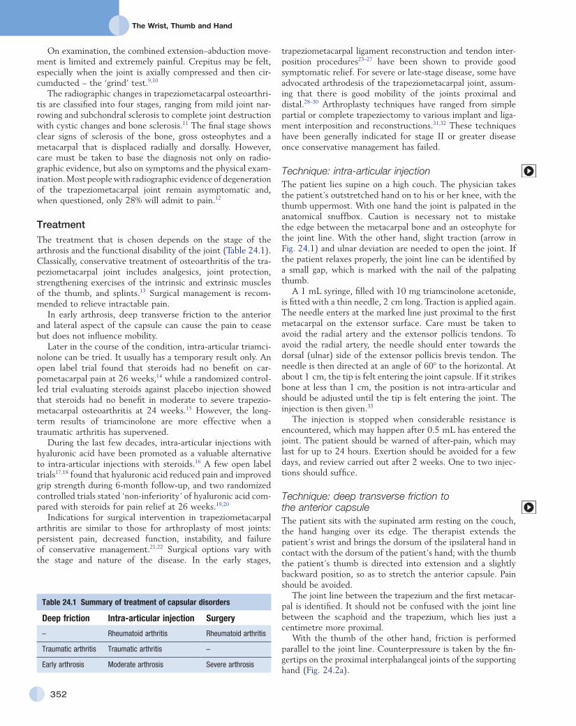
Technique: intra-articular injectionThe patient lies supine on a high couch. The physician takes the patient’s outstretched hand on to his or her knee, with the thumb uppermost. With one hand the joint is palpated in the anatomical snuffbox. Caution is necessary not to mistake the edge between the metacarpal bone and an osteophyte for the joint line. With the other hand, slight traction (arrow in Fig. 24.1) and ulnar deviation are needed to open the joint. If the patient relaxes properly, the joint line can be identified by a small gap, which is marked with the nail of the palpating thumb.
A 1 mL syringe, filled with 10 mg triamcinolone acetonide, is fitted with a thin needle, 2 cm long. Traction is applied again. The needle enters at the marked line just proximal to the first metacarpal on the extensor surface. Care must be taken to avoid the radial artery and the extensor pollicis tendons. To avoid the radial artery, the needle should enter towards the dorsal (ulnar) side of the extensor pollicis brevis tendon. The needle is then directed at an angle of 60° to the horizontal. At about 1 cm, the tip is felt entering the joint capsule. If it strikes bone at less than 1 cm, the position is not intra-articular and should be adjusted until the tip is felt entering the joint. The injection is then given.33
The injection is stopped when considerable resistance is encountered, which may happen after 0.5 mL has entered the joint. The patient should be warned of after-pain, which may last for up to 24 hours. Exertion should be avoided for a few days, and review carried out after 2 weeks. One to two injec-tions should suffice.
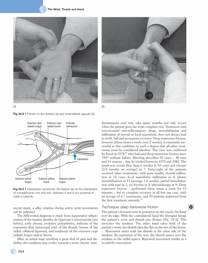
Technique: deep transverse friction to the anterior capsuleThe patient sits with the supinated arm resting on the couch, the hand hanging over its edge. The therapist extends the patient’s wrist and brings the dorsum of the ipsilateral hand in contact with the dorsum of the patient’s hand; with the thumb the patient’s thumb is directed into extension and a slightly backward position, so as to stretch the anterior capsule. Pain should be avoided.
The joint line between the trapezium and the first metacar-pal is identified. It should not be confused with the joint line between the scaphoid and the trapezium, which lies just a centimetre more proximal.
With the thumb of the other hand, friction is performed parallel to the joint line. Counterpressure is taken by the fin-gertips on the proximal interphalangeal joints of the supporting hand (Fig. 24.2a).
C H A P T E R 2 4 Disorders of the thumb
353
Disorders of the contractile structures
Pain
Resisted extension
This movement seldom causes pain in isolation. Because the extensor brevis and abductor longus tendons lie together in one tendon sheath, in the majority cases resisted abduction is also painful.
Abductor pollicis longus, and extensor pollicis brevis (first tendon sheath)
Intersection syndromeIntersection syndrome is a specific painful disorder of the forearm that is relatively common but sometimes not correctly diagnosed clinically. It has also been referred to in the literature by the terms ‘peritendinitis crepitans’, ‘oarsmen’s wrist’, ‘crossover syndrome’, ‘subcutaneous perimyositis’, ‘squeaker’s wrist’, ‘bugaboo forearm’ or ‘abductor pollicis longus syn-drome’.34 Dobyns et al introduced the term ‘intersection syn-drome’, an anatomical designation related to the area in which the musculotendinous junctions of the first extensor compart-ment tendons (abductor pollicis longus and extensor pollicis brevis tendons) intersect the second extensor compartment tendons (extensor carpi radialis longus and extensor carpi radialis brevis tendons), at an angle of approximately 60° (Fig. 24.3).35
The most plausible pathophysiology is that of a peritendini-tis at the intersection of the two tendinous compartments, which spreads upwards to the musculotendinous junction. The lesion may also lie somewhat more proximally in the muscle bellies; hence the name, ‘myosynovitis’.36 This is shown by magnetic resonance imaging (MRI) findings demonstrating the presence of peritendinous oedema concentrically surrounding the second and the first extensor compartments, beginning at the point of crossover, 4–8 cm proximal to the Lister tubercle and extending proximally.37
The lesion always results from occupational overuse or after unusual effort. It is often associated with sports-related activi-ties, such as rowing,38 canoeing, playing raquet sports, horse riding and skiing.39,40
The patient mentions crepitus during wrist movements. On examination, resisted extension and resisted abduction of the thumb are painful. Some passive wrist movements can also be painful. This puzzling phenomenon can be explained by the fact that discomfort is increased not only by movements that stretch the tendons (e.g. passive flexion of the thumb and ulnar deviation of the wrist), but also by every movement that pushes the tendon into the tendon sheath (e.g. thumb exten-sion, radial deviation and flexion or extension of the wrist).
On palpation, tenderness and swelling are found in a region about 4–8 cm proximal to the Lister tubercle, where the first and second extensor compartment tendons cross. In cases of
Technique: deep transverse friction to the anterolateral capsuleThe patient’s forearm is brought into neutral position. With the contralateral hand, the therapist brings the patient’s wrist into ulnar deviation and the thumb into flexion, which stretches the capsule.
Friction is imparted at the joint line and parallel to it with the thumb of the other hand. Counterpressure is given with the fingers on the ulnar aspect of the patient’s hand (Fig. 24.2b). It is important to ensure that the thumb remains palmar to the extensor pollicis brevis tendon, in that the lesion lies at the anterolateral aspect of the joint.
Friction is given 2–3 times a week for about 2–3 weeks. The results are good in traumatic arthritis and early arthrosis.
Fig 24.1 • Intra-articular injection.
The Wrist, Thumb and Hand
354
Spontaneous cure may take many months and only occurs when the patient gives the wrist complete rest. Treatment with non-steroidal anti-inflammatory drugs, immobilization and infiltration of steroid or local anaesthetic does not always lead to swift, full and permanent recovery. Deep transverse friction, however (three times a week, over 2 weeks), is extremely suc-cessful in this condition, to such a degree that all other treat-ments must be considered obsolete. This view was confirmed by Paton in 1978,41 who had used deep transverse friction since 1947 without failure. Bisschop describes 62 cases – 48 men and 14 women – that he treated between 1975 and 1982. The onset was recent (less than 6 weeks) in 55 cases and chronic (2.6 months on average) in 7. Forty-eight of the patients received other treatments, with poor results: steroid infiltra-tion in 16 cases; local anaesthetic infiltration in 4; plaster immobilization in 13 (average 1.8 weeks); partial immobiliza-tion with tape in 2; ice friction in 4; physiotherapy in 9. Deep transverse friction – performed three times a week for 15 minutes – led to complete recovery in all but one case, with an average of 6.7 treatments, and 39 patients improved from the first treatment onwards.42
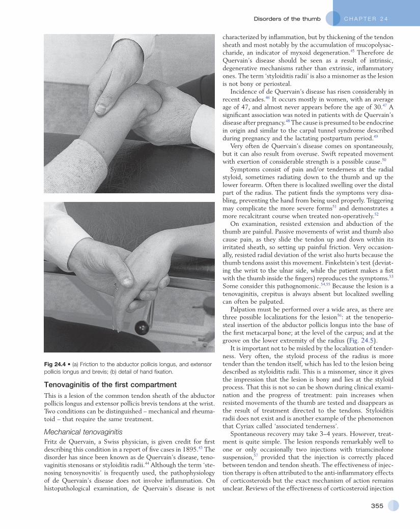
Technique: deep transverse frictionThe patient’s forearm rests in pronation on the couch, the hand over its edge. With the contralateral hand the therapist brings the patient’s wrist and thumb into flexion (Fig. 24.4). This stretches the tendons. The other hand takes hold of the patient’s wrist; the thumb then lies flat on the site of the lesion.
Movement starts with the thumb at the ulnar side of the tendons. By supination of the arm, the thumb passes over the tendons to the radial aspect. Repeated movement results in a to-and-fro movement.
recent onset, a silky crepitus during active wrist movements can be palpated.
The differential diagnosis is made from suppurative inflam-mation of the tendon sheaths, de Quervain’s tenosynovitis (see below), early chronic evolutive polyarthritis, arthrosis of the trapezium–first metacarpal joint of the thumb, lesions of the radial collateral ligament, and tendinosis of the extensor carpi radialis longus and/or brevis.
After an initial stage involving a great deal of pain and dis-ability, the condition may evolve towards a more chronic state.
Fig 24.3 • Intersection syndrome: the lesions lies at the intersection of compartments one and two, between 4 and 8 cm proximal of Lister’s tubercle.
Extensor pollicislongus
Extensor pollicisbrevis
Abductor pollicislongus
Intersection
Extensor carpiradialis longus
Extensor carpiradialis brevis
Extensorretinaculum
Fig 24.2 • Friction to the anterior (a) and anterolateral capsule (b).
(a) (b)
C H A P T E R 2 4 Disorders of the thumb
355
characterized by inflammation, but by thickening of the tendon sheath and most notably by the accumulation of mucopolysac-charide, an indicator of myxoid degeneration.45 Therefore de Quervain’s disease should be seen as a result of intrinsic, degenerative mechanisms rather than extrinsic, inflammatory ones. The term ‘styloiditis radii’ is also a misnomer as the lesion is not bony or periosteal.
Incidence of de Quervain’s disease has risen considerably in recent decades.46 It occurs mostly in women, with an average age of 47, and almost never appears before the age of 30.47 A significant association was noted in patients with de Quervain’s disease after pregnancy.48 The cause is presumed to be endocrine in origin and similar to the carpal tunnel syndrome described during pregnancy and the lactating postpartum period.49
Very often de Quervain’s disease comes on spontaneously, but it can also result from overuse. Swift repeated movement with exertion of considerable strength is a possible cause.50
Symptoms consist of pain and/or tenderness at the radial styloid, sometimes radiating down to the thumb and up the lower forearm. Often there is localized swelling over the distal part of the radius. The patient finds the symptoms very disa-bling, preventing the hand from being used properly. Triggering may complicate the more severe forms51 and demonstrates a more recalcitrant course when treated non-operatively.52
On examination, resisted extension and abduction of the thumb are painful. Passive movements of wrist and thumb also cause pain, as they slide the tendon up and down within its irritated sheath, so setting up painful friction. Very occasion-ally, resisted radial deviation of the wrist also hurts because the thumb tendons assist this movement. Finkelstein’s test (deviat-ing the wrist to the ulnar side, while the patient makes a fist with the thumb inside the fingers) reproduces the symptoms.53 Some consider this pathognomonic.54,55 Because the lesion is a tenovaginitis, crepitus is always absent but localized swelling can often be palpated.
Palpation must be performed over a wide area, as there are three possible localizations for the lesion56: at the tenoperio-steal insertion of the abductor pollicis longus into the base of the first metacarpal bone; at the level of the carpus; and at the groove on the lower extremity of the radius (Fig. 24.5).
It is important not to be misled by the localization of tender-ness. Very often, the styloid process of the radius is more tender than the tendon itself, which has led to the lesion being described as styloiditis radii. This is a misnomer, since it gives the impression that the lesion is bony and lies at the styloid process. That this is not so can be shown during clinical exami-nation and the progress of treatment: pain increases when resisted movements of the thumb are tested and disappears as the result of treatment directed to the tendons. Styloiditis radii does not exist and is another example of the phenomenon that Cyriax called ‘associated tenderness’.
Spontaneous recovery may take 3–4 years. However, treat-ment is quite simple. The lesion responds remarkably well to one or only occasionally two injections with triamcinolone suspension,57 provided that the injection is correctly placed between tendon and tendon sheath. The effectiveness of injec-tion therapy is often attributed to the anti-inflammatory effects of corticosteroids but the exact mechanism of action remains unclear. Reviews of the effectiveness of corticosteroid injection
Tenovaginitis of the first compartmentThis is a lesion of the common tendon sheath of the abductor pollicis longus and extensor pollicis brevis tendons at the wrist. Two conditions can be distinguished – mechanical and rheuma-toid – that require the same treatment.
Mechanical tenovaginitisFritz de Quervain, a Swiss physician, is given credit for first describing this condition in a report of five cases in 1895.43 The disorder has since been known as de Quervain’s disease, teno-vaginitis stenosans or styloiditis radii.44 Although the term ‘ste-nosing tenosynovitis’ is frequently used, the pathophysiology of de Quervain’s disease does not involve inflammation. On histopathological examination, de Quervain’s disease is not
Fig 24.4 • (a) Friction to the abductor pollicis longus, and extensor pollicis longus and brevis; (b) detail of hand fixation.
The Wrist, Thumb and Hand
356
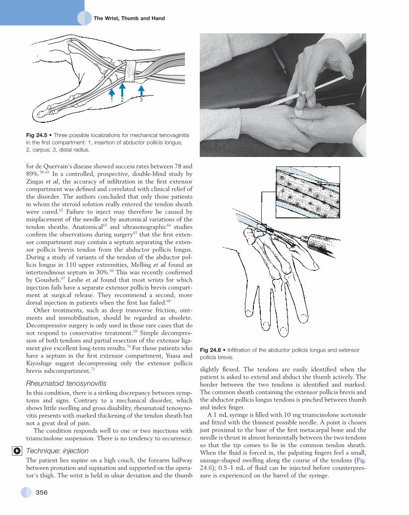
slightly flexed. The tendons are easily identified when the patient is asked to extend and abduct the thumb actively. The border between the two tendons is identified and marked. The common sheath containing the extensor pollicis brevis and the abductor pollicis longus tendons is pinched between thumb and index finger.
A 1 mL syringe is filled with 10 mg triamcinolone acetonide and fitted with the thinnest possible needle. A point is chosen just proximal to the base of the first metacarpal bone and the needle is thrust in almost horizontally between the two tendons so that the tip comes to lie in the common tendon sheath. When the fluid is forced in, the palpating fingers feel a small, sausage-shaped swelling along the course of the tendons (Fig. 24.6); 0.5–1 mL of fluid can be injected before counterpres-sure is experienced on the barrel of the syringe.
for de Quervain’s disease showed success rates between 78 and 89%.58–61 In a controlled, prospective, double-blind study by Zingas et al, the accuracy of infiltration in the first extensor compartment was defined and correlated with clinical relief of the disorder. The authors concluded that only those patients in whom the steroid solution really entered the tendon sheath were cured.62 Failure to inject may therefore be caused by misplacement of the needle or by anatomical variations of the tendon sheaths. Anatomical63 and ultrasonographic64 studies confirm the observations during surgery65 that the first exten-sor compartment may contain a septum separating the exten-sor pollicis brevis tendon from the abductor pollicis longus. During a study of variants of the tendon of the abductor pol-licis longus in 110 upper extremities, Melling et al found an intertendinous septum in 30%.66 This was recently confirmed by Gousheh.67 Leslie et al found that most wrists for which injection fails have a separate extensor pollicis brevis compart-ment at surgical release. They recommend a second, more dorsal injection in patients when the first has failed.68
Other treatments, such as deep transverse friction, oint-ments and immobilization, should be regarded as obsolete. Decompressive surgery is only used in those rare cases that do not respond to conservative treatment.69 Simple decompres-sion of both tendons and partial resection of the extensor liga-ment give excellent long-term results.70 For those patients who have a septum in the first extensor compartment, Yuasa and Kiyoshige suggest decompressing only the extensor pollicis brevis subcompartment.71
Rheumatoid tenosynovitisIn this condition, there is a striking discrepancy between symp-toms and signs. Contrary to a mechanical disorder, which shows little swelling and gross disability, rheumatoid tenosyno-vitis presents with marked thickening of the tendon sheath but not a great deal of pain.
The condition responds well to one or two injections with triamcinolone suspension. There is no tendency to recurrence.
Technique: injectionThe patient lies supine on a high couch, the forearm halfway between pronation and supination and supported on the opera-tor’s thigh. The wrist is held in ulnar deviation and the thumb
Fig 24.6 • Infiltration of the abductor pollicis longus and extensor pollicis brevis.
Fig 24.5 • Three possible localizations for mechanical tenovaginitis in the first compartment: 1, insertion of abductor pollicis longus; 2, carpus; 3, distal radius.
1 2 3
C H A P T E R 2 4 Disorders of the thumb
357
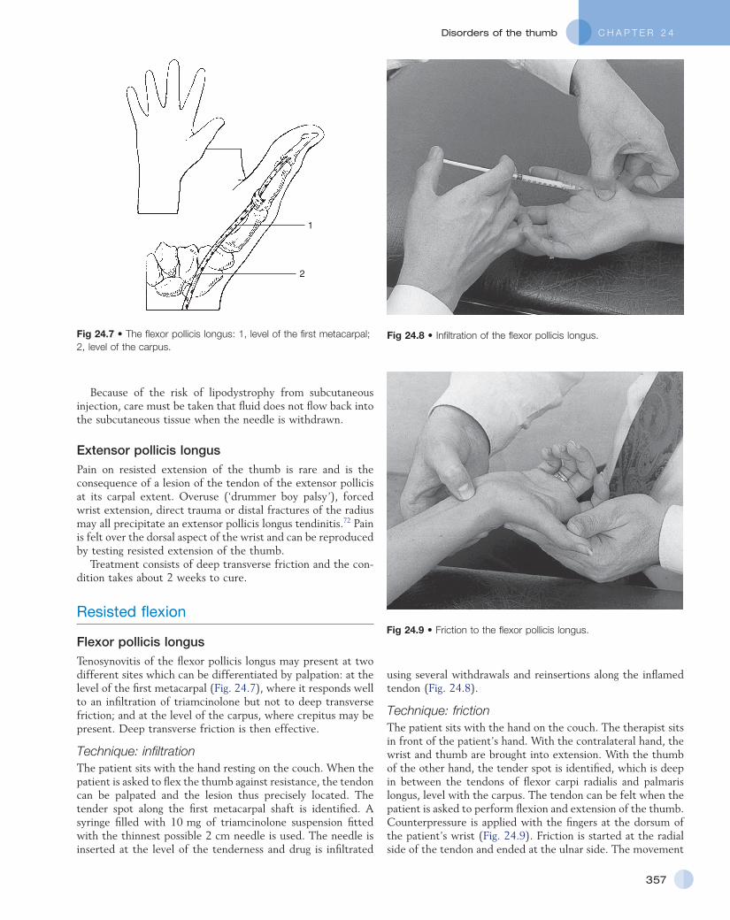
using several withdrawals and reinsertions along the inflamed tendon (Fig. 24.8).
Technique: frictionThe patient sits with the hand on the couch. The therapist sits in front of the patient’s hand. With the contralateral hand, the wrist and thumb are brought into extension. With the thumb of the other hand, the tender spot is identified, which is deep in between the tendons of flexor carpi radialis and palmaris longus, level with the carpus. The tendon can be felt when the patient is asked to perform flexion and extension of the thumb. Counterpressure is applied with the fingers at the dorsum of the patient’s wrist (Fig. 24.9). Friction is started at the radial side of the tendon and ended at the ulnar side. The movement
Because of the risk of lipodystrophy from subcutaneous injection, care must be taken that fluid does not flow back into the subcutaneous tissue when the needle is withdrawn.
Extensor pollicis longusPain on resisted extension of the thumb is rare and is the consequence of a lesion of the tendon of the extensor pollicis at its carpal extent. Overuse (‘drummer boy palsy’), forced wrist extension, direct trauma or distal fractures of the radius may all precipitate an extensor pollicis longus tendinitis.72 Pain is felt over the dorsal aspect of the wrist and can be reproduced by testing resisted extension of the thumb.
Treatment consists of deep transverse friction and the con-dition takes about 2 weeks to cure.
Resisted flexion
Flexor pollicis longusTenosynovitis of the flexor pollicis longus may present at two different sites which can be differentiated by palpation: at the level of the first metacarpal (Fig. 24.7), where it responds well to an infiltration of triamcinolone but not to deep transverse friction; and at the level of the carpus, where crepitus may be present. Deep transverse friction is then effective.
Technique: infiltrationThe patient sits with the hand resting on the couch. When the patient is asked to flex the thumb against resistance, the tendon can be palpated and the lesion thus precisely located. The tender spot along the first metacarpal shaft is identified. A syringe filled with 10 mg of triamcinolone suspension fitted with the thinnest possible 2 cm needle is used. The needle is inserted at the level of the tenderness and drug is infiltrated
Fig 24.7 • The flexor pollicis longus: 1, level of the first metacarpal; 2, level of the carpus.
1
2
Fig 24.8 • Infiltration of the flexor pollicis longus.
Fig 24.9 • Friction to the flexor pollicis longus.
The Wrist, Thumb and Hand
358
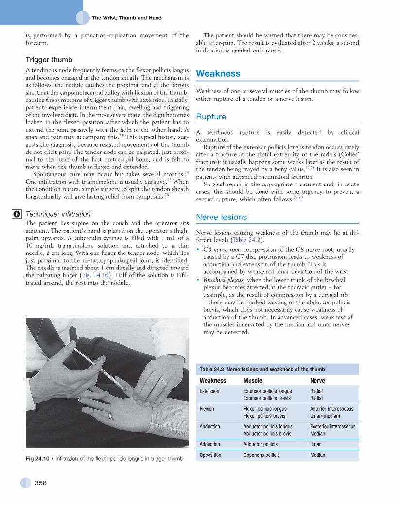
Fig 24.10 • Infiltration of the flexor pollicis longus in trigger thumb.
Table 24.2 Nerve lesions and weakness of the thumb
Weakness Muscle Nerve
Extension Extensor pollicis longus RadialExtensor pollicis brevis Radial
Flexion Flexor pollicis longus Anterior interosseousFlexor pollicis brevis Ulnar/(median)
Abduction Abductor pollicis longus Posterior interosseousAbductor pollicis brevis Median
Adduction Adductor pollicis Ulnar
Opposition Opponens pollicis Median
is performed by a pronation–supination movement of the forearm.
Trigger thumbA tendinous node frequently forms on the flexor pollicis longus and becomes engaged in the tendon sheath. The mechanism is as follows: the nodule catches the proximal end of the fibrous sheath at the carpometacarpal pulley with flexion of the thumb, causing the symptoms of trigger thumb with extension. Initially, patients experience intermittent pain, swelling and triggering of the involved digit. In the most severe state, the digit becomes locked in the flexed position, after which the patient has to extend the joint passively with the help of the other hand. A snap and pain may accompany this.73 This typical history sug-gests the diagnosis, because resisted movements of the thumb do not elicit pain. The tender node can be palpated, just proxi-mal to the head of the first metacarpal bone, and is felt to move when the thumb is flexed and extended.
Spontaneous cure may occur but takes several months.74 One infiltration with triamcinolone is usually curative.75 When the condition recurs, simple surgery to split the tendon sheath longitudinally will give lasting relief from symptoms.76
Technique: infiltrationThe patient lies supine on the couch and the operator sits adjacent. The patient’s hand is placed on the operator’s thigh, palm upwards. A tuberculin syringe is filled with 1 mL of a 10 mg/mL triamcinolone solution and attached to a thin needle, 2 cm long. With one finger the tender node, which lies just proximal to the metacarpophalangeal joint, is identified. The needle is inserted about 1 cm distally and directed toward the palpating finger (Fig. 24.10). Half of the solution is infil-trated around, the rest into the nodule.
The patient should be warned that there may be consider-able after-pain. The result is evaluated after 2 weeks; a second infiltration is needed only rarely.
Weakness
Weakness of one or several muscles of the thumb may follow either rupture of a tendon or a nerve lesion.
Rupture
A tendinous rupture is easily detected by clinical examination.
Rupture of the extensor pollicis longus tendon occurs rarely after a fracture at the distal extremity of the radius (Colles’ fracture); it usually happens some weeks later as the result of the tendon being frayed by a bony callus.77,78 It is also seen in patients with advanced rheumatoid arthritis.
Surgical repair is the appropriate treatment and, in acute cases, this should be done with some urgency to prevent a second rupture, which often follows.79,80
Nerve lesions
Nerve lesions causing weakness of the thumb may lie at dif-ferent levels (Table 24.2).
• C8 nerve root: compression of the C8 nerve root, usually caused by a C7 disc protrusion, leads to weakness of adduction and extension of the thumb. This is accompanied by weakened ulnar deviation of the wrist.
• Brachial plexus: when the lower trunk of the brachial plexus becomes affected at the thoracic outlet – for example, as the result of compression by a cervical rib – there may be marked wasting of the abductor pollicis brevis, which does not necessarily cause weakness of abduction of the thumb. In advanced cases, weakness of the muscles innervated by the median and ulnar nerves may be detected.
C H A P T E R 2 4 Disorders of the thumb
359
• Median nerve: long-standing compression of the median nerve in the carpal tunnel may lead to slight weakness and wasting of the thenar muscles.
• Posterior interosseous nerve: a lesion of the posterior interosseous nerve at the elbow results in weakness of thumb abduction and finger extension (see online chapter Nerve lesions and entrapment neuropathies of the upper limb).
• Ulnar nerve: entrapment of the ulnar nerve in the hand gives rise to weakness of thumb adduction.
Disorders of the thumb are summarized in Box 24.1.
Access the complete reference list online at www.orthopaedicmedicineonline.com
Box 24.1
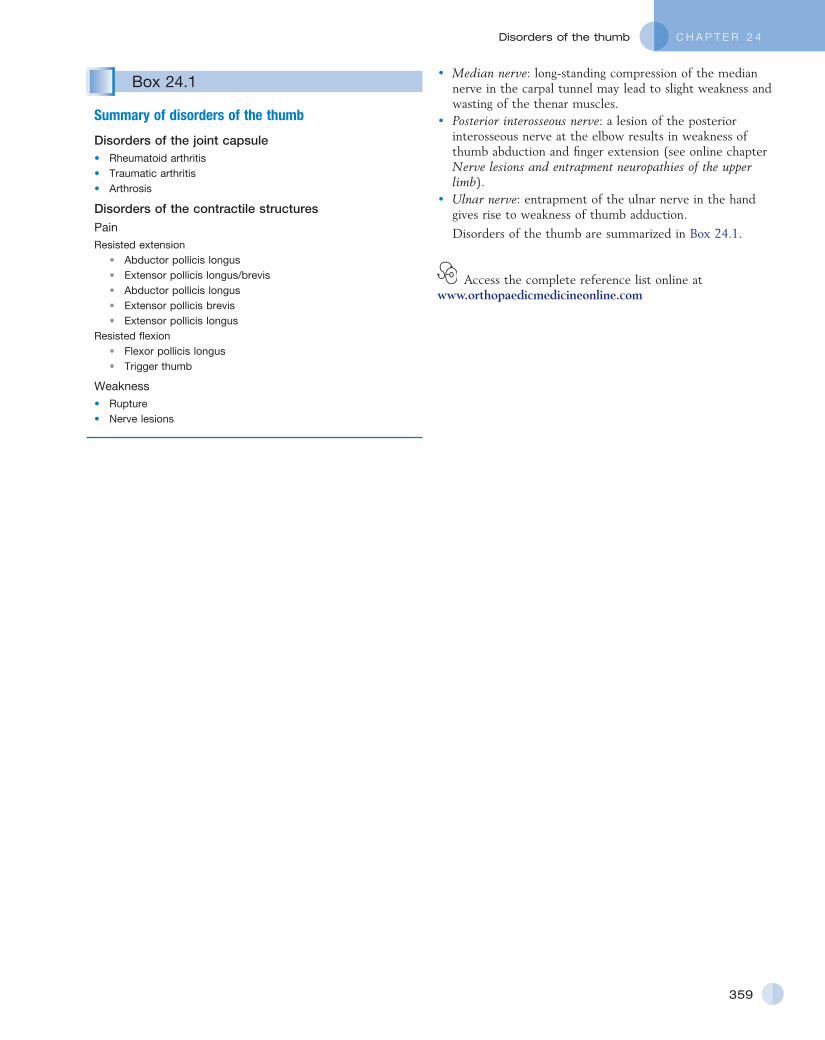
Summary of disorders of the thumb
Disorders of the joint capsule• Rheumatoid arthritis• Traumatic arthritis• Arthrosis
Disorders of the contractile structuresPainResisted extension
• Abductor pollicis longus• Extensor pollicis longus/brevis• Abductor pollicis longus• Extensor pollicis brevis• Extensor pollicis longus
Resisted flexion• Flexor pollicis longus• Trigger thumb
Weakness• Rupture• Nerve lesions
C H A P T E R 1 4Disorders of the thumb
359.e1
References
1. Gunther SF. Carpometarcarpal joint of the thumb. In: Lichtman DM, Alexander AH, editors. The Wrist and its Disorders. 2nd ed. Philadelphia: Saunders; 1997. p. 443–58.
2. Burton RI, Pellegrini VD Jr. Surgical management of basal joint arthritis of the thumb, Part II: ligament reconstruction with tendon interposition arthroplasty. J Hand Surg 1986;11A: 324–32.
3. Pellegrini VD Jr. Osteoarthritis at the base of the thumb. Orthop Clin North Am 1992;23(1):83–102.
4. Haara MM, Heliövaara M, Kröger H, et al. Osteoarthritis in the carpometacarpal joint of the thumb. Prevalence and associations with disability and mortality. J Bone Joint Surg Am 2004;86-A(7):1452–7.
5. Dahaghin S, Bierma-Zeinstra S, Ginai A, et al. Prevalence and pattern of radiographic hand osteoarthritis and association with pain and disability (the Rotterdam study). Ann Rheum Dis 2005;64:682–7.
6. Jónsson H, Elíasson GJ, Jónsson A, et al. High hand joint mobility is associated with radiological CMC1 osteoarthritis: the AGES-Reykjavik study. Osteoarthritis Cartilage 2009;17(5):592–5.
7. Hunter DJ, Zhang Y, Sokolove J, et al. Trapeziometacarpal subluxation predisposes to incident trapeziometacarpal osteoarthritis (OA): the Framingham Study. Osteoarthritis Cartilage 2005;13:953–7.
8. Menon J. The problem of trapeziometacarpal degenerative arthritis. Clin Orthop Rel Res 1983;175:155.
9. Swanson AB. Disabling arthritis at the base of the thumb: treatment by resection of the trapezium and flexible (silicone) implant arthroplasty. J Bone Joint Surg 1982;54A:456.
10. Merritt MM, Roddey TS, Costello C, Olson S. Diagnostic value of clinical grind test for carpometacarpal osteoarthritis of the thumb. J Hand Ther 2010;23(3):261–7.
11. Eaton RG, Glickel SZ. Trapeziometacarpal osteoarthritis. Staging as a rationale for treatment. Hand Clin 1987;3(4):455–71.
12. Armstrong AL, Hunter JB, Davis TR. The prevalence of degenerative arthritis of the base of the thumb in post-menopausal women. J Hand Surg Br 1994;19(3):340–1.
13. Gomes Carreira AC, Jones A, Natour J. Assessment of the effectiveness of a functional splint for osteoarthritis of the trapeziometacarpal joint on the dominant hand: a randomized controlled study. J Rehabil Med 2010;42(5):469–74.
14. Joshi R. Intraarticular corticosteroid injection for first carpometacarpal osteoarthritis. J Rheumatol 2005;32:1305–6.
15. Meenagh GK, Patton J, Kynes C, Wright GD.A randomised controlled trial of intra-articular corticosteroid injection
of the carpometacarpal joint of the thumb in osteoarthritis. Ann Rheum Dis 2004;63(10):1260–3.
16. Fuchs S, Monikes R, Wohlmeiner A, et al. Intra-articular hyaluronic acid compared with corticoid injections for the treatment of rhizarthrosis. Osteoarthritis Cartilage 2006;14:82–8 [PubMed].
17. Mandl LA, Hotchkiss RN, Adler RS, et al. Injectable hyaluronan for the treatment of carpometacarpal osteoarthritis: open label pilot trial. Curr Med Res Opin 2009;25(9):2103–8.
18. Figen Ayhan F, Ustün N. The evaluation of efficacy and tolerability of Hylan G-F 20 in bilateral thumb base osteoarthritis: 6 months follow-up. Clin Rheumatol 2009;28(5):535–41.
19. Stahl S, Karsh-Safriri I, Ratzon N, et al. Comparison of intra-articular injection of depot corticosteroids and hyaluronic acid for treatment of degenerative trapeziometacarpal joints. J Clin Rheumatol 2005;11:299–302 [PubMed].
20. Heyworth BE, Lee JH, Kim PD, et al. Hylan versus corticosteroid versus placebo for treatment of basal joint arthritis: a prospective, randomized, double-blinded clinical trial. J Hand Surg [Am] 2008;33:40–8.
21. Kuschner SH, Lane CS. Surgical treatment for osteoarthritis at the base of the thumb. Am J Orthop 1996;25(2):91–100.
22. Poole JU, Pellegrini VD Jr. Arthritis of the thumb basal joint complex. J Hand Ther 2000;13(2):91–107.
23. Eaton RG, Lane LB, Littler JW, Keyser JJ. Ligament reconstruction for the painful thumb carpometarcarpal joint: a long-term assessment. J Hand Surg 1984;9A:692.
24. Robinson D, Aghasi M, Halperin H. Abductor pollicis longus tendon arthroplasty of trapezio-metacarpal joint: surgical technique and results. J Hand Surg 1991;16A:504–9.
25. Sigfusson R, Lundborg G. Abductor pollicis longus tendon arthroplasty for treatment of arthrosis in the first carpometacarpal joint. Scand J Plast Reconstr Surg Hand Surg 1991;25:73–7.
26. Burton R. American Society for Surgery of the Hand Correspondence Newsletter, 1995 January.
27. Freedman DM, Clickel SZ, Eaton RG. Long-term follow-up of volar ligament reconstruction of the thumb. J Hand Surg Am 2000;25A:297–304.
28. Carroll RE, Hill NA. Arthrodesis of the carpometacarpal joint of the thumb. J Bone Joint Surg 1983;55B:292–4.
29. Stark HH, Moore JF, Ashworth CR, Boyes JH. Fusion of the first metacarpotrapezial joint for degenerative arthritis. J Bone Joint Surg 1977;59A:22–6.
30. Ishida O, Ikuta Y. Trapeziometacarpal joint arthrodesis for the treatment of arthrosis.
Scand J Plast Reconstr Surg Hand Surg 2000;34(3):245–8.
31. Bozentka DJ. Implant arthroplasty of the carpometacarpal joint of the thumb. Hand Clin 2010;26(3):327–37, v.
32. Badia A, Sambandam SN. Total joint arthroplasty in the treatment of advanced stages of thumb carpometacarpal joint osteoarthritis. Hand Surg Am 2006;31(10):1605–14.
33. Mandl LA, Hotchkiss RN, Adler RS, et al. Can the carpometacarpal joint be injected accurately in the office setting? Implications for therapy. J Rheumatol 2006;33(6):1137–9.
34. Grundberg AB, Reagan DS. Pathologic anatomy of the forearm: intersection syndrome. J Hand Surg 1985;10:299–302.
35. Dobyns JH, Sim FH, Linscheid RL. Sports stress syndrome of hand and wrist. Am J Sports Med 1978;6:236–54.
36. Howard NJ. Peritendinitis crepitans. J Bone Joint Surg Br 1937;19:447–59.
37. Lee RP, Hatem SF, Recht MP. Extended MRI findings of intersection syndrome. Skeletal Radiol 2009;38(2):157–63.
38. Wood MD, Dobyns JH. Sports related extraarticular wrist syndrome. Clin Orthop Rel Res 1986;202:93–102.
39. Palmer DH, Lane-Larsen CL. Helicopter skiing wrist injuries: a case report of ‘bugaboo forearm.’ Am J Sports Med 1994;22:148–9.
40. Servi JT. Wrist pain from overuse: detecting and relieving intersection syndrome. Phys Sports Med 1997;12:41–4.
41. Paton HO. Traumatic tenosynovitis of the wrist. BMJ 1978;i:789.
42. Missotten J, Stainier P, Bisschop P. Ténosynovite des extenseurs et abducteur du pouce. Kinésithérapie Scientifique 1992;311:35.
43. de Quervain F. Über eine Form von chronischer Tendovaginitis. Korresp Bl Schweiz Ärzte 1895;25:389.
44. de Quervain F. On a form of chronic tendovaginitis by Dr. Fritz de Quervain in la Chaux-de-Fonds. 1895. Am J Orthop 1997;26(9):641–4 (English translation of the original article).
45. Clarke MT, Lyall HA, Grant JW, Matthewson MH. The histopathology of de Quervain’s disease. J Hand Surg 1998;23B(6):732–4.
46. Walker-Bone K, Palmer KT, Reading I, Coggon D, Cooper C. Prevalence and impact of musculoskeletal disorders of the upper limb in the general population. Arthritis Rheum 2004;51:642–51. doi: 10.1002/art.20535.
47. Le Viet D, Lantieri L. Ténosynovite de de Quervain. Cicatrice horizontale et fixation du lambeau capsulaire. Rev Chir Orthop Repar Appar Mot 1992;75(2):101–6.
48. Anderson SE, Steinbach LS, De Monaco D, et al. ‘Baby wrist’: MRI of an overuse
The Wrist, Thumb and Hand
359.e2
syndrome in mothers. AJR Am J Roentgenol 2004;182(3):719–24.
49. Capasso G, Testa V, Maffulli N, et al. Surgical release of de Quervain’s stenosing tenosynovitis postpartum: can it wait? Int Orthop 2002;26:23–5.
50. Armstrong ThJ, Fine LJ, Goldstein SA, et al. Ergonomic considerations in hand and wrist tendinitis. J Hand Surg 1987;12A:830.
51. Witczak JW, Mesear VR, Meyer RD. Triggering of the thumb with de Quervain’s stenosing tendovaginitis. J Hand Surg 1990;15A(2):265.
52. Albertson GM, High WA, Shin AY, Bishop AT. Extensor triggering in de Quervain’s stenosing tenosynovitis. J Hand Surg 1999;24A(6):1311–4.
53. Finkelstein H. Stenosing tenovaginitis at the radial styloid process. J Bone Joint Surg 1930;12A:509.
54. Palmer K, Walker-Bone K, Linaker C, et al. The Southampton examination schedule for the diagnosis of musculoskeletal disorders of the upper limb. Ann Rheum Dis 2000;59:5–11.
55. Pick RY. De Quervain’s disease: a clinical triad. Clin Orthop Rel Res 1979;143:165.
56. Cyriax JH. Textbook of Orthopaedic Medicine, vol I, Diagnosis of Soft Tissue Lesions. 8th ed. London: Baillière Tindall; 1982. p. 188.
57. Weiss AP, Akelman E, Tabatabai M. Treatment of de Quervain’s disease. J Hand Surg 1994;19A(4):595–8.
58. Jirarattanaphochai K, Saengnipanthkul S, Vipulakorn K, et al. Treatment of de Quervain disease with triamcinolone injection with or without nimesulide. A randomized, double-blind, placebo-controlled trial. J Bone Joint Surg Am 2004;86-A:2700–6.
59. Peters-Veluthamaningal C, Winters JC, Groenier KH, Meyboom-DeJong B. Randomised controlled trial of local corticosteroid injections for de Quervain’s
tenosynovitis in general practice. BMC Musculoskelet Disord 200927;10:131.
60. Sawaizumi T, Nanno M, Ito H. De Quervain’s disease: efficacy of intra-sheath triamcinolone injection. Int Orthop 2007;31:265–8.
61. Richie CA, Briner WW Jr. Corticosteroid injection for treatment of de Quervain’s tenosynovitis: a pooled quantitative literature evaluation. J Am Board Fam Pract 2003;16:102–6. doi: 10.3122/jabfm.16.2.102.
62. Zingas C, Failla JM, Van Holsbeeck M. Injection accuracy and clinical relief of de Quervain’s tendinitis. J Hand Surg 1998;23A(1):89–96.
63. Aktan ZA, Ozturk L, Calli IH. An anatomical study of the first extensor compartment of the wrist. Kaibogaku Zasshi 1998;73(1):49–54.
64. Nagaoka M, Matsuzaki H, Suzuki T. Ultrasonographic examination of de Quervain’s disease. J Orthop Sci 2000;5(2):96–9.
65. Bahm J, Szabo Z, Foucher G. The anatomy of de Quervain’s disease. A study of operative findings. Int Orthop 1995;19(4):209–11.
66. Melling M, Wilde J, Schnallinger M, et al. Supernumerary tendons of the abductor pollicis. Acta Anat 1996;155(4):291–4.
67. Gousheh J, Yavari M, Arasteh E. Division of the first dorsal compartment of the hand into two separated canals: rule or exception? Arch Iran Med 2009;12(1):52–4.
68. Leslie BM, Ericson WB, Morehead JR. Incidence of a septum within the first dorsal compartment of the wrist. J Hand Surg 1990;15A:88.
69. Ta KT, Eidelman D, Thomson JG. Patient satisfaction and outcomes of surgery for de Quervain’s tenosynovitis. J Hand Surg 1999;24A(5):1071–7.
70. Scheller A, Schuh R, Hönle W, Schuh A. Long-term results of surgical release of
de Quervain’s stenosing tenosynovitis. Int Orthop 2009;33(5):1301–3.
71. Yuasa K, Kiyoshige Y. Limited surgical treatment of de Quervain’s disease: decompression of only the extensor pollicis brevis subcompartment. J Hand Surg 1998;23A(5):840–3.
72. Thorson E, Szabo R. Common tendinitis problems in the hand and forearm. Orthop Clin North Am 1992;23(1):65–74.
73. Wilhelmi BJ, Mowlavi A, Neumeister MW, et al. Safe treatment of trigger finger with longitudinal and transverse landmarks: an anatomic study of the border fingers for percutaneous release. Plast Reconstr Surg 2003;112(4):993–9.
74. Schofield CB, Citron ND. The natural history of adult trigger thumb. J Hand Surg 1993;18B(2):247–8.
75. Rhoades CE, Gelberman RH, Manjarris JF. Stenosing tenosynovitis of the finger and thumb. Clin Orthop 1984;190:238.
76. Turowski GA, Zdankiewicz PD, Thomson JG. The results of surgical treatment of trigger finger. J Hand Surg Am 1997;22(1):145–9.
77. Engkvist O, Lundborg G. Rupture of the extensor pollicis longus tendon after fracture of the lower end of the radius – a clinical and microangiographic study. Hand 1979;11:75.
78. Kozin SB, Wood MB. Early soft-tissue complications after fractures of the distal part of the radius. J Bone Joint Surg 1993;75A(1):144–53.
79. Schneider LH, Rosenstein RG. Restoration of extensor pollicis longus function by tendon transfer. Plast Recon Surg 1983;71:533–7.
80. Ferlic DC. Extensor indicis proprius transfer for extensor pollicis longus rupture. In: Blair W, Steyers C, editors. Techniques in Hand Surgery. Baltimore: Williams & Wilkins; 1996. p. 649–53.


![Abductor pollicis longus tendon division with swan neck ... · moment arms of thumb motor tendons restricting the movement at the base of the thumb [6]. Compensatory movements occur](https://img.pdfslide.net/doc/110x75/5f0dfe657e708231d43d182e/abductor-pollicis-longus-tendon-division-with-swan-neck-moment-arms-of-thumb.jpg)