Embed Size (px)
Citation preview
METHODS: Peripheral blood samples were collected beforeand 30 days following surgical removal of renal masses from 53patients with presumed RCC. Samples were analyzed for immunosup-pressive cell populations, including regulatory T cells, myeloid�derivedsuppressor cells, inflammatory monocytes and exhausted T cells, andcytokines (IL-1�, IL-5, TNF�). Patients were compared to 10 age andgender matched healthy controls.
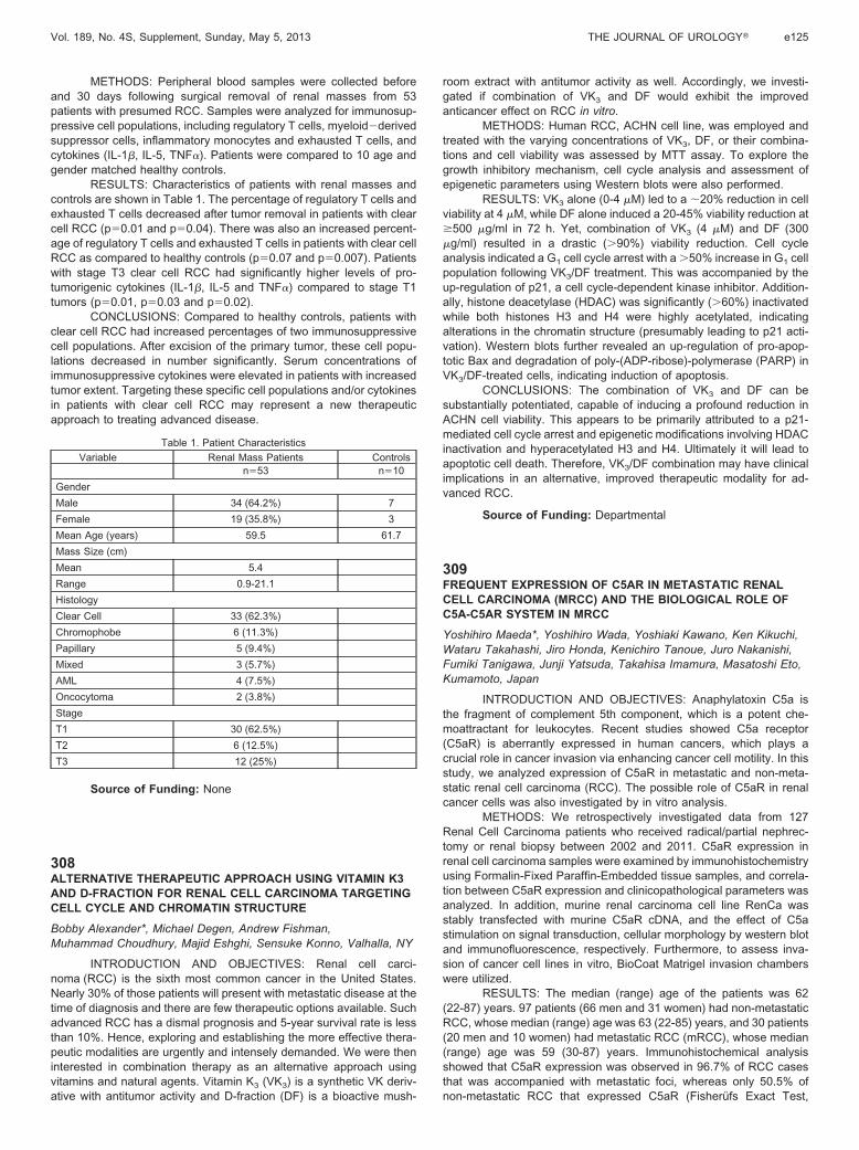
RESULTS: Characteristics of patients with renal masses andcontrols are shown in Table 1. The percentage of regulatory T cells andexhausted T cells decreased after tumor removal in patients with clearcell RCC (p�0.01 and p�0.04). There was also an increased percent-age of regulatory T cells and exhausted T cells in patients with clear cellRCC as compared to healthy controls (p�0.07 and p�0.007). Patientswith stage T3 clear cell RCC had significantly higher levels of pro-tumorigenic cytokines (IL-1�, IL-5 and TNF�) compared to stage T1tumors (p�0.01, p�0.03 and p�0.02).
CONCLUSIONS: Compared to healthy controls, patients withclear cell RCC had increased percentages of two immunosuppressivecell populations. After excision of the primary tumor, these cell popu-lations decreased in number significantly. Serum concentrations ofimmunosuppressive cytokines were elevated in patients with increasedtumor extent. Targeting these specific cell populations and/or cytokinesin patients with clear cell RCC may represent a new therapeuticapproach to treating advanced disease.
Table 1. Patient Characteristics
Variable Renal Mass Patients Controlsn�53 n�10
Gender
Male 34 (64.2%) 7
Female 19 (35.8%) 3
Mean Age (years) 59.5 61.7
Mass Size (cm)
Mean 5.4
Range 0.9-21.1
Histology
Clear Cell 33 (62.3%)
Chromophobe 6 (11.3%)
Papillary 5 (9.4%)
Mixed 3 (5.7%)
AML 4 (7.5%)
Oncocytoma 2 (3.8%)
Stage
T1 30 (62.5%)
T2 6 (12.5%)
T3 12 (25%)
Source of Funding: None
308ALTERNATIVE THERAPEUTIC APPROACH USING VITAMIN K3AND D-FRACTION FOR RENAL CELL CARCINOMA TARGETINGCELL CYCLE AND CHROMATIN STRUCTURE
Bobby Alexander*, Michael Degen, Andrew Fishman,Muhammad Choudhury, Majid Eshghi, Sensuke Konno, Valhalla, NY
INTRODUCTION AND OBJECTIVES: Renal cell carci-noma (RCC) is the sixth most common cancer in the United States.Nearly 30% of those patients will present with metastatic disease at thetime of diagnosis and there are few therapeutic options available. Suchadvanced RCC has a dismal prognosis and 5-year survival rate is lessthan 10%. Hence, exploring and establishing the more effective thera-peutic modalities are urgently and intensely demanded. We were theninterested in combination therapy as an alternative approach usingvitamins and natural agents. Vitamin K3 (VK3) is a synthetic VK deriv-ative with antitumor activity and D-fraction (DF) is a bioactive mush-
room extract with antitumor activity as well. Accordingly, we investi-gated if combination of VK3 and DF would exhibit the improvedanticancer effect on RCC in vitro.
METHODS: Human RCC, ACHN cell line, was employed andtreated with the varying concentrations of VK3, DF, or their combina-tions and cell viability was assessed by MTT assay. To explore thegrowth inhibitory mechanism, cell cycle analysis and assessment ofepigenetic parameters using Western blots were also performed.
RESULTS: VK3 alone (0-4 �M) led to a 20% reduction in cellviability at 4 �M, while DF alone induced a 20-45% viability reduction at�500 �g/ml in 72 h. Yet, combination of VK3 (4 �M) and DF (300�g/ml) resulted in a drastic (�90%) viability reduction. Cell cycleanalysis indicated a G1 cell cycle arrest with a �50% increase in G1 cellpopulation following VK3/DF treatment. This was accompanied by theup-regulation of p21, a cell cycle-dependent kinase inhibitor. Addition-ally, histone deacetylase (HDAC) was significantly (�60%) inactivatedwhile both histones H3 and H4 were highly acetylated, indicatingalterations in the chromatin structure (presumably leading to p21 acti-vation). Western blots further revealed an up-regulation of pro-apop-totic Bax and degradation of poly-(ADP-ribose)-polymerase (PARP) inVK3/DF-treated cells, indicating induction of apoptosis.
CONCLUSIONS: The combination of VK3 and DF can besubstantially potentiated, capable of inducing a profound reduction inACHN cell viability. This appears to be primarily attributed to a p21-mediated cell cycle arrest and epigenetic modifications involving HDACinactivation and hyperacetylated H3 and H4. Ultimately it will lead toapoptotic cell death. Therefore, VK3/DF combination may have clinicalimplications in an alternative, improved therapeutic modality for ad-vanced RCC.
Source of Funding: Departmental
309FREQUENT EXPRESSION OF C5AR IN METASTATIC RENALCELL CARCINOMA (MRCC) AND THE BIOLOGICAL ROLE OFC5A-C5AR SYSTEM IN MRCC
Yoshihiro Maeda*, Yoshihiro Wada, Yoshiaki Kawano, Ken Kikuchi,Wataru Takahashi, Jiro Honda, Kenichiro Tanoue, Juro Nakanishi,Fumiki Tanigawa, Junji Yatsuda, Takahisa Imamura, Masatoshi Eto,Kumamoto, Japan
INTRODUCTION AND OBJECTIVES: Anaphylatoxin C5a isthe fragment of complement 5th component, which is a potent che-moattractant for leukocytes. Recent studies showed C5a receptor(C5aR) is aberrantly expressed in human cancers, which plays acrucial role in cancer invasion via enhancing cancer cell motility. In thisstudy, we analyzed expression of C5aR in metastatic and non-meta-static renal cell carcinoma (RCC). The possible role of C5aR in renalcancer cells was also investigated by in vitro analysis.
METHODS: We retrospectively investigated data from 127Renal Cell Carcinoma patients who received radical/partial nephrec-tomy or renal biopsy between 2002 and 2011. C5aR expression inrenal cell carcinoma samples were examined by immunohistochemistryusing Formalin-Fixed Paraffin-Embedded tissue samples, and correla-tion between C5aR expression and clinicopathological parameters wasanalyzed. In addition, murine renal carcinoma cell line RenCa wasstably transfected with murine C5aR cDNA, and the effect of C5astimulation on signal transduction, cellular morphology by western blotand immunofluorescence, respectively. Furthermore, to assess inva-sion of cancer cell lines in vitro, BioCoat Matrigel invasion chamberswere utilized.
RESULTS: The median (range) age of the patients was 62(22-87) years. 97 patients (66 men and 31 women) had non-metastaticRCC, whose median (range) age was 63 (22-85) years, and 30 patients(20 men and 10 women) had metastatic RCC (mRCC), whose median(range) age was 59 (30-87) years. Immunohistochemical analysisshowed that C5aR expression was observed in 96.7% of RCC casesthat was accompanied with metastatic foci, whereas only 50.5% ofnon-metastatic RCC that expressed C5aR (Fisherufs Exact Test,
Vol. 189, No. 4S, Supplement, Sunday, May 5, 2013 THE JOURNAL OF UROLOGY� e125
p�0.001). There is no correlation between C5aR expression and otherclinicopathological parameters. RenCa cells overexpressing mC5aRshowed increased ERK activation upon C5a stimulation compared tocontrol cells. F-actin staining using Alexa-conjugated Phalloidin re-vealed formation of actin stress fiber in mC5aR expressing RenCa cellsby C5a stimulation. Matrigel chamber assay demonstrated the invasiveability of RenCa cells overexpressing mC5aR were enhanced signifi-cantly by C5a stimulus, compared to control cells.
CONCLUSIONS: These results suggests that aberrant expres-sion of C5aR may be a pivotal step for cancer cell metastasis andinvasion.
Source of Funding: None
310MTOR PATHWAY REGULATES THE EXPRESSION OFANGIOTENSIN 2 TYPE 1 RECEPTOR IN RENAL CELLCARCINOMA
Gou Kaneko*, Akira Miyajima, Takeo Kosaka, Ryuichi Mizuno,Eiji Kikuchi, Mototsugu Oya, Tokyo, Japan
INTRODUCTION AND OBJECTIVES: The potential role ofrenin-angiotensin system in angiogenesis and promotion of tumorgrowth has been growing. We previously showed that angiotensin 2-angiotensin 2 type 1 receptor (AT1R) signaling led to potent inductionof vascular endothelial growth factor in urogenital cancers. In addition,we reported that up-regulation of phosphorylated Akt contributed toelevated AT1R expression in castration-resistant prostate cancer. Inrenal cell carcinoma (RCC), AT1R expression was recently reported asa useful predictor of survival, however, the regulatory mechanism ofAT1R expression has not yet been elucidated. The aim of the presentstudy was to evaluate the regulatory mechanism of AT1R expression inRCC.
METHODS: AT1R expression was evaluated using proteinlysates from human RCC specimens by western blot analysis, andanalyzed a relationship between tumor grade and AT1R expression.AT1R and phosphorylated (pS6) expression in 6 human RCC cell lines(ACHN, A498, 786-O, 769-P, Caki-1, Caki-2) was evaluated by westernblot analysis. We evaluated whether mammalian target of rapamycin(mTOR) inhibitor (RAD001) regulates AT1R expression in vitro and invivo.
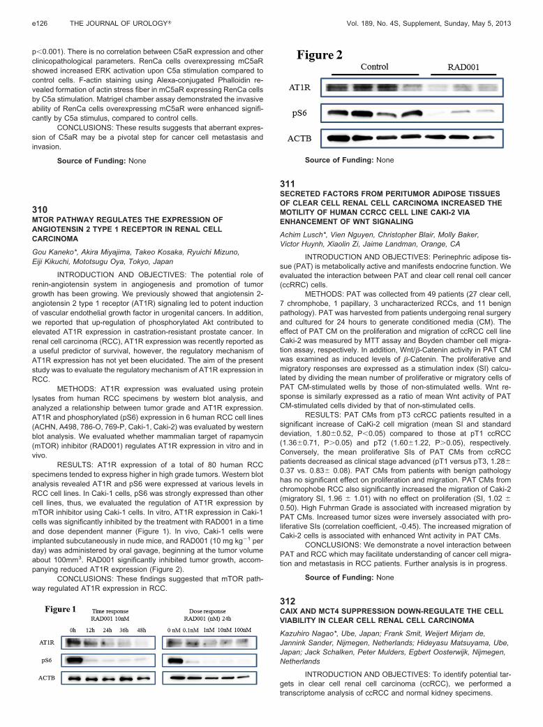
RESULTS: AT1R expression of a total of 80 human RCCspecimens tended to express higher in high grade tumors. Western blotanalysis revealed AT1R and pS6 were expressed at various levels inRCC cell lines. In Caki-1 cells, pS6 was strongly expressed than othercell lines, thus, we evaluated the regulation of AT1R expression bymTOR inhibitor using Caki-1 cells. In vitro, AT1R expression in Caki-1cells was significantly inhibited by the treatment with RAD001 in a timeand dose dependent manner (Figure 1). In vivo, Caki-1 cells wereimplanted subcutaneously in nude mice, and RAD001 (10 mg kg�1 perday) was administered by oral gavage, beginning at the tumor volumeabout 100mm3. RAD001 significantly inhibited tumor growth, accom-panying reduced AT1R expression (Figure 2).
CONCLUSIONS: These findings suggested that mTOR path-way regulated AT1R expression in RCC.
Source of Funding: None
311SECRETED FACTORS FROM PERITUMOR ADIPOSE TISSUESOF CLEAR CELL RENAL CELL CARCINOMA INCREASED THEMOTILITY OF HUMAN CCRCC CELL LINE CAKI-2 VIAENHANCEMENT OF WNT SIGNALING
Achim Lusch*, Vien Nguyen, Christopher Blair, Molly Baker,Victor Huynh, Xiaolin Zi, Jaime Landman, Orange, CA
INTRODUCTION AND OBJECTIVES: Perinephric adipose tis-sue (PAT) is metabolically active and manifests endocrine function. Weevaluated the interaction between PAT and clear cell renal cell cancer(ccRRC) cells.
METHODS: PAT was collected from 49 patients (27 clear cell,7 chromphobe, 1 papillary, 3 uncharacterized RCCs, and 11 benignpathology). PAT was harvested from patients undergoing renal surgeryand cultured for 24 hours to generate conditioned media (CM). Theeffect of PAT CM on the proliferation and migration of ccRCC cell lineCaki-2 was measured by MTT assay and Boyden chamber cell migra-tion assay, respectively. In addition, Wnt/�-Catenin activity in PAT CMwas examined as induced levels of �-Catenin. The proliferative andmigratory responses are expressed as a stimulation index (SI) calcu-lated by dividing the mean number of proliferative or migratory cells ofPAT CM-stimulated wells by those of non-stimulated wells. Wnt re-sponse is similarly expressed as a ratio of mean Wnt activity of PATCM-stimulated cells divided by that of non-stimulated cells.
RESULTS: PAT CMs from pT3 ccRCC patients resulted in asignificant increase of CaKi-2 cell migration (mean SI and standarddeviation, 1.80�0.52, P�0.05) compared to those at pT1 ccRCC(1.36�0.71, P�0.05) and pT2 (1.60�1.22, P�0.05), respectively.Conversely, the mean proliferative SIs of PAT CMs from ccRCCpatients decreased as clinical stage advanced (pT1 versus pT3, 1.28�0.37 vs. 0.83� 0.08). PAT CMs from patients with benign pathologyhas no significant effect on proliferation and migration. PAT CMs fromchromophobe RCC also significantly increased the migration of Caki-2(migratory SI, 1.96 � 1.01) with no effect on proliferation (SI, 1.02 �0.50). High Fuhrman Grade is associated with increased migration byPAT CMs. Increased tumor sizes were inversely associated with pro-liferative SIs (correlation coefficient, -0.45). The increased migration ofCaki-2 cells is associated with enhanced Wnt activity in PAT CMs.
CONCLUSIONS: We demonstrate a novel interaction betweenPAT and RCC which may facilitate understanding of cancer cell migra-tion and metastasis in RCC patients. Further analysis is in progress.
Source of Funding: None
312CAIX AND MCT4 SUPPRESSION DOWN-REGULATE THE CELLVIABILITY IN CLEAR CELL RENAL CELL CARCINOMA
Kazuhiro Nagao*, Ube, Japan; Frank Smit, Weijert Mirjam de,Jannink Sander, Nijmegen, Netherlands; Hideyasu Matsuyama, Ube,Japan; Jack Schalken, Peter Mulders, Egbert Oosterwijk, Nijmegen,Netherlands
INTRODUCTION AND OBJECTIVES: To identify potential tar-gets in clear cell renal cell carcinoma (ccRCC), we performed atranscriptome analysis of ccRCC and normal kidney specimens.
e126 THE JOURNAL OF UROLOGY� Vol. 189, No. 4S, Supplement, Sunday, May 5, 2013