Embed Size (px)
Citation preview
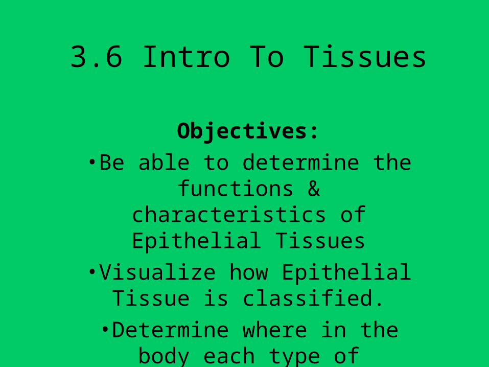
3.6 Intro To Tissues
Objectives:
•Be able to determine the functions & characteristics of Epithelial Tissues
•Visualize how Epithelial Tissue is classified.
•Determine where in the body each type of Epithelial Tissue is located.
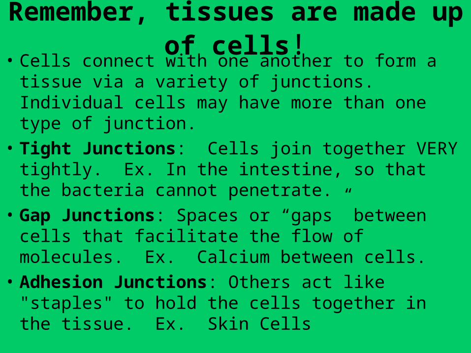
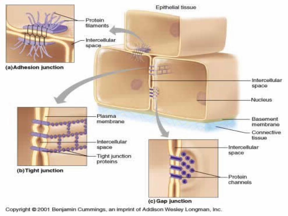
Remember, tissues are made up of cells!• Cells connect with one another to form a tissue via a
variety of junctions. Individual cells may have more than one type of junction.
• Tight Junctions: Cells join together VERY tightly. Ex. In the intestine, so that the bacteria cannot penetrate.
• Gap Junctions: Spaces or “gaps” between cells that facilitate the flow of molecules. Ex. Calcium between cells.
• Adhesion Junctions: Others act like "staples" to hold the cells together in the tissue. Ex. Skin Cells
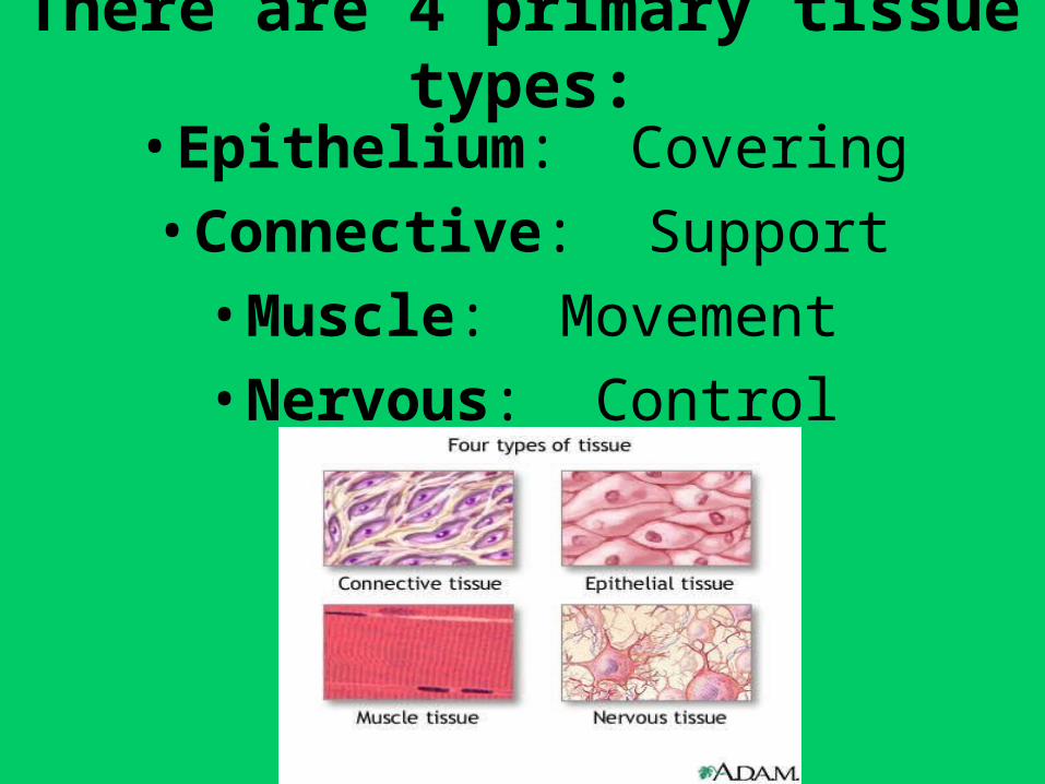
There are 4 primary tissue types:
• Epithelium: Covering
• Connective: Support
• Muscle: Movement
• Nervous: Control
Epithelium• There are 2 types of Epithelial Tissue:
1. Covering & Lining Epithlium covers the surface of the outside of the body and lines internal organs.
2. Glandular Epithlium secretes hormones or other products.
• Functions: Protection, Absorption, Filtration, & Secretion
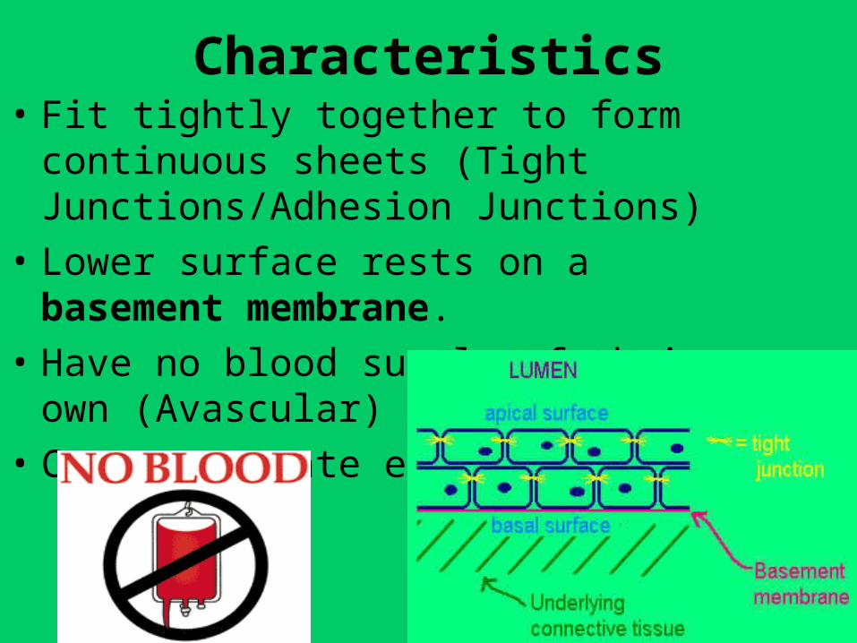
Characteristics• Fit tightly together to form continuous
sheets (Tight Junctions/Adhesion Junctions)
• Lower surface rests on a basement membrane.
• Have no blood supply of their own (Avascular)
• Can regenerate easily
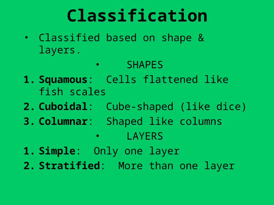
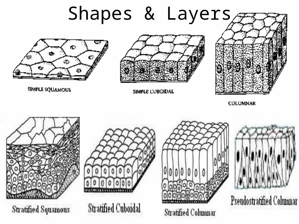
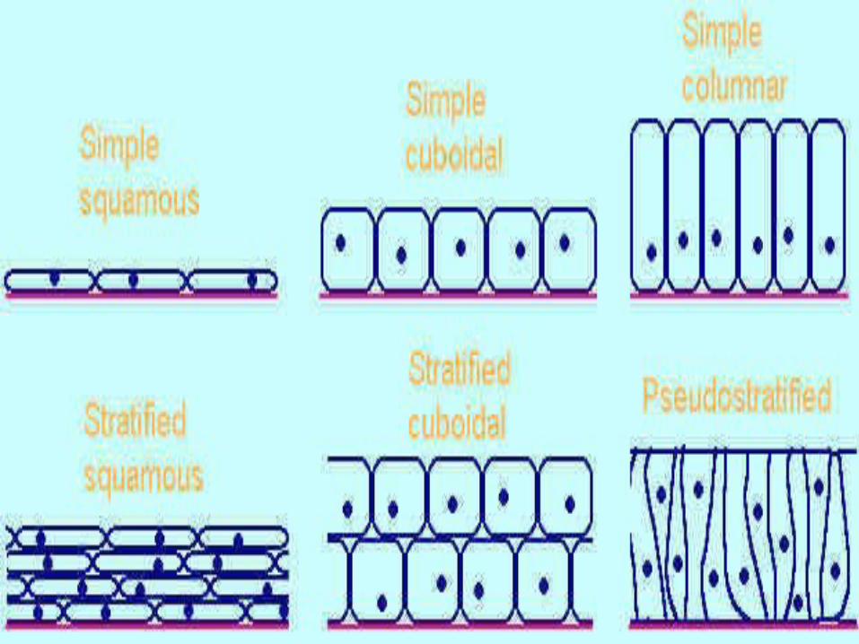
Classification• Classified based on shape & layers.
• SHAPES
1. Squamous: Cells flattened like fish scales
2. Cuboidal: Cube-shaped (like dice)
3. Columnar: Shaped like columns
• LAYERS
1. Simple: Only one layer
2. Stratified: More than one layer
Shapes & Layers
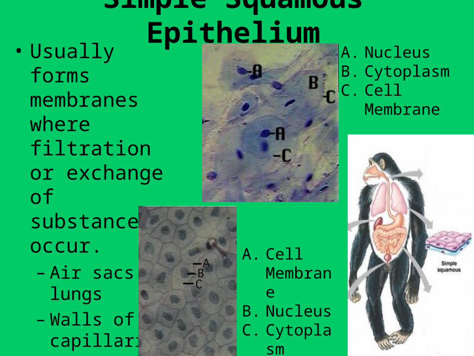
Simple Squamous Epithelium• Usually forms
membranes where filtration or exchange of substances occur.– Air sacs of lungs– Walls of
capillaries
A. NucleusB. CytoplasmC. Cell
Membrane
A. Cell Membrane
B. NucleusC. Cytoplasm
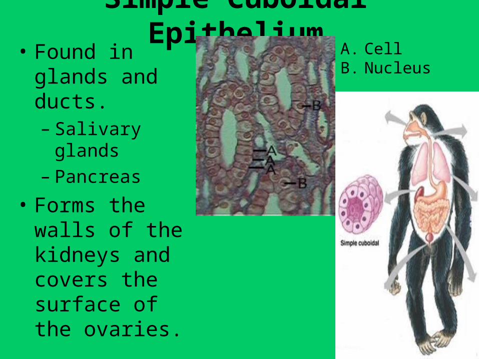
Simple Cuboidal Epithelium• Found in glands
and ducts.– Salivary glands– Pancreas
• Forms the walls of the kidneys and covers the surface of the ovaries.
A. CellB. Nucleus
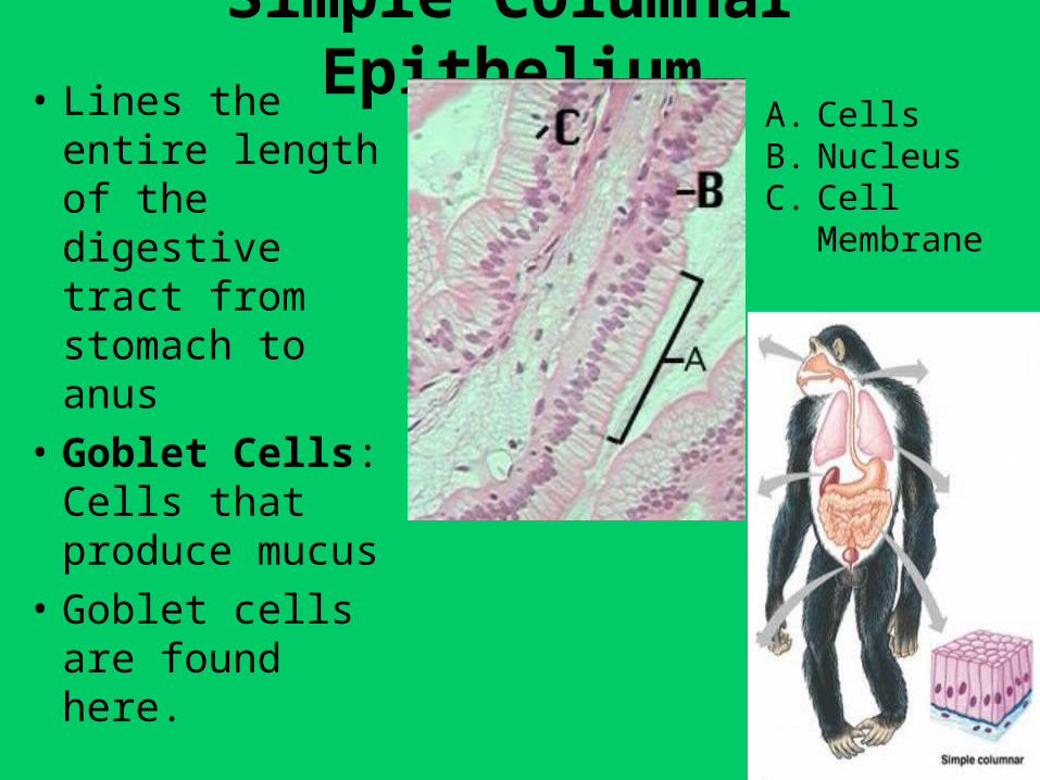
Simple Columnar Epithelium• Lines the entire
length of the digestive tract from stomach to anus
• Goblet Cells: Cells that produce mucus
• Goblet cells are found here.
A. CellsB. NucleusC. Cell
Membrane
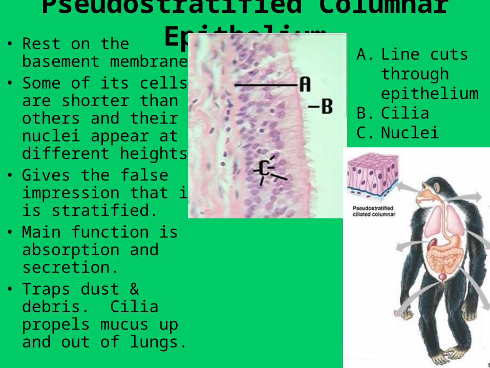
Pseudostratified Columnar Epithelium• Rest on the basement
membrane.• Some of its cells are
shorter than others and their nuclei appear at different heights.
• Gives the false impression that it is stratified.
• Main function is absorption and secretion.
• Traps dust & debris. Cilia propels mucus up and out of lungs.
A. Line cuts through epithelium
B. CiliaC. Nuclei
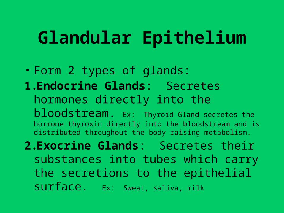
Glandular Epithelium
• Form 2 types of glands:1.Endocrine Glands: Secretes hormones
directly into the bloodstream. Ex: Thyroid Gland secretes the hormone thyroxin directly into the bloodstream and is distributed throughout the body raising metabolism.
2.Exocrine Glands: Secretes their substances into tubes which carry the secretions to the epithelial surface. Ex: Sweat, saliva, milk
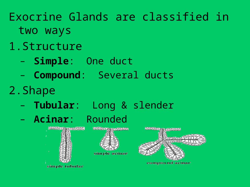
Exocrine Glands are classified in two ways
1.Structure– Simple: One duct– Compound: Several ducts
2.Shape– Tubular: Long & slender– Acinar: Rounded