1
39.22 Myocardial Perfusion Imaging in Patients with Syncope: Cardiac Risk Factors and Coronary Artery Disease Predict Perfusion Findings LM Phillips, D Gold, R Druz North Shore University Hospital, Manhasset, NY Background: Syncope often results in referral for myocardial perfusion imaging (MPI) to assess for coronary artery disease (CAD) in otherwise asymptomatic patients. It is unknown if patients with syncope benefit from referral to MPI. We investigated age, cardiac risk factors (CRF: hypertension, diabetes, dyslipidemia, family history, smoking), gender, and known CAD in predicting MPI findings in patients referred for evaluation of syncope. Methods: 684 pts (mean 64 yrs old; age range 35-89 years; 337 male; 347 female) with referral diagnosis of syncope were identified retrospectively from MPI database records. Age, gender, CRF, baseline heart rate, baseline blood pressure and resting ECG rhythm were known for all patients. Gated MPI was performed as per dual-isotope protocol. MPI findings were classified as: normal; fixed perfusion defect; reversible perfusion defect; fixed and reversible perfusion defect. Hierarchical linear regression was used to determine relative contributions of age, gender, CRF, known CAD, baseline rhythm, and stress ECG for MPI findings. Results: 555 patients had known CAD (29 with documented prior myocar- dial infarction) and 129 had no known CAD. Most patients did not have associated chest pain or dyspnea at the time of their syncopal event (35 chest pain, 31 dyspnea). Exercise MPI was performed in 371 patients, and pharmacologic in 313 patients. MPI findings were normal in 495 patients; fixed defects in 96 patients; reversible defects in 128 patients; fixed and reversible defects in 25 patients. Left ventricular ejection fraction was estimated at 57.221.2 % (range 54-80%). Female gender (R0.15, p0.0001), CRF (R0.31, p0.0001), known CAD (R0.41, p0.0001), baseline rhythm other than sinus (R0.42, p0.007), and positive stress ECG (R0.44, p0.001) were all independently predictive of MPI find- ings. Age, symptoms, baseline heart rate and blood pressure and ejection fraction were not predictive of MPI findings. Conclusions: In patients with syncope and no other symptoms, referral to MPI is warranted for those with known CAD, high-risk CRF, and baseline ECG reflecting non-sinus rhythm. A larger cohort of patients will be needed to determine contribution of gender. 39.23 The Presence of ST Depression During Adenosine Myocardial Perfusion Imaging Cannot Localize Coronary Artery Disease P Rao, T Pilgram, RJ Gropler Washington University, St. Louis, MO Background: It is well established that localization of coronary artery disease (CAD) on exercise stress ECG is inaccurate presumably because patients are upright during testing. Since patients are recumbent during vasodilator stress, theoretically, ECG changes indicative of ischemia should be able to localize CAD if it is present. Whether this presumption is correct is unknown. Methods: A retrospective database search was performed on 9,813 patients studied between January 2004 and 2007 who underwent dual-isotope vasodilator myocardial perfusion imaging (AMPI) with a 4-minute adeno- sine protocol. Data were based on the clinical interpretation. Subsequent cardiac catheterization was performed at the discretion of the patient’s physician. Patients were identified by meeting ECG criteria for ischemia with 1mm or more horizontal or downsloping ST depression in two or more contiguous leads. Localization to anterior, septal, lateral, and inferior distributions and the corresponding coronary arteries were assigned using standard criteria. Corresponding perfusion data was compared for presence of ischemia and location of abnormality based on the standard 17-segment model. The corresponding catheterization data was compared for the presence of stenosis 70%. Results: Of the 9,813 studies only 252 (2.6%) met ECG criteria for ischemia. Of these, 161 studies (64.8%) had a corresponding perfusion study interpreted as positive for ischemia. 106 (42%) patients underwent coronary angiography. There was no agreement between the ECG and AMPI for localization of CAD (p .05). Similarly, no agreement was found when ECG findings were compared with coronary angiography (p .05). The lack of agreement was independent of the location of the ECG abnormalities. Conclusions: Ischemic ECG changes during adenosine myocardial perfu- sion stress imaging are infrequent and cannot accurately localize coronary artery disease. 39.24 Myocardial Perfusion Imaging Evidence of Functionally Complete Revascularization by Minimally Invasive Direct Coronary Artery Bypass in Two-Vessel Coronary Artery Disease R De Maria, 1 A Repossini, 2 A Bestetti, 3 M Parolini, 1 V Arena, 2 O Parodi 1 1 CNR Clinical Physiology Institute-Milan, Cardiology Department, Niguarda Ca ` Granda Hospital, Milan, Italy, 2 Cardiovascular Department, Humanitas Gavazzeni, Bergamo, Italy, 3 University of Milan, Chair of Nuclear Medicine, S. Paolo Hospital, Milan, Italy Background: Whether, in patients with two-vessel coronary artery disease (CAD) and complex LAD lesions, patency of a second diseased vessel still impacts on myocardial perfusion when complete revascularisation of the left anterior descending coronary artery (LAD) territory has been achieved, is currently undetermined. We evaluated in these patients the impact of single LAD or integrated revascularisation on SPECT-assessed reversible myocar- dial ischemia. Methods: Thirty-five candidates for revascularisation with two-vessel disease, including the LAD, were studied. SPECT perfusion imaging was performed in pharmacological washout by Tc-99m tetrofosmin with a single-day stress-rest protocol. A 4-point semi-quantitative perfusion score was derived for each set of images using a 17-segment LV model. A summed score was calculated by adding the scores of the 17 segments in the stress and rest images, respectively. The ischemic area was derived as difference between stress and rest scores for the total LV and, separately, for the LAD and second vessel territories. Revascularisation was performed by minimally invasive direct coronary artery bypass (MIDCAB) of the LAD alone (n15) or by an integrated procedure with second-vessel angioplasty, either on admission (n13) or at 2 months (n7) according to extent of reversible perfusion defects (ischemic area 2 or 2) in the second vessel territory. Results: At one year, total ischemic area decreased from 9.3 5.1 to 0.81.5 in MIDCAB only patients and from 8.24.9 to 1.62.9 in the integrated group (group P .87, time P .001, group*time P .37). The ischemic area in the second vessel territory similarly decreased in both groups: from 3.932.58 to 0.470.99 in MIDCAB only patients and from 4.503.86 to 1.402.76 in the integrated group (group P .23, time P .001, group*time P .81) as shown in Figure 1. Conclusions: In two-vessel CAD involving LAD, MIDCAB alone achieves, in a substantial proportion of patients, functionally complete revascularisation even in the non-revascularised second vessel territory. 39.25 A Noninvasive Approach in Predicting Adverse Cardiac Events in Heart Transplant Recipients: Role of Dipyridamole Tc-99m Agent Single Photon Emission Computed Tomography O Parodi, 1 B De Chiara, 1 R Sara, 2 E Roubina, 1 M Bianchi, 1 M Parolini, 1 M Frigerio, 3 E Vitali 3 1 CNR Clinical Physiology Institute-Milan, Cardiology Department, Niguarda Ca ` Granda Hospital, Milan, Italy, 2 Nuclear Medicine Service, Niguarda Ca ` Granda Hospital, Milan, Italy, 3 Cardiology Department, Niguarda Ca ` Granda Hospital, Milan, Italy S128 Abstracts Journal of Nuclear Cardiology July/August 2007

39.22Myocardial Perfusion Imaging in Patients with Syncope: Cardiac Risk Factors and Coronary Artery Disease Predict Perfusion Findings

  • Upload
    r

  • View
    216

  • Download
    4

Embed Size (px)

Citation preview

39.22Myocardial Perfusion Imaging in Patients with Syncope: Cardiac RiskFactors and Coronary Artery Disease Predict Perfusion FindingsLM Phillips, D Gold, R DruzNorth Shore University Hospital, Manhasset, NYBackground: Syncope often results in referral for myocardial perfusionimaging (MPI) to assess for coronary artery disease (CAD) in otherwiseasymptomatic patients. It is unknown if patients with syncope benefit fromreferral to MPI. We investigated age, cardiac risk factors (CRF: hypertension,diabetes, dyslipidemia, family history, smoking), gender, and known CAD inpredicting MPI findings in patients referred for evaluation of syncope.Methods: 684 pts (mean 64 yrs old; age range 35-89 years; 337 male; 347female) with referral diagnosis of syncope were identified retrospectivelyfrom MPI database records. Age, gender, CRF, baseline heart rate, baselineblood pressure and resting ECG rhythm were known for all patients. GatedMPI was performed as per dual-isotope protocol. MPI findings wereclassified as: normal; fixed perfusion defect; reversible perfusion defect;fixed and reversible perfusion defect. Hierarchical linear regression wasused to determine relative contributions of age, gender, CRF, known CAD,baseline rhythm, and stress ECG for MPI findings.Results: 555 patients had known CAD (29 with documented prior myocar-dial infarction) and 129 had no known CAD. Most patients did not haveassociated chest pain or dyspnea at the time of their syncopal event (35 chestpain, 31 dyspnea). Exercise MPI was performed in 371 patients, andpharmacologic in 313 patients. MPI findings were normal in 495 patients;fixed defects in 96 patients; reversible defects in 128 patients; fixed andreversible defects in 25 patients. Left ventricular ejection fraction wasestimated at 57.2�21.2 % (range 54-80%). Female gender (R�0.15,p�0.0001), CRF (R�0.31, p�0.0001), known CAD (R�0.41, p�0.0001),baseline rhythm other than sinus (R�0.42, p�0.007), and positive stressECG (R�0.44, p�0.001) were all independently predictive of MPI find-ings. Age, symptoms, baseline heart rate and blood pressure and ejectionfraction were not predictive of MPI findings.Conclusions: In patients with syncope and no other symptoms, referral toMPI is warranted for those with known CAD, high-risk CRF, and baselineECG reflecting non-sinus rhythm. A larger cohort of patients will be neededto determine contribution of gender.

39.23The Presence of ST Depression During Adenosine Myocardial PerfusionImaging Cannot Localize Coronary Artery DiseaseP Rao, T Pilgram, RJ GroplerWashington University, St. Louis, MOBackground: It is well established that localization of coronary artery disease(CAD) on exercise stress ECG is inaccurate presumably because patients areupright during testing. Since patients are recumbent during vasodilator stress,theoretically, ECG changes indicative of ischemia should be able to localizeCAD if it is present. Whether this presumption is correct is unknown.Methods: A retrospective database search was performed on 9,813 patientsstudied between January 2004 and 2007 who underwent dual-isotopevasodilator myocardial perfusion imaging (AMPI) with a 4-minute adeno-sine protocol. Data were based on the clinical interpretation. Subsequentcardiac catheterization was performed at the discretion of the patient’sphysician. Patients were identified by meeting ECG criteria for ischemiawith 1mm or more horizontal or downsloping ST depression in two or morecontiguous leads. Localization to anterior, septal, lateral, and inferiordistributions and the corresponding coronary arteries were assigned usingstandard criteria. Corresponding perfusion data was compared for presenceof ischemia and location of abnormality based on the standard 17-segmentmodel. The corresponding catheterization data was compared for thepresence of stenosis �70%.Results: Of the 9,813 studies only 252 (2.6%) met ECG criteria for ischemia.Of these, 161 studies (64.8%) had a corresponding perfusion study interpretedas positive for ischemia. 106 (42%) patients underwent coronary angiography.There was no agreement between the ECG and AMPI for localization of CAD(p � .05). Similarly, no agreement was found when ECG findings werecompared with coronary angiography (p �.05). The lack of agreement wasindependent of the location of the ECG abnormalities.Conclusions: Ischemic ECG changes during adenosine myocardial perfu-sion stress imaging are infrequent and cannot accurately localize coronaryartery disease.

39.24Myocardial Perfusion Imaging Evidence of Functionally CompleteRevascularization by Minimally Invasive Direct Coronary Artery Bypassin Two-Vessel Coronary Artery DiseaseR De Maria,1 A Repossini,2 A Bestetti,3 M Parolini,1 V Arena,2

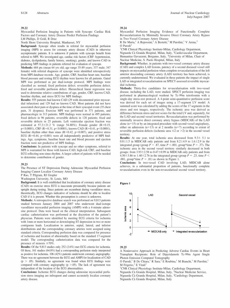
O Parodi11CNR Clinical Physiology Institute-Milan, Cardiology Department,Niguarda Ca Granda Hospital, Milan, Italy, 2Cardiovascular Department,Humanitas Gavazzeni, Bergamo, Italy, 3University of Milan, Chair ofNuclear Medicine, S. Paolo Hospital, Milan, ItalyBackground: Whether, in patients with two-vessel coronary artery disease(CAD) and complex LAD lesions, patency of a second diseased vessel stillimpacts on myocardial perfusion when complete revascularisation of the leftanterior descending coronary artery (LAD) territory has been achieved, iscurrently undetermined. We evaluated in these patients the impact of singleLAD or integrated revascularisation on SPECT-assessed reversible myocar-dial ischemia.Methods: Thirty-five candidates for revascularisation with two-vesseldisease, including the LAD, were studied. SPECT perfusion imaging wasperformed in pharmacological washout by Tc-99m tetrofosmin with asingle-day stress-rest protocol. A 4-point semi-quantitative perfusion scorewas derived for each set of images using a 17-segment LV model. Asummed score was calculated by adding the scores of the 17 segments in thestress and rest images, respectively. The ischemic area was derived asdifference between stress and rest scores for the total LV and, separately, forthe LAD and second vessel territories. Revascularisation was performed byminimally invasive direct coronary artery bypass (MIDCAB) of the LADalone (n�15) or by an integrated procedure with second-vessel angioplasty,either on admission (n�13) or at 2 months (n�7) according to extent ofreversible perfusion defects (ischemic area �2 or �2) in the second vesselterritory.Results: At one year, total ischemic area decreased from 9.3� 5.1 to0.8�1.5 in MIDCAB only patients and from 8.2�4.9 to 1.6�2.9 in theintegrated group (group P � .87, time P �.001, group*time P � .37). Theischemic area in the second vessel territory similarly decreased in bothgroups: from 3.93�2.58 to 0.47�0.99 in MIDCAB only patients and from4.50�3.86 to 1.40�2.76 in the integrated group (group P � .23, time P �.001, group*time P � .81) as shown in Figure 1.Conclusions: In two-vessel CAD involving LAD, MIDCAB aloneachieves, in a substantial proportion of patients, functionally completerevascularisation even in the non-revascularised second vessel territory.

39.25A Noninvasive Approach in Predicting Adverse Cardiac Events in HeartTransplant Recipients: Role of Dipyridamole Tc-99m Agent SinglePhoton Emission Computed TomographyO Parodi,1 B De Chiara,1 R Sara,2 E Roubina,1 M Bianchi,1 M Parolini,1

M Frigerio,3 E Vitali31CNR Clinical Physiology Institute-Milan, Cardiology Department,Niguarda Ca Granda Hospital, Milan, Italy, 2Nuclear Medicine Service,Niguarda Ca Granda Hospital, Milan, Italy, 3Cardiology Department,Niguarda Ca Granda Hospital, Milan, Italy

S128 Abstracts Journal of Nuclear CardiologyJuly/August 2007