Embed Size (px)
Citation preview
416 SPO Abstracts
433 VAGAL TONE AS A MARKER OF INDMDUAL DIFFERENCES IN CENTRAL NERVOUS SYSTEM ACTMTY IN NORMAL HUMAN FETUSES. U Groome, DM Mooney', LS Bentz", JD Wilson". University of South Alabama, Mobile, and the University of Arkansas, Little Rock. OBJECTIVE: High-frequency, rhythmic fluctuations in heart period, termed parasympathetic (or vagal) tone, occur as a result of changes in vagal activity in association with respiration. Considerable success has been achieved in neonates, infants, and older children using vagal tone (V) as a marker for individual differences in central nervous system (eNS) activity. In this study we developed the fetal acquisition instrumentation necessary to non-invasively determine V for 12 normal human fetuses in stste 2F at 36-40 wks of gestation. STUDY DESIGN: For each fetus, breathing activity was observed with real-time sonography and recorded on videotape, and the fetal heart rate (FHR) was sampled 800 times/sec over S channels in 3-min blocks. An interactive graphical display was developed to extract fetal R-waves during periods of stste 2F. Digital signal processing techniques were used to convert the raw data to equal intervals and low - frequency variations were removed using a moving polynomial. A FastFourier transform was then performed on the residual signal, and V was determined by calculating the area under the peak of the power density spectrum corresponding to the frequency of fetal breathing (0.5 - 1.5 Hz). For each fetus, V measured during periods of breathing were compared to V measured during periods of no breathing using the Student t-test. RESULTS: An average of 23 (range 16-32) 3-min blocks of data were obtained for each fetus during stste 2F; 16 (range 8-22) blocks were obtained during breathing and 8 (range 4-10) blocks were obtained in the absence of brestbing. For each of the 12 fetuses, there was a statistically significant difference (p < 0.(05) between V measured during periods of breathing (range of means: 18-97) during periods of no breathing (range of means:8-16). CONCLUSION: We have found, using spectral decomposition techniques, that there is considerable variability in V between normal human fetuses. These findings suggest that even among normal fetuses, significant differences in eNS activity exist. Further studies are required to determine if differences in V are predictive of differences in subsequent neurobehavioral outcome.
43~
January 1993 Am J Obstet Gyneco1
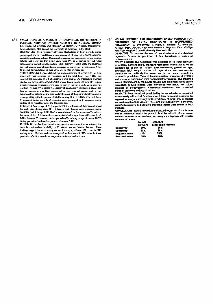
NEURAL NETWORK AND REGRESSION BASED FORMULA FOR PREDICTION OF FETAL HEMATOCRIT IN ISOIMMUNIZED PREGNANCY. A. Lysikiewicz S. Inglis, L. Bracero, F.Chervenak, N.Tejani. Dept. Ob/Gyn. New York Medical CoUege and Dept. Ob/Gyn New York Hospital, Cornell University New York, NY. OBJECTIVE: To compare the use of neural network and a standard regression formula for prediction of fetal hematocrit in cases of isoimmunization. STUDY DESIGN: The hematocrit was predicted in 46 cordocenteses by a neural network and by standard regression formula based on an expected fall of Hct of 1 %/day. Last hematocrit, gestational age, estimated fetal weight, number of days since last intravascular transfusion and antibody titer were used in the neural network as parametric predictors. Type of isoimmunization, presence of hydrops and number of transfusion were nonpararnetric variables. 1lIe predicted values of hematocrit by the neural network and prediction based on the regression derived formula were correlated with actual Hct values obtained at cordocentesis. Correlation coefficient was calculated between predicted and actual values. RESULTS: Fetal hematocrit predicted by the neural network correlated more closely with actual fetal hematocrit than hematocrit predicted by regression analysis although both predictors showed only a modest correlation with actual values (R=0.3 and 0.2 respectively). Sensitivity, specificity, positive and negative predictive VIIlues were similar for both methods. CONCLUSIONS: Neural network and standard regression formula have similar predictive abiHty to project fetal hematocrit. Since neural network includes more varables, accuracy may improve with greater numbers of cases.
Sensitivity Specificity Neg.pred.vlllue Pos.prad. value
Neural NetworK
80% 33% 77% 36%
standard regression fonnuls
85% 111% 74% 28%