Embed Size (px)
Citation preview
Journal of Applied Pharmaceutical Science Vol. 8(02), pp 001-010, February, 2018Available online at http://www.japsonline.comDOI: 10.7324/JAPS.2018.8201
ISSN 2231-3354
© 2018 Kiran Kemkar et al. This is an open access article distributed under the terms of the Creative Commons Attribution License -NonCommercial-ShareAlikeUn-ported License (http://creativecommons.org/licenses/by-nc-sa/3.0/).
*Corresponding AuthorSathiyanarayanan L.; Associate Professor, Department of Pharmaceuti-cal Chemistry, Bharati Vidyapeeth Deemed University, Poona College of Pharmacy, Pune-411 038, Maharashtra, India.E-mail: pharmsathiya @ gmail.com
6-shogaol from ginger oleoresin loaded liposomes using DMPG-Na as a carrier enhances the in-vitro and in-vivo anticancer activity
Kiran Kemkar, Sathiyanarayanan L.*, Arulmozhi Sathiyanarayanan, Kakasaheb MahadikBharati Vidyapeeth University, Poona College of Pharmacy, Pune-411 038, Maharashtra, India.
ARTICLE INFO ABSTRACTArticle history:Received on: 26/11/2017Accepted on: 08/01/2018Available online: 27/02/2018
Ginger oleoresin (GO) plays an important role on the attenuation of complications associated to the cancer therapy which is attributed to 6-shogaol (6-SGL). The major challenge in using 6-SGL for therapeutic applications is its poor aqueous solubility, low stability in GI and low bioavailability. Considering the potent anticancer nature of 6-SGL and its synergistic activity with other constituents in GO, there is a need to develop a suitable drug delivery system. Thus in the present study, 6-SGL rich GO (6-SRGO) was incorporated into liposomes by solvent injection technique using lipid (DMPG-Na) as carrier. The prepared 6-SRGO loaded liposomes (6-SRGO-LLPS) were characterized physically and chemically using FTIR, DSC, surface morphology, drug content (DC), encapsulation efficiency (EE), particle size (PS), zeta potential and further evaluated for stability study, in vitro release study, in vitro cytotoxicity study and in vivo anticancer activity in comparison with 6-SRGO. 32 factorial designs was used for optimization of formulation variables and the results of DC, EE and PS obtained were 89.92 ± 1.56, 79.36 ± 1.05 and 356.11 ± 4.07 respectively. Considering the above findings, 6-SRGO-LLPS exhibited significant in vitro (GI50- <10 μg/ml) and in vivo anticancer activity in comparison with 6-SRGO.
Key words: 6-Shogaol, Liposomes, Fac-torial designs, Cytotoxicity, Breast cancer.
INTRODUCTIONNow a day’s natural dietary foods including vegetables,
fruits, and spices aliments have been achieved a great attraction from the scientist of various health care departments, food industries and the normal public because of their safety, efficacy and availability at the comparatively lower price. In addition they are believed to have no or very less toxic effects than the synthetic drugs (Kelly et al., 2016; Shukla and Singh, 2007). However, the low aqueous solubility, poor bioavailability, and the stability of phytoconstituents lead to hamper their use in the treatment of various diseases. Therefore, various delivery system including nanotechnology-based drug-delivery systems have been designed for different naturally occurring phytoconstituents to increase the aqueous solubility, bio-availability and reduce the dose without hampering the efficacy of the drug, safety and the compatibility with patients (Bothiraja et al., 2013).
6-shogaol rich ginger oleoresin (6-SRGO) is an oleoresin extracted from Zingiber officinale Rosc., containing higher content of phenolic ketones; gingerols and shogaols (Oriania et al., 2016; Singh et al., 2008). Traditionally 6-SRGO used for the treatment of cough, stomach upset, inflammation, diarrhea, nausea and vomiting. Among the various phenolic constituents of ginger oleoresin (GO), the analogs of shogaols and gingerols are known for their potent antioxidant, anti-inflammatory, and anticancer activities (Chen et al., 2012). Various studies have discovered that 6-shogaol (6-SGL) possesses most potent anti cancer activity than the other analogs of the shogaols and the gingerols (Wu et al., 2010). Various in vitro studies have demonstrated the role of 6-SGL in suppressing the different cancer cells like ovary (Kim et al., 2008), lungs, colon, gastrointestinal tract and neuroblastoma. The metabolites of 6-SGL are also known to possesses anticancer activity (Shukla and Singh, 2007; Surh et al., 1998).
The major challenge of using 6-SGL is its poor aqueous solubility; which limits its therapeutic efficacy (Nikam et al., 2013). High dose of 6-SRGO is required to reach the therapeutic efficacy, which may lead to the several side effects such as diarrhea, nausea and many times the tolerance of the
Kemkar et al. / Journal of Applied Pharmaceutical Science 8 (02); 2018: 001-010002
6-SRGO. There are few attempts have been made to develop nanoformulations using conventional extract of ginger but detail investigation, characterization and anticancer potential are not reported (Kumar et al., 2011; Uthumpa et al., 2013; Ratcharin et al., 2012). Prompted by the above facts, present research aims at incorporating 6-SRGO within polymers to develop a drug delivery system displaying enhanced drug solubility, achieving site-specific tumor accumulation while reducing systemic toxicity of 6-SRGO.
Nanoparticular drug delivery systems using liposomes is an emerging technologies for the rational delivery of cytotoxic drugs in the treatment of cancer (Gao et al., 2011; Li et al., 2011; Tong et al., 2012; Muthu et al., 2012; Saxena and Hussain, 2012). Their use offers solubilization of water insoluble drugs, high drug loading, improved stability, increases circulation time, improved tissue targeting, control and extended drug release and lower systemic toxicity (Malam et al., 2009). Liposomes are closed spherical vesicles consisting of a lipid bilayer that encapsulates poorly water soluble drugs into the inner hydrophobic core thus improving solubility, stability and bioavailability (Akbarzadeh et al., 2013). Due to its outer hydrophobic shell and nano size particles they sometimes evince prolonged circulation times in vivo and can hoard in tumoral tissues. The liposomes size varies from 200-400 nm adduce rival physical and chemical properties that can be exploited for drug delivery by conjugation with drugs. The commercial availability of liposomal Doxil and albumin-nanoparticle-based Abraxane has existed for the treatment of the cancer. Due to above applications, liposomes offer increased precision in chemotherapeutic targeting of breast cancer.
To the best of our knowledge no previous studies have attempted the preparation of 6-SRGO-loaded liposomes (LLPS) to improve its anticancer efficacy. Therefore, this study was attempted to investigate the potentials of 6-shogaol rich ginger oleoresin-loaded liposomes (6-SRGO-LLPS), which may enhance solubility, physiological stability and anticancer efficiency of 6-SRGO.
MATERIALS & METHODS
Carrier and core materialsLiposomes were produced using a carrier 1,2-Dimyristoyl-
sn-glycero-3-phospho-rac-glycerol, sodium salt, (DMPG-Na)lipid that was supplied by Lipoid, Cologne, Germany, with melting point at 122oC. The reference standard (pure isolated) 6-shogaol (>96% purity w/w) was procured from Natural Remedies Pvt. Ltd. Bangalore, Karnataka, India. 6-SRGO was purchased from Nisarg Biotech, Satara, Maharashtra, India. Dialysis bags (MW cut-off 12,000) were purchased from Sigma-Aldrich Chemical Private Ltd (Bangalore, India). All other reagents and chemicals used in this study were of analytical grade and procured from Merck Specialties Private Limited, Mumbai, India.
Quantification of 6-SGL present in GO6-shogaol present in ginger oleoresin was quantified
using RP-HPLC method which was reported in our earlier study (Nikam et al., 2013).
Preparation of 6-SRGO-LLPS6-SRGO-LLPS were prepared according to solvent
injection technique (Laouini et al., 2012). Extraction of 6-SGL was
performed by adding the ginger oleoresin to a chloroform containing a specific amount of DMPG-Na followed by mixing with a suitable quantity of cholesterol under stirring at room temperature. To the sufficient distilled water, above mixture was added rapidly under magnetic stirring at 2000 rpm for 45 min followed by sonication for 10 min to obtain liposome suspension. Blank liposomes (devoid of 6-SRGO) were prepared in the similar manner.
Optimization of 6-SRGO-LLPS by 32 factorial designs 32 factorial designs was adopted to optimize the liposomes
composition while studying the effect of lipid and cholesterol (independent variables) on Drug Content (DC), Entrapment Efficiency (EE) and Particle Size (PS) (three dependent variables or responses) by obtaining a response surface plot.
Characterization of 6-SRGO-loaded liposomes
Liposomes size distribution & zeta potentialThe size of blank and 6-SRGO-LLPS was determined
by laser diffraction technique (Malvern 2000 SM; Malvern Instruments, Malvern, UK). The particle size measurements were carried out at a 90° scattering angle. The samples were dispersed in distilled water and the average PS was determined and expressed in terms of d(0.9) μm. The zeta potential was measured with the laser doppler electrophoretic mobility measurements using Zetasizer 3000 (Malvern Instruments) at a temperature of 25°C.
DC & EEThe concentration of 6-SGL in the liposomes was
determined by rapid and sensitive RP-HPLC method. The superficial 6-SGL was determined by washing 6-SRGO-LLPS with ethanol and measuring the 6-SGL content collected in the washing solution. EE was determined as the ratio between the encapsulated 6-SGL (total — superficial) and the feeding amount of 6-SGL (active compound). The DC was determined as the ratio between the mass of liposomes collected at the end of the process and the mass of carrier added to 6-SRGO used in the production of liposomes.
Physical characterization
Fourier transform infrared spectroscopyThe pure 6-SRGO, DMPG-Na and 6-SRGO-LLPS
were characterized by FTIR spectroscopy in the 4000 to 400 cm−1 region using FTIR Spectrophotometer (Jasco 4100).
Differential scanning calorimetryThermal analyses were performed by differential
scanning calorimetry (DSC 821, Mettler Toledo). Approximately 5 mg of 6-SRGO-LLPS were heated in hermetically sealed alluminium pan with heating rate 10oC per min under nitrogen atmosphere (flow rate 50 ml/min). The pure 6-SRGO was also analyzed for comparison.
Surface morphologyThe morphology of 6-SRGO-LLPS was performed using
transmission electron microscopy (TEM). The sample preparation for the TEM was followed by (Bothiraja et al., 2013).
Kemkar et al. / Journal of Applied Pharmaceutical Science 8 (02); 2018: 001-010 003
Evaluation of 6-SRGO-LLPS stability6-SRGO-LLPS were transferred in glass vials with
plastic lids, protected from the light, and stored at 25°C for 90 days for short term stability studies. Liposomes were evaluated with regard to the content of 6-SGL, PS and EE.
In vitro release of 6-SRGO from liposomesThe in vitro release of 6-SRGO-LLPS was carried out
in 0.1 N HCL (pH 1.2) and in phosphate-buffer (pH 7.4) using dialysis bag diffusion technique. A formulation equivalent to 1 mg of 6-SRGO or 1 mg 6-SRGO solution (1 mg/ml in 50% w/w mixture of PEG 400 and water) as control was introduced into a dialysis bag (cellulose membrane, MW cut-off 12,000 Da), sealed and immersed into 50 ml of release medium. The temperature of entire system was maintained at 37 ± 0.5°C with continuous magnetic stirring at 100 rpm. At previously planned time intervals, the sample was removed and replaced with an equal volume of fresh medium in order to maintain sink conditions. 6-SGL present in the solution was quantified by using rapid and sensitive RP-HPLC method. Analysis of data was performed using PCP Disso software (V3; Poona College of Pharmacy, Pune, India).
In vitro cytotoxicity studyIn vitro cytotoxicity study of free 6-SRGO, blank liposomes
and 6-SRGO-LLPS were evaluated against human breast cancer MCF-7 using in vitro SRB assay at Advanced Centre for Treatment, Research and Education in Cancer (ACTREC), Mumbai. The results of MCF-7 were compared with that of marketed Adriamycin (Doxorubicin). The cytotoxicity protocol for SRB assay was followed by method described by (Kaushik et al., 2015).
Acute toxicity studyThe acute toxicity study was performed for 6-SRGO and
6-SRGO-LLPS on swiss albino mice. The animals were randomly divided into eleven groups (n = 6). The first group (control group) received feed and distilled water orally. Groups 2–6 were orally treated with 6-SRGO with doses 55 mg/kg, 175 mg/kg, 550 mg/kg, 1750 mg/kg and 2000 mg/kg body weight, respectively. Groups 7–11 were orally treated with 6-SRGO-LLPS with doses 55 mg/kg, 175 mg/kg, 550 mg/kg, 1750 mg/kg and 2000 mg/kg body weight, respectively. The animals were continuously observed for general behavioral changes, sign of toxicity, and mortality for 1 h after treatment and then intermittently for 4 h and thereafter over a period of 24 h. Mice were further observed for up to 14 days for behavioral change and sign of toxicity.
In vivo anti cancer activity of 6-SRGO and 6-SRGO-LLPS against Dalton’s Ascitic Lymphoma (DAL) in mice
Selection grouping and acclimatization of laboratory animal Male Swiss albino mice having the weight range of 20
gm to 25 gm were used to conduct this study. They were kept in to micro nylon boxes at control temperature (temp 25 ± 2°C).
“The protocol of the study was approved by Institutional Animal Ethics Committee (IAEC) constituted in accordance with the rules and guidelines of the Committee for the Purpose of Control and Supervision on Experimental Animals (CPCSEA),
India (approval number CPCSEA/Q.A./01/2014-15; institutional approval no- CPCSEA/1999/100). The Declaration of Helsinki as amended in Seoul 2008 for humans, and the European Community guidelines as accepted principles for the use of experimental animals, was followed.”
Induction of cancer cellsDLA cells were procured from Amala cancer research
center, Trissur, Kerala, India. The cancer cells implants in vivo in mice by intraperitoneal route. Before transferring the cancerous cells to mice, by using saline solution the DLA cells were taken from peritoneal cavity of the mice. The total cells count was maintained up to 1 × 10⁶ after dilution and was given intraperitoneally. The treatment was started after seven days of induction of tumor cells (Sathiyanarayanan et al., 2006; Unnikrishnan and Kuttan, 1990).
Grouping of the mice and outline of the treatmentAnimals were grouped in to nine groups with each group
contains six animals. The animals of eight groups have given DLA cells (1 × 10⁶ cells per mice) intraperitoneally, and one group is remains as a control group.
Group 1 and 2 represents normal control and tumor control respectively.
Group 3 represents positive control, (Received 5-fluoro uracil injection at 20 mg/kg body weight)
Group 4, 5, and 6 represents treatment control, (Received 6-SRGO at a dose of 100 mg/kg, 200 mg/kg and 400 mg/kg body weight respectively through orally).
Group 7, 8, and 9 represents treatment control group, (Received 6-SRGO-LLPS at a doses of 100 mg/kg, 200 mg/kg and 400 mg/kg body weight respectively through orally).
Evaluation of clinical parametersDuring this study, the cancer cells were inoculated in to
the animals and the treatment for cancer was started after 24 hrs once a day for 14 days. Once after all the doses were finished, animals were sacrificed by euthanasia. The samples of the blood were withdrawn from each mouse at predetermined time from retro orbital plexus bleeding. Various parameters were checked which include, count of the cancer cells, hematological parameters consist of content of Hb, RBC count, WBC count, platelet count and the volume of packed cells, serum enzyme and lipid profile including count of total cholesterol (TC), triglycerides (TG), aspartate amino transferase (AST), alanine amino transferase (ALT) and alkaline phosphatase (ALP) and the derived parameters such as total life span (%) of the animal, body weight of the animal used and the cancer cell count.
Statistical analysisAll data are expressed as means ± SEM. One way
ANOVA followed by Dunnettspost test was used for in vivo anticancer activity. P < 0.01 was considered as significant. Prism 5 Demo software has been used for the statistical analysis.
RESULTS AND DISCUSSION6-SGL is well known for its anticancer potential.
However its usage is limited due to its low solubility, low stability
Kemkar et al. / Journal of Applied Pharmaceutical Science 8 (02); 2018: 001-010004
in GI and low bioavailability. GO consists of higher amount of shogaols and gingerols compare to normal solvent extracts. Hence usage of GO is ideal for anticancer purposes especially due to the presence of higher amount of 6-SGL. Further, synergistic effect of herbals is always plays a role in therapeutic applications. Although the pharmacological activities are attributed to active constituents, practically isolation and formulation of pure compounds from herbals are expensive processes and there are possibilities of reduction in therapeutic activity due to lack of synergism while using single compound. So it is worthwhile to develop drug delivery systems for multi chemical compositions from herbals rather than single compound. Considering these facts in present
study, 6-SRGO-LLPS have been developed and investigated as nanocarriers for oral administration in order to enhance both in vitro and in vivo anticancer activity of 6-SGL in GO.
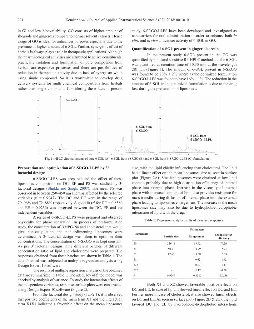
Quantification of 6-SGL present in ginger oleoresinIn the present study 6-SGL present in the GO was
quantified by rapid and sensitive RP-HPLC method and the 6-SGL was quantified at retention time of 10.30 min at the wavelength 281 nm (Figure 1). The amount of 6-SGL present in 6-SRGO was found to be 20% ± 2% where as the optimized formulation 6-SRGO-LLPS was found to have 16% ± 1%. The reduction in the amount of 6-SGL in the optimized formulation is due to the drug loss during the preparation of liposomes.
Fig. 1: HPLC chromatograms of pure 6-SGL (A), 6-SGL from 6SRGO (B) and 6-SGL from 6-SRGO-LLPS (C) formulation.
Preparation and optimization of 6-SRGO-LLPS by 32 factorial designs
6-SRGO-LLPS was prepared and the effect of these liposomes composition on DC, EE and PS was studied by 32 factorial designs (Shukla and Singh, 2007). The mean PS was observed in between 250–450 nm and was affected by the selected variables (r2 = 0.9247). The DC and EE were in the range of 79–96% and 72–88% respectively. A good fit (r2 for DC = 0.8580 and EE = 0.9236) was observed between the DC, EE and the independent variables.
A series of 6-SRGO-LLPS were prepared and observed physically for phase separation. In process of preformulation study, the concentration of DMPG-Na and cholesterol that would give non-coagulation and non-sedimenting liposomes were determined. A 32 factorial design was taken to optimize their concentrations. The concentration of 6-SRGO was kept constant. As per 32 factorial designs, nine different batches of different concentration ratio of lipid and cholesterol were prepared. The responses obtained from these batches are shown in Table 1. The data obtained was subjected to multiple regression analysis using Design Expert 10 software.
The results of multiple regression analysis of the obtained data are summarized in Table 1. The adequacy of fitted model was checked by analysis of variance. To study the interaction effects of the independent variables, response surface plots were constructed using Design Expert 10 software (Figure 2).
From the factorial design study (Table 1), it is observed that positive coefficients of the main term X1 and the interaction term X1X1 indicated a favorable effect on the mean liposomes
size, with the lipid chiefly influencing than cholesterol. The lipid had a linear effect on the mean liposomes size as seen in surface plot (Figure 2A). Smaller liposomes were obtained at low lipid content, probably due to high distribution efficiency of internal phase into external phase. Increase in the viscosity of internal phase with increased amount of lipid also provides resistance for mass transfer during diffusion of internal phase into the external phase leading to liposomes enlargement. The increase in the mean liposomes size may also be due to hydrophobic-hydrophobic interaction of lipid with the drug.
Table 1: Regression analysis results of measured responses.
CoefficientsParameters
Particle size Drug content Encapsulation efficiency
β0 356.11 89.92 79.36
β1 50.33 +1.79 +5.21
β2 12.67 +1.58 +5.58
β11 - -0.62 -1.42
β22 - -0.50 -1.17
β12 - +0.12 -0.38
r2 0.9247 0.8580 0.9236
Both X1 and X2 showed favorable positive effects on DC and EE. In case of lipid it showed linear effect on DC and EE. Further more in case of cholesterol, it also showed linear effects on DC and EE. As seen in surface plot (Figure 2B & 2C), the lipid favored DC and EE by hydrophobic-hydrophobic interactions

Kemkar et al. / Journal of Applied Pharmaceutical Science 8 (02); 2018: 001-010 005
with drugs leading to formation of interpenetrated network chain. However, cholesterol exerted an opposite effect as it led to
solubilization of the drug in the external phase.
Fig. 2: Response surface plot illustrating effect of factorial variables. (A) Particle size, (B) Encapsulation Efficiency, (C) Drug Content.
Based on the results of the factorial designs, 6-SRGO-LLPS having acceptable PS, DC and EE was selected as an optimized batch. The size of the optimized 6-SRGO-LLPS was 380 ± 3 nm – which was not significantly different from its blank counter part. Large molecules of more than 40 kDa in size and certain particles ranging from 10 to 400 nm can leave the vascular bed and accumulate inside the interstitial space of the tumor (EPR effect) (Gao et al., 2011). Drug delivery to specific sites of the body is influenced by size of the liposomes; smaller liposomes may tend to minimize the particle uptake by non targeted cells, including their premature clearance by the mononuclear phagocytic system (Brigger et al., 2002). It is hypothesized that liposomes developed in the present study are of appropriate size to be able to passively target the tumor site.
Physical characterization
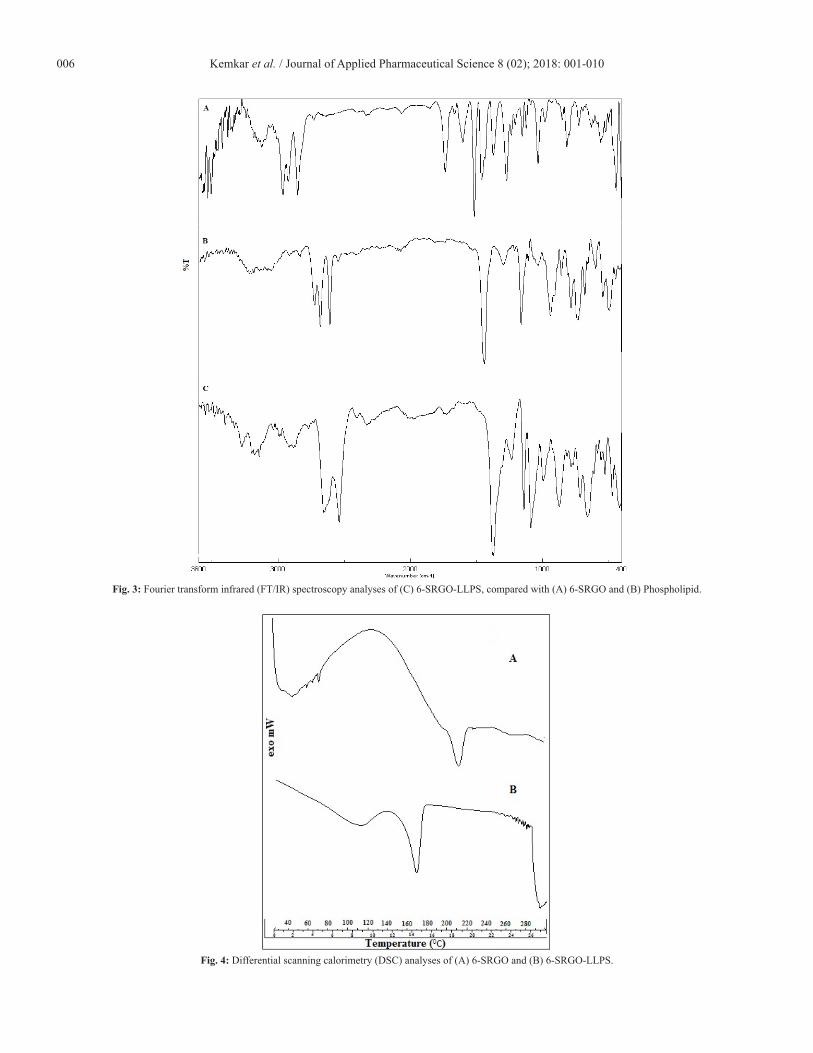
Fourier transform infrared spectroscopy6-SRGO, DMPG-Na, and 6-SRGO-LLPS were analyzed
by Fourier transform infrared (FTIR) spectroscopy and the results are showed in (Figure 3).
In the IR spectra (Figure 3A) of 6-SRGO following peaks were assigned, -OH stretching (~3124 cm−1), Aromatic –CH stretching (~2994 cm−1), Aliphatic –CH stretching (~2852 cm−1), –C=O stretching (~1736 cm−1) and OH bending out of plan (~1373 cm−1), while in the formulation (Figure 3C), 6-SRGO incorporated with polymer lipid, remarkable peaks of active drug
such as –OH stretching at (~3124 cm−1) were not observed. Thus final IR spectra of 6-SRGO-LLPS revealed that 6-SRGO has been completely encapsulated with lipid in the formulation.
Differential scanning calorimetryDifferential scanning calorimetry was performed with
6-SRGO-LLPS to determine the onset, maximum temperature and enthalpy of 6-SRGO-LLPS and 6-SRGO (Figure 4). According to the results, melting point of free 6-SRGO was observed at around 240oC with the enthalpy 31.11 J/g. In case of 6-SRGO-LLPS thermogram, the free 6-SRGO peak was disappeared and shifted to 169oC indicating molecular dispersion of 6-SRGO inside liposomes. Thermograms of 6-SRGO-LLPS exhibited a sharp endothermic peak starting at approximately 145oC and ending at approximately 185oC. Onset temperature at approximately 145oC and melting point around 169oC are desirable for particle stability at room or lower temperatures and digestibility in the gastrointestinal tract, respectively, acting as a trigger to release the active core during the digestion process (Tulini et al., 2016).
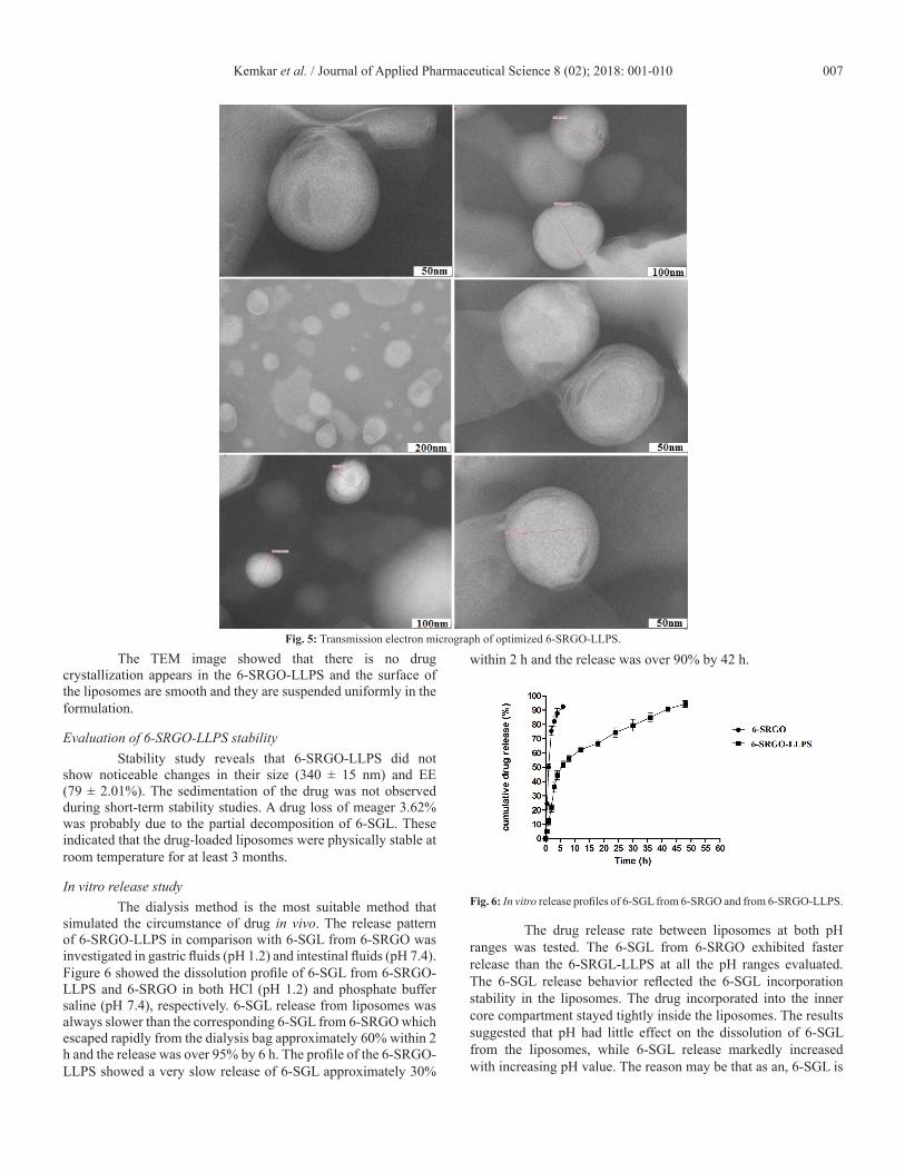
Surface morphologyThe obtained 6-SRGO-LLPS solution appeared clear
and light yellowish in color. Figure also revealed that the prepared formulation was monodispersed in water phase, tightly packed and spherical shaped liposomes whose size measured by laser scattering technique was correlated well with that by TEM (Figure 5).
Kemkar et al. / Journal of Applied Pharmaceutical Science 8 (02); 2018: 001-010006
Fig. 3: Fourier transform infrared (FT/IR) spectroscopy analyses of (C) 6-SRGO-LLPS, compared with (A) 6-SRGO and (B) Phospholipid.
Fig. 4: Differential scanning calorimetry (DSC) analyses of (A) 6-SRGO and (B) 6-SRGO-LLPS.
Kemkar et al. / Journal of Applied Pharmaceutical Science 8 (02); 2018: 001-010 007
Fig. 5: Transmission electron micrograph of optimized 6-SRGO-LLPS.
The TEM image showed that there is no drug crystallization appears in the 6-SRGO-LLPS and the surface of the liposomes are smooth and they are suspended uniformly in the formulation.
Evaluation of 6-SRGO-LLPS stabilityStability study reveals that 6-SRGO-LLPS did not
show noticeable changes in their size (340 ± 15 nm) and EE (79 ± 2.01%). The sedimentation of the drug was not observed during short-term stability studies. A drug loss of meager 3.62% was probably due to the partial decomposition of 6-SGL. These indicated that the drug-loaded liposomes were physically stable at room temperature for at least 3 months.
In vitro release studyThe dialysis method is the most suitable method that
simulated the circumstance of drug in vivo. The release pattern of 6-SRGO-LLPS in comparison with 6-SGL from 6-SRGO was investigated in gastric fluids (pH 1.2) and intestinal fluids (pH 7.4). Figure 6 showed the dissolution profile of 6-SGL from 6-SRGO-LLPS and 6-SRGO in both HCl (pH 1.2) and phosphate buffer saline (pH 7.4), respectively. 6-SGL release from liposomes was always slower than the corresponding 6-SGL from 6-SRGO which escaped rapidly from the dialysis bag approximately 60% within 2 h and the release was over 95% by 6 h. The profile of the 6-SRGO-LLPS showed a very slow release of 6-SGL approximately 30%
within 2 h and the release was over 90% by 42 h.
Fig. 6: In vitro release profiles of 6-SGL from 6-SRGO and from 6-SRGO-LLPS.
The drug release rate between liposomes at both pH ranges was tested. The 6-SGL from 6-SRGO exhibited faster release than the 6-SRGL-LLPS at all the pH ranges evaluated. The 6-SGL release behavior reflected the 6-SGL incorporation stability in the liposomes. The drug incorporated into the inner core compartment stayed tightly inside the liposomes. The results suggested that pH had little effect on the dissolution of 6-SGL from the liposomes, while 6-SGL release markedly increased with increasing pH value. The reason may be that as an, 6-SGL is
Kemkar et al. / Journal of Applied Pharmaceutical Science 8 (02); 2018: 001-010008
unstable in low pH i.e. in gastric condition, which may lead to a low bioavailability of 6-SGL from 6-SRGO.
Fig. 7: In vitro cytotoxicity study on breast cancer cell line (MCF 7), (A) MCF 7 control, (B) Treatment control, (C) Treated with 6-SRGO, (D) Treated with 6-SR-GO-LLPS.
Table 2: In vitro anticancer effect of 6-SRGO-LLPS on breast cancer cell MCF-7.
Drug concentration (µg/ml) calculated from graph
MCF 7 LC50 TGI GI50
6-SRGO >80 56.4 26.8
Blank >80 >80 >80
6-SRGO-LLPS >80 38.4 <10
ADR 60.8 29.5 <10
Each point represents an average ± SD (n = 3).
In vitro cytotoxic activityThe in vitro cytotoxic activity of 6SRGO-LLPS was
investigated and compared with free drug in solution, blank liposomes and marketed Adriamycin (Doxorubicin) against human breast cancer MCF-7 cells using in vitro SRB assay. The LC50, TGI and GI50 values were illustrated in Table 2, indicated that 6SRGO-LLPS displayed better cytotoxic activity than the 6-SRGO and the blank liposomes. The GI50 concentration against MCF-7 was found to be <10 μg/ml, 26.8 μg/ml and >80 μg/ml for 6-SRGO-LLPS, 6-SRGO and blank liposomes respectively (Fig. 7).
Table 3: Effect of 6-SRGO and 6-SRGO-LLPS on Hematological parameters.
TREATMENTTotal WBC Rbc Count Hb
PCV %Platelets
Cells/ml × 103 Mill/cumm gm/dl Lakhs/cumm
G1 10.35 ± 1.30 4.30 ± 1.85 12.50 ± 1.34 14.25 ± 2.44 3.30 ± 0.95
G2 15.22 ± 2.64a** 2.68 ± 0.72a** 6.80 ± 0.95a** 38.36 ± 3.35a** 1.70 ± 0.42a**
G3 12.32 ± 1.30b** 4.05 ± 1.40b** 11.90 ± 1.48b** 16.40 ± 1.40b** 2.94 ± 0.65b**
G4 12.90 ± 2.04b** 3.45 ± 1.05b** 11.60 ± 1.22b** 17.34 ± 2.30b** 2.88 ± 0.54b**
G5 13.08 ± 2.26b** 3.85 ± 1.34b** 12.35 ± 1.66b** 18.08 ± 2.66b** 3.04 ± 0.68b**
G6 12.66 ± 2.28b** 4.10 ± 1.45b** 12.15 ± 1.36b** 17.84 ± 2.46b** 2.94 ± 0.62b**
G7 11.74 ± 1.38b** 4.25 ± 1.82b** 12.40 ± 1.64b** 17.18 ± 2.26b** 3.40 ± 0.90b**
G8 11.66 ± 1.30b** 4.30 ± 1.85b** 12.44 ± 1.68b** 16.70 ± 2.18b** 3.46 ± 0.92b**
G9 11.58 ± 1.26b** 4.35 ± 1.88b** 12.55 ± 1.74b** 16.65 ± 1.98b** 3.54 ± 0.95b**
G1 – Normal Control, G2 – Cancer Control, G3 – Positive control, G4 to G6 – Treatment control (6-SRGO 100, 200, 400 mg/kg), G7 to G9 – Treatment control (6-SR-GO-LLPS 100, 200, 400 mg/kg).All values are expressed as mean ± SEM for 6 animals in each group and results were analyzed by using One way ANNOVA, followed by Dunnettspost test.**aValues are significantly different from normal control (G1) at P < 0.01**bValues are significantly different from cancer control (G2) at P < 0.01.
The enhanced cytotoxic activity of 6-SRGO-LLPS may be attributed to greater cellular uptake of liposomes via phagocytosis or the fusion process of lipid liposomes. Therefore, 6-SRGO-LLPS might have served as a potential nanocarrier to improve the in vitro cytotoxic activity of 6-SGL. The lower cytotoxic activity of free drug in solution, blank liposomes may be due to its efflux by P-glycoprotein pumps.
Acute toxicity studyNo death or toxic effect on tested animals were recorded
during first 24 h as well as 14 days of observation after oral treatment
of 6-SRGO and 6-SRGO-LLPS at the doses of 55 mg/kg, 175 mg/kg, 550 mg/kg, 1750 mg/kg and 2000 mg/kg body weight.
The present study showed that oral administration of 6-SRGO and 6-SRGO-LLPS in acute dose up to 2000 mg/kg body weight did not produce any sign of toxicity or death in mice, suggesting a lethal dose 50% (LD50) above 2000 mg/kg. An acute toxic study provides a guideline for selecting doses for in vivo study (1/10th and 1/20th of maximum dose in mice) which may be more clinically relevant (Mehta et al., 2009). Thus, derived doses of 100 mg/kg, 200 mg/kg and 400 mg/kg for both 6-SRGO and 6-SRGO-LLPS have been selected.
Kemkar et al. / Journal of Applied Pharmaceutical Science 8 (02); 2018: 001-010 009
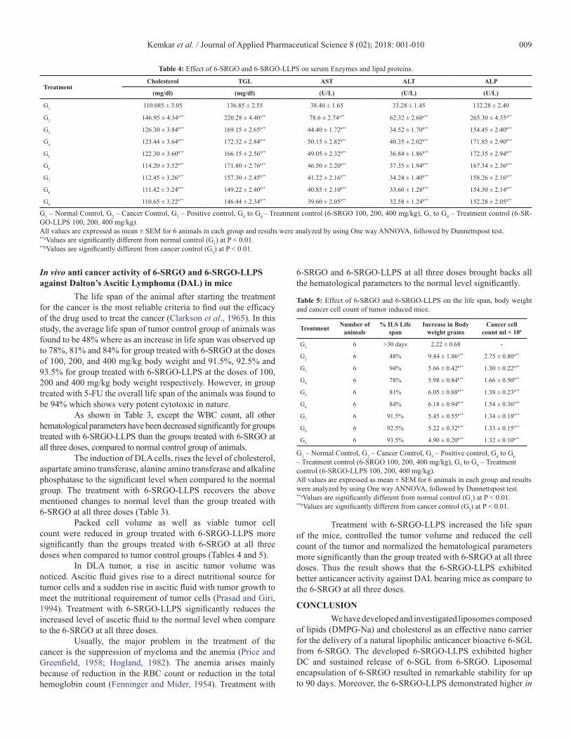
Table 4: Effect of 6-SRGO and 6-SRGO-LLPS on serum Enzymes and lipid proteins.
TreatmentCholesterol TGL AST ALT ALP
(mg/dl) (mg/dl) (U/L) (U/L) (U/L)
G1 110.085 ± 3.05 136.85 ± 2.55 38.40 ± 1.65 33.28 ± 1.45 132.28 ± 2.40
G2 146.95 ± 4.34a** 220.28 ± 4.40a** 78.6 ± 2.74a** 62.32 ± 2.60a** 265.30 ± 4.35a**
G3 126.30 ± 3.84b** 169.15 ± 2.65b** 44.40 ± 1.72b** 34.52 ± 1.70b** 154.45 ± 2.40b**
G4 123.44 ± 3.64b** 172.32 ± 2.84b** 50.15 ± 2.82b** 40.35 ± 2.02b** 171.85 ± 2.90b**
G5 122.30 ± 3.60b** 166.15 ± 2.56b** 49.05 ± 2.32b** 36.84 ± 1.86b** 172.35 ± 2.94b**
G6 114.20 ± 3.52b** 171.80 ± 2.76b** 46.50 ± 2.20b** 37.35 ± 1.94b** 167.34 ± 2.36b**
G7 112.45 ± 3.26b** 157.30 ± 2.45b** 41.22 ± 2.16b** 34.24 ± 1.40b** 158.26 ± 2.16b**
G8 111.42 ± 3.24b** 149.22 ± 2.40b** 40.85 ± 2.10b** 33.60 ± 1.28b** 154.30 ± 2.14b**
G9 110.65 ± 3.22b** 146.44 ± 2.34b** 39.60 ± 2.05b** 32.58 ± 1.24b** 152.28 ± 2.05b**
G1 – Normal Control, G2 – Cancer Control, G3 – Positive control, G4 to G6 – Treatment control (6-SRGO 100, 200, 400 mg/kg), G7 to G9 – Treatment control (6-SR-GO-LLPS 100, 200, 400 mg/kg).All values are expressed as mean ± SEM for 6 animals in each group and results were analyzed by using One way ANNOVA, followed by Dunnettspost test.**aValues are significantly different from normal control (G1) at P < 0.01.**bValues are significantly different from cancer control (G2) at P < 0.01.
In vivo anti cancer activity of 6-SRGO and 6-SRGO-LLPS against Dalton’s Ascitic Lymphoma (DAL) in mice
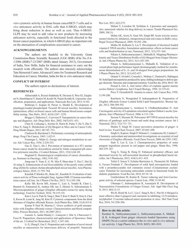
The life span of the animal after starting the treatment for the cancer is the most reliable criteria to find out the efficacy of the drug used to treat the cancer (Clarkson et al., 1965). In this study, the average life span of tumor control group of animals was found to be 48% where as an increase in life span was observed up to 78%, 81% and 84% for group treated with 6-SRGO at the doses of 100, 200, and 400 mg/kg body weight and 91.5%, 92.5% and 93.5% for group treated with 6-SRGO-LLPS at the doses of 100, 200 and 400 mg/kg body weight respectively. However, in group treated with 5-FU the overall life span of the animals was found to be 94% which shows very potent cytotoxic in nature.
As shown in Table 3, except the WBC count, all other hematological parameters have been decreased significantly for groups treated with 6-SRGO-LLPS than the groups treated with 6-SRGO at all three doses, compared to normal control group of animals.
The induction of DLA cells, rises the level of cholesterol, aspartate amino transferase, alanine amino transferase and alkaline phosphatase to the significant level when compared to the normal group. The treatment with 6-SRGO-LLPS recovers the above mentioned changes to normal level than the group treated with 6-SRGO at all three doses (Table 3).
Packed cell volume as well as viable tumor cell count were reduced in group treated with 6-SRGO-LLPS more significantly than the groups treated with 6-SRGO at all three doses when compared to tumor control groups (Tables 4 and 5).
In DLA tumor, a rise in ascitic tumor volume was noticed. Ascitic fluid gives rise to a direct nutritional source for tumor cells and a sudden rise in ascitic fluid with tumor growth to meet the nutritional requirement of tumor cells (Prasad and Giri, 1994). Treatment with 6-SRGO-LLPS significantly reduces the increased level of ascetic fluid to the normal level when compare to the 6-SRGO at all three doses.
Usually, the major problem in the treatment of the cancer is the suppression of myeloma and the anemia (Price and Greenfield, 1958; Hogland, 1982). The anemia arises mainly because of reduction in the RBC count or reduction in the total hemoglobin count (Fenninger and Mider, 1954). Treatment with
6-SRGO and 6-SRGO-LLPS at all three doses brought backs all the hematological parameters to the normal level significantly.
Table 5: Effect of 6-SRGO and 6-SRGO-LLPS on the life span, body weight and cancer cell count of tumor induced mice.
Treatment Number of animals
% ILS Life span
Increase in Body weight grams
Cancer cell count ml × 106
G1 6 >30 days 2.22 ± 0.68 -
G2 6 48% 9.44 ± 1.86a** 2.75 ± 0.80a**
G3 6 94% 5.66 ± 0.42b** 1.30 ± 0.22b**
G4 6 78% 5.98 ± 0.84b** 1.66 ± 0.50b**
G5 6 81% 6.05 ± 0.88b** 1.38 ± 0.23b**
G6 6 84% 6.18 ± 0.94b** 1.54 ± 0.36b**
G7 6 91.5% 5.45 ± 0.55b** 1.34 ± 0.18b**
G8 6 92.5% 5.22 ± 0.32b** 1.33 ± 0.15b**
G9 6 93.5% 4.90 ± 0.20b** 1.32 ± 0.10b**
G1 – Normal Control, G2 – Cancer Control, G3 – Positive control, G4 to G6 – Treatment control (6-SRGO 100, 200, 400 mg/kg), G7 to G9 – Treatment control (6-SRGO-LLPS 100, 200, 400 mg/kg).All values are expressed as mean ± SEM for 6 animals in each group and results were analyzed by using One way ANNOVA, followed by Dunnettspost test.**aValues are significantly different from normal control (G1) at P < 0.01.**bValues are significantly different from cancer control (G2) at P < 0.01.
Treatment with 6-SRGO-LLPS increased the life span of the mice, controlled the tumor volume and reduced the cell count of the tumor and normalized the hematological parameters more significantly than the group treated with 6-SRGO at all three doses. Thus the result shows that the 6-SRGO-LLPS exhibited better anticancer activity against DAL bearing mice as compare to the 6-SRGO at all three doses.
CONCLUSIONWe have developed and investigated liposomes composed
of lipids (DMPG-Na) and cholesterol as an effective nano carrier for the delivery of a natural lipophilic anticancer bioactive 6-SGL from 6-SRGO. The developed 6-SRGO-LLPS exhibited higher DC and sustained release of 6-SGL from 6-SRGO. Liposomal encapsulation of 6-SRGO resulted in remarkable stability for up to 90 days. Moreover, the 6-SRGO-LLPS demonstrated higher in
Kemkar et al. / Journal of Applied Pharmaceutical Science 8 (02); 2018: 001-010010
vitro cytotoxic activity in human breast cancerMCF-7 cells and in vivo anticancer activity in DAL cells than 6-SRGO, which may bring about reduction in dose as well as cost. Thus 6-SRGO-LLPS may be used to add value to new products by increasing anticancer activity, especially in functional foods directed to the breast cancer population, in which 6-SRGO play an important role on the attenuation of complications associated to cancer.
ACKNOWLEDGMENTSThe authors are thankful to the University Grant
Commission-Basic Scientific Research (UGC-BSR) (File No.:F.4-1/2006 (BSR)/7-23/2007 (BSR) dated January 2013), Government of India, New Delhi, India for financial assistance to carry out the research work efficiently. The authors also sincerely acknowledge Tata Memorial Center, Advanced Centre for Treatment Research and Education in Cancer, Mumbai, India for the in vitro anticancer study.
CONFLICT OF INTERESTThe authors report no declarations of interest.
REFERENCESAkbarzadeh A, Rezaei-Sadabady R, Davaran S, Woo SJ, Zargha-
mi N, Hanifehpour Y, Samiei M, Kouhi M, Nejati-Koshki K. Liposome: clas-sification, preparation, and applications. Nanoscale Res Lett, 2013; 8:102.
Bothiraja C, Kapare H, Pawar A, Shaikh K. Development of plumbagin-loaded phospholipid–Tween® 80 mixed micelles: formulation, optimization, effect on breast cancer cells and human blood/serum compat-ibility testing. Ther Deliv, 2013; 4(10):1247–1259.
Brigger I, Dubernet C, Couvreur P. Nanoparticles in cancer ther-apy and diagnosis. Adv Drug Deliv Rev, 2002; 54(5):631–651.
Chen H, Lishuang L, Soroka D, Warin R, Parks T, Hu Y, Zhu Y, Chen X, Sang S. Metabolism of [6]-Shogaol in Mice and in Cancer Cells. Drug Metab Dispos, 2012; 40:742–753.
Clarkson B, Burchenal J. Preliminary screening of antineoplastic drugs. Prog Clin Cancer, 1965; 1:625-9.
Fenninger L, Mider G. Energy and nitrogen metabolism in can-cer. Adv Cancer Res, 1954; 2:229-253.
Gao Z, Tian L, Hu J. Prevention of metastasis in a 4T1 murine breast cancer model by doxorubicin carried by folate conjugated pH sensi-tive polymeric micelles. J Control Release, 2011; 152(1):84–89.
Hogland H. Hematological complication of cancer chemothera-py, Seminars in Oncology, 1982; 9:95-102.
Jiang-nan Y, Yuan Z, Li W, Min P, Shan-shan T, Xia C, Hui Q, Xi-ming X. Enhancement of oral bioavailability of the poorly water-soluble drug silybin by sodium cholate/phospholipid-mixed micelles. Acta Pharma-cologica Sinica, 2010; 31:759–764.
Kaushik P, Khokra SL, Rana AC, Kaushik D. Evaluation of anti-cancer activity of Pinus roxburghii Sarg. Against IMR-32 human neuroblas-toma cancer cell line. Int J Pharm Clin Res, 2015; 7(1):105-108.
Kelly SB, Kevin MS, Aranee C, Luis A, Vondel R, Chen L, Bennett D, Emmanuel K, Andrea GB, Ian T, Zhimin X, Subramaniam S. Microencapsulation of ginger (Zingiber officinale) extract by spray drying technology. Food Sci Technol, 2016; 70:119-125.
Kim J, Lee S, Park H, Yang J, Shin T, Kim Y, Baek N, Kim S, Choi S, Kwon B, Leem K, Jung M, Kim D. Cytotoxic components from the dried rhizomes of Zingiber officinale Roscoe. Arch Pharm Res, 2008; 31(4):415-8.
Kumar P, Paul W, Sharma C. Green synthesis of gold nanopar-ticles with Zingiber officinale extract: Characterization and blood compati-bility. Process Biochem, 2011; 46:2007-2013.
Laouini A, Jaafar-Maalej C, Limayem I, Sfar S, Charcosset C, Fessi H. Praparation, characterization and applications of liposomes: State of the art. J Colloid Sci Biotechnol, 2012; 1:147-168.
Li X, ZhangY, Fan Y. Preparation and evaluation of novel mixed micelles as nanocarriers for intravenous delivery of propofol. Nanoscale
Res Lett, 2011; 6(3):275.Malam Y, Loizidou M, Seifalian A. Liposomes and nanoparti-
cles: nanosized vehicles for drug delivery in cancer. Trends Pharmacol Sci, 2009; 30(11).
Mehta AK, Arora N, Gaur SN, Singh BP. Acute toxicity assess-mentof choline by inhalation, intraperitoneal and oral routes in Balb/c mice. Regul Toxicol Pharmacol, 2009; 54:282–286.
Muthu M, Kulkarni S, Liu Y. Development of docetaxel-loaded vitamin E TPGS micelles: formulation optimization. effects on brain cancer cells and biodistribution in rats, Nanomed, 2012; 7(3):353–364.
Nikam A, Sathiyanarayanan L, Mahadik K. Mapping Pharmaco-kinetic and Tissue Distribution Profile of [6]-Shogaol from Ginger Oleores-in. Intl J Pharm Pharm Sci, 2013; 5(1):185-189.
Nikam A, Sathiyanarayanan L, Mahadik K. Validation of re-versed-phase high-performance liquid chromatography method for simulta-neous determination of 6-, 8-, and 10-shogaol from ginger preparations. Int J Pharm Pharm Sci, 2013; 5(1):432-437.
Oriania V, Alvimb I, Consolia L, Molinac C, Pastored G, Hubingera M. Solid lipid microparticles produced by spray chilling technique to deliver gin-ger oleoresin: Structure and compound retention. Food Res Int, 2016; 80:41–49.
Prasad S, Giri A. Anti tumor effect of cisplatin against murine ascites Dalton’s lymphoma. Ind J Expetl Biology, 1994; 32:155-62.
Price V, Greenfield R. Anemia in cancer. Adv Cancer Res, 1958; 5:199-200.
Ratcharin N, Wongtrakul P, Indranupakorn R. Preparation of Zingiber officinale Extract Loaded Solid Lipid Nanoparticles. Biomat Ap-pls, 2012; 506:389-392.
Sathiyanarayanan L, Arulmozi S, Chidhambarnathan N. Anti Carcinogenic activity of Leptadenia reticulataI against Dalton’s ascitic lym-phoma. Iran J Pharmacol Toxicol, 2006; 6:133–136.
Saxena V, Hussain M. Poloxamer 407/TPGS mixed micelles for delivery of gambogic acid to breast and multi drug resistant cancer. Int J Nanomed, 2012; 7(2):713–721.
Shukla Y, Singh M. Cancer preventive properties of ginger: A brief review. Food Chem Toxicol, 2007; 45:683–690.
Singh G, Kapoor, Singh P, Heluani C, Lampasona M, Catalan C. Chemistry, antioxidant and antimicrobial investigations on essential oil and oleoresins of Zingiber officinale. Food Chem Toxicol, 2008; 46:3295–3302.
Surh Y, Lee E, Lee J. Chemoprotective properties of some pungent ingredients present in red pepper and ginger. Mutat Res, 1998; 402:259–267.
Tong S, Xiang B, Dong D. Enhanced antitumor efficacy and decreased toxicity by self-associated docetaxel in phospholipid based mi-celles. Int J Pharmceut, 2012; 434(1):413–419.
Tulini F, Souza V, Echalar-Barrientos A, Thomazini M, Pallone E, Favaro-Trindade C. Development of solid lipid microparticles loaded with a proanthocyanidin rich cinnamon extract (Cinnamomum zeylani-cum): Potential for increasing antioxidant content in functional foods for diabetic population. Food Res Int, 2016; 85:10–18.
Unnikrishnan M, Kuttan R. Tumor reducing and Anti-Carcino-genic activity of selected species. Cancer Lett, 1990; 51:85-89.
Uthumpa C, Indranupakorn R, Asasutjarit R. Development of Nanoemulsion Formulations of Ginger Extract. Adv Appl Mat Elect Eng II, 2013; 684:12-15.
Wu H, Hsieh M, Lo C, Liu C, Sang S, Ho C, Pan M. 6-Shogaol is more effective than 6-gingerol and curcumin in inhibiting 12-O-tetradeca-noylphorbol 13-acetate-induced tumor promotion in mice. Mol Nutr Food Res, 2010; 54:1296-306.
How to cite this article: Kemkar K, Sathiyanarayanan L, Sathiyanarayanan A, Mahad-ik K. 6-shogaol from ginger oleoresin loaded liposomes using DMPG-Na as a carrier enhances the in-vitro and in-vivo antican-cer activity. J App Pharm Sci, 2018; 8(02): 001-010.