Embed Size (px)
Citation preview
Volume 16~ Number 1. Pdrt ~
627 FETAL THERAPY FOR A SACROCOCCYGEAL TERATOMA. Kent Heyborne,: M.O.x, Richard P. Porreco, M.D., Presbyter1~nlSt. lUke'sl PeMnatal Program, University of Colorado He~lth Sciences Center,! Denver, Colorado i
628
Sacrococcygea 1 teratom~s (SCT), the most common tumor: occurrin~ in the newborn, have an incidence of one in 40,000,1 with 80\ of affected infants being female. The majority of SCTsl are exophytic, benign growths t~ought to derive fro. Hensen' !I node. The newborn ~urvival for SCTs is excellent and varies froml 88-97\, with most mortality occurring from operative helROrrh~ge.1 Prenata lly ascertained cases, on the other hand, manifest ~ muchl higher mortality. In one recent review, 7 of 27 patients underwent elective termination; 9 of the rellllJining 20 patientsl (45\) experienced periMta 1 mortality. Seven of 7 fetuses rleve10ping hydrops in utero suffer~d TUFOs We report in uterol feta 1 therapy of a large, so lid, sacrococcygeal teratoma.l Ascertained at 21 weeks 3 (jays gestation, there was no evidence· of fetal comoromise. At 22 weeks 0 days gestation, the patien~ presented in preterm labor liIith significant oolyhydr~lIIlios.1 Moderate fetal hydrops was noted with marked skin thickening ~nd a pericardia 1 effusion. Co lor flow Doppler was used to identify! the dominent arterial supply to the tumor, probably representing the middle s~cral ~rtery. Four thrombogenic coils were placed in: the area of the arterial supply oercutaneously under ultrasound guidance. Color flow Doppler following the procedure showedl marked diminution of blood flow to the tumor. Although this moribund fetus suffered an intrauterine fet~l demiSe! approximately 24 hours following the procedure, ~utopsy showed marked compromise of the teratoma's vascu lar pedic le. Wei consider this case to represent ~ techn i ca 1 success. Given the high periMtal mortality rate for pren~tally ~scert~in~dl sacrococcygeal teratomas, in utero ther~py, especially 1n compromised fetuses at orev;~ble gestations, represents al treatment ootion. Technical success in interrupting the tUl1IOr'g vascular pedicle was achieved in this case using color floW! Dopp ler 9uided percutaneous placement of thrombogenic coils.
PREDICTIVE VALUE OF INVASIVE FETAL TESTING IN FETAL URINARY TRACT OBSTRUCTION
Authors: Paul C, Browne, MD Lewis H. Hamner III, MD, Bruce Broecker, MP-Emory School of Medicine, crawford Long Hospital, Atlanta, Georgia
Urinary tract obstruction 18 one of the most common fetal abnormalities detected by ultrasound. Urinary tract abnormalities were detected at a rate of 2.4/1000 ultrasound examinations performed at The Emory Regional perinatal center in Atlanta, Georgia. Of 28 fetuses demonstrating urinary tract obstruction, 24 underwent invasi ve fetal testing consisting of urine aspiration comhlned with either amnlocenteSls or umbilical blood sampling or both. Invasive testing was successful in 22/24 attempts. Electrolyte stUdies obtalned from fetal urine, amniotic fluld and serum demonstrate that fetal urinary chloride, urlnary sodium, and urinary osmolarity correlate best with neonatal ur1nary tract function. Amniotic fluid electrolytes more closely resemble fetal serum than fetal ur1ne with the exception of urinary nitrogen. Gradients between fetal serum and urine or between fetal urine and amniotic flU.ld were no more predictive of neonatal renal function than were urinary electrolytes alone. AmniotlC fluid volume as measured on ultrasound examination is also accurate in predicting residual neonatal renal function. Aspiration of fetal urine may be helpful in patient counselling and delivery planning of patlents with fetal urinary tract
o,",~oUO" Im~lLM_1 .... OOIl[/U.'N( s.rl~"/."'OO"C S£I",,/UI'NI
SPO Abstracts 417
629 FETAL THORACENTESIS. Cynthia J. Sims, Harlan R. Giles, Stephen Hasley". Medical College of Pennsylvania, Allegheny General Hospital, Pittsburgh, Pennsylvania.
630
In utero management of ultrasonographically diagnosed fetal malformations has historically been utilized with central nervous system or genitourinary anomalies. We report a case involving the fetal lung A 34 year old woman, gravida 3, para 2, was referred for diagnostic ultrasound at 20 weeks gestational age because of uncertain dating criteria. Ultrasound examination revealed a unilateral left pleural effusion, without a shift of the mediastinum. No other abnormalities were noted. An ammocentesis was performed which later showed a normal female karyotype. At 24 weeks gestational age progression of the effusion was noted with a shift of the mediastinum. A fetal thoracentesis was performed and 20 cc of yellow fluid was withdrawn. AnalYSIS of the fluid showed 97% lymphocytes, suggesting a chylous effusion. A rapid reaccumulation of the pleural fluid was noted. Repeat procedures were performed at 26, 28, and 30 weeks estimated gestational age with the collection of 40, 48 and 88 cc of fluid respectively. After the fourth drainage, minimal reaccumulation of fluid was observed. Within two weeks, the small effusion had resolved. The remainder of the pregnancy was uncomplicated. The patient subsequently delivered an 81b 5 oz female infant, vaginally. The infant had a normal examination and chest radiograph. At three months of age, the child and mother were doing well. A previous case of fetal pleural amniotic shunting of macrocystic adenomatoid malformation was complicated by a tension pneumothorax requiring right upper lobectomy. This complication has not been reported with sequential thoracentesis of congenital chylothorax.
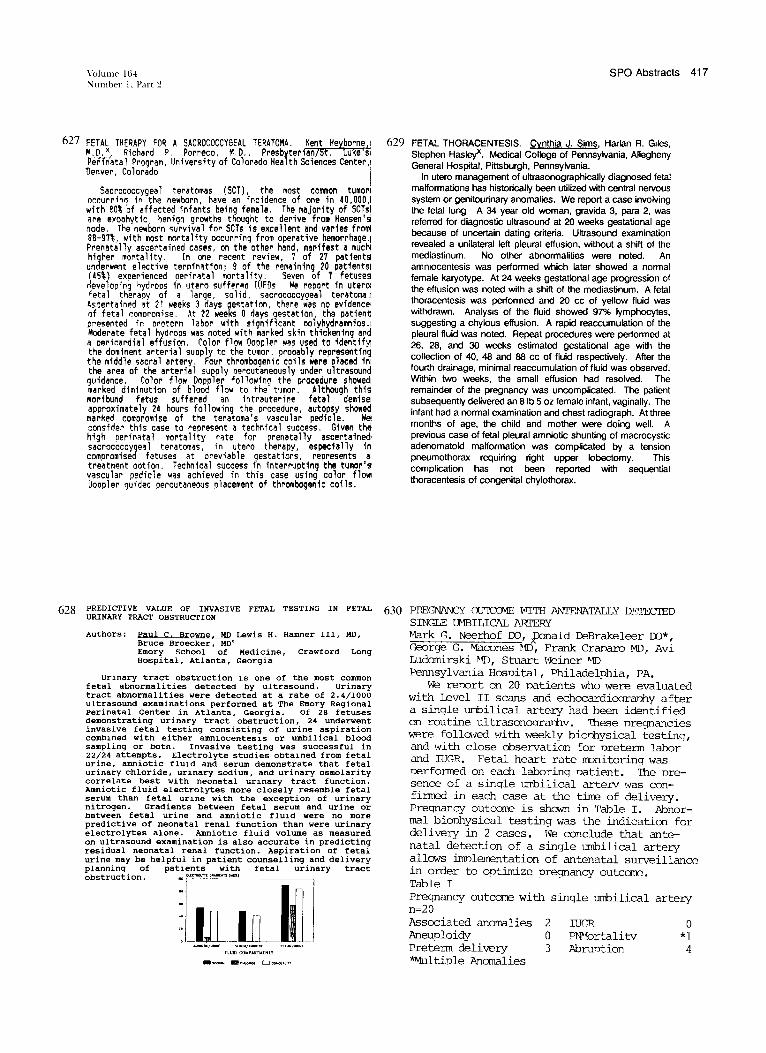
PP-EGNANCY 0U'tC01E WITH ANTENATALLY DETECIED SINGLE Ul'1BILICAL AmERY ~1ark G. Neerhof 00, Ibnald DeBrakeleer 00*, ('.,eorge G. ~cones riD, Frank Cranaro MD, Avi Ludomirski ~1]), Stuart lveiner '1])
Pennsylvania Hosuital, Philadelohia, PA. we renort on 20 patients who w-ere evaluated
with Level II scans and echocardioarauhy after a single uMbilical artery had been identified on routine ultrasonoaranhv. ~ese nreqnancies were folla·,ed 1·lith weekly bio'lhysical testing, and lvith close observation for nreterm labor and IUGR. Fetal heart rate IlDnitorinq lvas nerformed on each laboring natient. The oresence of a single unbilical arterv lvas confirmed in each case at the time of delivery. Pregnancy outcane is shawn in Table I. Abnormal biophysical testing was the indication for delivery in 2 cases. l~e conclude that antenatal detection of a single unbilical artery allows imnlementation of antenatal surveillance in order to optimize pregnancy outcomP.. Table I Pregnancy outcome with single umbilical artery n=20 Associated anomalies 2 Aneuploidy 0 Preterm delivery 3 ~ltiple Anomalies
IUGR PNI''Jortali tv Abruntion
o *1
4