Embed Size (px)
Citation preview
PBMWRAg M SURgE~Ar U$G&2 7%
(av flEy j7Ao'A, ____________top
DWTRUMN3X hTATIENTf
I MM~tC DTICIusrmCllorl LECTEf
AV AUItLffYJI tZ5WDEiT -AVAIL AMDM~ WSPAL
DATE Act3&iK*4K
DWMrMuVnTM VAJOF
86~ OAT% RET"IJnE
i~~~Rau oftT alRCE No,~ DinCO
DTIC79~~w7A DOCR MENTM M=OC4 A SUTM TOIOOII MA'' 3 DS(O
76A DUM¶ar0.OTIC SI
AD
Report *Ndver 2
Pathogenesis of Cell Injury byRtkettstia cohorii
Anlual Swmry Report
David 14. walker, M.D.
pay 17. 1985
Supported by
U.S. Army MedIcSl Research and OevelOtSfntt C.OMA
Fort getrick, Frederick, Maryl nd 21701
Contrict No. On 17-83-C-3122
University of North Carolilna at Chapel t111
chapel Sill, ,Nrth Carolina 27,14
Approved for public Teloaee diuttibutino unlited
The findings in this rept e nt be be construed as an official
Departwlfnt of the Army position unless so designated by other witiiled
documents.
Ire&d tA
REPORT DOCUMENTATION PAGEI& ASOkeST 35CURITI CLASSWICAlIOtl A 11L ASSTRICTIVUI WARN111"
___________________________________ Approved for public-release; distribution
SIP ".CLAMPJC^TIohON00 m 1ADMOSNG COUL8, unlimited
0011POMI 04ANI1TOPt 5591005? NOUM55A11411 MONIONI40 O 1AZATION 114905? "U'1044 P"111
Of NANO OP PnopooNMISG ONGA.IZTION OePSCI SYMOOL 71L PGAUS OP QONITORING ONGA#401ZATI105tknivereity of North Carolina ~"04=64)at Chapel Hill
Chapel Hill, North Carolina 27514 S ONE IIiT~TCTOS INUI
dP!t7rmy Med. Res.fl~,.lllfl6
&L A*005GM 40C0 . SUN 40 ZIP CO-ft $41. SOURCI 0f PU0401"a "Of.
Fort Detrick, Frederick, M'aryland 21701 INI1N. N.N
* i IT ME S*wIfy CUNtuio~is
tahngesnoo.if of roll Tn~sjrx hx Rrnngi"* i2PlSONAU A"'14N
h.TVTI 09 505 11sa. 13 TiNSve9 560s 4, CIAT 09 50 ? GIT (0, Me, *iI. 0"0 &04 COUNT
interim ýannual suminar mlow 84/04/11 To L.= ' WV1W IS SU9I.1INSNAR? NOTATION
,COSATIc~ools I&. sUeJECT T101S~ #cNlo on 09" V femn-V wed P 60af byS 66941*%o
'li~ ONOO lUGS I rickettsia, rickattuisi disease, boutonneuse fever,Rickettsis conorii, ticks, pathology
¶5. ATMACT WxitoIme. m.W of fteMoo di '.UftII, Wv "-.4w##
This work waa undertrken to determine the pathogenic mechanism by which !.Sc3.ttasiconorii causesa disease. R. coriorii, an organism that has been neglected in spite of its,ordesre.I diatribution enZd pathogenic qualities, was studied in human subjects, animalmodels, and in vitro. The purpose of the work is to elucidate the pathology of boutonneusfever and the pa-thoijenic mechanisms which eight be blocked therapeutically or prophylac-tically. Hu~na tissues were investigated by light microscopy, hiatochemistry, immuno-fluorescence, anid electron microscopy. In vitro models of cell injury by R. conoriiincluded the plaque modi~l and cell culture -rele'ase of lactate dihydrogenase.
Of biopsies of lesions compatible with tache noires from 22 patients in Sicily, 16have been documented a. Br, 1 was shown not t~o hav~ernd 5 haive incomplete data atpresenit. Evaluation of the documtented cases semiquantitatively for presefrce and severityof specific pathologic features yielded the followingi cutwleoua necrosis vsa presentin 10 of 15 evaulatable taches noires; vesculitim was severe or moderate in all 16;
34D OaaN$ SuT1O./AVAi LAGILI?'V0 XO TINACt al. ASGONACT ISCuUItT CLAS11101CATION
UNC.AIIIAJN.IIT5 0SAPA As ? 0 *TIC Umms 0
"k) NA^110 Of 0GSPONSI5LI 11NO,'ilI0AL mf ?S,.SPsomS NU'se Wiliof SeAG111094b A Me C.,
0o VCo~m 1473,83 APR 46I.ON 09 1i~ PAN 7 to *SOLO?%.
SSCUNIT', CLANSIV 1CAIO*4 00 Twil 9*06
- -----.--- * ----. *- - l-- *Pit-
AI
i CUOiTY CI I00 TWOI PASI II
thrombosis was severe in only 1, moderate in only 1, mild in 4, and absent in 10; dermoledema was moderate in 12, and mild in 4. The predominant leukocytes were lymphocytesand macrophages; immunofluorescent Rickettsia conorit were demonstrated in 12.
These results indicate that vascular injury by rickettsiae is the major lesionand that dermal edema is the important result. Thrombosis was generally absent or onlyfocal and mild.
Seven t~nsecutive Sicilian patients with boutonneuse fever who consented to liver
biopsy had hepatic lesions. This suggests that R. conorii is frequently viscerotropicand in patients with particular risk factors poses a 8erious threat. Clinicoepidemiologicstudies with European collaborators depict boutonneuse fever as geographically widelydistributed and at times quite severe. The problem of developing a good animal modelfor boutonneuse fever has been solved only for R. conori_ hepatitis in which our studiesof the mouse model have progressed. We may conclude that the pathogenic mechanisms andpathophysiology of R. conorii infection are being defined at the tissue level and thatthe cellular level Ts our current goal.
UCWMUr5'V CL"t.adOP'CA? O ?wOM PASQ
e;
5
AD
Report Number 2
Pathogenesis of Cell Injury byRickettsia conor i
Annual Summary Report
David H. Walker, M.D.
May 17, 1985
Supported byU.S. Army Medical Research and Development Command
Fort Detrick, Frederick, Maryland 21701
Contract No. DAM) 17-83-C-3122
University of North Carolina at Chapel HillChapel Hill, North Carolina 27514
The findings In this report are not be be construed as an officialDepartment of the Amy position unless so designated by other authorizeddocuments.
'A
i6
Summary
This work was undertaken to determine the pathogenic mechanism by whichRickettsia conoril causes disease. R. conorli, an organism that has beenneglected in spite of its widespreaddldstrlution and pathogenic qualities,was studied in human subjects, animal models, and in vitro. The purpose ofthe work Is to elucidate the pathology of boutonneuse Teve r and the patho-genic mechanisms which might be blocked therapeutically or prophylactically.Human tissues were investigated by light microscopy, histochemistry, immuno-fluorescence, and electron microscopy. In vitro models of cell injury by R.conorii included the plaque model and cefT-c-f-ure release of lactatedehydrog enase.
Of biopsies of lesions compatible with tache noires from 22 patients inSicily, 16 have been documented as BF, 1 was show not to have BF, and 5have incomplete data at present. Evaluation of the documented cases semi-quantitatively for presence and severity of specific pathologic featuresyielded the following: cutaneous necrosis was present in 10 of 15evaluatable taches noires; vasculitis was severe or nwderate in all 16;thrombosis was severe In only 1, moderate in only 1, m- ]d In 4, and absentIn 10; dermal edema was moderate in 12, and mild in 4. The predominantleukocytes were lymphocytes and macrophages; immunofluortscent Rickettsiaconor1i were demonstrated in 12.
, hese results indicate that vascular injury by rickel.tsiae is the majorlesion and that dermal edema is the important result. Thrr,nbosis wasgeneral ly absent or only focal and mild.
Seven consecutive Sicilian patients witt: boutonneuse fever whoconsented to liver biopsy had hepatlc lesions. This suggests that R.conorli is frequently viscerotropic and in patients with particular-riskfactors poses a serious threat. Clinicoepldemiologic studies with Europeancollaborators depict boutonneuse fever as geographically widely distributedand at times quite severe. The problem of developing a good animal modelfor boutonneuse fever has been solved only for, R. conori1 hepatitis in whichour studies of the mouse model have progressed.- We may conclLde that thepathogenic mechanisms and pathophysiology of R. conoril infection are beingdefined at the tissue level and that the ce ul arl -e'vel is our' current goal.
4,4
" ", ' "• ,'. , , •" ¾a,; .. • "
¼t
S ' '7
Foreword
For the protection of human subjects, the investigator has adhered topolicies of applicable Federal Law 45CFR46.
'In conducting the research described in this report, the Investigatoradhered to the "Guide for the Care and Use of Laboratory Animals," preparedby the Committee on Care and Use of Laboratory Animals of the Institute ofLaboratory Animal Resources, National Research Council (DHEW Publication No.(NIH) 78-23, Revised 1978.
,.'-'V
4' ?
.. I;*
8 -
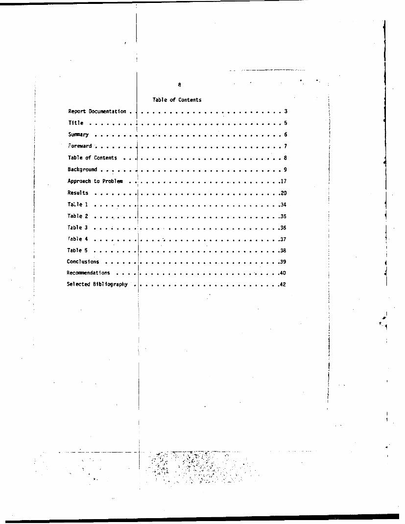
Table of Contents
Report Documentation .......... ........................... 3
Title .............. ............... .................. 5
Summary ................ ................................ 6
Foreward. ............. ............................... 7
Table of Contents ............ ............................ 8
Background ............................... 9
Approach to Problem ......... ............................ 17
Results . .............................................. 20
TaLle 1 ................. ........................... 34
Table Z ....... . ......... ............................. 35
Table 3 ... .......... ...... . . . . . . . ...... ... ...... 36
'rable 4 ................................................. 37
Table 5 .............. ................................. 38
Conclusions ............................................. 39
Recommendations ............ ....................... ..... 40
Selected Bibliography.. ......... .......................... 42
O~1
9
Statement of Problem
Spotted fever group rickettsiae including Rickettsia conorli, R.sibirica, and R. akari are important potential causes of mlitatTiry heilthproblems. In ordrto meet the challenges of these diseases to the health ofgroups of soldiers who enter zoonotic areas, methods of effective prevention,improved diagnosis, and optimal treatment are required. Development of aneffect've vaccine offers the best hope for prevention uf boutonneuse fever andother spotted fever group rickettsioses. No 2ffective vaccine exists for anyof these rickettsial diseases. Because most effective vaccines forprokaryotic organisms rely upon interdiction of the specific pathogenicmechanism of the organism, e.g., diphtheria and tetanus, it is important toelucidate the pathogenic mechanism of cell injury by R. conorli. The failureof killed rickettsial and bacterial vaccines, e.g., Rocky Mountain spottedfever, typhoid fever, and cholera, may be a result of a lack of stimulation ofthe immune system to block crucial pathogenic steps. The goal of thisresearch contract is to determine the pathogenic mechanism for R. conoril.Laboratory research on hypothetical rickettsial pathogenic effects must ecompared with observations on the -human disease in order to assure as well aspossible the relevance and reality of working models of the host-parasiteinteraction. The problems of lack of information on the pathology ofboutonneuse fever, the human ultrastructural lesions for any rickettsiosis,and the composition of the immune and inflammatory cell populations actuallypresent in foci of rickettsial infection in humans are addressed in thisresearch project. Diagnosis of boutonneuse fever, North Asian tick typhus,and rickettsialpox is an unsure affair with considerable room for error.Misdiagnosis and delayed diagnosis result in prolonged illness, need for morecare often including nursing and hospitalization and failure to instituteepidemiologic preventive illness. Yet, clinical features are variable and donot always lead to a timely correct diagnosis. There has been no rapid, acutelaboratory diagnostic method. Serologic diagnosis is a retrospective toolemployed during convalsescence or in the late stage of the illness. There arefew facilities in the world for isolation of R. conoril, and the laboratoryprocedure for isolation is both cumbersome anc long. A-diagnostic test thatcan be applied during the acute stage of illness is an expected spinoff ofthis research project.
Background
Rickettsial diseases occur over a wide geographic distribution, arefirmly entrenched ecologically, and pose an important threat to both militaryand public health.
Members of the genus Rickettsia are classified into three groups on thev basis of shared group antigens: spotted fever group, typhus group, and scrub
typhus group. All are obligate Intracellular bacteria which spend at least aportion of their life in arthropod hosts such as ticks, mites, fleas, or lice.They all affect man in a similar fashion with hematogenous spread and infec-tion of vascular endothelium producing increased vascular permeability andvasculitis In multiple organ systems. These rickettsiae include the etiologicagents of diseases that have been documented as major military healthproblems. Rickettsia prowazekli has affected the outcome of numerous militarycampaigns for centuries. R. ttsotsugamushi was a severe problem in Asia andthe western Pacific theaters during Wor d War H1 and infected soldiers in theViet Nam War. These rickettsiae have continied to attra't research support.Although R. conorli has received far less attention, it too has been docu-
-
10
mented as an important cause of illness among troops in South Africa. R.conorii is a member of the spotted fever group of rickettsiae along witf-otherhumanp athogens including R. rickettsli (Rocky Mountain spotted fever), R.akari (rickettsialpox), R.-sibirrca (North Asian tick typhus), and R.
tralis (Queensland tick typhus). Isolates of spotted fever group"rickettsiae from the Mediterranean basin, where the disease Is known asbcutonneuse fever, East Africa (Kenya tick typhus), South Afriri (SouthAfrican tick typhus), and the Indian subcontinent (Indian tick typhus), wereall shown to be members of the same species, R. conorli, by the mouse toxinneutralization test. Data presented by Myers and Wisseman on DNA hybridiza-tions among the spotted fever group rickettsiae have documented close rela-tionships among various strains of R. conorlil including rickettsiae associatedwith the severe disease occurring in Isra-el nd . rickettsii. Many of thesehybridizations were in the range of 90-100% homology.
Infection of man with various strains of R. conoril occurs in a wide-spread geographic distribution in the Old World with well-documented diseasein the Mediterranean basin, Africa, and the Middle East from Israel to India.In the Mediterranean basin, the disease is endemic in Portugal, Spain,southern France, Italy, Greece, Romania, Turkey, Morocco, Algeria, Tunisia,Libya,, and Egypt as well as in the margins of the Black Sea and the Caspianbasin. More recently it has been reported from South Africa, Kenya, India,Pakistan, Togo, Ethiopia. Cameroun, and Israel.
In the majority of the areas where the disease is endemic, it occurs assporadic cases during the summer months with little variation in the annualnumbers of cases reported. Scafidi notes that there were 107 cases in IsraelIn 1974, around 30 annual cases in Tunisia from 1961-1975, and 20 annual casesin Marseille from 1925-1930. He and Bourgeade et al, however, point out thatthese numbers do not reflect the reality since-the great majority of patientsare treated at home and are not reported. This is also an explanation for thescarcity of information about the prevalence of the disease.
The low endemicity that prevails in the majority of the affected areashas changed significantly in Italy where, since 1975, there was a sharpincrease in the incidence of the disease. Indeed, frcm an average of lessthan 10 cases per year up to 1972 the number of cases in Sicily increasedprogressively to reach 219 cases in 1979. Similar increases were observed inother regions of Italy as Liguria, Sardinia, and Lazzio; in this last men-.tioned region that includes the city of Rome, there were 369 cases reported in1979 . Besides in Rome, the disease has also been reported in suburban and.urban Marseille, and there are data that It is also increasing in Spain andPortugal. A large number of reports of boutonneuse fever have been publishedrecently in Spain. Many cases are seen in southern France around Marseilleevery year.
The causes for such a rapid increase in the incidence of boutonneusefever in Italy are not apparent. The Italians have suggested several possibleexplanations: 1) increase in the vector tick population, 2) introduction ofnew vectors, and 3) changes in the ecosystem. There have been some veryinteresting observations on the isle of Ustica where, after the recent intro-duction of wild rabbits, there was An explosive proliferation of Hexcavatum, a tick that had rarely been found In the island previously. Gilot,et al also mention the possibility of adaptation of certain species of ticks,parasites of wild animals, to human dwellings and the potential consequencesof the transmission of boutonneuse fever.
What is happening in Italy, France and Spain may occur in other regions.Weyer, reviewing the subject of rickattsioses in 1978, said, "Despite thegreat successes in control, none of the rickettsioses pathogenic for man have
)"
11r'
been eradicated. Therefore, it is necessary to preserve the knowledge aboutthese once devastating and important diseases because the present situationcould change suddenly.0
Indeed, recent data have demonstrated that several different species ofticks harbor R. conorli not only in the known, endemic ar^.as but also Inregions whereithe uman disease is not recognized including Pakistan, Armenia,Thailand, areas of France, Czechoslovakia, Austria, and Germany.
Boutonneuse fever is transmitted to man from ticks, most frequently byRhipiceealus sanguineus. Infected ticks transmit the disease through theirinfected salivary secretions euring the bite; exceptionhally the agents mayinvade the human host from infectious tick material through abrasions in theskin or through the conjunctivae. There are references that report thedisea3e being acquired by persons who rubbed their eyes after deticking dogsand, in fact, the principal investigator has observed just such a case. Theagent appears innocuous to the tick which also serves as reservoir for R.conoril which is transmitted transovarially in ticks. Small wild manuafs arethe source of blood meals for immature forms of R. sanuiLneus. Dogs, and onoccasion man, are the source of blood meals for the adult stage. Thefollowing species of ticks, besides the common vector Rhipicephalus sanguineushave been reported to harbor R. conoril: Ixodes ricinus, _.
Dermacentor marginatus, and D. reti-cuatus n--rance; Haemaphysa Is Ieachii,AiTlomma hebraeum, Rhipiceha usappendiculatus, R. evertsi, and HTalommamarginatus ,u-p-edes in South Africa; Amnblyoma var ega-a nd Hy t'aalbiparamatu, in Kenya; Ixodes granulatus in Malaysia; Rhipicephalus simus,Amblyomma variegatum, A. co--aerens, and A. gema in Ethiopia; andRhfpicepha us bursa, Hao arma iarglnatumý, H. usitanicum, and Haemaphysalis
ata In Sicily.Moreover, seroiogical tests in wild and domestic animalshnve shown that antibodies against R. conorii are present in several speciesin many regions, some of them far aw-ay-fr-m--I'e known endemic areas. InSicily, 20% of dogs hlarbor R. sanqguneus and 29-71% of themhave antibodies toR. conorii identified by indirect immunofluorescence assay. Serologic testsWFave identified antibodies against R. conorli in large numbers of healthypersons: in Africa, 13% of sera contai•ed antibodies in an investigation inCameroun and similar results were reported from Niger, Zaire ard CentralAfrican Republic; in Greece 16% of 560 sera from healty persons were positive;data from France indicate that positive serology in healthy persons has beenobserved in Caen, Nantes, and Lyon. In one endemic area of Sicily 19.3% ofhealthy subjects had positive immunofluorescence assay for anti-R. conorliantibodies. Not all of these studies employed the same serological tests, andthere is variation in specificity among different tests. Some, however, usedspecific immunofluorescence techniques.
All the data above presented confirm the suggestion of Weyer that thestage is set for an increase in the frmquency of boutonneuse fever and thatthis may occur in many different areas of the world.
Recently there have been reports of cases of boutonneuse fever in Germanand Swiss tourists who had returned from endemic areas and even of cases inAmerican tourists returning from Africa. Interestingly, a tick was found onone of these patients that might, if circumstances had been favorable, havebecome established in an American ecological niche. Cases have also beenreported in persons living in Paris and other parts of Europe that are notnear the Mediterranean Sea.
Human illness caused by R. conorli infection is usually an incapacitatingfebrile exanthem. Death has Seen reported more frequently in recent years,and some strains of R. canoril possess the capability of producing severedisease requiring hospita-ization and critical medical and nursing care. The
::4
12
disease usually resolves spontaneously in one or two weeks, ti•is pcr•od beingreduced by appropriate antibiotic therapy which may be given at home. It isnecessary to emphasize that even when mild the I l ness is incapacitating andin a minority of cases cas, be severe or even fatal; moreover, in certainregions, as apparently is the case in Israel, South Africa, France, and Spain,it can assume a more severe course similar to the picture of Rocky Mountainspotted fever. Severe disease has been associated with G6PD deficiency,alcoholism, older age, ano diabetes. Men are slightly more frequentlyaffected than females, and the disease occurs at all ages being. however,uncokion in the very young end very old. Most of the patients repott contactwith dogs, ticks, or recent visit to endemic nreas; others are farmers orhunters. The incubation period varies from 7 to 14 days, but can be as shortas 4 or as long as 2.' days. In the majority of the cases the patientr'-enbers being bitten by a tick and irom 33% to 921 cf them have an eschar(tac Wire) at the site of the tick bite. Les. frequently they have acuteunfiatieiWaTonjunctivit's.
The disease begins with sudden increase in temperature to levels as highas 40*C; at the same time the pAtients complain of joint and muscle pain andviolent, persistent headache that is frequentl' retroorbital. There is also acongestion of the conjunctivae and mild lymphacenopathy. These manifestationscoincide with the appearance of the eschar. Four to five days after thebeginning of the fever the typical rash appears; it is first observed on thelimbs but rapidly expands to trunk and face with palms and soles also beinginvol ved. In some cases even the oral mucosij presents an exanthem. In thebeginning the rash appears as erythematous macules that rapidly change to amaculopapular pattern and eventually become nodular or button-like, as thename describes. The early lesions are light pink, but some of the older onesmay become darker or hemorrhagic. The rash occurs in successive bouts so thatlesions in different phases may be observed side by side.
Fever persists for 7-14 days, and during this period A6% of the patientsdevelop splenomegaly, 20% hepatomegaly, and some patients, signs of pulmonary.congestion. Diarrhea, constipation and vomiting may also occur. Neurologicalsigns of meningecl irritation as nuchal rigidity or Kernig's sig- as well asobtundation and even coma can be observed in a minority of the cases. Thesemore severe manifestations occur mainly in older or debilitated persons; theyare exceptional in children. Recovery is unevertful witheut any sequelae.Mortality is low. In a few cases, however, compicatons occur, they are rareand, as stated, tend tn occur in older debilitated Dersons. Scafidi et aldescribe cases of hypirtoxic, "dermatotifosa" and hemorrhagic disease•,t-helast form being assoc'ated with severe gastrointestinal or genital bleeding.Fatal gastrointestinal hemorrhage with ri, ettsial vascu!itis of the stomachhas been described. Scafidi et al describd cases vith atrial fibrillation,myocardial ischemia, and ren-alT-'lications. A series of French piblicationsdescribe "atypicx' rickettsiosis" with pericarditis, pleuritis, andpneumonitis. -...e ,f the cases, however, did not present with eschars and thefinal diagnosis was made by positive microagglutination tests according to themethod of Giroud, thus raising doubts concerning the diagnosis. In Israel,hc ver, there have been some very interesting cases of tick-bornerickettsiosis with severe renal insufficiency requiring dialysis; in thesecases, there are questions about tne exact classification of the etiologicagent that did not conform exactly with the antigenic stricture of R. conorli.More recently severe and fatal cases have been described tn South Africa,Spain, and France.
The clinical feature that is most significant diagnostically in R.conoril infection is the tache no'-e which develops at the site of tic-bite in
4 mmm?!
13
dpproxImately SO% of cases. The tacho noire, or black spot. Is azone ofdermal and epidermal necrosis whi~my appear prior to onset of fevor andrash. Conor and Burchl did not describe eschars in tile original report ofhuman L cono'.1i infection in 1910. Tache nor is al French term and wasIntrodc.W-4TRTI2 by Pieri to refer'to the tkcxbite sit. eschao inboutonneuse fever. Thereafter, the toem tat.ne noi re seems to. have been usedcontinuously. Sim~lar eshars, are frituinTy-17-o~rvid in scrub typhus .
tsusumsi) North Asian tick typhus (R. sibiricaj, rickettsla! pox, n.a ari), and Qensl and tick typhus (A. oustrirTs .1schars are rarely-Ysi*ved in Rocky Mountain spotted favor andd not occur in typhus teveranmurine typhus. Thus, @%chars are soerw only in ficko¶tsioses transmitted byinoculation of infected salivery secretions by ticks and sites and are notobserved in rickettsioses transmitted by scrrotching rlckettsia-coataininglouse or flea feces into the skin. Patients who develop aoutaotnuse fevarafter accidental introduction of infected ticK constItutents Into theconjunctiva do not have escilars, but manifest conjunctivitis at the portal 0f
"Oryur laboratory has described 'the clinical featurs brightfield micro-scopic pathology, and distribution of L. rickottili in echars which occurredin two fatal cases of Rocky Mcunttain i~otted feT*ver examined at autopsy. Theseetchars consisted of a 8 x 10 an oval region of necrotic epidermis and under-lying dermis. The necrotic zone was surrounded by a zone of blood veselsthat ware injured with extensive th. ~sis and intramural and perivascularmononuclear Inflwimatory cells. lamnohistochomicallexamination revealed veryl arge, quantities of L. rickettsii iIn the endothel ium and , scul ar wallI ofthese blood vessels.-
There is some degree of c*6g. iversy about the role -,f constituents oftick salivary secretions such as erzymes associated with tickbite in thepathogenesis of the tache noire. Experimental studies suggest that the doseof Inoculum of r~ckef~ert~ than the tickbite itself is crucial.Inoculation of a large drs-. of R. rickottsii a generally nonescharogenic"'ý,Irttsls. into human sf.in by ir~~i~l produco"s eschars. Inocula-tion of R. conorli Into the skin of syphilitic subjects as pyrotherapy pro-duced ta-cliet -noires prf.portional to the quantity of rickettsia* injected.E-ien nns-char-ogenic A.. mooseri produces #%chars In the skin of guinea pigsinjected intradermlIV lb~y y~rine and needle with a laMrr doSe of rickettsia*.Not allI monkeys inocul ated with R. t~usugamshi devel c tit aeshar at theinjection site; some develop only $ pule$ whirch do mat -^.j#.9o epidersalnecrosis and ulceration. Rsibits inoculated intracutaneoisly with a high doseof R. Libirica developed an eschar; rabbits Inoculated with 1% of the escharo-genTc dos* do v~oped only cutaneous orythoem without necrosis or formation ofa dark crust. Thus, the tache noire appears to be ant ac.essiblo leson thatcontains the pathogenic QWsnie R. conorii and' the i.mmne and inflam.."mtory mechanisms of the host that h~aZ t~oI;_iiTnq.,b
tlypothetical rickettsial pa'.hogenic mechanisms jinclude both those thatare host-mediated and rickettsia-mediated. Kost-*.adlated mechanisms of injurywhich have been oroposed include imumnopathology. blood coagulation, andInflammtion. Rickettsia-inediated mechanisms might Include endotoxin, exo-toxin, wnzymet that destroy host components, met abollic coieptition for thehost's intracellular substrates, ATP parasitism, and~ host cell mewbrane injuryon rickottsial penetration Into and/or release fr~imuthe target cell.
Experimental evidiewci indicates that host-miediated pathogenic mechanismssuch as iinanopathnloqy. Shwartzman phenomenoni-like blood coagulation, andinflanution are not the primary mechanisms of Injuey In i... :tion by R.rickettsii. Localized effects of kal 1ikruin are probably events seonaary to
the Wriary pathogenic mechanism(s). Occlusive vascular thrombosis ft infre-quent and has not been demionstratcd as a primary pathogenic mechanism.
Dinon the hypothetical rickettsia-mediated mechanisms of injury.currently no toxin of L. rickettsii has been identified, and there Is evidenceagainst the existence of -a toin as an important pathogenic mechanism. Theconfusion regarding this hypothesis has originated in tho so-cal led moust'toxin phenomeno and in erroneous analogies drawni between endotnxin andrictettsits. Mouse toxicity depends on viable. metabolically active
rickettsioe and is prevented by heating (60%t for AO minutes), exposure todilute formalin, rickettsial starvation. ultraviolet irradiation, soecificanti-rickettsial antiserum neutralization, and a beita-llpoproteln present insew normal humlan sera. The pathogenesis of this phenomienon may be relatad tothe pathophysiology of the rickettsia-h~ost cell Interaction, e.g., massiverickettsial penetration of endotholilm. Rickettsiae, of both the typhus andspotted faver groups have been shown to contain 1 ipopolysaccharides. owever,the endotoxin activity in bioassays including the Shwartzman phenomenon andLimulvs assay was considerably less than that of potent bacterial endotoxins.Moreover, study of the adrenal In fatal RMSF has not demwonitrazed the patholo-gic lesions expectee of endotoxln-aediated pathogenesis. Further evidence
against the hypothesis of ,-ickottsial toxin has boen demonstrated in thet;-ýuo model. Thus, the evidence for a rickettsial toxin of pathogenic lopor-tnce Is quite meager.
The plaque model has been established as a useful tool for Investigationef pa-hogenic mechanisms of cellI injury by R. rickettsi 1. !Nocul ation ofconfl u~nt monolaeyers of primary chick uubryi~cif1f-sder-ed from 12-day oldspecifi. ;athogen-free, antibiotic-free, embryonated hen's eggs with a definedquantity 0~ Q, rickettsil results In , predictable course of infection andpathologic aTterat~ios n vitro. Eac~h Infectious unit under agoars* overlayproduces contiguouis contr~ruiT sprrad of intracel lular Infection and injuryto the host cellI monol ayer. This m~oul produces a grossly visible plaque onday 5 after inoculation when a second overlay of agarose-'ontaininq thesuprovital dye neutral red is added. The plaque provid't. a temporal andspatial cross-section of the rickettsia-host cellI int-oraction includingrickettsia] penetration, proliferation and riiat- and host cellcytopathologic alterations and necrosis. Morphometric analysis of the plaqueand surrounding infected and uninfected cells has been performed maintainingthe topographic rel ationships of t~he cellst as a monol ayer. The results haveshown the association of Intensity of Infection and cytopathology at themicroscopic and ultrastructural levels. There is a statistically highlysignificant relationship between the Intensity of Infection as measured by thequantity of intracellular rickettsila And the presence of cellular injury asjudged by cytopatholo-4y and necrosis. This relationship Is valid indepen-dently of the apparent duration of infection. That Is to say, more heavilyparasitized host cells are more likely to exhibit pathologic alterations, evenif they are located at the margin of the plaque, than those cells whichcontain fewer rictottsiae and are nearer to the center of the plaque. Thisstudy also confirms the observation of Silverman and Wisseman that the typicalcytopatholoqic Change in chick ebryo cellIs infected with R. rickettsii isdistinct dfiation of the cisternat of e~ooplasmic retictilti Thiultrastructural finding Is characteristic of the response of an injured ceillto the influx of water. The utilization of the t~chnique of maintarining thetopograohy of the monolayer Intact enaolod us to determine th&t the isninf--tedcellIs of the monolayer even within 1 No of the intensely infected marginalzone of the plaque were normal 4y ultrestructural and suprevital 0*e stainingCriteria even though they were exposed to the sare milieu of extrecellular
nut riti onel factors , nonspec if ic toxic products of metabol ism and substances
spread of ric'ittsiae and yet al lows for diffusion of macromolecules, demon-strtedtha ee Iinjury wa mtdt h eeheavi ly parasitized eIsan
that there was no toxic effect on uninfected cells, even those Immediatelyadjacent. This Is strong evidence that L. rickettsil does not elaborate anextracel lular toxin which effects chick emFY-o-c-ells. Further studies in ourlaboratory have extended this observation and conclusion to Vera cells whichare of primate origin and to human umbilical vein endothelial cells.
Another strong inJication that R. rickettsii does not produce an impor-tant toxin resulted from observations utilizing parabiotic chambers.Specially designed flasks contained coverslips with monolayers of cells withfluid overlay in separate chambers which were separated by an 0.22 ummillipore filter. R. rickettsll was inoculated into one chamber of se-veralflasks; other contril flasks -weret observed without rickettsiae In eitIrchamber. Inoculated monolayers developed cytopathic effect associated withheavy rickettsial infection. On the other hand, the cellIs In the oppositechamber remained viable with the sawe appearance as monolayers of unmanipu-Ilated parabiotic chambers. No toxic, macr omlcules injured the side of thechamber which was protected from rickettsial infection by the 0.22 umi filter.The filter offered no barrier to the free passage of molecules between the
* infected and uninfected chambers. Thus, in an experimental system In whichrickettsiae injured infected host cell's, we demonstrated no effect of putativetoxin, which would have been in equal concentration in the extracellular fluid
* ~of both chaWvdors if It were present.Examination of the hypothesis of competition for metabolic substrates
has also failed to produce evidence to support it as a pathogenic mechanismin plaque nodal experiments with supplemental glutamate and glutamine.Although rickettsiae are capable of generating ATP for penetration of hostcellIs by oxidation of glutamate, exogenous ATP from the host cell Iisutilized for biosynthesis of proteins and lipids by rickettsiae. Thisenergy parasitism is mediated by an efficient rickettsial ATP/ADP transporbsystem., No experiment has yet been designed and executed to test the hypo-thesis of energy parasitism as a pathogenic mechanism.
Experiments reported principally by Wink ler and co-worters suggest thatrickettsia) penetration-associated phospholipase activity injure-% the hostcell membrane. The work of Winkler and associates on hemolysis by viable R.prrowazekii has led to an understanding of the rickettsia-host cell .memranieT~e-ra-c on wtich probably forms the basis of penetration and a-mechanism of
cell injury. Rickettsia] hemolysis may be divided into two steps,adsorption and lysis. Hemolysis is inhibited by cyanide (1 mM KCM, ininhibitor of the electron transport system), low temperature (0*C), andstarvation of R. prwzei for glutamate. Ghosts of erythrocytes exposedto Amhotericin 5or dgitonin, comounds which bind to the cholesterol-containing receptor sites in the erythrocytic membrane, are no longer ableto adsorb rickettsia*. Adsorption and hamolysis are Inhibited by adeninenucleotides, ADP, ATP, arsenite, which is a Krebs cycle Inhibitor, and 2,4-dinitrophenol and m-cOIlora-phonylhydrazone. which are oxidativephosphorylation uncouplers. when rickettsiae are unable to generate ATP bymetabolism of glutamate because of cyanide or arsenita inhibition, added ATPrestores hmowlytic activity of the rickettsiae. ATP. however, does notrestore hemolytic activity inhibited by uncouplers. Fluoride (10 mm NaF)prevents henmolysis by inhibition of erythrocytic glycolysis without
affecting adsorption or rickettsial metabolism Recý.ntly, rikattsial
16
hinolysis has been shown to be Associated with phospholipase A activity,which resulted in hydrolysis of fatty acids from the glycerophospholipids ofthe red blood cell memrane. Inhibition of either adsorption or lysis alsoprevented the release of free fatty acids.
Penetration by rickettsiae has many correlates' with rickettsialhemolysis. Inactivation of R. tsutsugamushi by heat (56*C for, 5 minutes),exposure to ultraviolet irradiation, or Incubation with 0.1% formalin pre-vents penetration into host cells. Penetration of 1. cells by R. prwzeicomprises two steps, adherence and interniali1zatio~n, and requires activeparticipation by both the rickettsia and the host cell. Treatmnt Ofrickettsiae with ultraviolet irradiation, 3% formaldehyde, or I NM KCNinhibited adherence to and internsai Ization Into L cellIs. The0 few inacti-voted rickettsiae, found associated with L. cellIs wert mostly adherent ratherthan internal ized. Treatment of L cellIs with NaO (an .nhibitor of metabo-lism), N-ethylmaletimid@, or cytochalasin 8 inhibited Internalization ofrickettsiae. Similar studies of the entry of !. prwzei Into endothelialcells support the hypothesis of induced phagocyto-sTi-771iiulatic.. of R.prrowazekil onto L cells at large multiplicities of Infection induced lime-da~t~e-cytooxI city. This cytotoxic effect was associated with phospholipase,
A activity and hydrolysis of fatty acids from host cell phospholipids.Cytotoxicity and phospholipase were inhibited in a parallel manner by KCN,N-*thylmaleimide, NaF, and low temperature.
Furtt'.r studies in our laboratory of pathogenic mechanisms in theplaque model employed chemical agents, which have a sound theoretiial besisof Inhibiting rickettsial penett'ation either at the stop of adsorptiolt ofthe rickettsia to the host callI (AaphoterIcin 8 and digitonin) or at thestop of internalization associated with phospholipase A activity, hsve beendemonstrated to reduce plaque formation. Amphotericin 8 and digitonin havebeen reported to inhibit thoe attachment of R. prowatkj to ebratrcyticcell membrmnes oy binding to a cholesterol-repo the .memrane.DAphOteritln 8 was introduced In concentrationt. of 5 and 10 ug/mi to theoverlay after the establishment of Infected foci on day 4 after inoculationof R. rlckettsii. In order to maintain active levels of this drug which hasa doca-y of 01 per 24 hours, at 370C, Avehotericin S was re')leished insequential overlays on days S and S. On day 6 Amphotericin 8 caused plaquereduction of 42-45% at both concentrations. More plaques appeared on day 7with plaque reduction of 165-23%. A similar experiment with digitonin at thesame concentrations caused similar p~aqiac reduction (38-401). Plaque reduc-tion was not observed on day 7. 'then the levels of cholesterol receptor-binding drugs are maintained, plaque reduction can be deownstrated. Th"issuggests that inhi'bition of rickettsia] adsorption delays the cytopathiceffect of R. rickettsii In primary chick embryo cells.
Phente'rine Is a drug which has been shown to have phospholipase A2inhibitory activity. A dose response study with this drug was performed inthe plIaque modelI. Pla que r -duct Ion was demonst rated at all doses of phen-termine: 691 plaque reduction at 0.5 mg/mI; 54% at 0.1 mg/ml; 251 at 0.05mg/mI; and 321. at 0.CI mq/ml. These results demonstrate that phenterminereduces the cytopathic effect of R. rickettsli and s'ugtj'-t that phosphol i-pas* activity may be a pat hogen Ic mecha~nirs-m or R. -Icte-1iii. These dataextend and support the obse'vations of Winkler t~at `pho-s-p-oTpase activityis associated with hemolysis and immediate cytotoxicity of a large Inoculumof R. prwazekil.
Prrvvio_,.s eports have documented th~at R. c -onoril forna distinct planuelsimi 1ar to those of k. rickettsii iIn the plasque mod#T. McOade at al pro-duced distinct plaquets with R. conorli In chick embryo cellIs w tntTfirst
7
"17
overlay of medium 199 containinq 5% calf serum and 0.5% agarose and a latersecond overlay of medium 199, no calf serum, 0.U% agarose, and 0.01% neutralred. tike et &I studied the critical variables in the plaque assay systemfor ricketts-Taie-nd also showed that R. conoril (Malish strain) produceddistinct plaques in the standard chicf reryo monolayer with nutrient over-lay containing agarose. Thus, the plaque model offers an opportunity toexamine quantitatively and predictably the pathogenic mechanisms of R.conorii in an in vitro system that may be manipulated experimentally toexamine hypothesessuch as phospholipase-mediated injury.
Because one hypothetical explanation for the apparent rarity of severevisceral involvement in BF as compared with RMSF (encephalitis, hepatitis,pneumonitis) is lower temperature sensitivity of R. conor1i we are inter-ested in the effect., of temperature on the physiofoa apathogenlcity ofthe organism. Oaks and Osterman have investigated the effects of tempera-ture on the opti*2l growth of R. conorii. This species of rickettsia has anoptimal range foor growth in gaia- rrTated L cel ls of 32-380C withInhibited proi'feration at 400C. The low rate of proliferation at 400Cmight explain the minimal visceral involvement in febrile patients whosebody core temperature is about 38*C and may exceed 40"C. An unansweredquestion is the effect of temperature on the pathogenic mechanism of R.conorii.
Approach to the Problem
Many features of boutonneuse fever have been investigated to a far lessdegree than typhus fever and Rocky Mountain spotted fever. In particular,pathogenic mechanisms, immune mechanisms against R. conori, and the labora-tory diagnwois of boutonneuse fever have not been Investigated sufficiently.There are Avantages of studying human cases, animal models, and call cul-ture models of R. conorti infection.
The local iie-Thisi at the site of the tick bite, the eschar or tachenoire offers an excel lent opportunity to extend our knowledge of path5F €cm ehanisms, imuine mechanism, and lab•ratory diagnosis of. BF in humans. Incontrast to typhus and Rocky Mountain spotted fever In which the lesions,althouoh nurerous and widespread, are extremely focal, the tache noire issufficiently large and contains a large contiguous network of se -e"iiinjured blood vessels that will allow prodictable saimpling and qualitativeand quantitative analysis of rlckettsial Infnction, host cel I injury, andhost Infla tory and Immune cellular response. Thus, although thebrightfield microscopic lesions are better described in typhus fever, RockyMountain spotted fever and scrub typhus than In boutonneuse fever, thesereports are not quantitative, often do not demonstrate rickettsiae with theefficiency and specificity of Imunnidstochemical techniques, and do notevaluate the ultrastructure of the human lesions. Surgically excised, well-fixed eschars should allow these studies in'boutonneuse fever.
As yet no significant in vivo ultrastructural study of the human host-rickettsia) relationship h•is aiI reported. There are two major reasons:1) the infection In human skin Is extremely focal, in the exact center ofthe maculopapular rash of RMSF and typhus and, thus, is difficult to find byelectron microscopy; 2) intensely Infected visceral tissues from fitalcases of RMSF and typhus are not suitable for ultrastructurel Investigationbecause of postmortem autolysis that occurs prior to pe. . roance of thenecropsy. Surgical biopsy of the tache noire of BF should providew l I1-
preserved lesions containing intaniserconorI infection for ultrastruc-tural investigation A report of the- iatrastructural aspects of an eschar
.4I
18
In Rocky Mountain spotted fever described rickettsiae in the iesion.However, the published electron micrographs were of poor quality, and norickettsiae were identifiable in them. Correspondence with the authorsdirectly in an attempt to obtain copies of the original electron micrographsor the EM grids for examination personal ly has not been answered.
A sample of the tache noire is col lected by sterile skin biopsytechnique under local anesthesia after obtaining the palient's informedconsent. The specimen is divided Into three small 1 mW blocks and fixedfor electron microscopy by ismrsion In cold buffered glutaraldehyde-formaldehyde solution. The fixed specimen may be held in this solution forthe period of shipping from Italy to our laboratory. On arrival at theInfectious Pathogenesis Laboratory in the Department of Pathology of theUniversity of North Carolina, the specimen will-be postfixed in 1% osmiumtetroxide, dehydrated in graded alcohol concentrations, embedded In a mix-ture of Epon and Araldite, ultrathin sectioned on an ultramicrotome, andstained with uranyl acetate and lead citrate. Sections will be examined ona high resolution Zeits 10 A electron microscope. Other transmissit•n elec-tron microscopes and a high resolution scanning electron microscope are alsoavailable within the departmental ficilites should the need arise.
The remainder of the specimen is fixed in neutral buffered-4%fnrmaldehyde for routine histology, histochemistry, and immunohisto-chemistry. Fixed tissue wi Il be embedded in paraffin and a ribbon of serial3actions will be cut at 4 um thickness. Adjacent sections will be mounted'for staining by hematoxylin- eosin (H E) for routine evaluation of patho-logic lesions, by phosphotungstic acld-hematox~ylin (PTAH) for fibrinthromi, by Voerhoff-van Gleson technique (VV) for evaluation of integrityof vascular elastic tissue, by modltied Brown-Hopps (BH) technique forhistochemical demonstration of rickettslte, and by Gtemsa technique andm'tt'yl green pyronin (MGP) for identification of host immune and inflam-matory cells. Among these stains, PTAH and VV yield highly sensitiveresults, BH demonstrates intracellular rickettsiae but -vith less stnsi-tivity, consistency, and specificity then tmmunofluorescence, and Gimsa andNGP assist In identification of eosinophils, basophils, neutrophils, acti-vated lymphocytes, and plasma cells but le, ve a large portion of unidenti-flied munonuclear lymphocytes.
Adjacent sections from the ribbon are processed for iqmunofluorescentdemonstration of R. conorli. Sections are affixed onto clean glass slideswith nonautofluorescent Le tige Bond Fast Resin Glue to prevent them frombeing washed off the slide af*-r digestion with trypsin. Sections affixedto slides with glue are heated in an oven at 60.C for 1 hour, deparafflnizedin three changes of xylene for 10 minutes each, and rehydrated through .serial changes of ethanol it, concentrations of 1001, 95%, 701, 50M, and 3F'and finally In distilled water. Sections are then incubated in 0.1% trypsinwith 0.1% CaCl?, p14 7.8, at 37*C for 4 hours. The slides are washedthoroughly in disti lled water, washed for 30 minutes In phosphate-bufferedsaline, and reacted with the specific itunofluorescent system for R.conorli. We have used anti-SFG rIckettsial conjugate in the direct-lmmuno-fluorescence system and indirect immunofluorescence wAth guinea pig Iuneanti-R. conorit serum followed by anti-guinea pig imme-noglobulin conjugateto demonstrate structures which have the expected vascular location andcoccobacil I ary morpho ogy of rickettsiae.
An animal model of boutonneuse fvver is needed for examination ofimmnity Including prospective evaluation of new vaccines, for Investigationof R. conorii pathogenic mechanisms and pharmacologic intervention withtheis pathogenic mechanisms in vivo. anl for study of organ systemic patho-
.4
19
physiology now that a picture of the visceral lesions Is emerging forboutonneuse fever. The distributions of lesions and P.. conoril need to bedetermined for various strains of R. conorli in mice,-gif iinapigs, and otherspecies of mammals. Studies of exirf-_entaT-animals in our laboratory andothers have shown some of the qualitative ultrastructural aspects of therickettsia-host interaction. Our ultrastructural analysis of rickettsialinfections demonstrated R. rickettsii in endothelium, vascular smoothmuscle, and phagocytes of nfected guinea pigs in three investigations:sal ine-hydration prolonged survival, anti lymphocyte serum immunosuppression,and tetracycline-treated rickettsia clearance.
Further studies of pathogenic mechanisms of R. conorit are performed inthe plaque model which we have exploited in investigat-ons of pathogenicmchanisms of tR. rickettsli. Aliquots of R. conoril stock are thawed anddiluted in sucrose phosphate buffer to con'ain-3M'-aque forming units(pfu) per ml. Confluent monolayers of Vero cells are inoculated with either0.1 ml of diluted rickettsial stock containing 50 pfu of R. conorli oruninfected diluent. Rickettsial plaque technique is per rU according tothe method of Wike and Burgdorfer and Wike et al. After 30 minutes forabsorption to occur and penetration to oenInl, It-e monolayer is overlaid with0.5% and agarose In mlnimun essential *ditum with 5% fetal bovine serum andincubated at 35*C. On day 4 after inoculation, 4 ml of second overlay with1% neutral rad is added, and the flasks are allowed to Incubate in the darkat 35*C. F'asks are examined daily for plaques afterwards with observationsof monolayers by inverted microscope and with collection of specimens forexamination by immunofluurescent and transmission electron microscopy.
The sides of the 25-sq cm Falcon flasks opposite the monolayers areremoved by cutting the plastic. Agarose gel overlays are gently removed byseparating the overlay from the sides of the flask with a sharp spatula edgeand allowing the gel to detach under the force of gravity. Exposed mono-layers are fixed in 70% ethanol for 20 minutes prior to direct immuno-fluorescent staining for R. conorli with a specific anti-R. conoril conju-gate. After incubation ofmonoTayers with conjugate for o minutes, theyare washed in phosphate-buffered saline for 30 minutes, washed in distilledwater, and mounted with 90% glycerol in phosphate-buffered saline (pH 9) andcover glass. Monolayers are oxamined on a Leitz Ortholux ultraviolet micro-scope equipped with the appropriate barrier, exciter, and edge filters forfluorescein I sothiocyanate fluoresccnce microscopy.
Monolayers with overlays removed as described for immuncfluorescenceare fixed by cot tring the cells with a solution of buffered 2.5%glutaraldehyde for 1 hour. Cells are maintained on the plastic surfacethroughout postfixation in osmium tetroxide, dehydr3ted through a gradedseries of ethanol and hydroxypropyl methacrylate solutions, and embedded inMollenhauer's Epon-araldite No. 2 followed by aclynerization In an oven at37*-45*C for 24 hours and then 60*C overnight. Embedded monolayers areseparated from the plastic flasks. At this point, rickettsial plaques maybe observed with the unaided eye as distinct clear zones surrounded by agrey-black carpet of cell s. Plaques and adjacent cel' s are cut out andrembedded In flat molds with the monolayer perpendicular to the plane ofsectioning. Ultrathin sections are cut on an LKB ultrýTicrotome using adiamond knife. Observation of the block during sectioning reveals the exactrelationship of the section to the plaque. Sections are stained with leadcitrate and uranyl acetate and examined on a Zeiss 1OA transmission electronmicroscope.
Plaque size, time of appearance and phase contrast morphology areobserved. The relationship of f. conorti to plaques is observed by inmwuno-
-i
20
fluorescence microscopy. The cytopathology of injured cells is describedIncluding state of rough endoplasmic reticulum, mitochondria, plasma mem-brane, and nucleus. For each plaque zone (center, margin, and periphery),the quantity of R. conorli in cytopathologic and cytologically normal cellswill be counted and subjected to statistical analysis for association ornon-association of cytopathology with intensity of intracellular infection.The cytology of uninfected cells adjacent to the plaque w.11 be examined.
Plaque model studies of penetration-associated pathogenic mechanismemploy the plaque model, Auphotericin 3 (5 and 10 ug/ml), and digitonlrf (5and 10 ug/ml). compounds which have been shown to bind to cholesterol-containing menibrane receptors, to block attachment of R. prowazekii toerythrocyte plasma membrane and to reduce poaque format7ioinbyR.r•ckettsiwhen incorporated into the agarose-nutrient medium overlay. The secondoverlay contains the same concentration of the chclesterol-receptor bindingdrug. Plaques are enumerated at the time of appearance of distinct plaquesIn untreated plaque assays of the same inoculum for statistical analysis.In a second set of similar eiperiments Amphotericin B and digitonin areintroduced only in the second overlay on day 4 after inoculation, at whichtime infected foci will be well established. Significant plaque reductionIn this experiment indicates blocking of a pathogenic mechanism, not justabortion of initial infection.
One hypothe.tis which can be tested in the plaque model is that thepaucity of signs and symptoms poirting to visceral involvement is due to alower threshold of temperature sensitivity of R. conorii. The inability toproduce pathogenic effects at temperatures great-er th-an 8% could explainthe relative lack of severity of BF when compared with RMSF despite the 91-94% relatedness of the etiologic agents. The plaquing efficiency of variousstrains of R. conorti and R. rickettsii are compared at 320C, 340C, 369C,38*C, and 4Oc71--CrTtion-in number of plaques formed, plaque size (areameasured morphometrically by computer assisted image analysis), and time ofonset are examined. These results reflect the effect of temperature onpathogenic effects.
The hypothasis of secretion of a potent extracellular toxin by R.conorii can be examined In an experimw-:1 utilizing parabiotic tissue-cultuteMniris. Parabiotic chambem contaiving cell monolayers are separated by afilter with 0.22 m pcre size. This f'Iter prevents the passage ofrickettsiae from o-u chamber to the adjacent chamber, but allows freepassage of macromo. -cules such as metabolites, putative toxic products, orenzymes. In some pairs of chambers, one chamber is inoculated with 10plaque-forming units of R. connrii. Other pairs of chambers are maintainedwith both chambers uninoZulated as co-tnols. On days 3, 5, and 7 postinocu-lation, trypan blue is added to selecteti pairs of chambers, and selectedchambers are examined by Immunofluorescence for R. conor1l. The degree ofcell injury in infected chambers, uninfected-ric-ttslai prducts exposedchambers, and control chambers is evaluated blindly by estimation of per-centage of cells failing to exclude trypan blue. Immunofluorescence for R.conorli confirms the limitation of infection to inoculated chambers andallows estimatlion of the percentage of the monolayer that is infected.
Results
The study of taches noires from patients with boutonneuse fever hasbeen performed in EVTIao-raiTn with physicians at the University ofPalermo. This collaboration has proven successful with opening of severalavenues for the continued investigation of the pathology, pathophysiology,
"-4
21
and clinical apoects of R. conorli infection in humans. The collaborativerelationships with Ors. iains~uetoringali, and others Is a valuable andprnd-tcttve resource for the ttudy of R. conorii and boutonneuse fever. Theyare very interested in the scientific questions related to the tache noire
boutonneuse fever, and R. conori . They have made great effort -to o-taincIi niral materi al. conduc cop-emt- e clinical patient fol lowup and convales-cent laboratory diagnosis confirmation and to establish a rickettsiologylaboratory. Ongoing ar&I future investigations of pathogenetic mechanisms,epidemiology, pathophyslology, diagnostic methods anu possibly preventivemeasures are mutually achievable goals. Considerable progress has been madetoward these goals. The clinical and professional resources available inSicily are valuable; they comprise interdependent personal and scientificrelationships in a setting which offers excellent opportunities for comple-tion of designed irvestigations. Drs. Mansueto and Tringali have a strongcommitment to these studies, and they command an impressive ability todirect their staffs and follow their patients in the classic European stylethat brings about a thorough completed study.
During the past year, col laboration with the clinical group of Dr.Mansueto and the rickettsiology-epillemiology group of Dr. Tringali hasachieved seven principal investigations resulting in a paper presented atthe American Society of Rickettsiology in Laguna Beach, California, inMarch, 1985; a manuscript on the frequent occurrence and clinicopathologicanalysis of hepatic lesions In boutonneuse fever which has been submittedfor publication; a manuscript on cold weather-season cases of boutonneusefever with the collaboration of Dr. Raoult of Marseille which has beensubmitted for publication; a manuscript on familial cases of boutonfleusefever which has been submitted for publication; a manuscript on acute phasereactants in boutonneuse fever which has been submitted for publica.ion; anda submitted manuscript on the analysis of the distribution of ticks infectedwith SFG rickettsiae, dogs seroposit!ve for R. conoril antibodies, and humancases of boutonneuse fever in western Sicily.
The ASR paper on taches noires traced the history of the term andpresented the results Fo• investigations. The term tache noare, meaninga cutaneous dark spot or eschar, was introduced in 1925-'-er- and Boinetin France. A work published by Pieri in 1933 recounts the origin of theterminology as translated from the French publication: "In October 1925, wereported the presence of an eschar that was called the tache noire by Boinetand by ourselves and which represents, in our opinion, te e-oa---of inocu-lation of the illness. The characteristic version of thG tache noire com-prises a black necrotic crust surrounded by an erythematous-"7rng-7-lhetachp noire was illustrated In a series of slides of boutonneuse feverp-ati'n-sa" the ASR meeting.
Of biopsies of lesions comp~tible with tache noires from 22 patients inSicily, 16 have been documented as BF, I wass-'•i-owni not to have BF, and 5have incomplete data at present. Evaluation of the documented cases semi-quantitatively for presence and severity of specific pathologic features(Table 1) yielded the following: cutaneous necrosis was p"sent in 10 of 15ovaluatable taches noires; vasculitis was severe or moderate In all 16;thromoosls was severe in only 1, moderate in only 1, mild in 4, and absentin 10; dermal edema was moderate In 12, and mild in 4. The predominantleukocytes were lymphocytes and macrophages; immunofluorescent Rickettsiaconorli were demonstrated In 12.
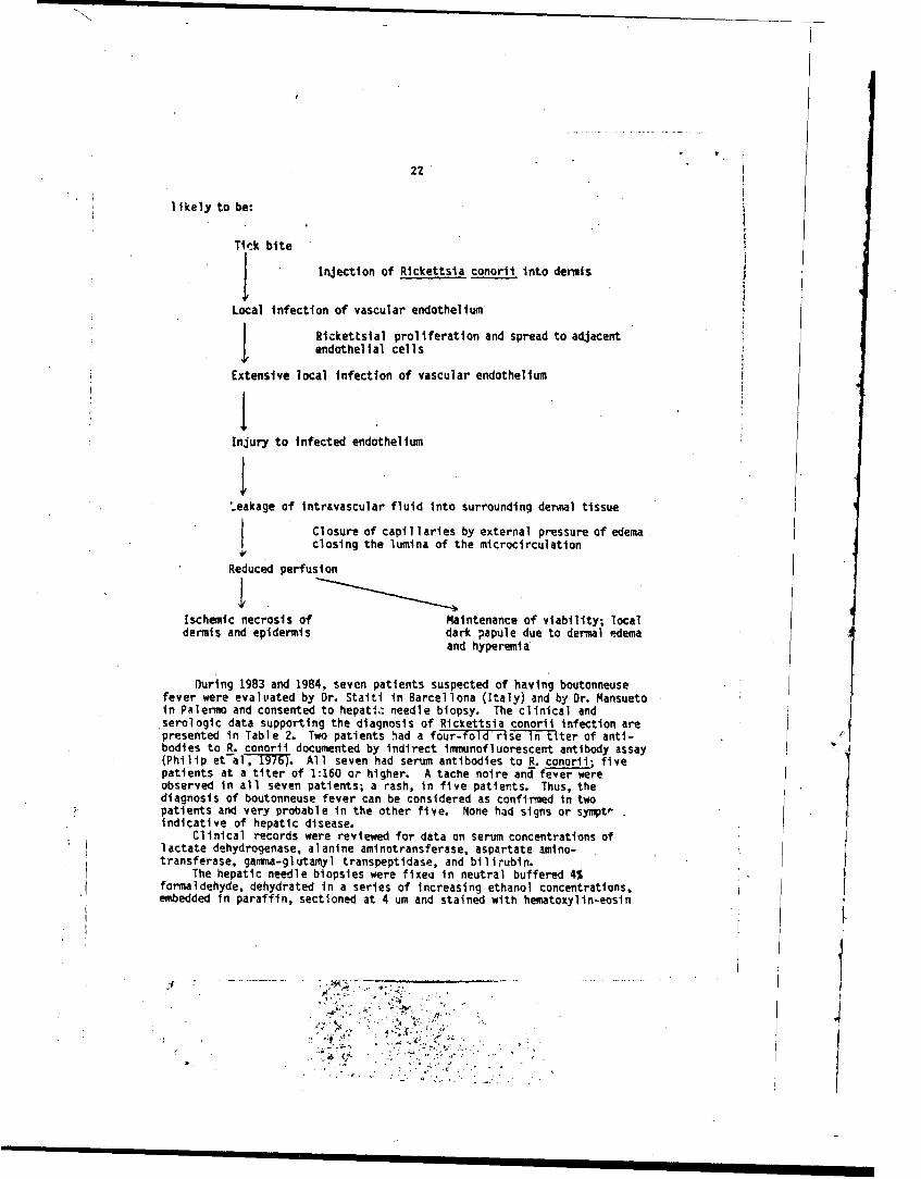
'Tgcse results indicate that vascular injury by rickettsiae is the majorlesion and that dermal edema is the important result. Thrombosis wasgenerally absent or only focal and mild. Thus, the pathogenetic sequence is
___
A I1
22
likely to be:
Tick bite
Injection of Rickettsia conoril into dermis
Local infection of vascular endotheliumSRickettsial prol iferation and spread to adjacent
endothelial cells
Extensive local infection of vascular endothelium
Injury to infected endothelium
'.eakage of intravascular fluid Into surrounding derual tissue
Closure of capillaries by external pressure of edemaclosing the lumina of the microcirculation
Reduced perfusion
Ischemic necrosis of Maintenance of viability; localdermis and epidermis dark papule due to dermal edema
and hyperemia
During 1983 and 1984, seven patients suspected of having boutonneusefever were evalvated by Dr. Staiti in Barcellona (Italy) and by Or. Mansuetoin Palermo and consented to hepatic needle biopsy. The clinical andserologic data supporting the diagnosis of Rickettsia conoril infection arepresented in Table 2. Two patients had a four-fold r s~e-T-l-•-ter of anti-bodies to R. conorli documented by indirect immunofluorescent antibody assay(Philip et-al. 1976) All seven had serum antibodies to R. conorli; fivepatients at a titer of 1:160 or higher. A tache noire and fever wereobserved In all seven patients; a rash, In five patients. Thus, thediagnosis of boutonneuse fever can be considered as confirned in twopatients and very probable in the other five. None had signs or sympto'indicative of hepatic disease.
Clinical records were reviewed for data on serum concentrations oflactate dehydrogenase, alanine aminotransferase, aspartate amino-transferase, gamma-glutamyl transpeptidase, and bilirubin.
The hepatic needle biopsies were fixej in neutral buffered 4%formaldehyde, dehydrated in a series of increasing ethanol concentrations,embedded in paraffin, sectioned at 4 um and stained with hematoxylin-eosin
*............... . :.. -• .- -...-
23
as wel l as phosphotungstic acid-hematoxylin for fibrin, Verhoeff-Van Giesonfor elastic tissue, reticulin, aisd Perl's Prussian blue for iron. Anadjacent serial section was affixed to a microscopic slide with Elmer'sglue, deparaffinized in xylene, digested with trypsin, and stained by directimmunofluorescence with a conjugate reactive with It. conorli as previously
described (Walker and Cain, 1978; Montenegro et al,-The hepatic laboratory data and results of evaluation for presence of'
hepatic lesions and R. conoril are presented in Table 3. The moderateelevatiTcs of serum concentrations of LDH, AST, And ALT are compatible withthe presence of scattered foci of hepatocellular necrosis, which Involvedonly a small proportion of hepatocytes. The lobular location ofthese lesions and the absence of involvement of portal triads 1s reflectedin that there were no striking deviations of laboratory measurements forserum hilirubin and alkaline phosphatase during the acute phase of theillness. Nevertheless, it is remarkable that all biopsies contained lesionswhich appeared to fit into the sequence of hepatocellular necrusis followedby focal, predominantly mononuclear leukocytic, inflammatory reaction. Yet,in no case were intact immunofluorescent SFG rickettsiae identified in thetissue. It is probable that alcoholic or other hepatic injury may haveoccurred as an underlying condition in some of these patients in whom fattychange was observed. This investigation confirms the reports of Guardia etal (1974) and Faure et al (1977) who have described hepatic lesions Inpatients with boutonneuse fever. It is our interpretation that theselesions do not fit the designation granulomatous hepatitis, but ratherconsist of foci of hepatocellular necrosis and predominantly mononuclearleukocytic reaction to the necrosis and probably former site of R. conoriiinfection. The lesion differs from a true granuloma in that it Ts not anavascular aggregate of activated macrophages in contrast to thegranulomatous hepatitis of Q fever In which aggregates of macrophages areobserved I n a peculIar doughnut arrangement (PIcchi et al, 1960; Pel l egrinet al, '1980). C. burnetli resides within the phagolysosome of macrophages,the target cel Io Q-fever (Burton et al, 1971), whereas endothel ial cel l sare the primary target of R. conorii In most organs (Walker and Gear, 1985).The target cel I of R. conorTi In I -ve r awaits further study of human casesand animal models. In fat-a cases of R. conorii infection in South Africa,.immunofluorescent rickettsiae have beeF'dgenst rated in hepatic sinusoidal
IOning cells that may have been Kupffer cells or endothelial cells (Walke!rand Gear, 1985). Adjacent necrosis was associated with foci of rickettsialinfection In those fatal cases, thus suggesting a role for rickettsiae inhepatocel 1 ul ar injury and the probabl e cl earance of rickettsiae from I esionsin the biopsies by effective host immune and phgocytic mechanism.
These lesions resenmle multifocal hepatocellular necrosis and infl am-mation observed in mice infected experimentally with R. conorli (Montenegroet al, 1984). The observations that similar lesions occur in-both imuno-compettnt and T-lymphocyte deficient mice although more rickettsiae persistin the immunodeficient mice suggests that immunopathologicmechanisms are notimportant in the pathogenesis of these lesions. Likewise, the fatal SouthAfrican cases contained recrotic hepatic cells in foci that did not containa cellular response. Future studies of human and experimental animalmaterial by electron microscopy and immunohistochemistry will be importantfor characterization of the populations of inflammatory cells and subpopula-tions of T-lymphocytes as well as for identification oi the hepatic targetcell of R. conorli.
Finally, the most important conclusion of this study Is thatboutonneuse fever must not be considered as a benign disease with rare
4V
"¼ k !
I.t
24extracutaneous involvement. The demonstration of hepatic lesions in sevenconsecutive patients evaluated by liver biopsy suggests that R. conorti isfrequently viscerotropic and in patients with particular risk faic-tors posesa serious threat.
Our other paper presented at the ASR meeting on the pathogenests of SFGrickettsioses is an extension of rEcent reports of gastrointestinal involve-ment in boutonneuse fever and R4SF. Two Spanish patients with severe gas-trointestinal hemorrhage have been reported by the group at the Universityof Salamanca. One underwent gastrectomy to control hemorrhage and subse-quently died. The resected gastric specimen co,,tained rickettsial vascularleisons as the basis for gastric hemorrhage.
Patients with RMSF frequently have nausea, vomiting,,diarrhea, orabdominal pain leading to an initial misdiagnosis of gastroenteritis.Nausea, vomiting, or diarrhea early in the course were reported in 63% of aseries of 131 well documented cases of RMSF. Abdominal pain was present in34%. A lack of awareness or the gastointestinal manifestations of RMSF bythe physician compounds the difficulty of dignosis during the first 2-5 daysof illness prior to onset of the rash. In a study by Hattwtck of 44 fatalcases of RMSF, 32% of cases, as compared with 4% of nonfatal control cases,had gastrointestinal complaints as the presenting symptoms leading to theincorrect diagnosis of gastroenteritis. In some RMSF patients, thesesymptoms lead to the differential dignosis of an acute surgical abdomen, andexploratory laparotomy is performed. Such manifestations of RMSF leading tolaparotomy have been reported previously in at leant six patients (Table 4).R. rickettsli were demonstrated by direct immunofluorescence in vascularTesicns of the appendix of one patient; organism compatible with rickettsiae jstained by Pinkerton's method were described in the lesions of the patientwith acute hemorrhagic ulcerative ileitis.
In addition to these case reports, a recent autopsy study of the gas-trointestinal tract and pancreas in RMSF was published from our laboratory.It documented rickettsial vascular lesions of vasculitis or thrombosis instomach in 89% of the cases, in the small intestine in all cases, in thelarge intestine in 92%, and In the pancreas of 70%. Although it is possible 4for rickettsial vasculitis to result in gastrointestinal necrosis, thisappears to be a rare event. We have recently investigated five patientswith RMSF with interesting abdominal findings.
A, 76 year old man appeared in good health until he was found drowsy andconfused at his home. At a nearby hospital in western North Carolina he wasnoted to be febrile and extremely confused. According to his wife, he hadbeen quite well without headache, diarrhea, nausea, vomiting, abdominal painor rash. Clinical and laboratory evaluation during the subsequent four daysfailed to establish a diagnosis. There was no skin rash. He remainedfebrile and was treated empirically with cefoxitin, gentamicin, and erythro-mycin for presumed sepsis. On his fifth day of hospitalization, he com-plained of pain in the right side of the abdomen, and an abdominal x-rayrevealed ileus and possible cholelithiasis. Because of the abdominal painand abnormal radiograph, acute choleuystitis was suspected and exploratorylaparotomy was perf.rmed revealing cholelithiasis, a non-obstructive stonein the common bile duct, and hepatomegaly, but no evidence of cholecystltis,abdominal or retroperitoneal abscess, or appendicitis. The stone wasremoved. Cholecystectomy and appendectomy were performed. His postopera-tive course included persistent fever, seizures, coma, hypotensinn,acidosis, thrombocytopenia, and oliguria progressing to anuria. Postopera-tive laboratory data Included elevated concentrations of hepatic enzymes,and serum bilirubin. Metronidazole, cefoperazone, and nlatelet transfusions
SL - . .
I
25
were added to his treatment regimen. After transfer to another hospital forhemodialysis, physical examination revealed a suggestion of an erythematousmacular rash with scattered petechiae, purpurae, ankd peripheral pittingadema. A skin biopsy contained immunofluorescent Rickwettsia rickettsii.The was treated with hemoldalysis, vasopressors with Swan-Ganz mon-orTin-g,chl oramphenicol , and methylprednisolone, but died on the tenth day ofI I1I ness.(
In view of the skin biopsy finding, the gal Ibladder and appendix weeexamined by direct iutunofluorescence and revealed R. rickettsii in thewalls of the blood vessels in each organ. Rickettsfal vasculitis wasobserved in each layer from the lamina propria to the subsarosa of -achorgan.
A 71 year old woman from eastern Tennessee developed headache, nausea,and vomiting for 2-3 days. She had a fever, right costovertebral angletenderness, and pyuria. Upon refusing hospitalization, she was treated with,cefamandole nafate and trimethoprim-sulfamethoxazole for presumedpyelonephritis. Two days later she was admitted with nausea, vomiting.weakness, and dehydration. Laboratory data included severethrombocyutopenia, elevated liver enzymes, creatinine, and mild hypoxemia onoxygen therapy. She developed noncardiogenic pulmonary edema on her secondhospital day. Ultrasonography of the right upper quadrant of the abdomenrevealed a normal liver and a thickened wall of the gallbladder, surroundedby a sonolucent zone that was interpreted as a pericholecystic abscess oredema. Because of suspected pericholecystic abscess, exploratory laparotamywas performed on the second hospital day with cholecystectomy, intraopera-tive cholangiogram, and liver biopsy. The gallbladder contained no stones,and the cholangiogram was normal. There were 500-1000 ml of bile-stainedascites. Her postoperative course was characterized by persistent fever,respiratory failure, metabolic acidosis, renal failure, and cutaneoushemorrhages. Prior to laparotony she was treated with various antibioticregimens which included ampicillin, doxycycline, gentamiciln. ;efoxitin,piperacillin, and clindamycin. Doxycycline, an active ant'.rickettsial drug,was given for less than a day. Postoper:cIvely, she received gentamicin andchloramphenicol. Although the diagnosis if RMSF was considered during theillness, it was not made. She died on her sixth day of hospitalization.
Direct immunofl.orescent examina'ion of the surgically removed gal l-bladder and postmortem skin revealed R. rickettsii in blood vessel walls.Rickettsial vascular injury was observcd: -
Case 3 was a 55 year old man who developed fever, headache, andmyalgia, six days before death. Over the period 2-4 days prior to death,he had intermittent nausea and Yomiting. One day before his demise, hevcmited a large blood clot and was hospitalized. Hematemesis continuedrequiring transfusion of six units of red blood cel ls. After transfer toChapel Hill he had hematocrit 37%, platelets 18,000/ul, acute renal failure,seizures, cardiopulmonary arrest, hypotension, and postarrest hematocrit19.5%. He was transfused with red blood cells and platelets. Endoscopyrevealed a massive amount of blood in the stomach and esophagus, but nodiscrete bleeding focus. Despite immediate treatment with choramphenicol,the patient died one day after admission. Autopsy demonstrated gastricrickettsial vascular infection and injury with hemorrhage, the immediatecause of death.
Case .4 was a 4 year old girl in the preantibiotic era, 1933, in fact.Postmortem examination revealed, in addition to the typical findings ofRMSF, a perforated appendix, the wall of which contained numerous injurc!blood vessels infected by immunfluorescent R. rickettsii. This case illus-
i
* . ,•-> : - .
26
trates that R. ricketts~i can actually cause lesions requiring surgicalintervention.
Case 5 was a 10 year old male who do.teloped fever, nausea, vomiting,and abdominal pain leading to appendectomy. Seven days later a history oftick bite was obtained and a ra~h was noted and demonstrated upon biopsy tocontain immunofluorercent R. rlckettsii. The hospital course was comli-cated by corA, seizures, acute renal failur!. noncardiogenic pulmonaryedema, cardiopulmonLry arrest, skin necrosis, and sevo.re thrombocytopmia,but the patient survived and was discharged from the t.osp~tal after morethan a month with convalescent IFA of 1:8,192. Review of the resectedappendix did not reveal any rickettsial vascular injury or inmunofluorescentR. rickettsii. Thus, our dilemma is that RMSF may be nisdiagnesed as acutesurgfc-alabomen or gastoenteritis without a crucial intraabdominal lesionthat would benetit from surgery or R. rickettsii-associated lesions mycause fatal gastrointestinal hemorrnage or caus. life threatening gastroin-testindl lesions such as ruptured appendix, a lesion requiring surgicalintervention. Finally, although we believe the g.i. manifestations of RMSFare associated with the abdrminal rickettsial vasculitis, demonstrablerickettsial infection an6 lesions may not always be pr..ent In the tissueexamined, particularly a surgircii 5oecimen.
A manuscript on the acute phase reaction in boutonneuse fever has beenprepared and submitted for publication. This work was performed in Palermo,n collaboration with the Clinic r. Tropical and Subtropical Diseases andthe Institute of Hygiene. Evaluat.ion of specific serum proteins from 44patients in Sicily with boutonneuse fever revealed that C-reactive protu',haptoglobin, alpna-l-antitrypsin, C3, and C4 are elevated at the time ofpresentation for medical attention in varying proportions of the patients.C-reactive protein is invariably elevated as a consequence of active injuryand inflammation during the first week of illness. Th:se increased serumprotein concentrations fol low the pattern of the acute phase revcztion andsuggest that immunopathologic phenomena and intravascular hemilysis do notoccur in most patients with biutonneuse fever.
The haptoglobin concentration was significantly higher during the firsttwo weeks of illness than subsequently. Only three determinations hadvalues below the normal range, one during the first week of illless an$ twoduring late convalescence. Serum alpha-l-antit-ypsin levels were high-tduring the first two weeks of illness and had diministhed to normal levelsby 4 w'p",s after onset of illness. Concentrations of C3 convertae and C4were elevated acutely with 16/25 measurements of C4 and 8/25 medsurefts ofC3 convertase abode the reference interval during the first week and 20/26assays of C4 and 5126 assays of C3 convertase above the reference intE...lduring the secorn wmek. In only one patient during 'he first week of
I ness were the concentrations of C3 convertase and Z4 below the referenceinterval. C-reactive prote,- (CRP) was at or ative the upper limit ofnornvii in all patieihts during the first week and in 20/26 patient. duringthe s.acond week. By late convalescence 1-2 months after onset, 15/20patients had no detectible %erum CRP.
This study has demonstrated ti'at the acute phase reaction ocurs duringLoutonneuse fever. Serum concentrations of certain hepatic synthesizedproteins are elevated as a starotype. response following surgical operation,several acute Infectljias, and other situations that have acute Inflammation ortissue nqcrosis. CRP concentration has s)een shown to rise from undetectiblelevels within 4-6 hours after acute injurv. Haptoglobin, fibrinogen, andalpha-l-antitrypsin are increased in concen.ration by 24 ho,,rs after i-Jury,and C3 convertase is elevated durinq the first week. Our measurements were
N,, -
27
not made early enough in the course of BF to determine how mayn hours afteronset were required before the rise occurred.
The e'facts of the elevated concentrations of these serum proteins onthe pathophysiology of OF is uncertain. It may be hypothesized that C3convertase and C4 might be consumed by binding to circulat~ng immne com-plexes, which have been demonstrated to occur in BF. Although the observa-tion that C3 convertase and C4 concentrations are mostly elevated or normaldoes not exclude that possiblity entirely, it does not support thathypothesis. Other observations and experimental data suggest that Imiuno-pathologic mechanism are not important in spotted fever group rickettsioses(Bradford et al, 1979; Jerrells and Eisemann, 1983; Kenyon and Pedersen, 1980;Kokorin etf17,-1976; Montenegro et al, in press; Moser at al, 1977; Walkerand Hendi-s-ii, 1978). monspecifTc opsonization of spotted"ever rickattsiaeby direct reaction with components of complement or CAP has never beendemonstrated; however, elevated concentrations, particularly of CAP, suggestthis as a possible host defense mechanism as It may be in other bacterialinfections. The persistent elevation of CRP in some patients Indicates thattissue injury or inflamation is ongoing.
The observation of a low plasma concentration of haptoglobin in onepatient during the acute phase of OF indicates that significant hemolysis Isnot a frequently occurring complication. The recent denorstration ofhemolysis in a patient with glucose-6-phosphate dehydrogtnase (G6PO)deficiency and fatal RMSF vJggests that hemolysis may play a role in theenhanced severity of RMSF in G6PO deficient men (walker at al, 1983). Sincemales with 66PO Mediterranean have been shown to have a Ugf-r incidence ofspecific compl•cations in SF than G6PD normal males (Piras et al, 1983), thepossibility of hemolysis should be now evaluated in this sur'-p--1ation ofpatients with OF.
In general, the acute phase reaction appears to be an immediateresponse which precedes the generation of a specific immune response. Likefever, this response is highly likely to have resulted from evolutionaryselection and to have an effect that is generally favorable to the host.The exact interectipns of C3 convertase, C4, and CRP with L conorii waitfurther study.
A manuscript on familial cases of boutoonnuse fever has been preparedand submitted for publication In coll aboration with the same groups ofInvestigators in Sicily. Pairs of cases of boutontuse fever occurred inthree families. The illness appeared nearly simultaneously in both Wersof *.e family, but generally was more serious in one as judged by clinicalaid laboratory parameters. The possibility of a abed rickettsiosis," thatIs, reactivation of rickettsiae by the blood meal obtained from the firstIndividual by the sam tick which fed upon the second Individual, can beexcluded in two of the three pairs of cases. Four of the six patients weresleeping in different beds.
On the other hand, the differing severity may be reflected I" thedifferent immunological patterns of the two patients. In o" couple it wasdocumented that the first and more seriously Ill had antibodies of the IgMclass, presumably a result of the first exposure to Rickettsia Conorit. Thesecond and less ill patient had antibodies of the 1gG class only, preumablythe result of reexposure after prtvious asymptomatic Infection with aspotted fever group rickettsia.
The cases reported hee. particuiarly the two sisters, coprise aninteresting exmale from the point of view of clinical at laboratoryobservations. Clinically, the course was riemrtable more severe, as judgedby maningeal signs, and more extensive heorrhagic rash, hepatomegaly,
,' •4 " I.
28
lymphadenopathy, in the younger (S4 year ole) sister who did not have asignifican prior medical history in comparison with her sister (74 year oldwith a four year history of hypertension). In general, rlckettsioses havebeen documnted to be more severe in older patients and clinical Judgementwould suggest In those with underlying medical problems. Also thelaboratory data demonstrated more marked abnormalI ties In the fIrst of thetwo patients to become ill.
The hypothesis that a single source of infection, that Is to say thesame tick, first bit the older sister and then the younger sister withprogressive *reactivationO of the rickettsiae after the first blood meal ofthe tick does not seem very probable. This hypothesis has been proposed isseveral cases of Rocky Mountain spotted fever occurring in married couples,familial rickettsioses of the bed (Parker, 1933; Schaffner et a1, 1965), butthis proposition Is not supported by our cases since the tZosTsters,although living In the sae house, had separate bedrooms.
It Sm to us instead a probable that the explanation has animmunologic basis supported by the course of development ofimmunofluorescent antibodies of a particular class. The younger sistar hadpredominantly 1gM antibodies early In her course, and her sister had 9gGantibodies. This suggests that the more severe case encountered infectioawith R. conorli for the first time while the milder case had an ancuensticimaune response with rapid rise in the IgG class of antibodies, possioly aMindication of previous asymptomatic Infection with a spotted fever grouprickettsia. This Imeediate iiune respoise, therefore, allowed a fasterantibody rise, and thus, better control of the pathogenic events. It isclear that this hypothesis goes beyord the documented evidence; however, werecently observed a typical 4tache noire" (in which biopsy was demonstratedthe presence of R. conor1i by miunofluorescence) in a patient who did notdavevlup subsequint TTT-iis.L Meanwhile, monitoring of the Immne responseshowed seroconversion of only 1%G antibodies. This observation supports thehypothesis that preexisting asymptomatic infection allowed an anamnsticimmune response sufficient to circumscribe the Infection at the site ofinoculation (Burgeots et al, 1982; Mansueto at al, 1964a).
Also in this repo6 are two paitrs of casesi1at appear to document adynamic analogue. Indeed, In the subjects with a predominant initial IgGresponse the illness was less severe. The role of humorsa itsunity in thepathogenesis of rickettsial diseases is not coletely clear; nevertheless,several experimental studies have demonstrated a role for antibodies in aneloquent manner. Gambr 11I and Wisseman have observed that human timuneanti-R, pro. zekii serum augmmnts the opsonizi power and the destructionof ricketts ae by eukocytes and macrophages. Topping (1940, 1943)demonstrated protection by prophylactic administration of immune sera. Themilitary significance Of Otise observations is that there art probably 'hotspots* of R. conorli prevalence in the ecosystem into which nonimmune troopsmight enteoF ar ---ome infected with a substantial attack rate for thegroup.
A manuscript has ben prepared and submitted for publication oan theepidemiology of boutonneuse fever in western Sicily that correlates thedistribution and prevalence of R. conorli infection in Rhipce!jh~lussanquwneiS tth canine sero•ositfvT•y•a-nd human cases 'f boutonneuse fever.
The aistribution and prevalence of spotted fever group rickettsialinfection in the ixodid dog tick Rh!iceiphalus sanIuineus were found tooccur at a rate of 19.71 with variation rated to o graphical and socio-occupational factors. A higher rate of infection was dmonstrated in ticksremoved from dogs associated with documented cases of boutonneuse fever.
X '
29
Out of the 1078 ticks removed from dogs, L. san uineus was predominantwith few other species (4 Ixodes ricinus, 4 aHie ma ncum'2ftbus) AllI stages of develTopmenftwere obtal od frw shee-d.4i, whTle onlyiduits were observed in domestic dogs living in urban areas and. in latesummer, in dogs of the mountainous areas. Only adult ticks were tested inthis study. In each place the. occurrence of SF has been documented.
A total of 212 (19.21) if 1078 ticks was found infected with SF6rickettsiae, Not all the areas investigated, however, showed the' sawe rateof infection but a gradient was observed wtich increased from urban(Palermo, Trapani, Agrigento) to rural areas (Carini, Castel lI amare, Vicari,Santo Stefano di Quisquina, Alcam)' and decreased again with a rise inaltitude (Petralia and the Madonie Mountains). Ticks obtained from dogsliving in sheepfolds in rural areas frequent-y contained SF6 rickettsziae;23/24 (95.8%) of these dogs were found to harbour infected ticks, and all ofthem where soropositive. In urban areas 14 (43.7%) of 32 dogs were free ofInfected ticks (100.01) although antibodies reactive with Lt copor[ weredetected in 2S/32 (78%). Higheor pearcentages of dogs carrytngi5Ti~f actedticks and seoiegative dogs were found in mountainous areas: 14/21 (67%)and 10/21 (47%), respectively (110.001). Seventy-three dogs (67.6%) werefound to harbour Infected ticks overall. Antibodies reactive with R.conorii were present in 88/106 (83%); yet only 20 of 35 (57.1%) dogiwlithuninfected ticks were seronegative (1)(0.001).
Investigation of 8 human cases of SF where association with dogs wasdocumented revealed that 16 out of 22 (73%) dogs tested harboured ticksinfected with R. conorli as demonstrated by direct examination of the ticksand seroconverision of gInes pigs. Al 1 the dogs were found to have anti-R.conoril antibodies. It is intriguing to note that the case from SantoStefano di Quisquine ras a shepherd and the patients in Casteliammare andVicari, a school teacher and a grocer, respectively, had a sheepfold verynear the land surrounding their hawes. Controls were chosen and taken fromthe so neighborhood from houses where no cases of OF had occurred; asignificantly lower percentage of seropositive dogs and Infected ticks wasobserved in thes controls (P(O0.01). A tick LL* sanquineais) removed from thesite of bite on the shoulder of an agricul tural w-o-rer contai ned SFGrickettsiae demonstrated by microscopic observation and Imunfl ucrescentstaining of hemolymph; seroconversion occurred In the Inoculated guinea pig.The dog owd by this patient was also seropositive. Ail Iguinea pigs Inocu-lated with hemolymph positive ticks showed seroconversion after 28 days.
The essential role of the tick is emphasized by the high rate of tickInfection (19.7%) and the uneven distribution of SF6 rickettsial Infectionof ticks ranging from 41 to 35% In different areas. These variationscorrelated with geopgrapftic factors: high infection rates In rural areasand lower altitudes and, conversely, lower Infection rates in urban andmountainous areas.
These observations of high proportions of rickettsia-infected ticks andseropositive dogs in areas of Sicily with higher incidence of OF are similarto the evidence for greater rickettsial activity In dogs in France and theUSA in areas of higher human attack rates for OF and Rocky Mountain spottedfever, respectively.
There was an even higher rate of SF6 rickettsial infection of ticksIfrom dogs that were associated with human cases of SF in Sicily than theaverage rate of tick infection-, 731 of such dogs had SF6 iickettsia-containing tictks. The stimulationt of seroconversion of guinea pigsInoculated with tick-rickettsis suspensions strongly suggests that those SFG
rickettsiae were, In fact, pathogenic Lt cooi This association of R..
30
conorli-infected R. sanguineus with huaan BF in western Sicily was mostconclusively dociTenitd"1jith isolation of a SFG rickettsia from a R.sangutneus tick which was removed from a patient with BF.
Amanusc, Ipt reporting four cases of boutonneuse fever that occurredduring the cold season of the year has been prepared and submitted forpublication in collaboration wtth investigators at the University of Palermoand Dr. Raoult of Marseille. These documented cases occurred in Sicily andFrance between November and February. It cannot, therefore, be assumed thatboutonneuse fever is limited to the warm weather seasons of the year. Thediagnosis must be considered. for febrile patients in the Mediterranean basinthroughout the year.
A manuscript that documents the occurrence of hemolysis and severeboutonneuse fever In a previously healthy 27 year old ran with glucose-6-phosphate dehydrogenase deficiency tas been prepared and submitted forpublication in collaboration with the Infectious Diseases Unit of the BoignyHospital and the Pediatric Hematology Unit of La Timone Hospital inMarseille. A 27 year old previously healthy Algerian man was hospitalizedon August 3, 1984, after two days of fever, headache, myalgias, asthenia andweight loss of two kilograms. Several days before hand he had noticed ascrotal lesion with painful inguinal lymphadenopathy. On admission he hadtemperature 39.50C, pulse 100/mmn, blood pressure 110/60 mmlg, and a scrotaleschar. Laboratory data included normal urinalysis, hematocrit 45%,erytlocyte count 4.6 x 10⁹/ul, hemoglobin 14.65 g/dl, white blood cells 8.4x 10/ul, with 77% polymorphonuclear leukocytes, platelet count 128 x10O/ul, serum sodium 137 mmol/1, serum potassium 3.7 mmol/l, serum chloride99 m0/oll, serum calcium 2.4 mmol/l, prothrombin time 11 seconds (control 12seconds), unconjugated bilirubin 11 mc mol/l, aspartate aminotransferase(SGOT) 25' IU/I, and blood urea nitrogen 5.1 mool/I. He became stuporous andconfused. A maculopapular eruption involving the palms and soles andpurpuric on the legs and trunk was observed on August 7, 1984. The diagnosisof Mediterranean spotted fever was made, and intravenous treatment withdoxycycline, 200 mg/day, was begun on August 7. On August 10, 1984, thepatient was asthenic and stuporous but apyretic. Laboratory data showedhepatic involvement (SGOT, 145 IU/l and SGPT, 142 IU/i) and evidence ofhemolysis with decreased hemoglobin and haptoglooin but normal bilirubin.The subsequent course was uneventful and did not require blood transfusions.G6PO phenotype was determined to be G6PD S' by low W6PO activity determinedby spectrophotometry and by electrophoretic mobility, The #$agnosis ofMediterranean spotted fever was confirmed by immunofluorescent antibodytest. The investigation of the family revealed two other cases of G6PDdeficiency, a brother and a maternal uncle.
Severe cases of Mediterranean spotted fever have been reportedpreviously. These cases were usually males, with underlying diseasessuch as diabetes, cardiac insufficiency, or alcoholism. WPO deficiency wassuspected to be one of the risk factors for severe disease. G6P0 deficiencyis rare in France, but in the southern France especially in Marseille, apopulation of diverse origins is present. In Marseil le 10% of the popula-tion originally, cam from Corsica and 18% from North Africa where G6POdeficiency is prevalent. The rate of G6PO deficiency is 10% in Algeria.G6PO deficiency nas previously been associated with severity in m.rinetyphus, scrub typhus and Rocky Mountain spotted fever. Regarding !Mediterranean spotted fever, Meloni and Forteleoni found no statisticaldifferences in patients with and without G6PO deficiency although no parti- 3cular criteria that were evaluated were stated. In contrast, Piras at alreported an evaluation of Sardinian males with Mediterranean spotted fever
1
31
for complications of particular organ systems which occured more frequentlyin G6PO deficient patients. Neither study ciented on examination forhemolysis.
Our patient had a quite severe form of serologically confirmedMediterranean spotted fever with stupor, confusion, purpuric exanthemthromuocytopenia, hepatic involvement, and evidence for hemolysis. Thehemologlobin .'all from 14.7 9/dl to 9.9 g/41, and serum haptoglobin wasdepleted (0.16 9/1) acutely and rose to normal concentration (1.5 9/1) duringconvalescence. This is the first reported case of hemolysis in a G6PDdeficient patient suffering from Mediterranean spotted fever and the firstreported case of hemolysis with rickettsiosis in a G6PO B (Mediterranean)patient. The mechanism of enhanced severity of rickettsioses in G6PDdeficiency is unclear. Two hypotheses are drug induced hemolysis andincrease of rickettsial activity due to defective host defense orstimulation by constitutents released from lysis of red blood cells.In our case hemolysis occurred before tetracycline treatment. In ourexperience this man was the only patient with documented G6PO deficiencyamong 49 miles with Mediterranean spotted fever where the G6PO activity wasdetermined. Furthermore, he is the only patient utnder 30 years of age tohave a severe form of the disease. In our opinion, in addition toalcoholism, diabetes and old age, G6PD deficiency could be considered as acause of enhanced severity In Mediterranean spotted fever as wll a s inother rickettsioses.
This report suggests that servicemen, particularly those of African andMediterranean ancestry, should be evaluated for giucose-6-phosphatedehydrogenese phenotype before assignment to duty in southern Europe,Africa, or the Middle East. Documentation o. this genetic condition mayallow particular preventive measures to be taken, e.g. vaccination, If itbecomes available, or anticipation of severe disease if boutonneuse fever isnot diagnosed and treated early in the course.
Other Investigations involving study of human rickettsioses that waresubjects of wort during the past year Include the publication of thearticle, TCorrelation of the Distribution of Rickettsia conortl, MicroscopicLesions, and Clinical Features In South African Tick 8iteFe n incollaboration with JJM.S. Gear, and autopsy case study of a fatal case ofboutonneuse fever from Salamanca, a study of a riche noire biopsy from acase of boutonneuse fever in a tourist who retuw--?,rThe Mediterraneanregion to San Franscisco, and Investigation of fatal cases of epidemictyphus in 'Jorth Africa during World War It. The South African case, whichwas described In last year's annual report, was completed and presented as apaper at the annual meeting of the American Society of Tropical Medicine andHygiene and an origInal research article in the Journal of the AmericanSociety of Tro•pical Medicine and Hygiene (see encmo-sedri--prnts Theiatalcas• - STaanca Ts -currefM B iTnghprepared for imunofluorescent demon-stration of R. conorti. Antigen of R. conorit was detected in sparse quan-tity In the iac'e-•-nvTre sent from SAnr -Frnssco. The method for demonstra-tion of R. p z-e(=fT" fixed human tissues that have been stored embeddedin paraf~in- e s--eT early 1940's has been developed using a corjugatereactive with typhas group rickettsiae that was obtained from the CDC.Tissues of three cases from the AF!P archives are In the procesc of prepara-tion in our laboratory for a clinicopathologic study in col laboration withDo. Ted Woodward. Preliminary results of sensitive, imunofl'orescentexamination for the distribution of R. p. zekit have shown rickettsiae Inendothelial locations in kidney and'heair-t.7W pe to elucidate severalpathophysiologic aspects of tpidemic typhus and to coare the distribution
?iF,!
S. . .. . . . . .. .. -
32
of rickettsiae tith RMSF and boutonneuse fever.Our work on the mouse model of R. conoril infection that was begun
prior to the inittatijn of this contract was prepared as a completed manu-script and published this year (Montenegro, M.R., Walker, O.H., Hegarty,B.C., Infection of genetically immunodeflcient mice with Rickettsia conorii.Acta Viral 28:508-514, 1984). It includes descriptions of a model o .--on-or7-Fe-patitis that is analogous to human boutonneuse fever hepatic-
les-To"ns, particularly as it occurs in itmmunocompetent mice. Work is inprogress toward further elucidation of hepatic injury by R. conoril and tkelocal immune response 1V ,iepatic rickettsial infection by eiettronmicroscopy and imuunohi.- "ochemistry. A good animal model for documented,reproducible severe R. anorii Infection of organs pertinent to severe huroninfection other than-Mhier--a-s nut been described and must be developed.Study of C3H and Balb/c mice with R. conor1i Inoculated by Intraperitoneal,silbcutaneous, or tntradr.mal routes has been conducted in collaboration wirJoDr. Tom Jerrells at Walter Reed Army Institute of Research. We haveexamined by light micro~copy the tissues of mice susceptible (C31) a%1resistant (Balb/c) to lethal R. conori1 infection by the intraperitoreal(lethal -for C3H), subcutaneous (resistant in both mouse strains), and intra..dermal (resistant In both mouse strains) routes over the first 12 days afterinoculation. Lesions are consistently present in hepatic tissues of thesemice (Table 5). Renal and CNS lesions are less consistent. Imumnofluores-cent determination of the presence, distribution, and relative quantity ofrickettsiae in these tissues by the deparaffinizatlon, trypsin digestionmethod revealed that rikettsiae were present in the peritoneum and liver oflethally infected CBH mice inoculated intraperitoneally when sacrificed onday 7. Systemic endothelial infection was not observed in any of the mice.The lethal cmdbination is, like the other combinations, therefore, ieither&n acceptable model of R. conorti endothelial injury nor of imminity toendothel al Infection.
During the period of a visiting professorship by Ors. Mansueto andTriali in March of 1985, tissues of guinea pigs Inoculated with f. conortiwere examined for lesions and immunofluorescent rickettsiae. Leisons werefew and Inconsistently observed except for periorchitis and multifocalhepatic Inflamation. Immunofluorescent R. conor1i were detected in themesothelitum of the tmnita vaginalis but notc iFnsystemic and pulmonaryvascular endotheliun. Thus, guinea pigs, In which even fever is frequentlyabsent or of short duration, are not a model for study of thepathophysiology and pathogenesis of R. conorii infection.
Because cotton rats have been repor"-eW-iasmodel s for several humanviral infections and R. prowszek11 infection and latency, this species,
'Si on hisPidus was'TInoi~u7aTdwi th R. conor1i, R. sibirica, and R.r t- t T *-e resu I t s were d I sapo Inti ng with- o Id ea-iT--T few eI tonsor demonstrated SFG rickettsiae after an extensive histologic and imno-fluorescent examination.
Coalaborative assistance has been provided to Dr. T. S. Walker Inprobing the role of prostaglandins In the pathogenesis of SFG rickettsioses.
Plasma and sera from experimentally infected guinea pigs and humai caseshave been collected and sent to this laboratory for analysis. The resultsare pending at this time.
Investigations of cel l culture models of R. conorii Injury includedfinal preparation of the manuscript and publ irllairn-of t article "Injuryrestricted to, col Is infected wth spotted fever group rickettsiae in para-biotic chambers,* Acts T_'sro 41:307-312, 1984, by Walker, 0. H., Firth,W. T., Iegarty, L.C.", prev!'Z!-Tly reported In annua1 report number 1
-4
33
(reprints enclosed). evaluation of the effect of temperature on cell Injuryby R. conorl and work towa;,d development of a model of cell injury withfluId overlay by measurement to lactate dehydrogenase concentration inculture medium. Plaques were not appreciably different in quantity at 320,34* and 36C.c At 38% fever R. conori1 plaques were observed. The LDHmodel has been characterized-In that predominantly the lsoenzyme LDH-5 iscontained in and released by Vero cells. An assay that preferentil] lymeasures this isoenzyme Is under investigation. The. major problem atpresent relates to the relativ ely small amount of LDH released andstandardization of a protoocol for precision In determining the concentra-tion in the medium. The model is not currently ready for use In pursuingthe mechanisms of cell injury related to phospholipase A and trypsin-likeprotease.
Some effort has been ey.par 4ed on review and analysis of the publishedmedical and scientific literat~lre on rickettsial disease problems. Thiseffort has been useful in focusing on the critical issues that thesediseases pose. A major product of this work is the writing of a comprehen-sive chapter for a CRC Handbook on viral hemorrhagic fevers and rickettsialdiseases edited by Or, JJt.S. Gear. *The chapter entitled aPathology andPathogenesis of the Hemorrhagic State in Viral Hemorrhagic Fevers andRickettsial Diseases* encompasses correlation of clinical and hemorrhagicmanifestations, human and experimental animal pathologic lesions, and inves-tigations of hemostatic and pathogenic mechanisms in these diseases. Asecond major work on synthesis of the current state of knowledge Is theplanning and editing of a CRC Handbook, Biology of Rickettsial Diseases.It is my goal to produce a comprehensive work to encompass the meaning ofthe recent advances in rickettsiology and their implications for under-standing rickettsial ecology. physiology, imunity. pathology, and clinicaldiseases. The field lacks a book that presents its Interesting problems asthey currently exist in a form that transcends the Individual article orcollection of research publications. The results should be useful formilitary medical planping and will include contributions from rickettsiolo-gists with a perspective of rickettsiae that include military significance.Ors. Gregory Dasch, Thomas JerrelI$, Jim William, Emillo Weiss, and I aremong the authors of the various chapters.
A*I
U
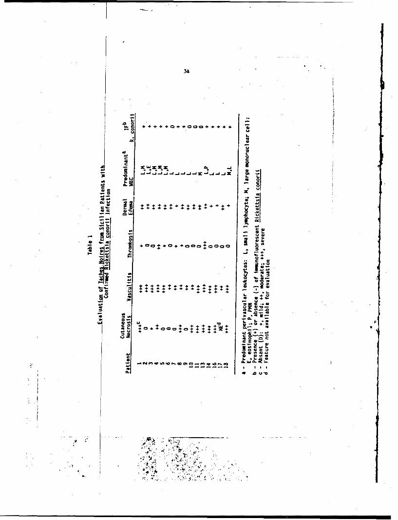
34
414
or
04
c41
C UL
44+ +... 4)
.C U
-0 a.
VI:; oL
-l - "a
ik 1; i
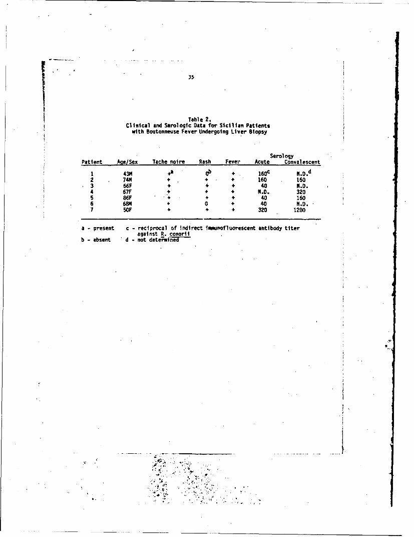
35
Table 2.Clinical and Serologic Data for Sicilian Patients
with Boutonneuse Fever Undergoing Liver Biopsy
SerologyPatient Age/Sex Tache noire Rash Fever Acute Convalescent
1 43M + 160c N.D.d2 74M + + + 160 1603 66F + 4 ÷ 40 N.D.4 67F + + N M.D. 3205 86F 4 + + 40 1606 6814 + 0 + 40 N.D.7 5OF' + + + 320 1280
a - present c - reciprocal of indirect timnofluorescent antibody titeragainst R. conorli
b - absent d - not determi1
(Af
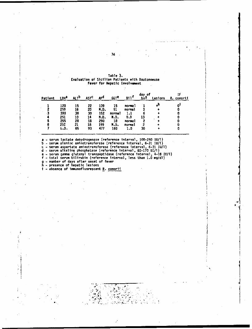
36
Table 3.Evaluation of Sicilian Patients with Boutonneuse
Fever for Hepatic Involvement
day of IFPatient LDHa ALTb ASTc APd GGT Bilf bx g Lesions R. conorit
1 120 15 22 139 15 normal 1 +h 012 259 16 20 N.D. 51 normal 3 + 0.3 390 38 30 152 normal 1.1 6 + 04 251 10 14 N.D. N.D. 0.9 13 + 05 255 28 18 290 18 normal 2 + 06 21C 21 18 199 N.D. normal 2 + 07 N.D. 85 93 477 160 1.0 30 + 0
a - serum lactate dehydrogenase (reference interval, 100-240 IU/1)b - serum alanine aminotransferase (reference Interval, 6-31 IU/I)
- serum aspartate aminotransferase (reference Interval, 6-31 IU/1)d,- serum alkaline phosphatase (reference interval, 60-170 UI/1)• - serum gamma glutamyl transpeptidase (reference interval, 4-18 IU11)f - total serum bilirubin (reference interval, less than 1.0 mg/dl)g - number of days after onset of feverh - presence of hepatic lesionsi - absence of Immunofluorescent R. conorii
<p
¢.
i.
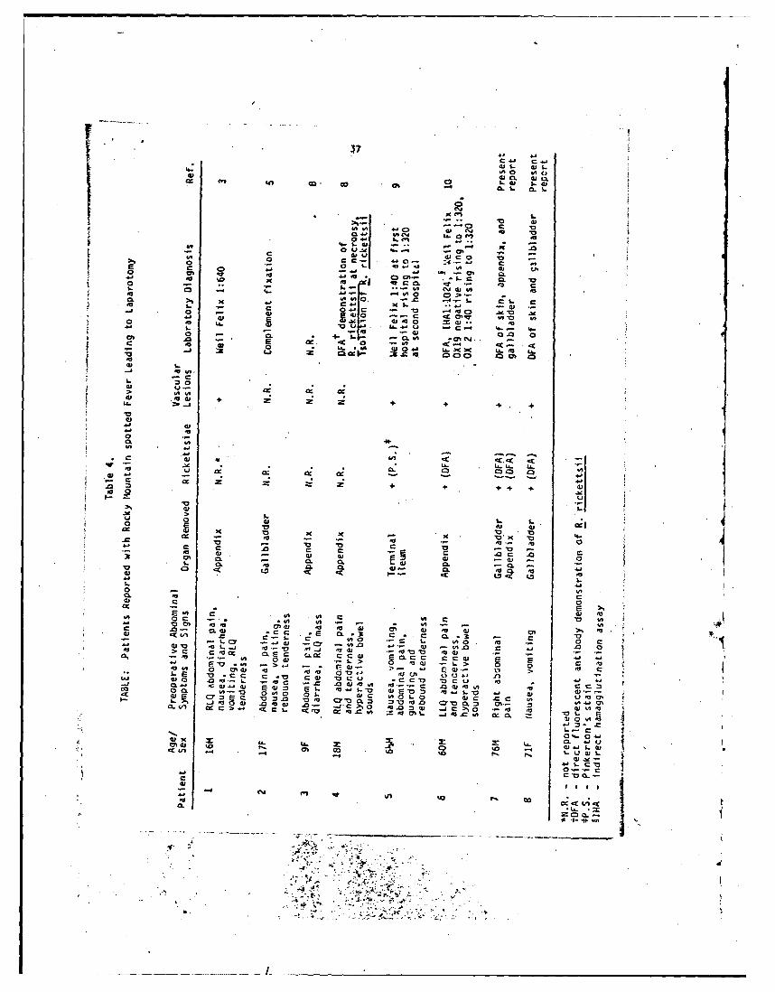
em tnco40 L. 4(9L 41x1.
L.0 4p0U.1 L
1 4-0
L. C 0U. 41 x . 40I
0 LA 4JCO.m 0 06.;.... 4--~ ut -. .- 0 (7% c%
1041 0C -4D1 CaC C3..
0 41- 1 0 ~ .
Q - j-W* M 0 0 0 1
1. u
41 .041 a9 f I94*
+ ++
*0wAt
4- a
06CL CA. 4)-
C .
00 0 6 Q) l 1 00
L > %. >.
0 e I0* -jc t4) C Cu m0 C0 Cu a :30 Q 1 -o w1 41
1. 0 . 0 0. 00.a
L. 0
2 4 V 7 0 .4 C- c r C 2 43cA: aC a, -Af -j, C~ -, 0 _j.*4-~~$ <0 Or_4 0 0 4 0 1 0 1
tm 4 1 0 0 1 . . 0 - - 0 40 0.I %~gC
0.4 ~ ~ r en11 00 o~ 1 41.2 .C C 0 C41 0~ U CC0 OCU - - 4410 00-1 0C A .0 10.4 0
-- C .041 .40.4- 201 21. 04-L-0 4 20~ 4-LO 4- 4
00. 1() 0110 01. 41C 401 0 41 CC 11 14-0
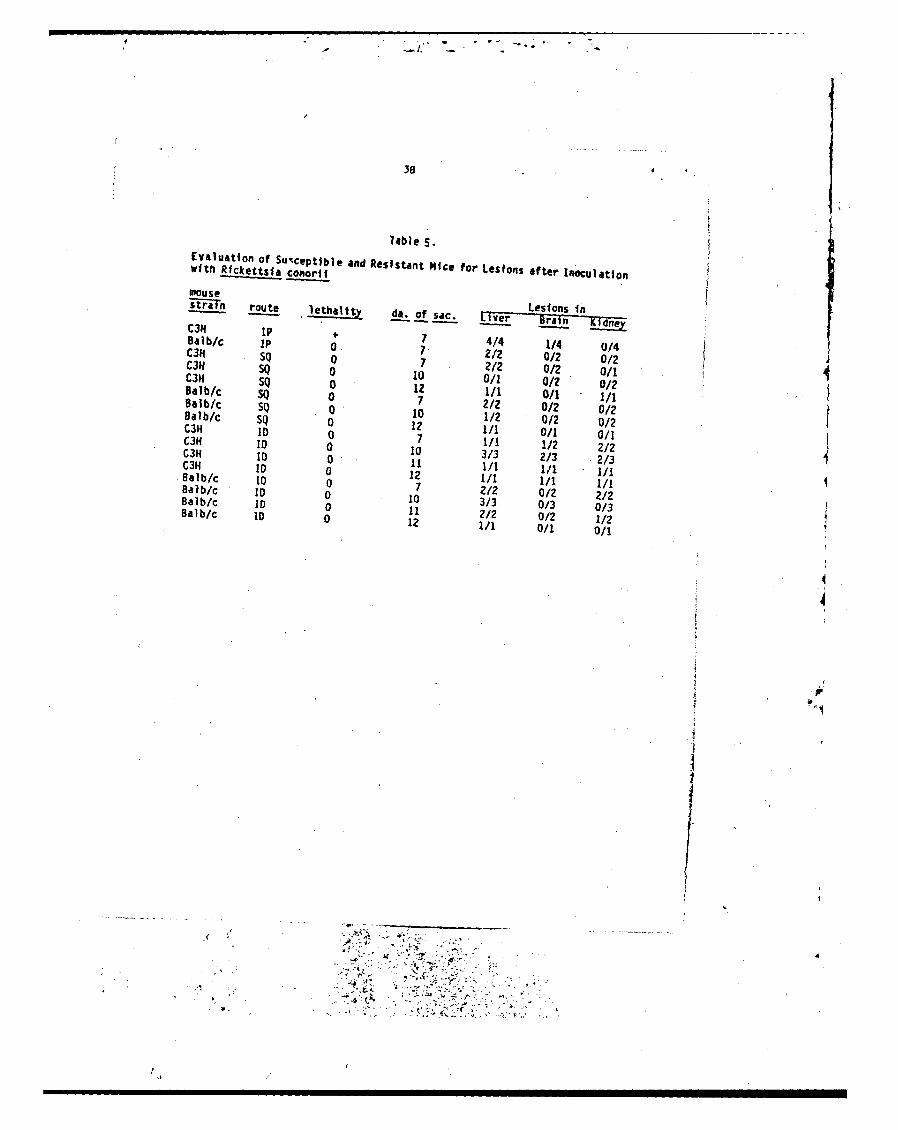
3 8 o
7able S.Evaluation of Susceptible and Resistant Nice for Lesions after inoculationWith Rfckettsla ¢onori
MousestraIn route aettlli t oa if Lisac.insIC3O IP 7 4/4 1/4 0/4Balb/c IP 0 7 2/2 0/2 0/2C3H SQ 0 7 2/2 0/2 0/1C3A SQ 0 10 0/1 0/2 0/2C3H SQ 0 12 1/1 0/1 1/1Balb/c SQ 0 7 2/2 0/2 0/2Balb/c SQ 0 10 1/2 0/2 0/2Balb/c SQ 0 12 1/1 0/1 0/i 1C3H ID 0 7 1/1 1/2 2/2C31 ID 0 10 3/3 2/3 2/3C3H 10 0 11 1/1 1/1 1/1C3R D 0 12 I/1 1/1 I/1
8galb/c 10 0 7 2/2 0/2 2/2,Balb/c ID 0 10 3/3 0/3 013Balb/c ID a 11 2/2 0/2 1/2Balb/c ID 0 12 1/1 0/1 0/1
.-A
(t
V. -
39.
Conclusions:
The currently recognized high rate of R. conoril infections in Spain,Portugal, France, Israel, and Italy is likeTy to continue. The absenca ofdata from northern, eastern, and western Africa and the Middle East reflectthe failure of clinical and epidemiologic systems in these areas to focusthe necessary laboratory methods and organizational efforts on the problem.The disease is likely to be or to become an important unrecognized cause ofincapacitating febrile illness in these areas. i Boutonneuse fever is aneglected disease that has recently attracted investigation in Sicily,Marseille, and Salamanca. Col laborativw -aiationships with the.e labora-tories encourages study of the epide-i.ologic, clinical, pathophysiologic,and rickettsiologic questio:;ns at a adm4rable level of effort and expertise.This contract has fostered a significant part of this result and cancontinue to play an important role in maintainfnq enthusiasm for elucidatingthe unknown factors that boutonneuse fever encomeasses.
It is probable that R. conorli infections are a greater cause ofmorbidity and mortality tfn-fa s now appreciptsd4 In Marseille, Salamanca,Israel, and South Africa, severn i llness is regularly recognized in a sub-stantial portion of those affected. The application of immunofluorescenceto autopsy specimens is revealing the rickettsial and clinicopathologicbasis for the pathophysiologic observations such as meningoencephalitis,hepatitis, and other visceral involvement. The documentation of hepaticlesions in seven consecutive Sicilian patients with hepatic biopsies duringboutonneuse fever demonstrates the serious nature of the disease. Tinerecognition that boutonneuse fever may cause an! incapacitating febril eillness with neither rash nor tache noire suggests that the high incidenceof antibodies to R. conorli in various populations may very well be due toprior undiagnosed siptomatic R. conoril infection. Specific dignostictests are not applied to patie~ts-Wo-lo-not present at least some of theclassic manifestations of the disease, particularly a rash. Such a situa-tion is obviously a potential threat to military health of soldiers in the.wide geographic distribution of R. conorli. This situation is analogous to 4the problem of scrub typhus in VoWrtdWar I, e•g. the Assam-Burma theater ofoperations. d e
Particular problems that have been ldentirled recently are the frequenthepatic involvemuent, lifethreatening gastrointestinal hemorrhage, severityof ll ness in gl ucose-6-phosphate dehydrogenase deficient men even if theyare young and previously healthy, the occurrence of cases even in coldweather months, and occurrence of familial cases that suggest 'hot spots" ofR. conorli endemicity.
Ou rcareful study of the tachenoires has elucidated important factorsin the pathogenesis that conform tu current clinical features of R. conoriipathogenesis: 1) R. conorit is present in the lesions and appears to 7paya direct cytopathic-roTle n vascular injury, 2)vasculopathic edema is amajor pathophysiologic effect, 3) ischemic necrosis is not necessarily theend result of vascular injury, 4) thrombosis is~not the major pathologiceffect of vascular injury and, thus, should noti be treated with anticoagula-tion, and 5) in human disease the local antirickettsial Immune and inflamma-tory response is predominantly lymphocytes and m'crophages.
Electron microscopy and immunofl uo escence! havt not yet demonstratedlarge quantities of R. conorli in tache noires or liver biopsies. Thepossible explanations- iGlue 1) t-h-e-ho--Fninune and phagocytic tesponsein these lesions may be effectivu t the stage of illness when roatients arereferred for tertiary subspecialty medical caret 2) antirirct.tjial anti-
0 i- -4 ,-.-
40
microbial therapy may have been administered prior to collection of speci-mens, 3) ultrastructural sampling may be a problem. Planned approaches tothis problem are attempts f-ý obtain biopsies from patients earlier in thecourse and assignment of labor 1tory personnel to the task of greater effortat examining samples of th biopsies by electron microscopy and utilization 4of dep,,rafflnization electron microscopy of foci in paraffin blocks that aredem~nstrited to contain R. conoril by immunofluorescence. More skin puncheshave been distributed to periphera collaborative sites and more collabora-tive institutions have been recruited.
Mouse R. conoril hepatitis is a good model for human boutonneuse feverhepatic lesTons.-T heTmouse model should be examined to determir, the targetcell type and host immune response by immunohistochemistry and electronmicroscopy for endothelial cell s, B-cel l s, T-cel l subsets, and macrophages.The target cel l of Immunodeficient murine R. conoril hepatitis is a goo!candidate for in vivo ultrastructural evalUation of the relationship ofrickettsial quantity to cytopathology and the ultrastructural features ofcytopathol ogy.
Other mouse models of SFG rickettsial infection must be evaluated for amodel of SFG endothelial infection and injury in order to correctly investi-gate pathogeaesis, pathophysiology, and immunity.
Monoclonal antibodies to R. conoril have been developed under aseparate grant from the Naitona Institute of Allergy and Infectiousdiseeses and will be characterized as to their molecular targets as afunction of the purposes of that research project on rickettsial antigensand vaccine devlopment. These monoclonals should be used to studypathogenic mechanism of R. conorii under this Army contract in cell cultrueart, dnimal models. The ONH grant does not have the funds or goals tosupport investigation of specific blockinge of pathogenic mechanism(s).However, if pathogenic mechanisms that may be blocked by monmclonal anti-bodies are discovered, the most effective approaches to prevention andadjunctive treatment may be developed.
Recommendat 4•.ns:
1. Offer skin biopsy immunofiuc-escent demonstration of R. conorii in tachenoire as a reference military laboratory dignostic test f~r Foutonn--fever.
2. Study the rickettsial hepatitis of boutonneuse fever and experimental R.conorli infection as models for visceral tissue injury by R. conoril.
3. Initiate a search for nonpathogenic R. conorii or R. conori!-likeisolates in tiý.s in Sicily.
4. Develop a more rapid, quantifiable assay system for growth of and cellinjury by R. conorii for screening amidine type trypsin-like proteaseinhibitors as potential prophylactic or adjunctive therapeutic agents.
5. Screen amidine type protease inhibitors for inhibition of cell injury byR. conorii.
6. Continue studies of tache noires for role of thrombosis in cell injury
and specific identificatT-n-of-TloaT immune cell types.
7. Continue in vitro study of pathogenic mechanisms of cel 1 injury by R.
- -7;1 ..
41
S. Initiate epidemtologic surveillance of troops in Spain, Italy, and otherplaces in the geographic distribution of R. conorit for acute cases ofboutonjeuse fever and for seroprevalence iof 7 antbodies to f. conori1.
9. Obtain sevropidmiologic data on the prevalence of R. conorli infec-tion In indigenous populatics in Africa and the Middl"iEast.
, |
L•___... ... ... __
42
Selected Bibliography
1. Adams JS, WalIker ON: The liver in Rocky Mountain spotted fever. As JClin Path 75:156-161, 1961.
2. BellI EI, Stoenner HG: Immunologic relationships amon the spottedfever group of rickettsias determined by toxin neutral ization tests inmice with convalescent animal serums. J Imminol 64:171-182, 1960.
3. Bourgede A, Joan Pastor MS. Rauult 0: Notes sur Is fievre boutonneuse,Had iterranee (A propos de 40 cas recent a Ma rseilleI s). Bull Sac Pat.Exot 72:308-314, 1979.
4. Burgeois AL, Olson 3G. Fang RCY, Whang J, Wang CL, Show L. Seckthold 0,Dennis OiT. Coolbaugh JC, Weiss E: um~oral and cellular reponses Inscrub typhus patients ref lecting primary Irfection and reinfection withRickettsia tsutsugamushi. Am J Trop Med Ilyg 31:532.540, 1962.
5. Comiesco 0: Sur une epidemli do fitvrte xanthinatique: fievreboutonnetise. Arch Roun Path Liper 2:311, 1932.
6. Faure J, Escande J-C, Grataloup G, Massot C, Faure H: Hepatitogranulomateuse rickettsienne interest do Ia biopsie, hepatique NftvPress* Medicalet 6:2896, 1q77.
7. Sear WM, Miller GO, Martins H, Swenepoel R, Volstenholme, B, Coppin A:Tick-bits fever in South Africa. The occurrence of severe cases on theWitwatersrand. S Afr Mod J 63:807-410, 1963.
S. Gear 3145, Miller GO, Martins H, Swanepoel A. Wolstenholme B, Coppin A:Tick-bite fever In South Africa. SA Med J 63:807-410, 1943.
9. Grl -Rome n A., Pmrz-JImawzF, Escauri aza J, Jimonez-Al zonso J,Mol ina-PadiIa J. Jimenez-Preperez JA: Fiebre botonosa: estudio dolos factores pronosticas. Rev Cl In Espan. 166:61-43, 1982.
10. Guardia J, Martinez-Vazquez 3M, Moregas A, key C, Vilasoca J, Torio J,Bel tran M, Barcardi R: The liver in boutonneuse fever. Gut 15:549-551, 1974.
11. Gutman A, Schrelbor H, Toregan R- An outbreak of tick typhus in thecoastal plains of Israel. trans Roy Soc Trop Mod Piyg 67;112-121,1973.
12. Houvenaeghel M, Ottameni A, ~Aou It D, Rouzaud M. Manell I 3C, Bluer J:Une forms grave do fitvro boutonneuse mediterraneenne. Ann Fr AresthReanlm 3:134-136, 1984.
13. Kelly 0W, Ost%.rmani JV, Stephenson EM: Rocky Mountain spotted faver inareas of high and low prevalence: survey for canine antibodies tospotted fever rickettsiae Am J Yet Res 43::429-1431. 1962.
14. Kokorin IN, rKabanovs EA, Shirokova EM, Abrosimova GE. Rybkina MNf,Pushkareva .,I: Role of T-lymphocytes in Rickettsia conorli Infection.Acts ýirol 26:9i-97, 182.
15. Lanotte 6, Urvolgyl J, Raiux JA, at al. Lo chlte dans la RickettsioseNediltrr~aneena. RIsultas d'vne etude seroiminologiquo sur 634ochmntillons recoltes dans i'Herault, lI Vaucluse at I haute Corse.Amales do Parassitologle (Paris) 56:237-239, 1961.
166 1a Rosa F, Ruggeri P, Stvrnlolo 6, Calvo R: Saggto di alcuni onzinls.r ci (MON, L014, ICH) in corso di febbre bottonosa. Acta Mad PatolIof Trap 1:419, 1962.
17. Ls Ross F, Sturni0lo 6, Caruso R, Ricciardi F, Ruggeri P:Ccmportarm to di OCT a SON del stare in pazianti con febbre ottomosa.Giornal Mad Inf Parass 33:819, 1981.
18. Mansuato S, Tringall 6, Do Leo R, Naniscalco N, Montenegro MR, WalkerOH: Tacha noire without other signs of Illness follwting tick-bite:dIm-stretoun of Rickettsla conoril in the tache noire byisunof 1ereosccAn. 7• 'Tq--i% yg 33:479-482, 1984.
19. Mansueto S, Vitale G, Miceli PD, Tringall G, Quartararo P, Picoae NO,Occhino C: A sero-epideMological survey on asymptmat¢c cases ofboutonmmse fever in Western Sicily. Trans Roy Soc Trop Ned fyg78:16-18, 1964b.
20. olelon T, Forteleaom G: O)ucos.-6-phosphate dehydra•gese deficiencyand Mediterranean fever in Northern Sardinia. J Infect Dis 146:301-302, 1962.
21. M•onciamont 0, Miro I, Simonin R, D. Micco C: Maeatitr grnamlostausea Rickettsia conor.l. Nouv Presse Mad 10:36S1, 1961.
22. Montenegro ME, Hegarty KC, Tringeli , Walker ON: Identification ofickettsiacj in tissues by imunofle oresconce. Act& Med PatoloTW Tra9' ''f: 1W-T IM3.
23. Montenegro MR, Mansueto S, Nagarty 1, Walker ON: The histology ofptaches noire" of boutnnawuse fever and donstration of Rickettsiac ..•t tn them by tmnsofloaorenc. Virdws Archlv 400:3-317,
24. Montane• a MR, Walker ON, "larty KC: Infection of geneticallyimmanodeflclmnt mice with Rlckettsia cmorii, Act&a Viral 26:5B-614,1964.
2S. Morroale S, Monsueto S: Studio di alcuni parintri enzintici in corsodi fabbre bottonosa III - traisamisi 1,TQ)-TGP) a fosfatasi alcalinAct& Madit Patol 1ay Trop 2:133. 1983.
26. Oaks SC Jr, Ostomqn JV: Tha Influence of tariture and pH on thegrowth of Rickettsia conoli in Irradiated omals calls. Pcta Viral23:67-72, 1969.
2Y, Pallot At, Voisin S, C•ahuwv P, Fadel N, Kliwnknecht 4, Marial C: Iaf2 r da1 Igne do Ia fievre boutonhwuso oaditervtneome. Press Med
13:13M, 1964.
f
44
28. PHert J, Mosinger N: Etude anatom pathologique do la papuleboutomwnia. Press* Mid 74:1425, 1935.
29. Pires MA, Cali& G, Saba F, Gakis C, Andreoni G: Glucose-6-phosphattdehydrogerase deficiency in male patients with Mediterr•s•an spottedfewvr in Sardinla. J Infect Dis 147:607-608, 1963.4
30. Raoult 0. Gallais H, OttomIni A, Resch JP, Tichadou D, DeWicco Ph.,Casanova P: La forme maligne do Ia fievre boutonneuse mIditerraneenno:six observattons. Presso Med 12:2375-2378, 1963.
31. Rosenthal T, Michaill 0: Murine typhus and spotted fever in Israel Inthe seventies. Infect 5:82-84, 1977.
32. Rutz R, Kerrero J.!, Martin AN, Sanz F, Nateos A, Harnandez A, Querol R,"Ptugal J: Vascvlar perbiability in boutonnouse fevor. J Infect 01s149:1036, 1964.
32. Ruiz R, Hertero JI, Martin AM, Vincent VC, San: F, Mateos A, Quml R,Portugal J: Forms graves do fievre exantematica mediterrones.Analisis prospectivo do 71 enfermos. Acceped for publication inAnnales de Medicina Interns (Madrid).
33. Ruiz R, Martin AN, Sanz F. Mateos A, Becares N. Ouerol A. Cunedo A.PortuqaI J: Compl icactones do l a flevre botonosa medlterrane& Aproposito do dos observaclones. Ann Ned Intern 1:32-36, 1963.
34. Scafftdi V: Attuale espa.tsione endemoepidevica della febbre bottonosain Italia.Min Med 72:2063-2070, 1981.
35. Scoffidl V: Contsmp.-.-noita lel I a recente espansione eodemoepidemicsdells febbre bottonosa in Italia a in Isrmale. Giomi gal Inf Paans34:677-481, 1982m.
36. Scaffidl A, Furttano 6, Seaffidi L: LI complicanze della fabbrabottmnosa. Min Red 722053-o2062. 1981.
37. Torregossa MY, 01 Fetta C, Z1btto G: Indagini sulla prtsenza dianticorpi anti L. conri (reazione di micro-Itmofluoresconza) tistni di soggetfi sant viventi nella SicilIi Occidentale. 19 Mod79:22S-231, 1963.
38. Vigo C, Lets GP. Piper Pj: Mechanisrs of Inhibition of phospholipes.A2. BSochm PMlrm•col 29:623-627, 1M.
39. Vitale G, Tri"IalI G, 01 Rosa S, et &I: MappagglIo geogrficomOnitore•glo siero•idemlolo•ico del Is infeztone da R. conorli nellepopolazione canine della Sicilta Occtdentale. Rev ress-P--"w'36-41,1384.
40. Walcer ON, Cain 9G: A method for specific diagnosis of Rocky Mountainspotted fever on fixed, partffhn-ib.dded tissue by imunofluoruscence.J Infect 0is 137:206-209, 1978.
4k.
A,
41. Walker OH, Cain 96: The rtckettsial plaque: evidence for directcytopathic affect of Rickettsila rickettsii. Lab Invest 43:388-396,19w.
associated penetration mechanism in cell injury by Rlckettsia
rickettsti. Infect Immu 40:840-842. 1963.
43. Walker OH. Firth WT, EdgelI C-JS: Human endothelial cell cultureplaques induced by Rickettsia rickettsii. Infect Immun 37:301-306,19a2.
44. Walker OH, Gay RM, Valdes-Ospena M: The occurene of eschars in RockyMtountain spotted fever. J Amer Acad Dermatol 4:571l-76, 1981.
4S. Walker OH, Gear JHS: Correlation of the distribution of Rickettsiaconorli micrscopic lesions, and clinical features in SoUthAFft~cantick-bite fever. Am J Trap Hed Hyg 34:361-37!, 1965.
46. Walker ON, Haq~ins ig, Hudson P- Fulatnant Rocky Mountain spottedfever: its p4thologic characteristics associated with glucose-6-phosphate dehydragenase deficiencey. Arcii Pathol Lab Med 107:121-12S,1963.
47. Walker OH, Kirkman HN: Rocky Mountain spotted fever and deficiency inglucose-6-phosphate dehydrogease. J Infect Di's 1?:771, 1980.
48. Walker OH, Kirkman, HIN, Wlttenber PIH: Genetic states possiblyassociated with enhanced severity of Rocky Mountain spotted fever. 'InRickettsiae and Rickettsial Diseases Surgdorfer W, Anacker RL, Eds.,A5edwic PMs-S, % YorOkR, !S*f, P9 21-430.
49. Walker OH, Radisch OL, Kirkmn HIN: Haemlysis with rickettsiosis andglucose-4-phosphato dehydrogenas. deficiency. Lancet 2:217, 1983.
SO. Whelton A, Dopadio 3W, Elisb"r BL: Acute renal failure compl icatingrickettsial infectiont in dehydrogenase deficientindividuals. 69:323-328, 1968.
S. Wike DA, Tallent 6, Peacock MG, Omsbo RA: Studies of the ricettstalplaque assay techoique. Infect biom 5:715-722, 1972.
S2. Winkler MH: Immediate cytotoiicity and phospholipase A: the role of A
phospholipase A in the intcrection of LR Prowgzeki and L-cells. InRickettsiae and Rtckettsial Diseases, W Srgdorfer and RL Anacker, Ed%.X TFZFtc Ress, Now York, 196I, pp 327-333.
4
t
ACTA TROPICAJournal of Biomedical SciencesRevue de Sciences Biomcdicales
Zeitschrift Mbr Biomedizinische Wissenschaften
Published in chargeof the Swiss Tropical Institute, Basel
wj C
Reprint ! Tiri-e-part / Sonderdruck
j SCHWABE & CO LTD PUBLISHERS. BASEL/SWITZERLAND
'd
ACTA Ti OPICA. an international journal of biomedical sience&Sives aitention to.-very aspect of this field relevant to human hk ifth. including veterinary medicine andbiology in the tropics. The subjects dealt with range from medicine and communityhealth to epidemiology and the biology of parasites and vectors as well as to toxinology.The journal is opmtn to contrbutions concerning either basic or applied rewi~rch indisciplines such as taxonomy. morphology, biochemistry. physiology and immunology.Contributions may be in the form o original popers review articles, or short communi-cations,
EDITONIAL NOAaD
A. DEGREMONT. T FREYVOG EL. H. H EC KER. L. JENN I.
D. STORCHLER. N. WEISS
10A!4FA Of' COLLAWOKATONS
M. A. Amin. Khartoum. Sudan. K. Awadzi. Tamale. Ghana. J. R. Baker. Cambridge.England: K. Bienz. Baael. Switzerland: D. J. Bradley. London. England: Z. Brener.Belo-Ho1rizonte. Brazil. W. Bturgdorfer. Hamilton. Montana. USA: E. Bursell. Lang-ford. Bristol. England: T. Chmipuphaiaisiddhi. Bangkok. Thailand: D. H. Connor.Washington D.C., USA: G. A.M. Crtvs. New York. N.Y. USA: A. M, Deeider.Leiden. The Netherlands. P. Diehl. Neuch~tel. Switzerland: M. Dietrich, Hamburg.F~ederal Republic of Germany: J. J. Dayle. Nairobi. Kernya: J. Eckert. Zt1rich. Switzer-land: A. Fain. Antwerpen. Belgium: E.' Gander. Brasilia. Brazil: M. Gentilini. Paris,France: H. M. Gilles. Liverpood. England. K. Gyr. Basel. Switzerland: B. Ho~rning.Bern. Switzerland: J. Msay. Ramat Aviv. lsrae': W. L. Kilama. Dar es Salaam.Tanzania: S. M. Kra.t~ncr. Irvine. Valiforniia. USA. M. Laird. Auckland. New Zea-land: F. McCullough. Geneva. Switzerland: J. Martin. Lausanne. Switzerland:J. Maisel. Epalinges Switzerland: 6 F. Mitchell., Victoria. Australia: D. H. Molyneux.Safford. England: T_ P. Monailh. Fort Collins. Colorado, USA: K. D. Murrell. Beltsville.Maryland. USA: A. Rougemiont. Geneva. Switzerland:, F. Smntoro. Lille. France:,H. Scfxoler. Base!. Switzerland: P. H. Silverman. Orono. Maine. USA: R. E. Sindent.Awt-4. Berlis. England: H. Tanaka. Tokyo. Japan. D. A. Worrell, Bangkok. Thailand:,V. Zaman. Singapore: It. Zeledon. Tres Rio&. Comta Rica.
4 man, Me war/so4vne.Sathirrpowrn ma:y tw ordered through sfl? tioofletrv of directly froim the pubhlihers-
S4 mw~ss. & Co. Lid. Sim~etiiorurmw !). CH.A4010 Bawl. Swqitrefand.
e Acta Tropic 194. All rep"imiuion r~igts reserved.
Acta Tropica 41, 307-312 (1994)
Department of "~itholor. University of North Carolina SLhool of Medicine.Chapel HilL N.C. 27514. USA 
Injury restricted to cells infected with spotted fever grouprickettsiae in parabiotic chambers
D. H. WmALExa. W. T. FiRT, BARBARA C. HEGARTY
One chamber of paired parabiotic chambers separated by 0.2umi pore-sized membrane filters which prevented passage of rickettsiae were infectedwith either Rickettsia rickettsii or R. conorii. Infected VERO cell monolayersunderwent necrosis. Uninfected monolavers in adjoining chambers whichshared the same extracellular milieu including soluble rickettsial products didnot undergo necrosis.
Key woo : Rickettsia rickettfii: Rickettsia conornii rickettsial pathogenesis.parabiotic chamber.
The hypt~hesis that cell and tissue injury are mediated by, a rickettsialtoxin has been suggested (Moe et al.. 1976; Murray. 1980) although an exotoxinhas never been demonstrated and rickettsial lipopolysaccharides do not havepotent toxic activity (Schramek et al.. 1977). Much of the confusion concerningrickettsial pathogenesis is the result of the name given to the phenomenon of Iaelethal effect of large doses of viable rickettsiae when inoculated intravenouslyinto mce (Bell and Pickens. 1953: Gildenmeister and Haagen. 1940). Tradition-ally. this rickettsial laboratory assay has hben termed the -mouse toxinphenomenon- although it cannoq be produced by rickettsiae that are metaboli-cally inactive or dead (Bovarnick and Allen. 19"_ and 1957). and this toxicityhas never been produced by a purified component of rickettsiae.
Recent investigations by Winkler and coworkers and in our laboratoryhave suggested that a rickettsial enzyme may mediate rickettsial injury to host
Correvuodence: Dr D H. Walker. De'ariment of Patho4or,, 229-H.U mverty of Ntwih CarolinaL Chapel Hill. N.:. 27514. USA
307
cell membranes. Phospholipase activity appears to play an important rote inRickettsia prowazekii-induced hemolysis (Winkler and Miller. 1980). in theimmediate cytotoxicity of a large inoculum of R. pmowa-zekii on cell monolayers I(Winkler and Miller. 1982). and in plaque formation by R. nickettsii (Walker etal.. 1983). Phospholipase activity is a plausible hypothesis for explanation of themouse toxicity phenomenon. In this study parabiotic chtambers were employedvv determine whether any soluble rickcttsial product would injure uninfectedcells sharing the same culture medium with cells infected and killed by R.
Twcalv pains of sterik- parabiotic chiambiers (Belico Glass. Vineland. NJ) were sepiarated by25 min diameter cellulose triacetate membrane filters (GclIan Sciencms Ann Arbor. Ml) with0.2 min pore ste sealed between the chambers with silicone suejcock grease. Covermlips measuring10.5 x 35 mm were placed in each chamber and were %ceded with 5>e 101 VERO cells (CDC TissueCulture Unit. Atlanta. GAl. A01er incubation at 37*C in minimum essential medium w~th 5% heat-inactivated fetal calf serum and 10%t trypboqe phophate broth to. 24-4 h. monoilavers wereconfluent. 7be m~itum was removed, and 11 -40 pladque-forming units of R. ncekerrsif (Sheila Sm~ithstrain) were inoculated into one chamber of each of 13 pains of parabiotic chambers. After 34)-Ainin for adsorpai-m of inoculum. 10 ml of themsme medium was added. Five pairs of chambers, werenot inoculated with rickettsiae. CoversImps from adoining inoculated and uninoculated chamberswere examined for evidence of cell death as determined by trypnbu stainn 1(arve 1i a.77),
on days 3. 4. 5.6 and 7 postinoculationi and for presence and distribution of R. ,riketim liy directimmunoifluorescenice (Walker and Cain. 198M) on days 5. 6. 7 and 9 pmosnoculation. Uninoculatedpairs o(chamberswere esAmgned as cotntrols onda 7 afer inoculation. Fo~ra poitive tosm ncontrol.
one chamber of each of two pain was inoculated with a fresh clinical isolate of Pseuikmewwtuarieomme with examination of chambers on day and on day 5 bNit thvan blue uaining. In awbseqlucrt experiment. 36.-360 plaue forming units of R. compfre (strain 7) were inoculated in asimilar manner into one of the matched pairs of parahsotic chambers contaminin covenslips withmonioiavertof VERO celks The coversltps% were examined on days 5 and 6 byv phase contrast micros-copy after trypan bHue staining and then &Afte acetone fixation by, direct immunotiluorescerice forriktwt
Remdts
By day 3 after inoculation. foci of trypan blue-stained necrotic cells werepresent in the parabiotic chamber inoculated with R. ricketsisi. Over the suc-ceeding days. the trypan blue-stained foci appeared to enlarge progressively. Byday 5 thewe foci consisted of 25-50) necrotic cells admixed with an equivalentquantity of viable cells. By day 7 the infection had become confluent and con-tained a majority of necrotic cells. The uninoculated monolayers both in chamn-bers adjoining infected cytopathic cells, and in control chambers exhibited asimilar appearance with only a few (< Ii%). single. randomly distributed trypanblue-stained cells. The presence and distribution of R. rickettsii as determinedby immunofluorescerce correlated with the dis~nbution of cell necrosis. Ondays 5 and 7. monolayers were examined by trypan blue staining and subse-
304
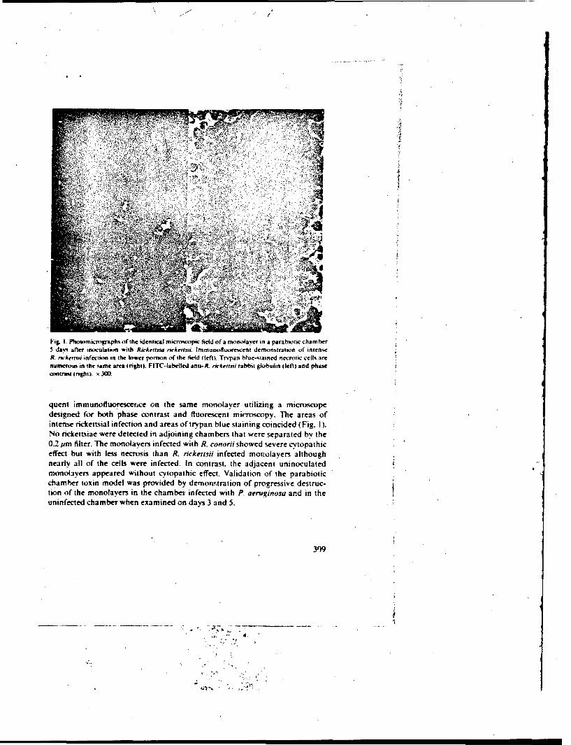
T-L
Fig. 1.P ftwimmuographa of the identical microscoipc field of a monol~ayer in a parabiotic chamnbet5 days after inoculatiim with Rickettst riwkeriss. lmmunotluores.cent demonstration of intenseR. rka&eervi infection in the lower portion of the field (left). Trvpan blue itained necrotic cells arenurnerous in the %ame area (tight). FITC-lahelled ana- R. nrketrsti rabbit globulin I left) and phase.cfltrast (nghtlt. x 300.
quent immunofluorescerice on the same monolayer utilizing a microscopedesigned for both phase contrast and fluorescent microscopy. The areas ofintense rickettsial infection and areas of trypan blue staining coincided (Fig. 1).No rickettsiae were detected in adjoining chambers that were separated by the0.2 pm filter. The monolayers infected with R. conorti showed severe cvtopathiceffec but with less necrosis than R. rickensii infected morwolayers althoughnearly all of the cells were infected. In contrast, the adjacent uninoculatedmonolayern appeared without cy'topathic effect.. Validation of the parabioticc hamber toxin model was provided by demort'tration of progressive, destruc-lion of the monolayers in the chambei, infected with P. aertiginosa and in theuninfected chamber when examined on days 3 and 5.
309
Dbcuno
In this model of severe cell injury and cell death caused by spotted fevergroup rickettsiae, uninfected cells of the same type as those injured and killedby rickettsiae were exposed to the same extracellular milieu including concen-tration of soluble rickettsial products which would have passed freely alongwith other macromolecules through the 0.2 pm pore filter. None of these ex-posed yet uninfected cells exhibited any more cellular necrosis than controlmonolavers in which both parabiotic chambers were not infected with rickett-siae. These data argue strongly against the existence of an important rickettsialexotoxin or soluble enzyme analogous to the phospholipase of Clostridiumpefringens in the pathogenesis of cell injury by R. rickettsii.
These data are compatible with the proposed phospholipase-associatedpenetration mechanism of cell injury by rickettsiae. R. prowazekii requiresattachment to erythrocytes for accomplishing rickettsial hemolysis (Ramm andWinkler. 1973 and 1976; Winkler. 1977). R. ickettsii appears to require attach-ment to cells by the cholesterol-containing receptor in the plaque model to exertcell injury (Walker et al.. 1983). Thus, prevention of passage of spotted fevergroup rickettsiae from one chamber to the adjoining chamber by the 0.2 pmpore-5ize filter would limit the phospholipase-associated penetration mechan-ism to the infected chamber. The more extensive frank necrosis with R. rickettsiiinfection than with R. cononi correlates with the greater incidence of complica-tions and mortality in Rocky Mountain spotted fever than in boutonneusefever.
This in vitro experiment does not exclude the possibility of a role for endo-toxin in Rocky Mountain spotted fever although the data for a lipopolysac-charide with in vivo endotoxin activity for rickettsiae are weak. Classical endo-toxin pathogenic mechanisms involve in vivo host-mediated mechanismsdependent on polymorphonuclear leukocytes and coagulation: these host-mediated pathogenic elements were not tested in this parabiotic chambermodel.
This parabiotic chamber model was chosen over exposure of uninfectedmonolayers to filtered supernatant of rickettsia-infected cells. Since the para-biotic system compares the effect of the products of rickettsial infection over thesame time course and concentrations as of the monolayer with rickettsial cyto-pathic effect, its negative results may be interpreted as valid. In contrast, acuteexposure of monolayer to filtered supernatant would not reflect such a dynamicinteraction.
The formation of enlarging foci of infection and necrosis occurs in thismodel with fluid medium that allows release of rickettsiae from infected cellsinto the medium and spread to infect randomly any cell of the monolayer. Thecontiguity of most infected and injured cells suggests that cell-to-cell spread ofrickettsiae ma) be important in the pathogenesis of spotted fever group rickett-
310
/t

sioises. Thbis contiguous distribution of rickettsia-infected and injured cells isanalogous to our observations of fickettsial distribution in human RockyMountain spotted fever (Adams and Walker, 198 1; Walker et al.. 1978) and iscompatible with the proposed mechianism of injury by the phospholipase-asso-diated penetration mechanism. F-,i her experiments should be designed to ex,ploic this and other direct fickettsial mechanisms of cytotoxicity other thansoluble exotoxins and enzymes.
Acknowiedpmeis
This work was st pported in part by Public Health Service grant AI-16023 from the NationalInstitute of Allergy and Infectious Diseases and in part by United SLates Army Contract DAMD17-83-C-3 122. We thank Jennie Lu Hollander for sccretarial assistance in thc preparation of themanuscript.
Adams J1. S.. Walker D. H.: The liver in Rocky Mountain spotted fever. Amer. 1. dlin. Path. 75. 156-161 (1981)i.
Bell E. I.. PickensE. G.: A toxic substance associated with the rickettsais of the spotted fever group.J. Immunoil. 70. 461-472 (1953).I
Bovamick MI. R.. Allen E. G.: Reversible inactivation of typhus rickettsiae. 1. Inactivation byfreezing. J. gen. Physiol. 38, 169-179(11954).
Bovarnick M. R.. Allen E. G.: Reversible inactivation of the toxicity and hemolytic activity ofryphus rickettsaie by starvation. J1. Bact. 74. 637-64A5 (1957).
Garvey J. S.. Cremer N. E.. Sussdorf D. H. (eds.l: Methods in immunology. 3rd ed.. p. 445-449.Bý,niamin-Cumminj 5s Publishing Co.. Reading. Mass. 1977.
Gildenmeiister E.. Hatigen E.: Fleck.4eberstudien. 'Nachweii cinies Tcxins in Rickettsien-Eikul-turen (Rickettsia mooseri). Dtuch. med. Wschr. 66. 87F,-8801(1940).
Moe J. B., Mosher 0. F.. Kenyon R. H.. White J. D.. Stookey J. L.. Bagley L. R.. Fine D. P.: Func-tional and mrorphologic changes during experimental Rocky Mountain spotted fever in guineapigs. Lab. Invest. 35. 2.35--245 11976).
Murray E. W.: Rickettsiae. In: Microbiology. ed. by B. D. Davis. R. Dulbecco. H. N. Eisen. H. S.Ginsberg. 3rd ed. p. 763-774. Harper & R ow. Publishers. Haigerstown 1980.
Ranim L. E.. Winkler H. H.: Rickettajal hemolysis: adsorption of rickettsiae to erythrocytes. Infect.Immun. 7. 93-99(1973).
Ramm L. F.. Winkler H. H.: Rickettsial hemolysis: effect of metabolic inhibitors upon hemolysisand adsorption. Infect. Immun. 7. 550-555 (.973).
Ramm L. E.. Winkler H. H.: Identification of c.tolesterol in the receptor site for rickettsiae on sheeperythrocyte membranes. Infect. Immun. 13. 120-126 (1976).
Schramek S.. Brezina R.. Kazar J.: Some biological properties otf endotoxic lipopolysaccharidefrom the typhu~s groups rickettsaie, Acta virol. 21. 439-441 (1977).
Walker D. H.. Cain B. G.: The rickettsial plaque. Evidence for direct cytopathic effecct of Rickettsiarirkenxii. Lab. Invest. 43. 388-396 (1980).
Walker D. H.. Cain B. G.. Olmstead P. NI.: Laboratory diagnosis of Rocky Mo'untain spotted feverby immunoifluirescent demonstration or Rickettsia rickettsii int cutaneous lesions. Amer. 1. dlin.Path. 69. 619--623(J1978).
Walker 0. H.. Firth W. T.. Ballard 3. G.. Hegarty B. C.: Role of the phospholipa~se-associatedpenetration mechanism in cell injury by Rickettsia ricketrtiti. Infect. I mmun. 40. 8.40--942 (1983).
Winkler H. H.: Inhibitory and restorative effects of~ adenine nucleotides ott rickettsial adsorptionand hemolysis. Infect. Immun. 9. 119--126 (1974).
311
Winkler H. H.: Rickettsial hemolysis: adsorption. desorphion, reabsorption. aad hemagglutination.Infect. Immun. 17. 607-613 (1977).
Winkler H. H., Miller E. T.: Phospholipase A activity in the hemolysis ofsheep and human erythro-cytes by Rickettsia prowazekii. Infect. Immun. 29. 316-321 (1980).
Winkler H. H.. Miller E. T.: Phcspholipase A and the interaction of Rickettsia prowazekii andmouse fibroblasts (L-929 cells). Infect. Immun. 38. 109-413 (1982).
4
1 ~1'
'4
312
Tropical Diseases Research Series AWPublished on Ix nalf of theUNDP / WORLD BANK / WHOSpecial Programme for Research and Training in Tropical Diseases
The series will deal with current outstanding problems in research related to thecontrol of major infectious tropical diseases. It is intet,1ed to attract scientistsworking in other disciplines to the problems and opportunities for research whichare offered by the infective agents of concern to the Programme, and to acquaintthose already working on these organisms with new awareness for investigationwhich advances in other biomedical sciences may offer.The Special Programme is a goal-oriented research and training programme withthe objectives: (I) Research and development to obtain new and improved toolfor the control of major tropical diseases, and (2) Strengthening of the researchcapability of the tropical countries.
No. I: The Role of the Spleen in the Immunology of Parasitic Diseases
No. 2: The Membrane Pathobiology of Tropical Diseases
No. 3: The in vitro Cultivation of the Pathogens of Tropical Diseases
No. 4: Modern Genetic Concepts and Techniques in the Study of Parasites
Order form IBestellkarie
Name and addrew:
Schwabe & Co. LtdPublisher
Steinentorstrass 13
CH-4010 BaselSwitzerland
,I
Request for sample copies and informationAs addressee of this reprint you are surely interested in the journal in which thispaper was published or another of our publishing house. For further informationwe will send you sample copies on request Please indicate the journal desiredwith a check and return the attached postcard to us.
Probeheft- und ProspektanforderungAls Emp(Anger des Sonderdruckes stnd Sie sicher an der Zeitschrift. in der dieseArbeit erchienen ist. oder an einer anderen Zeitschrift unseres Hauses interes-siert. Zur nmheren Information lassen wit lhnen auf Wunsch gem Probeheftezugehen. Bitte kreuzen Sit die betreffenden Zeitschriftlen an uLd senden Sic dieanhhngende Karte an uns zurack.
SCHWABE & CO AG.VERLAG. BASEL
ORDER FORM SCHI,4BE
Sam*p copies Information"o Act& Tropca 0 Tropial Diseases Researc-h Series
"o Act Psedopercbiamca Published on behalf of the[3 Acs Paeo1•iaricaUNDP/World Bank/WHO.
"o Heivetc Chirura Acts 0 Comete Cata e
"o Helvetica Paedistrica Acu 0 Catalu of P-riodk
0 SchweizerischehMedizinische Wochernmbrft
Aeft Vir"I. 23: 58-5-14, 1984
IiNFECTION OF GENETICALLY IMMUNODEFICIENT MICEWITH RICKETTIA CONOR1f
N. R. MdONTENEGOo, 013 H. WALKERt'). OB. C. REGAitTY
Univeruidadle Patodual Paulista. Botucatu 8P, Brasiland ODepartvwnt of Patholoay. University of North Carolina School of Medicine,
Romi~usd Apral 10, 1984
Summary. - In order to determine the definitive importanceof T- and B-lymphocyte. in immunity to Rickeft*ia cOOnt, mice'genetically deficient in T-oells, B-cella, or both T- and B-oellswere infected experimentally. T-lyrnphocytes rather than humoralantibodies were crucial to rickettajal clearance and a reducedmortality rate. Mice incapable of an antibody response to poly-saccharide capsular antigens effectively controlled rickettsialinfection with no mortality. In contrast, nude mice producedantibody to thymus-independent antigens early in the course ofinfection, yet experienced severe rickettsial infection resulting indeaths. The observed hepatic lesions are similar to those ofboutonneume fever. This model offers the opportunity to investi-gate rickettsial immune mechanisms and hepatic injury.Key wtortie: T-Jympkocyles; B-lyixphoe~ea, Rckettei4 eonorsiiboadonneuse fever; liver
fsgrodasdion
The pathologic lesions of the tewAe "wre and rash of boutonneuse feverand the inoculation site in guinea pigs experimentally infected with Rickdt*ia"coorii contain rickettsiae infecting the vascular endothelium and leukocyteso( the immune and phagocytic systems (Montenegro el at., 198.3a; b). Therolei of each of these elements (rickettsiae and host response) in the poatho-gene.oin of timstie injury were not determined. Likewrise, the rplative con-t~ribution of litymns dependent (T) lymphocytes and B-lymphocytes toimmune recot ,)ry from R. coitorii infection has not been defined. Investi-gations by K 'korin and co-workers suggested that T-lyniphoc-yteo are im-portant for i~nmunity to R. rooiorii (Kokorin Pil al., 1692). Inbred l)BA/2mice wero im~munosuppremmod by cyclophomphamide treatment and infectedwith R. ronorii; the animals were given spleen cells from immune and'non-immune dcnors. They demonstrated that immune spleen cells conferredprotection and stiggeted that the T-Jymphocytýe was crucial foi the de-
1) Whomn ,uaqtq rOw F.?)flfti mhomild h"w~ adr"^a*4 to.
,4ea
R. CONOR1! IN IM11UNODKIFICIENT MICE 9
velopment of immunity rgainst R. cwwsrii. This conclusion was based onthe lows of protection after pretreatment of transferred immune spleen cellswith antibodies inactivating T-cells.
Our work was designed to examine further the relative importance ofT-and B-lymphocyte. in the pathogenesis and immunity to R. caonoii. Weused mice genetically deficient in T-cells, B-cells as well as in both T- andB-cells, inoculated by intradermal route to examine the presence of rickettsaieand pathologic lesions at the inoculation site and in systemic tissues.'
Materials and MethodsA niowd. Thirty mice (seneproual) providd by I)P. D~om Mickety. University of North Carolina
at Chapel Hill), included six 46-day-old male 8-lymphoryte deficient (NIH 11) mice. (Group 1)(Amohaugh rt d.. 1972), six 46-day-old male T-lymphooc te deficient (NIH nude) mice (flroup 11).six 46-day-old T. and B-lymphoeyte deficient tNIH 11) mice (Group 1l1) (At-at o #41.. 19041)andl twelve 62-day -old male Swiss. outhred immunologically intact mice (Group'IV). Each groupwe& hou'sed in a neparato csae except Group IV which was divided between two capros.
Rickdteaso. Riekofasi, eotoski (strain 7) wais obtaiuned from ATCIX as a frowen 41% yolk mecsuopernsion. Stock R. coaerii woo pseparoid by inoculation of the vaolk me. of 3Sday-old specifliopat hogroi-fice- emhcy-onated hen's ega sand hariiested 3 days later. Aliquots wer@ storeod frove at- 70) C aAsa 100% susppension in sucrose phosphate buffor. These aliquots contained 6 x 1t)4 PPU/mlwhen titrasted in primary chock embryo cell (CEC) culture.
Seeolnqp. Serum was evaluated for antihoilies to R. conqnii by indirect immunofluorescent anti-body asooty (Philip ot zii.. 19761 using microilots of R. ceomenti (ostrian 7) cultivated in rabbitkidney-13 IRK-Li) cells. Slideso with mcit-odots were. sir-diried. fixed in secotos' tar It)min.andl 4torowd fro men ait -- 70) *C. Seortal dilut ions of mouseworisa wr incubated on micrtodotm for30 trmm at room temera tire in a moist chlambher. S4lideso were washed in pihosophatoe txff~modsaline (PHS) for 341 niin, reactedo with fluonw-'.in tsothitioaniate-tconjiigtedl rabbit tati-moup"'immunoitohulin l1AKO. Ac-urate Ctwenocaj and ScientificComnlianvr, Westliry. N.Y.). diluted
1 20, wasthed in PBS4 agaan, and mnounted in 90".' Klyceprol in 1'FlS. Slides were examined ona Leitz Oe~holux ultraviolet microwmioe with incident hewan illuminatiom equiplord with harrierand exiciter! filters far FITt'.
His~age and imsti~dc~i~~for apotted freer group rnceoliao,~. At thes time' of ideath,.the inoceulia tion site. splecin, andu liver wpme col-etaid. fixed in 406 neutral buffred formnalilehydefor 24 tipi. emlieddl.d into pa ~affln. andi cut at 4 j&m. Sectwins were otuineut with heomotox'ulin-Poosinanti bv the Brown-floploos method. O~ther secrtionsm were examitned for R. en~ane- by immunii-ftuwm"sc-ence using a .1r-ct conjugate for %pottedl fever group rickettsiae (Heeotl A4 at., 11154)afterdicp ffinixatitin and trypoin diarestion (Walker and Cain. 197,1: Monienegro .4 a.. IlI:ks. b).
Xr.-rwimieal doigr"sq. F~ive animalm fromt each t.% p.' of immunodoefci-ent micre andi ton intactAwiss ion" were inoc-ulated with 0).: ml of suspenstion containing 1.2),i 104' PiU (if Rt. cutsorii.O~ne nnimal in each of the six cagsir was markedt for idenotificat ion anit inoculated with t).2 mloftmedium tonitainimnit no ntckettoiae. O n day :1 oine(,- -te~l animal from each grr.uioi was oacrificedby culli-iting blood from the heart unider Atoiium-prtwnohaiibtal a-itea.On iday 7 two minfe-tedaninals wind ono- control antamal fr-mant eh uriti-op and cn day 15 the iemtainider of the animalsiwere '.imilarly "xmnsnitinated. Serum was w-parateit from clotted bloiod. Se-runm splecimenst fromthe same animal group colltected on the stame lay were poooled.
Resuts
Mortality due to R. conorii infection was observed only in T-cell deficientmice (2 deaths) and T- and B-cell deficient mice (1 death). The content ofrii~kettsise in tissues also followcd the same pattern with more R. t'onorsiiin the livers and spleens of the T-celI deficient mice and T- and B-cell deficientmice than in the B-cell deficient mice and normal mice (Table 1). The humoral
44
"- ,
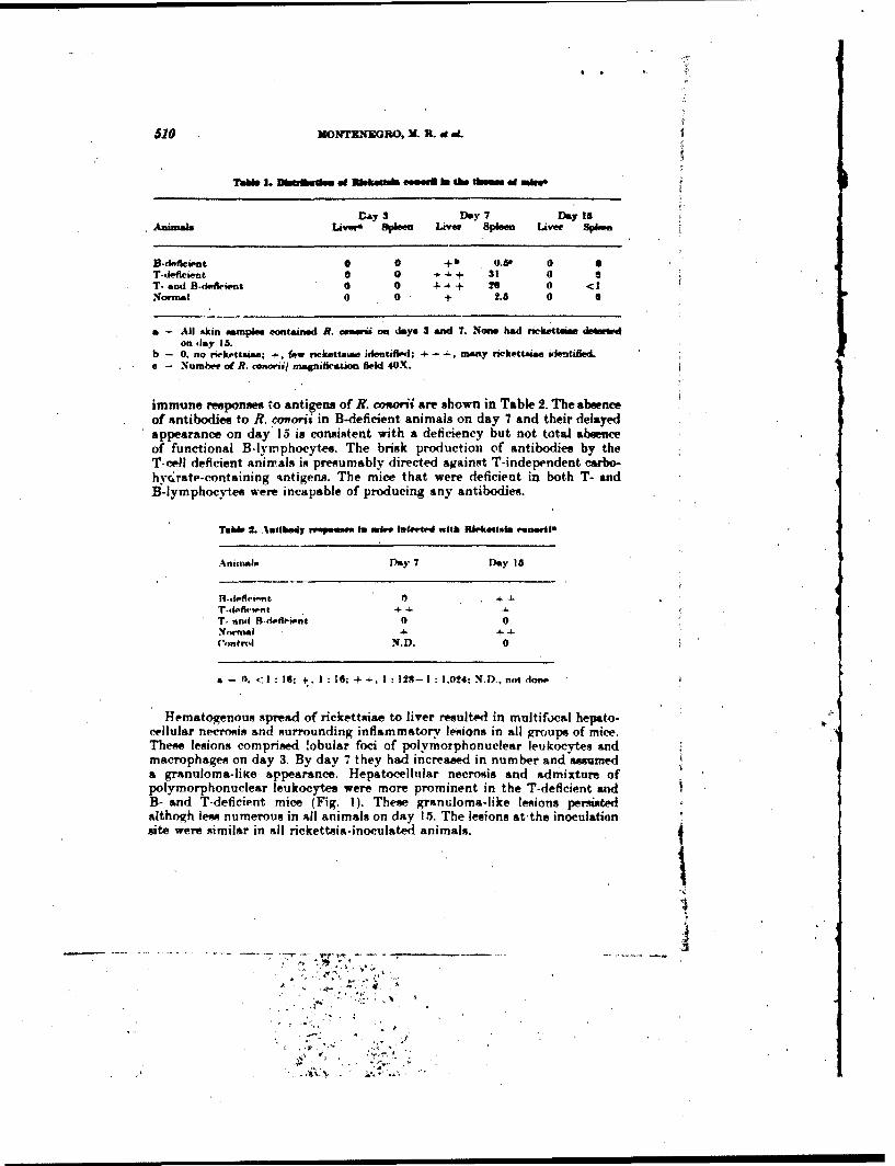
69MO~rNTIIGRO, M. PL aL
TaM. 1. Dbffiadenm 4d Mehat4 esmeeil JI the tba~ adm.
Day 3 Day 7 Day isAnimals Livers Spleen Liver Spleen Liver Spleen.
B-deficaiost 0 0 + b 0.60T-deficient 0 0 + -4.+ 31 0T- and B-deficwnt 0 0 + 4+ 26 0 <1Normal 0 02.6 0 S
a - All skin samples contained R. ceeerii on days 3 and 1. Nowe had neckettelee theeruPdon (lay 1S.
b - 0. no rvekettname; +t, few rickettsaea identdfwd: -4-+ many rickettsiae identalleda - Number of R. conorni/ magni&iration field 4OX.
immune response@ 'to antigens of R. maorii are shown in Table 2. The absenceof antibodies to R. "oorii in B-deficient animals on day 7 and their delayedappearance on day 15 is consistent with a deficiency but not total absenceof functional B-lymphocytes. The brisk production of antibodies by theT-cell deficient Animals it; presumably directed against T-independent carbo-hvc.rate-containing -,ntigens. The mice that were deficient in both T- andB-lymphocyttes were incapable of producing any antibodies.
Tubbe 2. Antibody reepeusom In mire luneIed with RoielaiettelerewiI
.Anirnal My 7 Day I16
T.defiseint 0&.~
T. anti H-deflheient I)0Normal -
Control N4.D. 0
Hematogenous spread of rickettsiae to liver resulte-d in multifocal hepato-cellular necrosis and surrounding inflammatory lesions in all groups of mice.The"e lesions comprised !obular foci of polymorphonuclear leukocytes andmacrophages on day 3. By day 7 they had increased in number and assumeda granuloma-litce sppearance. Hepatocellular necrosis and admixture ofpolymorphonuclear leukocytes were more prominent in the T-deficient andB- and T-deficient mice (Fig. 1). These granuloma-like lesions persbotdalthogh less numerous in all animals on day 15. The lesions at the inoculation
site were similar in all rickettaia-inoculated animals.
R. CONORII IN INNUNODIPFI7C T MIC 11
Presented experiments in gonetically immunodeficient mice document theimportance of T-lymphocytes in the clearance of R. cuorii from the tissuesof infected mice. On day 7 the spleens and livers of T-cell deficient as wellas of T- and B-cell deficient mice contained numerous rickettsae, whereasanimals with an intact T-lymphocyte immune system containked very fewrickettsiae in their hepatic and splenic tissues. The only animals that diedas a result of R. camoni infection in these studies were those deficient inT-lymphocytes whether or not they had B-lymphocytes. These resultssupport the conclusion of Kokorin ed al. (1982) that T-lymphocytes conferprotection against R. oasoii infection in mice. Our resuits, however, do notrely upon general immunosuppression after cyciophosphamide treatment,a drug which affects many tissues including not only T- and B-lymphocytesbut also proliferating cells of the bone marrow and other tissues, and re-constitution with a mixture of mouse spleen cells that were either untreatedor depleted of lymphocytes by an absorbed polyclonal antiserum principallydesigned to affect T-lymphocytes. Instead, theme studies employed geneticallyimmunodeficient mice that have deficiencies of ipecific components of theimmune system.
Moreover, the generation of a humoral immune response in our experimentsdid not correlate with clearance of rickettsiae and protection from death.T-lymphocyte deficient nude mice synthesized antibodies to R. aRooriidetectable on day 7 at which time the visceral organs contained manyrickettsiae. Despite the presence of antibodies in these animals, mortalitywas observed. Nevertheless, B-lymphocyte deficient mice had no antibodiesand yet had effectively restricted rickettsial proliferation in the liver andspleen on day 7; moreover, none of these mice died. These observationsconform to the conclusions of previous studies on immunity to members ofthe genus Rickettria, namely that T-lymphocytes are crucial for immunity(Shi rai eg al., 1976; Walker and Henderson, 1978; Murphy et al., 1979;Kenyon and Pedersen, 1980: Kokorin et al., 1982).
It is important to note that this does not exclude an auxiliary role forantibodies in opsonizing rickettsiae for phagocytosis and digestion particularlyby macrophages (Gambrill and Wisseman, 1973; Beaman and Wisseman,1976) or for specific antibodies to particular rickettsial proteins, e.g. a proteinfor attachment to host cells prior to entry into the cell, in blocking rickettsialpenetration or another rickettsial function of pathogenic mechanism. Jerrellsand Eisemann (1983) have suggested that the antibodies produced by nudemice to spotted fever grolip rickettsiae may be directed to the polysaccharide--containing slime layer described by Silverman et al. (1978). Nevertheless,this strain of B-deficient mice that has been shown to be defective in theproduct;on of antibodies to pneurnococcal capsular polysaccharide (Amsbaughet al., 1972) effectively clesrs its iissues of R. conorii. Protection o0 guineapigs against R. rickettai by specific antibodieq to a surface protein has beenshown by Anacker et al. (1983) and by monoclonal antibodies to surfaceantigens to Rickpjtima rickettaji (Lange, 1983, personal communication).
5 2 MONTENEGRO, )X. R. c ,d.
H wever, the natural generation of antibodies after initiation of infectiondses not seem to be as effective as incubation of rickettsiae with antibodiesprior to inoculation (Anacker et al., 1983) or prophylactic administrationo9 antibodies (Ricketts and Gomez, 1908; Topping, 1940).
The mechanism by which T-lymphocytes confer immunity against rickett-si'e has been suggested by recent in vitro studies of lymphokines (Nacyand Meltzer, 1979; Nacy and Osterman, 1979; Meltzer and Nacy, 1980;Meltzer et al.. 1982: Turco and Winkler, 1983a, b, c: Wisseman and Waddell,I 983). Lymphokines, y-interferon in particular, exert rickettsiostatic effectsoh intracellular organisms not only in macrophages but also in fibroblastsand endothelial cells. Further evaluation of the importance of y-interferoniiý vivo supports this as an important mechanism by which T-lymphocytesserve as a crucial host defense against rickettsiae (Palmer et a., 1984).
The immune response has not been shown in this or previous studies tob6a pathogenic mechanism of tissue injury in infections by members oftie genus Rickettsia (Moe et al., 1976; Mosher et al., 1977; Walker and Hen-dtrson, 1978; Kenyon and Pederson, 1984); Kokorin et al.. 1982). The cvtolyticeffect of lvmphokines on rickettsia-infected cells in vitro again raises theqriestion of an immunopathologic mechanism of tissue and cellular injuryiii rickettsial diseases (Turco and Winkler. 1983a, b: Wisseman and Waddeli,1983). Although this investigation does not exclude a contribution to cellinjury by lymphokine-mediated cytolvsis, it does document that in the0oerall balance the T-lymphocyte aff•ords protection against R. monorii.
her studies including the in vitro plaque model have demonstrated thatrickettsiae pogsegs direct cvtopathic activity that appears to be mediatedai least in part by the p)hospholipase-a.-s)ciated penetration mechanism(Walker and ('sin. 198O: WVinkJer and Miller, 1982: Walker et al., 1983) and
active in the absence of the immune system.The similarity of the hepatic lesions of these animals to hepatic lesions
in boutonneuse fever contributes to underStandrng of the pathogenesis ofthe human lesions of multifocal hepatocellular necrosis and associated focalhepatic inflammatory response (Guardia Pt al., 1974. Faure et al.. 1977;Walker, Staiti, and ilansueto, unpublished data). The finding of R. conoriiini these lesions in mice suggests that the rickettsiae play an important rolein their pathogenesis. The immunodelcient mouse model of R. conoriiinfection should be useful in further dissection of path,)genic mechanismsof hepatic injury by R. conorii and in evaluation of the importance ofstimulation of each component of the immune system by specific purifiedrickettsial antigens in protective immunity and vaccine design.
I R p ete ne.,,,
Ai.\nimuq~h. 1). F.. Hand-n. C'. T.. P?,4ýott, H... Atn, in.hk 1. WV.. HartholdI 1). R..,and Mtker.r PA.
(197): (,Gene't" oontr.i ,of i 1w antihtiv r"-..p.n.w, to tyvw III pne.'nm et.-'al lw', •-ivafharidiin mu,.,. 1. .videnev. tl',ni an % rinked w-n, e pu av a a ,et 'v n. role in detormining n'rtIr)nooeno, ."J P. AMed. I:3. 93:1 --41).
,anmeker. A. L., Philip, R. N.. (aup•.r EK. Toili, W. J., Mann, 1. i...lohnston. M. R.. andNauek. C. J. (19.11: Biolog,'ial propu'rti o,"rabbit antiho~tii. t, a •arfa-" ,untlijn of PJickett•i•
ricrkett*ii,. Ifect. Immun. 4C 292-291.
.
a _________ ____________
-• 1 -t .
R. CONORII IN INYL7NODEFICIRNT MI1CE .f
Azar, R. A.. Hanson. C. T.. a=W Cosita J7. (19W0): X:NIII(S)1I-nu/nu mirae with combinedinamunalde&,wrncv: A nw mociel for. human tumori hatziotransplantat ion. J. nam. Caomeslust. 61. 421- 430.
Boaman. L.. and Wisoataan. C. L. Jr. (1975): Me-ehanitims of immunity in typhus infection*.VI. Differential opuammsing and neutralizing action of human typhus r ckettisa-specific cyto.philic antibodiett in cultures of human macrophages. Itefec. iausauaa. 14. 1071-1076.
Four", J.. Ewandp. J. C.. Grataloup, 0.. Mamont. C.. and Faaur. Ht. (197): Hepatite granulo.mostruma rieliettsaenne interet de I& hiopair heptiqia.. Vowr.. Preset Medietal. 6, :*96.
Gambuill. M. H.. and Wi~ssrman. C. L. Jr. (1973): Mmertanuaensoadimmunity in typhus infections.fit. Intiurne- aaf huumn immune srum anti roanptamont on the fate, of RiceftioiE ssoomeriwithin human macrophaw-a.. Inafect. lissmma. 8. 631 - W4.
Guaratia. J-. Mart inva-Vazquez. J. M.. Moragaa.. A.. Re. C.. Vilavec,. J.. Tornw...J.. Eloltran. w.and Bawu-rdi. R. ( 1974): The liver in hoaatonaaaaaae fever. (out MI 549 - 551.
Heberat. G., A.. Tzianahoa. T.. Gamblile. W. C.. Auaal Claapis.l. W. A. (19011): D)evelopment awlchsarsteraunatiaan ot hmgh-tataeaed. gru-Aicticfuoerseent-,hittmbdy reagents fewr diraect iden.tification of ruaItettosae, in clinic'al tipecimanon. J. clin. Micra~aoh. 11. SW-57
Jerrells. T. Rt.. anti Eittemann. C. S. (19W3): Mile of T-lymphaaaeytuas in productiont of antibodlyto antigens of Ricke~ama £aL~aaaqgamaae" anti othe'r Riekeaeu, species. Iaafeaw. suaua. 41.6"4-674.
Kenyon. R. H.. andl Paaaraaern. U~. E. Jr. (10144)): Immune ai-spoawse to Rickeaei~a akann infection.n atiaangrinitally athyms- naude ma'.. nfaaed. lasauaa. 21. 314)- 313.
Kokoarn. 1. X.. Kahanova. E. A.. tlairokova. E. X1. Ahaasaamova, Ca. E.. Itylkina. N. N. andPusthkarva. V. 1. (1982): Hole of T-ymphoarvtes in Rickett~ia cwanai infection. .4r*v aired. 691- 97.
Meltzer., . .14.sanid Narv. C. A. I I 9811: Maarnophsame in rreatusnco to raaekettsiall anf~etiamn: Sus-cepttiblaaty it) lethal affir-ets of Rtirka-a1~ apkaar infection in moual. strains with defective manaro..phaga. funtmnaaa. Cr fassausai 34. 487 - 494).
Melts".~ M.S..XNarv. 'A., ttavaenso. M. M., and Skama'. R. (1"!!): Maeraaphagaes in resistance
to rockettasiall anf~etaoaa: Gienetic. analysis of susceptibility to leithal aoffects tif Ricksissio aikariinfection anal deavelopa~ment of activated. e.ytgozbe maerrophiagea in A anai HIQ.A maice. J.IMMAM4uao. 129. 1719-1 72,1.
Moe. J5. H.. Moniator. IX. F.. Kenyon. It. H.. White. 2. 1). Staaokey..J. L. Baittley. L. It.. andiFine.. 1). P. (1976): Fanaetionsa and morphoilogical changeus during experimental Hocky Mountainaapotttil fever in cuiona pigs. LýAi. Iaara.. 131. 235- %*43.
Montenegro, W4 It., H#'gartv. B. V... Trinuohl. G... and Walkier. D). H:. (lit83ai: Identification
of Riekaeusi~ariaaoaa, an tai-ues by ammnaiiaaa-ac.Ado .fa'l~il Pniao. Is!. Trop. 2, 161- 16t..Montene~gro. 31. R.. Man~Atieto. S.. Haogarav. Ri. C., and Walker, 1). H. (186:The histology
of -taarhon naaare- oaf baoutonnaeise fapvar anal demonaaotrataoaa of Rickeaai. reoaa~ri in themt,. immunotlaawra~oaa'rac. ViareAtowtArs.rhee. "G. 309-317.
Mosher. 1). F.. Fiae. 1). P., Main.. .1. R.. Kenyon. H. H.. Ruch. 0. -L. (1977): Stuaitle of the eo.agulataon anad 4aaamplemaent ,ayata-ma aduring experimental Rocky Mountain ispotted fever inrhesus monlcayo. J. inja~ec. IN a. 133s* 90a. -- 99~9.
Murphy. J. H.. Wiamwman. C. L.. Jr.. anat Ftuawt. P. (1979): -Mechanisms of immunity in typhusinfection: Adoptive trnaniaer aof imnsuaaaty to Riekeltsiia mos"a~a. Ismeal. Imasana. :1., 387 -393.
xa'v C. A. anal Meltzer. M).S.(1079): 3Iatrophagcaa an resistanc~e to riaekettsiaa infection: Macro-aphe altian eis at, foar killing of Rikatiatson tfaateagritnitaasaa. J. limaaaaaaan. 123. 2544-. 2549.
Nary,. C. A.. said Melt At-r. M. A. (1992): Maataaaphiteas in resistance to riekattiaiall infection: Stratinsof mii's susa'eptible tn the lethal effeeto af Rickensin akaart %how defective maaeraphagsa rickett-sicidal ai.-tivity in vitro. laafeei. Irsasaaa. .11. 1096-1 lot.N y.C. A.. antl Osaterman. .1. V. (1979): Waiat detenwimst in experimental scrub *yphus: role
of nrmld and activated maa~rophagea. Inf/ect. !amusaaa. 24. 744 - 7.50.
Palmer. B. A., Hetrick. F. 34.. antl Jerrells. T- J. (1984): Producition of gamma intert. 1% inmice immuno to Ricekts~iaiauqaaas. Ifaa/ac. 1 iaaawa. 4a. 5 - 6a,
Philip, R. %.. Casper. E. A.. Ormsbee. R. A.. Peacock, M. 0.. and Baargdorfer. W. (1975): Micro.* immunoifluoaeaacence test for the aaprologicala study of, Rocky Mountain "ptied fever and
typhus. J. elin. 31firaob"o. 3,5S1I- 61.',ickaetts. H. T.. and Caomesa.i 1. 1908): Studies oan immunity in Rockiy Mountain spotted fever.
First communication. J. iaajecg. Die. 3, 221 224.
Pa
.514MONTILVEGH~O. M. P_ . ade.
8hira4 A.. t'atanaw.,. P. J.. Phillips, S. M.. et al. (1976): Hoat deleniar in ezperumneitel scruhtyphius: Holue of cellular immniz~ty in he4.Uolopoua priit-.tioo. lInfecit.. Jlswm. 14, 39-46.
Siulverman. D. J., Wvpacna. C. L., Waddell. A. I).. Jonei... Jd 1 979): External 1aytis o(RckeumainproenuZkii: (hicurrnn.'. ofs, slime layer. Infect. Immuu.. U. 233 - U3.
Topping.N. H. (1940)t Rocwky M4ountain mtpolted fever. Trw~atnient of inhictod laboratory' animal*with immune rabbitsparumn. Puisbd Hfsa&A Repw.ts 33i. 41 - 441.
Turco, J.. a&d Winkler. H. H. (I983ou: Cwnmpariwon of the. pnnpertle. of antawiuketteWa activityand inte~rferon an mouusc lynblihokines. Jafirdi. Immnum. It. 27- 32.
Turro. J., sand Wiulklerý. H. H. (1983b): Inhibit Io of the. growth of Ricksawv4 ireenzekii in*cultured filwobleato ikv lvinphokine~s. J. exp. Mied. 117, 9714 -986.
Tumro.J.. and Winkler. H. H. (1910c): Clonral mouve inter4f.ron-y inhatbits the growth of RiukaUviusProaswrki in cultured moore fibroblAnst. J. .rzp. Med. I=8. 2139- 2164.
Walker. 1). H.. anal Cain. H. GZ. (197,14): A mcythtmi for peprefie diagnosis of Rocky 3lourttain
"*spteid fever on fixed. paaffin-enbiurlale tinautt by immunoflunrvw'-ence. J. sealed. Dia. 137.
W~alker. 1). H.. and Henderson. F. W. (1979): E111ect of immunivsuppnrreion on Brkncoia icrudf@,iinfiection in gwnewa pigs. lInfrd. Inwpmwa. W, ff! - 227.
Walker. 1). H., Firth,. W. T., Ballard. J. G.. andt Hegarty. B. C. (1983): Role of phiospholipsne."40 - 842.
Winkler. H. HI.. and 31ialter, E. T. (19842): Phoaphoidimop A and the interaction of Ricknitsiap~wiaweki andi moores libroblgata (L-.429 vilads. Infect. Isaunau. =. 109-I113.
Waaeman. C. L- Jr., and Waufriell. A. I(I9833: Intorfsmron -lke factors from antigen. and initogen.v-stimulatedl human leukocy"e withI antimckettmial and rytolytie actionsa oun Ricettira,~ pro.warrkis. J. exrp. Mid. 357, 17210- 1793.
Xr Luvf"t MacrogrpAe (Plate L'CIX):0
Fig. 1. (luafter. of immuanolluort-cont Rtirketuin' cnovwi (arrows) in a T-oehI de&ient moire.,(a) spieen (left) aid (b) liveer granuloma (right) 7 days" after' inoculat ion. Anti-spotted fevergr~iip rickettsaa.' FJTC-laheli~e i.onjtgnte, 3735-. e) Poleattei.. o pneerion surrotinderd hinoIiulr aceumimlat ion of polvmorplionu..l.r lpukorvtus ank: ".a.rophaiges in a T-meil deficientmourew 7 days after inoculation with Rickr*1.au coOrtis. Hemsntoxylin-Powin stain; x 375.
Bar reprerents 14) in.
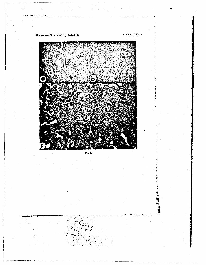
XManua gro.1 R. od a.e (!1. 0 -o 3141 PLATIh LXIX
2. '
IN
COP,,,i 0 156S by The AMO Sonno Trrupco 14.6c #d Hyo
CORRELATION OF THE DISTRIBUTION OFRICKETTSIA CONOPJI. MICROSCOPIC LESIONS, AND CLINICAL
FEATURES IN SOUTH AFRICAN TICK BITE FEVER
DAVID H. WALKER &-o .1. H. S. GEARDepartment of Pathology. Umwsevty of North Carolina. Chapel HUZL
North Carolina 27514 (USA) and Thor South Africanu Instiuteof Medical Research, Johannesburg (South AfrnJa
Abseroct. Three South African patients wi~th severe Rickettnia cononii inlection hadcomplicated courses of illness with 2 fatal cases and I with gangrene of multiple digits.Immunofluorescent organisms of R. conorii were demonstrated in vascular endotheliumnof brain, leptomeninges, renal glomerular arterioles and capillaries, renal arteries and veins,myocardial capillaries and arteries, pulmonary alveolar capillaries, pancreatic wepma splenticarterioles, and dermis. Rickettsiae we.. also observed in hepatic sinusoidal lining cells,splenic and lymph node macrophages, and the blood vessels of the partially vuiabe zoneof the amputated digits. Pathologic lesions included ceirebral and cerebellar perivascularmononuclear leukocytes, mild mononuclear leptomeningitis, glomerular arteriolitis, vas-cular and perivascular mononuiclear cell-rich inflammatory foci in the kidney, pancreas,skin, and myocardium, hepatocellular necrosis, and pulmonary edema, The sites of lesionsand rickettsiae showed strong topographical correlation. Thrombi and hemorrhage occurredin a minority of the sites of vascular injury. Rickettsiae were the apparent direct cauze ofmeningoencephalitis, peripheral gangrene, and other foci of vYscular injury. Fatal A. cotoriiinfection with disseminated organ involvement emphasizes the pathogenic potential of thisdisease.
Rickettsia conon,. a member of the spotted exist among these geographic and ecol ogicfever group of rickettsiae, is distributed widely sources. Although human infection with R. co-through southern Europe. Africa, and the Middle non: is generally considered to be a self-limitedEast. Strains of R. , fonoii from this broad geo- illness of moderate severity, the range of clinicalgraphic distribution have been assigned to this severity contains subjects with antibodies to R.species on the basis of quantitative measure- canonsi and no history of febtile exanthem, 2" aments oi'the capability of antisera to neutralize patient with tache noire and no other signs or-the lethal effect of intravenous inoculations of symptoms,4 patients with severe manifestationsthe respective rickettsiae. I The disease caused by such as acute renal faiure, neurologic signs, or-R. cononi has borne various names reflecting the peripheral gangene,3-' and fatal cases.'" Severegeographic occurrence including Mediterrant-an cases have been observed in Israel, South Africa,spotted fever, Kenya tick typhus, South Africs- France, and Italy. Host factors which may affecttick bite fever, and India tick typhus. The mt. ,,ýr the severity of illness are ghacose.-6-phosphateecologic niche o.ý the rickettsiae affecting ir --i is dehydrogenase deficiencyO and older age."~ Spe-the dog tick, Rhipicepholus sangutnettr which cif& rickettsia virulence factors have not yet beenmaintains the rickettsia by transnvzjial trans- identi'ried. The pathologic alterations associatedmission and transmits the rickettsia to man by with human R. cononii infection have been re-its bite. Ofthier ticks which have been reported to ported only in skin and liver.16- 3- n"-' We de-harbor R. cononi include members of the genera scribe the pathoiogy and rickettsia] distribution
j Rhipicephalus. Ixodes, Dermacentor. Hyalom- in the central nei vous system, kidney, liver, heart,ma. Ambb'ormna. and Hemaphysalis. It is pos- lung, spleen, lywrph node, pancreas, adrenal, andsible that some i-ickettsial geneo.tic diversity may skin. These are the first descriptions of the pa-
thology and rickettsiae in the tissues of fatal R.Accepted I I October 1934. canonsi infection.
361
362 WMA.Mt AND GEAA
MATMALS AND MET•ODS monocytes. and 7% lymphocytes, .4atelet count65,000/1A, prothrombin index 68%, serum so-
Paraffin sections of tissues from 3 cases Of dium 121 mEq/1. serum potassium 4.9 mEq/1,South African tick bite fever were stained by blood glucose 22 mg/dl, blood urea 42 mmol/Lroutine hematoxylin-eosin method, phosphl- and creatinine 415 smol/l. His urine containedtungstic rcid-hematoxylin technique, and deput- numerous red blood cells and white blood cells,affinization-trypsin digestion-immunofluores protei, and glucose. His cerebrospinal fluid wascenct method with a conjugate reactive with R. slightly xanthochromic and contained 1,280 redconovii as reported in detail previously. ' T4s- blood cells, 3 neutrophils, 1I lymphocytes, pro-sues examined were cerebrum, cerebellum, kid- tein 93 mg/dl, and glucose 11.2 mmoLl. No bc-ney, liver, spleen, heart, Jung. lymph node. anId teria were observed and bacterial culture yieldedadrenal gland (Case lI cerebrum, kidney, ver, no growth. Virus was not isolated from his bloodspleen, heart, lung. and pancreas (Case 2d and by the inoculation of litters of baby mice andgangrenous digits (Case 3). Positive cnntrol tis- tissue cultures. Two adult male guinea pigs in-sues for R. conorii immunofluorescence includ,!d oculated with his blood on the day of admissioneschar from guinea pig inoculated intradermally to Rietfontein Hospital developed a slight feverwith P. ct.--ni (strain 7), eschars from Patients associated with a slight scrotal reaction. No pa-witl serologically documented boutonneuse se- sages were made. When their sera were tested aver, and liver tissue from nude mouse inoculated month latu- in the rickettsial complement fixa-with R. conori (strain 7). tion test, a positive reaction with Rickettsia con-
orii var. pyjpen antigen was noted, thus confirm-ing the clinical diagnosis of South African tick
RESULTS .bite fever.
Clinical observations It -vas subsequently ascertained that his familyowned a dog which was allowed indoors and to
Case I. A 54-year-old miner employed on one sleep on the patient's bed. It was dipped regularlyof the gold mines of the Witwatersrand became to rid it of the ticks which it acquired in theill with "flu-like" symptoms ofheadache, muscle vicinity of the house. The patient had com-pain and fever. He became worse on each su6- plained of a -pimple" in the middle of his backceeding day and on the fourth day developed a 4 or 5 days before he became ill. A plaster wasprofuse rash and was admitted to the hospital. put on it and when it was removed left a non-On the ninth day, the rash became hemorrhagic. purulent sore. It seems likely that this was theand he was transfered to the Rietfontein Fever primary lesion of tick bite fever and that the tickHospital near Johannesburg. 1 responsible for transmitting the infection came
On admission he was desperately ill, being from the dog sleeping on his bed.semicomatose with stertorous breathing. An ex- Case 2. A 65-year-old man, after returning totensive hemorrhagic maculopapular rash cov- his home in Hillcrest. Durban, from a holidayered his body. A nonpurulent ulcer was seen in in the inland farming district of Mooi River, be-the middle of his back. His tongue wrs coated came ill with high fever and severe headache. Heand palate covered with blood-stained mucus. became delirious and developed a profuse mac-His chest sounds were obscured by stertorous ulopapular rash which became hemorrhagic. Be-breathing. heart rate was 100/min, and blood cause ofhis rash and other signs ofa hemorrhagicpressure, 140/80 mm Hg. He had slight neck and state, the possibility of one of the African hem-back stiffness. His deep tendon re,"exes were brisk, orrhagic fevers was considered. In spite of alland the Babinski sign was flexor. I: was suspected treatment, he died about 14 days after the onsetthat he had tick bite fever. 1 of his illness. At postmortem, specimens were
Because of renal failure and urgent need for taken from the varous organs and tissues. Serumdialysis, he was transfered to the intensive care specimens separated from blood taken on 18 andunit in the Johannesburg Hospital, but died soon 25 August, the day before he died, were alsoafter admission. evaluated. Indirect. immunofluorescent tests, for
Laboratory data included hemoglobin 15 g/dI, antibodies to Lassa. Marburg Ebola. Crimean-white blood cell count 10,5 0 0 /jl with 86% neu- Congo, and Rift Valley fever vir,ses gave neg-trophils, many showing toxic granulation. 7% ative results with botn -errum specimens. The
SV .
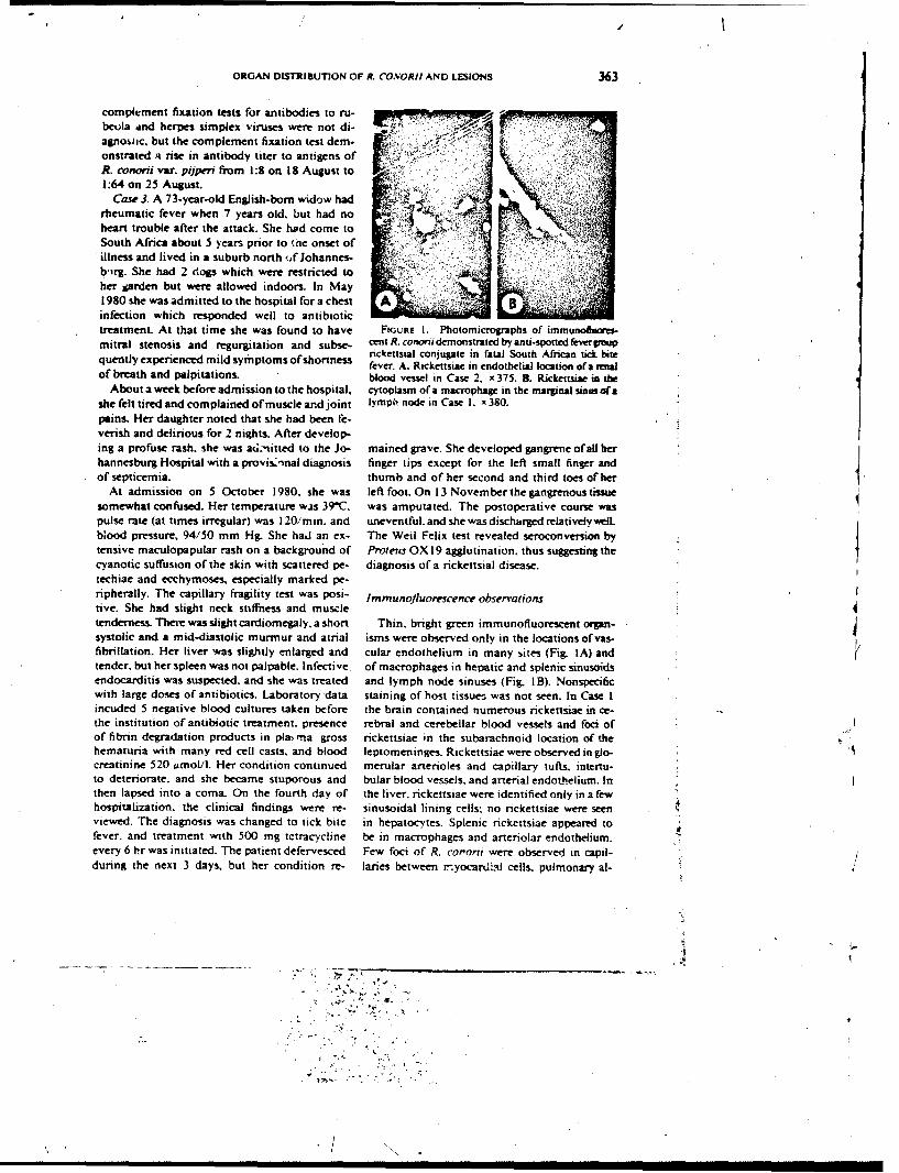
ORGAN DISTRIBUTION OF R. CONORI) AND LESIONS 363
complement fixation tests for antibodies to ru- I y ;beola and herpes simplex viruses were not di- F7,7.agnostic, but the complement fixation test dem-onstrated a rise in antibody titer to antigens ofR. conodi var. pijperi from 1:8 on IS August to1:64 on 25 August.
Case 3. A 73-year-old English-born widow had 'krheumatic fever when 7 years old, but had noheart trouble after the attack. She had come toSouth Africa about 5 years prior to (.he onset ofillness and lived in a suburb north ,f Johannes-b,irg. She had 2 dogs which were restricted toher parden but were allowed indoors. In May1980 she was admitted to the hospital for a chest Ainfection which responded well to antibiotictreatment. At that time she was found to have FIGURE I. Photomicrographs of immunoffioresmitral stenosis and regurgitation and subse- cent R. conorridemonstrated by anti-spotted feverpoup
rickettsia] conjugate in fatal South African tick bitequently experienced mild symptoms of shortness fever. A. Rickettsiae in endothelial location of a renalof breath and palpitations. blood vessel in Case 2. x 375. &. Rickettsiae in the
About a week before admission to the hospital, cytoplasm of a macrophage in the marginal sinus ofashe felt tired and complained ofmuscle and joint lymph node in Case . x 380.pains. Her daughter noted that she had been fe-verish and delirious for 2 nights. After develop-ing a profuse rash, she was aci.-itted to the Jo- mained grave. She developed gangrene of all herhannesburg Hospital with a provis.'nal diagnosis finger tips except for the left small finger andof septicemia. thumb and of her second and third toes of her
At admission on 5 October 1980, she was left foot. On 13 November the gangrenous tissuesomewhat confused. Her temperature was 39C, was amputated. The postoperative course waspulse rate (at times irregular) was 120/min. and uneventful. and she wasdischarged relativelywell.blood pressure, 94/50 mm Hg. She had an ex- The Weil Felix test revealed seroconversion bytensive maculopapular rash on a background of Proteus OX 19 agglutination, thus suggesting thecyanotic suffusion of the skin with scattered pc- diagnosis ofa rickettsial disease.techiae and ecchymoses. especially marked pe-ripherally. The capillary fragility test was posi- lmmuno]luorescence observationstive. She had slight neck stiffness and muscletenderness. There was slight cardiomepaly, a short Thin, bright green immunofluorescent organ-systolic and a mid-diastolic murmur and atrial isms were observed only in the locations of vas-fibrillation. Her liver was slightly enlarged and cular endothelium in many sites (Fig. IA) andtender, but her spleen was not palpable. Infective of macrophages in hepatic and splenic sinusoidsendocarditis was suspected, and she was treated and lymph node sinuses (Fig. I B). Nonspecificwith large doses of antibiotics. Laboratory data staining of host tissue- was not seen. In Case Iincuded 5 negative blood cultures taken before the brain contained numerous rickettsiae in ce-the institution of antibiotic treatment, presence rebral and cerebellar blood vessels and foci ofof fibrin degradation products in pla, ma gross rickettsiae in the subarachnoid location of thehematuria with many red cell casts, and blood leptomeninges. Rickettsiae were observed in glo-creatinine 520 umoUl. Her condition continued merular arterioles and capillary tufts, intertu-to deteriorate, and she became stuporous and bular blood vessels, and arterial endothelium. Inthen lapsed into a coma. On the fourth day of the liver, rickettsiae were identified only in a fewhospitalization, the clinical findings were re- sinusoidal lining cells; no rickettsiae were seenviewed. The diagnosis was changed to tick bite in hepatocytes. Splenic rickettsiae appeared tofever, and treatment with 500 mg tetracycline be in macrophages and arteriolar endothelium.every 6 hr was initiated. The patient defervesced Few foci of R. cororii were observed in capil-during the next 3 days, but her condition re- laries between rnyocardLal cells, pulmonary al-
S . .... . - -.- 9! °
1 , , . ,7
/,• .
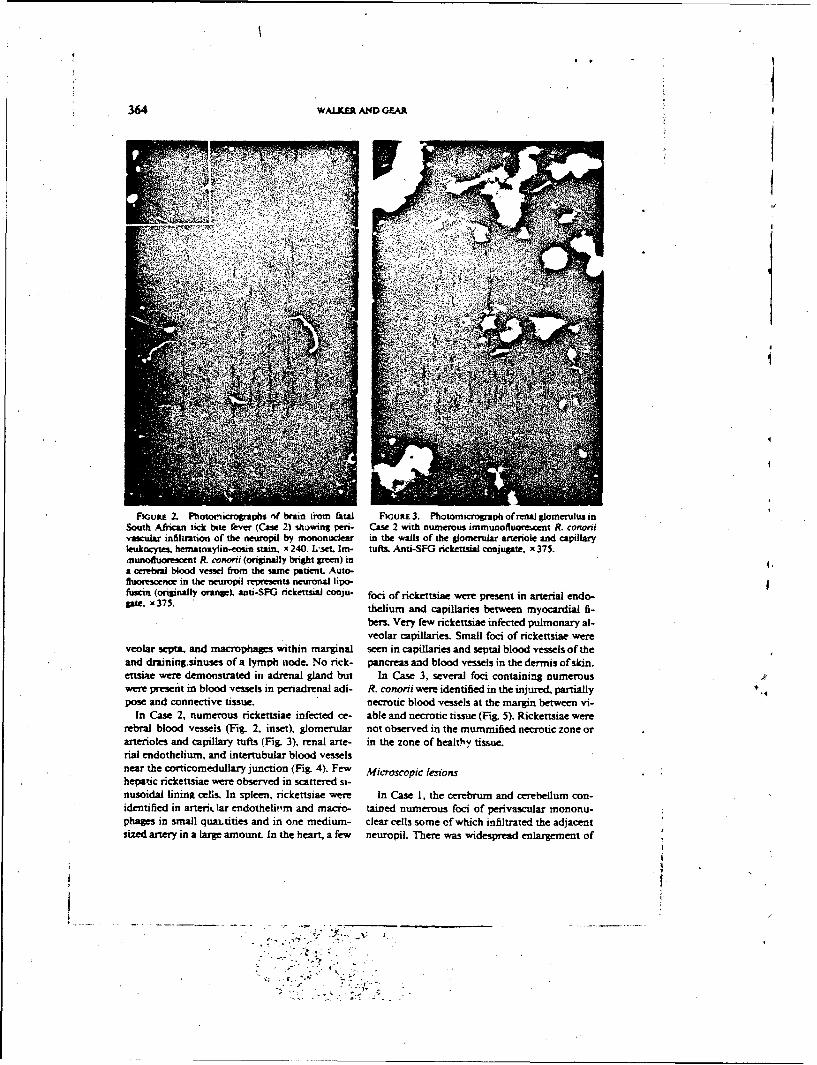
364 WALKER AND GEAR
_ Aý
FiouRE 2. Photornicrogpaphs of brain tioro fatal FtGutE 3. Photomicrograph of renal glomeruusinSouth African tick bite fever (Case 2) shawing pen- Case 2 with numerous immunofluorewcent R. cnonsivascular infiltration of the neuropii by mononuclear in the walls at the glomnerular arteriole and capillaryleukocytes. hematoxylin-eosin stain. x 240. L'set. Im- tufts. Anti-SFG rickeutsial conjugate. x 375.munoduorescent R. conord (originally bright green) ina cerebral blood vessel ftrom the samne patient. Auto-fluorescence in the neuropil represents neuror~AJ lipo-fuscin (originally orange), anti-SFG rickettsii coSju- foci of rickettsiae were present in arterial endo-pr, 35 thelium and capillaries between myocardial fi-
bers. Very few rickettsiae infected pulmonary al-veolar capillaries. Small foci of rickettsiae were
veolar sepMa and macrophages within marginal seen in capillaries and septal blood vessels of theand draining~sinuses; of a lymph node. No rick- pancreas and blood vessels in the dermis of skin.estsiae were demonstrated int adrenal gland but In Case 3, several foci containting numerouswere present in blood vessels in periadrenal adi- R. conorii were identified in the injured, partiallypose and connective tissue, necrotic blood vessels at the margin between vi-
In Case 2, numerous rickettsiae infected ce- able and necrotic tissue (Fig. 5). Rickettsiae wererebral blood vessels (Fig. 2. inset), glomerular not observed in the mummified necrotic zone orarterioles and capillary tufts (Fig. 3), renal arte- in the zone of health'y tissue.rial endotheliumi and intertubular blood vesselsnear the corticomedullary junction (Fig. 4). Few Miosoclsonhepatic rickettsiae were observed in scattered si-nusoidal lininK cells. In spleen. rickettsiae were In Case 1, the cerebrum and cerebellum con-identified in arterii. tar endotheliv,m and macro- tained numerous foci of perivascular inononu-phages in small quai~tities and in one medium- clear cells some of which infiltrated the adjacentsized artery in a large amount. In the heart a few neuropil. There was widespread enlargement of
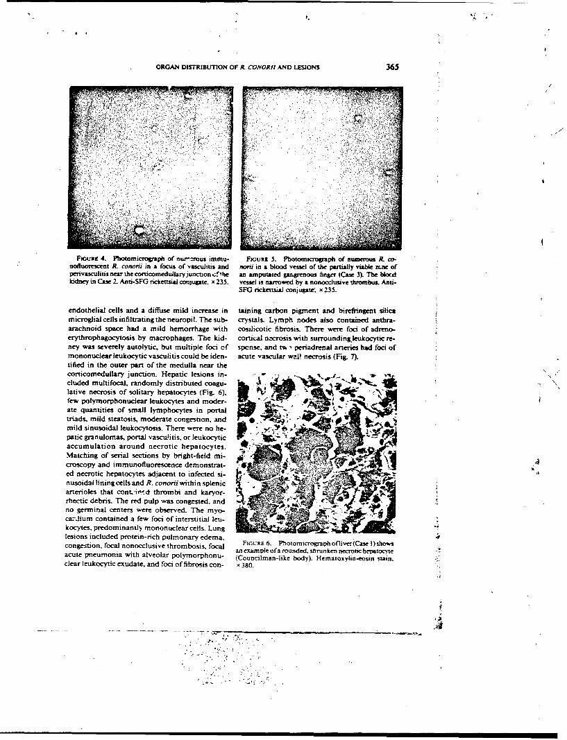
ORGAN DISTRIBUTION OF P_ CONORJI AND LESIONS 365
FiGuRE 4. Plotomicropa phi of nw'r~rous iznmu- FiouRE 5. Photomkcrograph of numerou R. cv-nofluorescent R. conorii in a focus of vasculitis and noni in a blood vessel of the partially viable zone ofpenivasculitus near dhe corticomedultary junction ./'he an amputated gangrenous finW (Case 3). The blor4kidney in Case I. Anti-SF0 nckettszsi conjugate, x 235. vessel is narrowed by a nonocclusive thrombus. Anti-
SF0 nickeutsWa con ugate, x ;35.
endothelial cells and a diffuse mild increase in tainung carbon pigment and bireffnngent silicamicroglial cells infiltrating the neuropil. The sub- crystals. Lymph nodes also contained anthra- -
arachnoid space had a mild hemorrhage with cosilicotic fibrosis. There were foci of adreno-erythrophagocytosis by macrophages. The kid- cortical ne-crosis with surrouinding leukocytic re-ney was severely autolytic, but multiple foci cf spcnse. and t% , periadrenal arteries had foci ofmononuclear leukocytic vasculitis could be iden- acute vascular wrJ! necrosis (Fig. 7).tified in the outer part of the medulla near thecorticomedullary junction. Hepatic lesions in INcluded multifocal, randomly distributed coagu-~ te.lative necrosis of solitary hepatocytes (Fig. 6)few polymorphonuclear leukocytes and moder.ate quantities of small lymphocytes in portaltriads. mild stealosis, moderate congestion, and amild sinusoidal leukocytosis. There were no hepatic granulomas. portal vascuhtits, or leukocyti c,accumulation around necrotic hepatocytes. tMatching of serial sections by bright-field mi p- ,-
croscopry and immunofiuorescence demonstrated necrotic hepatocytes adjacent to infected si-nusoidall lining cells and R. conorii within splenic ýarterioles that cont.,ii,'d thrombi and karyor-rhectic debris. The red pulp was congested, and 4 -.no germinal centers were observed. The myo-Ycazdium contained a few foci of interstitial leu-kocytes, predominantly mononuclear cells. Lung.4lesions included protein-rich pulmonary edema,congestion, focal nonocclusive thrombosis. foa FiGURE 6. Phiotomicrograph ofliver(Case 1) shown
an exampte of a rounded, shrunken necrotic hepatocyteacute pneumonia with alveolar polvmorphonu- (Councilman-like body). Henmatoxylin-eosin stain,clear leukocytic exudate, and foci of fibrosis con- x 380.
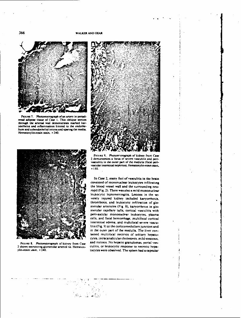
366 WALKER AND) GEAR
ýK11
U~
Foovz7.Phoomirogaphoan rtey i pevad
renal adpwtsu fCw1 hsolqescwthrogh he asenl wal dmonsrats makedkar
Fioumiti in them~rpad ofte par aoer ihe meell(ocadpri
miiiI adises 2.su oan Caci of Thislti inliu thectiontconiste toe artenuclal leuolle demnsilttrmakednagyonhthen alod vnesmmtel lwmlte ano the surounthge.
humlukcyi andoeinits sundoheia inim and w.n hemda
Frlyinure9dboo~roaho kidney fnlddkryom Caxse2hdembonstrts, an feuocysofseer infilcrtio ndo pen-vasclar interolstta (Fhnig. 8)ematyoxyeinos in stain
/In Case2 manilay tfts, ortiscaliiscuinthe brinh~ ~ ' 7 convsisltedo mononuclear leukocytes. infltating
thels baod vessel wemllrandge. srrioundln noru-a* f ropitl (i.2.Teremwas alsn id m ononucl sve e arcu
leukocyFic.9)a theptomeitis.edlr jusinction thndeeeyinjteutrpredo ktdey inluedula h liaryornei.
throedmbosisc andeukcytic onfsoitaryto of glo-myes nranlular arterioles, (Fild 8)saroreatis in l
Rouit S. Phtmcorp f kiyfo a n mirtaocaillar tuo s coraticganlomascuprtlts-wt2 sowsnerotzig Soreruarartno ti. emaox culiivscuOr lmononulclrearns toerotesc plasm
ylin-emin~' insx24.tcterstia edema ansrvd.h sultnocad seer capsula-
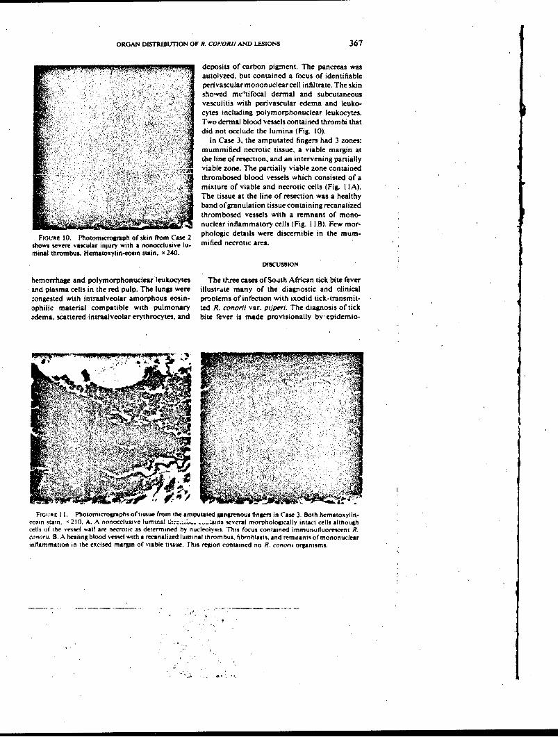
ORGAN DISTIBUTION OF A. COPORI! AND LESIONS 367
deposits of carbon pigmient. The pancra wasautolyzed. but contained a focus of identifiableperivascular mononuclear cell infiltrate. The skinsh~owed ml.atifocal dermal and subcutaneousvasculitis with perivascular edema and leuko-cytes including polymorphonuclear leukocytes.Two dermal blood vessels contained thrombi thatdid not occlude the lumina (Fig. 10).
In Cawe 3, the amputated fingers had 3 zones:S mummified necrotic tissue, a viable margin at
the line of resec-tion. and an intervening partiallyviable zone. The partially viable zone containedtl'rombosed blood vessels which consisted of a
S mixture of viable and necrotic Cells (Fig. I IA).The tissue at the line of resection was a healthy
* band of granulation tissue containing recanalized
- .~ thrombosed vessels with a remnant of mono-nuclear inflammatory cells (Fig. I I B). Few mor-
FIGURE 10. Phsotomicrograph of skin from Ca 2 phologic details were discernible in the mum-shows severe vascular injury with a nonocclusive lii- mified necrotic area.minal thrombus. Hematoxylin-.eosin stain, x 240.
DISCUSSIOM
hemorrhage and polymorphonuclear'leukocyies The three cases of South African tick bite feverand plasma cells in the red pulp. The lungs were illustrite many of the diagnostic and clinicalcongested with intraalveolar amorphous eosin- prioiems of infection wth ixodid tick-transmit-ophilic material compatible with pulmonary ted R. conorii var. ptjperi. The diagnosis of tickedema. scattered intraalveolar erythrocytes, and bite fever is made provisionally bv epidemiO-
a, 1*1 "
A
FIGURE 11. Photomicrographs of tissue from the amputauted gangrenous fingers in Case 3. Both hematoxylin-eoiin stain. '210. A. A nonocciusive lumir.=1 a-jn several morphsologically intact cells althoughcells of the veivel wall are necrotic as determined by nucfeoily-is. This focus contained immunolluoreicent R.conorri. B. A healing blood vessel with a recanalized luminal thrombus, fibroblasts, and remnant% of mononuclearinflammation in the excised margin of viable tissue. This region contained no R. cono"it organisms.
368 WAtXER AND GEAR
logic and clinical observations, treatme-nt with disease may develop a hemorrhagic state withtetracycline is administered, and subsequently bleeding into the elements of the rash, petechialthe diagnosis is confirmed by laboratory meth- hemorrhage and purpura of the skin, and bleed.ods. The patients often give a history of having in% from needle puncture wounds and from thebeen camping or picknicking in the bushveld, mucous membranes. The hemorrhagic state maywhere ticks abound, one week before becoming be associated with a marked thrombocytopenia,ilL Clinically the diagnosis is suggested by the prolonged prothrombin time, and evidence of apresentation of a primary sore with a black ne- consumption coagulopathy. The lesions in thecrotic center, associated especially in severe cases central nervous system from both diseases re-with tender regional lymphadenitis, fever, and semble the classic Frankel's noddles of typhusthe development of a maculopapular rash on the fever with cells compatible with macrophagesthird to fifth day of illness. The diagnosis can be and small and large lymphocytes permeating theconfirmed in the laboratory by serology, by the perivascular neuropil." -', A mild leptomenin-isolation of the causative rickettsiae, and by im- gitis is observed in both rickettsioses. The cere-munofluoresent demonstration of spotted fever brospiral fluid in Case I reflected the rickettsialrickettsiae in a biopsy of the eschar or rash."' meningoencephalitis with mild pleocytosis andReliance is usually placed on the results of the elevated protein concentration. Clinically, thereserological tests; however, these tests usually do may also be marked involvement of the centralnot give positive results until after the tenth day nervous system in both South African tick biteof illness. Isolation and characterization of R. fever and RMSF, progressing from severe head.conorii by intraperitoneal inoculation of guinea ache to mental confusion, delirium and coma.pgip with the patient's blood is performed only Other analogous lesions include multifocal peri-in specialized laboratories. As the specific im- vascular interstitial nephritis,10,2' focal intersti-munofluorescent demonstration of spotted fe-, er tial myocarditis, 22-23 dermal and subcutaneousrickettsiac in skin hiopsies is only available in a vasculitir in the skin of the rash, eschar, andfew laboratories, the earily diagnosis in most cases peripheral Sangrene10'- 3. 2. 11 and vasculitis in theof tick bite fever must be based on the clinical pancreatic interlobular septa 2l and periadrenalfindinp. Treatment with tetiacycline or chlor- adipose tissue. Lesions which were observed inamphenicol should be initiated immediately to fatal R. conorai infection and are not character-avoid the pathologic consequences illustrated in istic of RMSF are necrotizing glomerularthese patients. anteriolitis"0 "- 2, and multifocal necrosis of sin-
In a typical caw, the clinical course is char- gle hepatocytes. Although hepatocellular necro-acterized by complaint first of lassitude most sis other than centrilobular necrosis was presentmarked in the evening. A feeling of chillness with in 5 of 9 fatal cases of RMSFP' in one series andslight rigors may then occur, and the patient de- in 7 of 16 cases in another series (M. Jackson
velops fever and headache, which may become and W. D. Bradford, Duke University Medicalso severe as to be almost unbearable. The patient Center, C. Kirkman and. D. H. Walker, personalmay complain of insomnia and become deliri- communications), the major hepetic lesions ofous, most often at night. The temperature falls RMSF, portal vasculitis and triaditis, were noton about the tenth day. In children and young observed in the two patients presented. Clini-adults, there are few complications and recovery cally, hepatic involvement is reflected in someis rapid. In the absence of treatment, compli- cases of R. conoriu infection by jaundice and in-cations are frequent in older patients and may creased levels of blood bilirubin ,tnd hepatic en-occur even with treatment. zymes. The other major difiretnve is the presence
The pathologic lesions and pathophysiology of of interstitial pneumonia in RMSF"1 10 and itsfatal Rocky Mountain spotted fever (RMSF) and absence in South African tick bite fever.South African tick bite fever have both similar. The distribution of lesions correlates with theities and distinct diflerences. In general, the pat- locations of rickettsiae in each disease. Thus, thetern of mononuclear leukocyte-rich infiltration pulmonary capillaries and other small p'lmo-ofthe infected blood vessel wall and perivascular nary blood vessels are infected with numerousspace is the basic lesion in most affected organs R. rickeftsif in RMSF, but there were fcw R.in both RMSF and these two fatal cases of R. conordi infecting the pulmonary microcir.:ula-conorii infection. Severely ill patients with either tion.21 Conversely, the glomerular arterioits in
4J
ORGAN DISTRIBUTION OF &. CONORI! AND LESIONS 369
South African tick bite fever were the sites of days after the beginning of treatment with tet-intense R. conorii infection and necrotizing vas- racycline is remarkable. Identical findings wereculitis but are not a target in RMSF. 23-.27 In present in amputated leg specimens from a pa-both, renal failure may be reflected by rising blood tient in North Carolina with Rocky Mountainurea and creatinine levels and ultimately anuria. spotted fever who had been treated for three weeksThe location of R. conorii and lesions correlated with chloramphenicol (D. H. Walker, personalwell in brain, meninges, liver, kidney, heart, communication). In both cases the spotted fever Ispleen, skin, pancreas, and periadrenal arteries. group rickettsiae were seen only in the ischemic,These general observations were confirmed in partially viable zone where delivery of effectivespecific foci where R. conorii were observed in antirickettsial drug concentrations and host de-serial sections of loci of vascular injury. Thus, fenses such as T-lymphocytes, macrophages, andthese data support the theory of direct rickettsial interferon was probably inadequate. This phe-injury of the parasitized cells proposed by nomenon may be related to the well-known ca-Wolbach"4 which is also supported by recent in pability of spotted fever group rickettsiae to con-vitro experiments."-" A discrepancy in the cor- tinue to proliferate in the yolk sac of hen's eggsrespondence of the injured cell and parasitized for 48-72 ?-- after the death of the embryo."cell is noted in the liver. The description of R. Proof that these morphologically intact organ-conorui infection of sinusoidal lining cells is the isms are truly viable will require isolation ofrick-first report of direct rickettsial infection of the ettsiae from similar amputation specimens. Thisliver in this disease; nevertheless, necrosis occurs documentation of the pathologic potential of R.in the hepatocyte adjacent to the infected sinu- conorii in its most severe form indicaten thatsoidal lining cell. The mechanism by which this further investigations of the renal, hepatic, neu-injury to the hepatocyte is mediated ii not ap- rologic, and cutaneous pathophysiology of thisparent. Previous investigations of lesions in he- disease and of the pathogenic mechanisms of R.patic biopsies have demonstrated accumulations conori should be pursued.of leukocytes in foci of hepatocellular necro-sis.". ,6 We have been unable to demonstrate in- ACKNOWLEDGMENTS
tact R. conorii in any of these inflammatory le-sions in human liver biopsies. Experimental The authors wish to thank Ms. Zelda Kirsch,infections of mice with R. conorii have suggested Ms. Barbara Hegariy, and the pathology depart-that immune mechanisms clear the rickettsiae ments of the National Center for Occupationalefficiently from the foci of hepatocellular necrosis Health and the South African Institute for Med-and inflammation of mice with intact T-lym- ical Research for technical assistance in the prep-phocytes while these lesions in T-cell-deficient aration of specimens and Ms. Lorraine Zeiler formice contain numerous rickettsiaet." Moreover, secretarial assistance in the preparation of thethose experiments and the absence of lympho- manuscript. This research was supported in partcytes and macrophages in the foci of hepatocel- by the U.S. Army Medical Research and De-lular necrosis at the stage of development present velopment Command Contract DAMD 17-83-in these cases would make cell-mediated im- C-3122.munopathologic mechanisms seem unlikely. Itmay be hypothesized that the patchy hepatocel-lular necrosis is related to the infected adjacent REFERENCESsinusoidal lining which may cause reduced per-fusion of the liver lobule and diminished ex- 1. Bell. E. I., and Stoenner, H. G.. 1960. Immu-change of nutrients and metabolites between the nologic relationships among the spotted feverlumen of the sinusoid and the hepatocyte. group of rickettsiae determined by toxin neu-
tralizition tests in mice with convalescent se-The other lesions of particular interest are in rums. J. Immrrnol.. 84: 171-182.the amputated fingers. In addition to the ex- 2. Torregrossa, M. V.. DiFatta. C.. and Zambito, G.,pected observations of necrosis and wound re- 1983. Indagmni %ulla prsenza di anticorpi anti-pair, there was a zone of severely ischemic tissue R. conori, (reazione di micro-immunofluores-
cenia) in sieri di soggeti sani viventi nella Siciliain which a few viable cells persisted and organ- Occidteorsle. lgiene ,Ve derna. 79: 225-231.isms of R. conorii were identified. The obser- 3. Mansueto. S., Vitale. G.. Miceli, M. D., Tringali.vation of persistent rickettsiae in the tissues 36 G., Quartararo, P., Picone, D. M., and Occhino,
370 WuALE AND GEAR
C., 1984. A sero-epidemniologicait survey of 17. Walk,!, D. H., and Cain, B. G., 1978. A methodasyniptomastic cases of boutonneuse fever in for the specific diagnosis of Rocky Mountainwestern Siciy. Trams. R. Soc. Trop. Meif Hyg.. spotted fever on fixedt, parafn-embedded tissue78: 16-18. by immunofluoisucenice. J. Inrfect. Dis.. 137:
4. Mansueto, S., Trnngli, G., DiLeo. R.. Maniscalco. 206--209.M.. Montenegro. M. R.. and Walker, D. H., 1984. IS. Frankel, E.. 1914. Uber Fleckhieber anod Roseola.Demonsttation of spotted fever group rickettsiae M9.nchener Medimrniscme WocheriAbrW. 61:Iin the tachte noire of a healthy person in Sicily. 57--60.Am. J. Trop. Mted. Hyg.. 33: 479-482. 19. Lillie, R. D., 1941.' The pathology of Rocky
S. Gutmian, A., Terprn, R.. and Bernheim. Y., 1975. Mountain spote fe-ver Nat' Inst. Heafth Bull.,Acute renal failure in spotted fever. Harefuah. 177: 1-46.89: 289-300 (In Hebrew.). 20. Walker, D. H.. and Mattern W. D.. 1979. Acute
6. Rasoult, D.. Gailaois. H., Ottomani, A.. Resch, 1. rewta failure in Rocky Mountain spotted fever.P., Tichadou. D., DeMicco. P.. and Casaonova, Arch. Intern. Med.. 139: 443-44S.P., 1933. LaS kme maligne de Iaflvre bou- 21. Bradford, W. D.,Croker. B. P.. and Tither C. C.,tonneuae Mbditerrom6enne: six observationa. L~a 1979. Kidney lesions in Rocky Mountain spot-PresserMedicale, 12: 237-5-2378. led fever. Am. J. Pathol., 97: 381-392.
7. Gear. J. H. S.. Miller. 0. B.. Martins. H.. Swane- 22. Bradford. W. D., and Hackel. D. B., 1978. Myo-poel. R.. Woistenholme, B.. and Coppin. A_, cardial involvement in Rocky Mountain spotted1933. Tick bite fever in South Africa: the oc- fever. Arch. Pathol Lab. Med.. 102: 357-359.
currence of severe cases on the Witwaoterstan& 23. Walker, D. H., Paletta, C. E_. and Cain. B. G..SosuthAfrcan Med.i.. 63: 8074 10. 1930. Pathogenesis of myocarditia in Rocky
8. DiLeo, R.. Maniscalco, M.. Casanodrao. F., Corso. Mountain spotted fever. Arch. Pathdl Lab.D., and Mansucto, S., 1932. Studio di alcuni MoId,. 104:171-174.patrameuli enzimahici in corso di febbre botto- 24. Wolbach, S. B., 1919. Studies on Rocky Moun-nows. 1. aiprelurininarnivconportamento della lain spotted fever. J. Med. Res.. 41: 1-139.lattico-deidrogenasi (LDH). Adra MWedt. Paid. 2S. Walker. 0. H.. Gay. R. M.. and Dapena, M., 198 1.Inf Trap.. 1: 123-128. .The occurrenceL of eschosan in Rocky Mountain
9. Piras, M. A. Calia,. G., Saba, F., Gakia. C.. and spotted fever. J. Amn. Acad Dormatot, 4: 571 -Andreoni. G., 1983. Glucose-6-pdsospitate de- 576.hydrogenase deficienicy in male patients with 26. Randall, M. R., anod Walker. D. H.. 1964. RockyMediterranean spotted fever in Sardlinia. J. In- Mountain spotted fervew-. gastrointestinal andJeer. Dis.. 147: 607-608. panceatic lesiona and nicketitn~al infection. Arch.
10. Pieri. J.and Mosinger. M.. 1935. Etude anatomno Pati. Lab. Med.. 198: %63-Q7.pathologique de In papule boutonneuse. Pmvse 27. DeBrito. T.. Tiriba. A.. Godoy. C. V. F., Pennai.Med.. 43: 1425-1428. D. 0.. and Jordao. F. M., 19%8. Glomerular
11. Combiesco. D.. 1931. Suir Is nature de In -tache response in human and experimental rickettsial'wire" dicriie cAris is Miivre exanthitmauuque de disease (Rocky Mountain spotted fever groupYMarseille. Compley Pend Sor. Boot 4t8: 1231- a ihot and microscopic study. Paih. Microbial.1282. 31: 753-377.
12. Montenegro, M. R.. Hegoorty.9. C.. Trngeli. G.. 28. Adamns,1. S..and Walker, D. H.. 198 1. The liverWalker. 0. H.. and Mansueto. S.. 1983. Iden- in Rocky Mountain spotted fever. Am. J. Clin.tification of Rickettsia eaoporo in tissues by im- Pajama,.. 7 5: 15t*- 161.munsofluorescence. Acra Ms4dut. Pawl. Inf Trap.. 29. Walker. D. H.. Crawford. C. G.. and Cain. B. G.,2: 161-168. 1980. ,Rickettasal infection of the pulmonary
13. Montenegro, M. R.. Mansueto. S.. Hegarty. B. C.. micioc rculation: the basis for interstitial one-land Walker, D. H.. 1983. The histology of monanis in Rocky M muntain spotted fever. Hum.-taches noires- of boutonneuse fever and dem- PathwI.. ]1: 263-272.onstration of Rickerrsua conorit in them by im- 30. Ro~li. V. L. Keener, S.. Bradford, W. D., Walker.
unoftoluorescence. tri~ows Arch. A. 400: 309- D. H., and Pratt, P. C., 1985. Pulmonary pao-317, thology of Rocky Mountain spotted fever in chil.
14. Rioult, D.. de Micco. C.. Gallais. H.. anod Toga. dren. Pediatrk Pathology, (In press.)M., 1984. Laboratory diagnosis of Mediterra- 3,. Walker. D. H.. and Cain. B. G.. 1980. The rick.nean spotted fever by immunofluoretcent oem- ettsaal plaque- evidence kmw direct cytopathic ef-oristration of Rickettsia conorii in cutaneous le- fect o(Rkkerrsia nc~etsu. Lab. Inw.es..43: 388-sions. .1, Infect. Dis.. i30: 145--148. 396.
I5. Guardia. I., Msprunez-Vazquet. J. M., Morgass.t 32. Walker, D. H.. Firth, W. T., and Edgell. C.-i. S..A.. Rey. C.. Vilaseca. J1.. Tornos. I., Beftran. M.. 1982. Human endothelial cell culture plaquesand Bacardi, R., 1974. The liver in bouton- induced by Rick-emasa nckertrsa. Infect. Ian.netise fever. Guat. 15: 549-5 51. man,,, 37: 301-306.
16. Faure. I1., Escionde. J.-C.. Grataoloup. G.. Massot, 33. Walker, 0. H.. Firth, W. T., and Hegarty. B. C..C., and Faure, H., 1977. Hfgtette granuloma- 1964. Injury restnicte-d to cells infected withteuse nickettswiene inttret de la boopste h4pa. spocted fever group rickettsiae in parabiotictique. Nouvwelle Presse Medicaie, 6: 289%. chamrbers Adra Tropica. 41: 307-3 12.
ORGAN DISTRIBUTION OF R. CONORll AND LESIONS 371
34. Montenegro. M. R., Walker. D. H., and Hegarty. 35. Stcenner. H. G., Lackman, D. B., and Bell, EI., .B. C.. 1984. Infection of genetically immtno- 1962. Factors affecting the growth ofricketias'deficient mice with Rickettsia conorii. Acta •7- of the spotted fever group in fertile hens' eggs,rol. (In pres) J. Infect. Dis.. 110: 121-128.
i
Si
.,
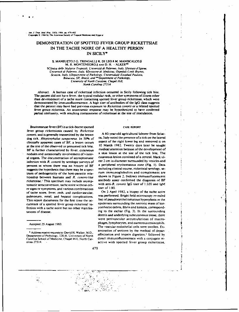
,A..J. T•P. Med. Hvx.. 33M31. 1934. w 479-4.82Copw% C 1934 by The Amencan Sonea of Trop" Moedm mWd H•l•j i
DEMONSTRATION OF SPOTTED FEVER GROUP RICKETTSIAEIN THE TACHE NOIRE OF A HEALTHY PERSON
IN SICILY*
S. MANSUETOAt G. TRINGALI.4 R. DI LEO.A M. MANISCALCOIM. R. MONTENEGRO.| ANO D. H. ý.ALKERI
tClinica delle Malatie Tropicali. Unn•ivritd di Palermo. Italy. tJtituao d'Igiene.Universtid di Palermo. Italy. WDivisione di Medicina. Ospedali C iwli Riuniti.Sciacca. Italy. I Department of Pathology. Univerzidade Estadusal Paulista.
Boaucatu, SP. Brazil, and "Department of Pathology.Universtty of North Carolina. Chapel Hill.
North Carolina 27514
Abstract. A human case of -ickettsial infection occurred in Sicily following tick bite.The patient did not have fever, the typical nodular rash, or other symptoms of illness otherthan development of a tache noire containing spotted fever group rickettsiae, which weredemonstrated by immunofluorescence. A high titer of antibodies of the IgG class suggeststhat the patient may have had previous exposure to Rickettsia conorii or a related spottedfever group rickettsia. An anamnestic response may be hypothesized to have conferredpartial immunity, with resulting containment of rickettsiae at the site of inoculation.
Boutonneuse fever (BF) is a tick-borne spotted CASE REPORTfever group rickettsiosis caused by Rickettsiaconorui. and is generally transmitted by the brown A 60-year-old agricultural laborer from Sciac-dog tick. Rhipicephalussanguineus. In 50% of ca. Italy noted the presence of a tick on the lateralclinically apparent cases of BF. a lesion occurs aspect of the right lower leg and removed it onat the site of the observed or presumed tick bite. 10 March 1982. Twenty days later he soughtBF is further characterized by fever, cutaneous medical attention because of the development ofnodules and occasionally involvement of viscer- a skin lesion at the site of the tick bite. The
al organs. The documentation of asymptomatic cutaneous lesion consisted of a central, black ul-infection with R. conordi by serologic surveys of cer 2 cm in diameter surrounded by vesicles and
persons in whom there was no history of BF a peripheral erythematous zone (Fig. I). Datasuggests the hypothesis that there may be a spec- including clinical course, rickettsial serology. se-trum of pathogenicity of the host-parasite rela- rum immunoglobulins and complement aretionship between humans and R. conorii-like shown in Figure 2. Indirect immunofluorescentrickettsiae.' This spectrum may include asymp- antibody assay confirmed the diagnosis of BFtomatic seroconversion. tache noire without oth- with anti-R. conorii EgG titer of 1:320 and igMer signs or symptoms, and various combinations titer of 1:80.of tache noire, fever, rash, and cardiovascular. On 3 April 1982, a biopsy of the tache noirepulmonary, renal, and hepatic complications, was performed. Bright field microscopy revealedThis report documents for the first time the oc- foci of pseudoepitheliomatous hyperplasia in thecurrence of a spotted fever group rickettsial in- epidermis surrounding the necrotic mass of kar-fection with a tache noire but no other manifes- yorrhecticdebris, fibrin and keratin, correspond-tations of disease. ing to the eschar (Fig. 3). In the surrounding
dermis and underlying subcutaneous tissue, there_____were perivascular accumulations of macro-
Accepted 25 August'1983. phages, lymphocytes, and numerous eosinophils.The vascular endothelial cells were swollen. Ex-
Address reprint requests to: David H. Walker, M.D., amination of sections by the method of depar-Department of Pathology, 228-H. University of North affinization and trypsin digestion.2 followed byCarolina School of Medicine. Chapel Hill. North Car- direct immunofluorescence with a conjugate re-olina 27514. active with spotted fever group rickettsiae.
479
.5
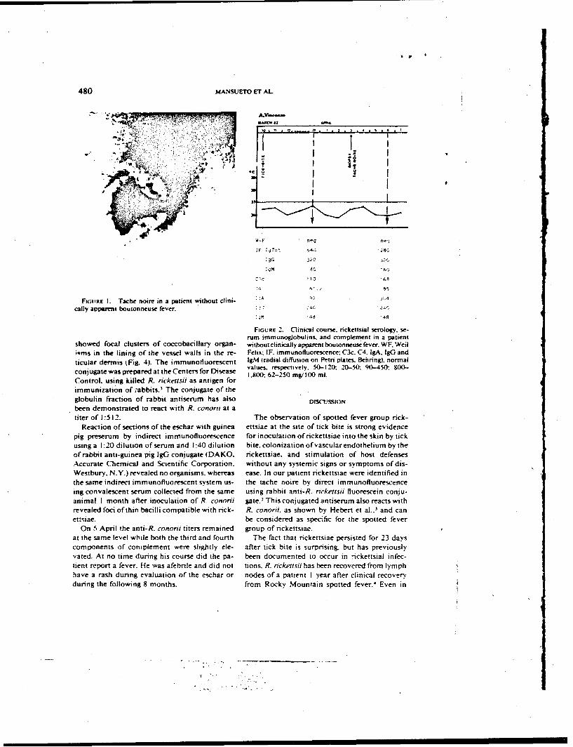
480 MANSUETO ET AL
IFI
IF ',Tot 64C.
:gG 320
.: ,'•0 '68
FilOP I. Tache noie in a patient without dlini- :;A
cally apparent boutonneuse fever.48 48
FIGURE 2. Clinical course. rickettsial serology, se-rum immunogJobulins, and complement in a patient
showed focal clusters of coccobacillary organ- withoutclinicallyapparentboutonneusefever.WFWeiliqms in the lining of the vessel walls in the re- Felix: IF. immunofluorescence: C3c. C4, IgA, I[G and
ticular dermis (Fig. 4). The immunofluorescent 1gM (radial diffusion on Petri plates. Behring), normalvalues. respectively, 50-120: 20-50;. 90-450:, 800-
conjugate was prepared at the Centers for Disease 1,800- 62-250 mgV100 ml.
Control. using killed R. rickemsii as antigen forimmunization of abbits. The conjugate of the
giobulin fraction of rabbit antiserum has also DISCUSSIONbeen demonstrated to react with R. conorri at atiter of 1:512. The observation of spotted fever group rick-
Reaction of sections of the eschar with guinea ettsiae at the site of tick bite is strong evidence
pig preserum by indirect immunofluorescence for inoculation of rickettsiae into the skin by tick
using a 1:20 dilution of serum and 1:40 dilution bite, colonization of vascular endothelium by theof rabbit anti-guinea pig IgG conjugate (DAKO. rickettsiae. and stimulation of host defensesAccurate Chemical and Scientific Corporation, without any systemic signs or symptoms of dis-
Westbury. N.Y.) revealed no organisms, whereas ease. In our patient rickettsiae were identified in
the same indirect immunofluorescent system us- the tache noire by direct immunofluorescenceing convalescent serum collected from the same using rabbit anti-R. ricketrsi fluorescein conju-
animal I month after inoculation of R. conorii gate.2
This conjugated antiserum also reacts with
revealed foci of thin bacilli compatible with rick- R. conorii, as shown by Hebert et al.,' and canettsiae. be considered as specific for the spotted fever
On 5 April the anti-R. conorti titers remained group of rickettsiae.
at the same level while both the third and fourth The fact that rickettsiae persisted for 23 dayscomponents of complement were slightly ele- after tick bite is surprising, but has previouslyvated. At no time during his course did the pa- been documented to occur in :'ickettsial infec-
tient report a fever. He was afebrile and did not tions. R. rickentsii has been recovered from lymph
have a rash during evaluation of the eschar or nodes of a patient I year after clinical recovery
during the following 8 months. from Rocky Mountain spotted fever.' Even in
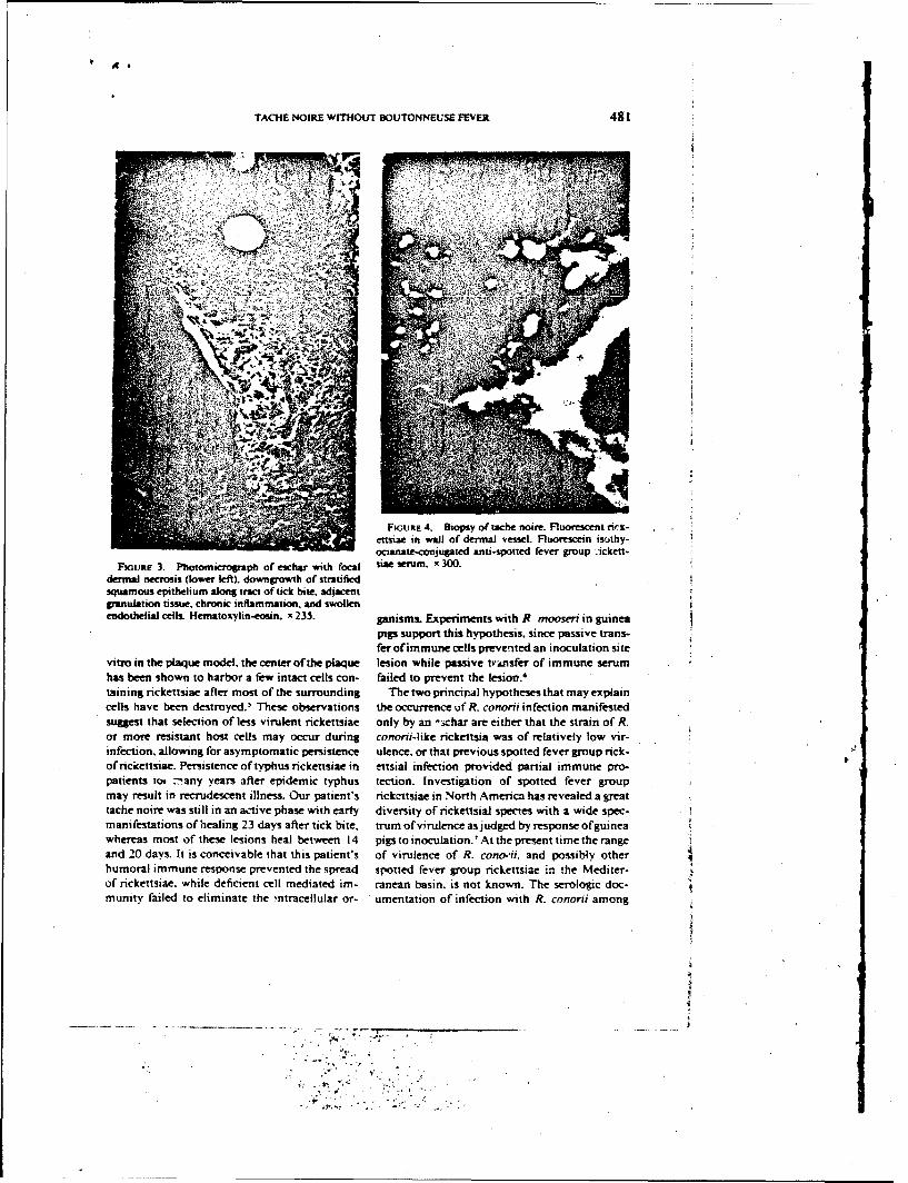
TACHE NOIRE WITHOUT BOUTONNEUSE FEVER 481
T
4,:J
4FIGuRE 4. Biopsy of tache noire. Fluorescent ncs7-ensiae in wail of dermal vessel. Fluorescein isothy-ocianate-conjugated anti-spotted fever group Ackett-
FiouRE 3. Photomicroaph of eschar with cocai sise serum. x 300.
dermal necrosis (lower left), downgrowth of stratifiedsquamous epithelium along tract of tick bite. adjacentgranulation tissue, chronic inflammation, and swollenendothelial cells. Hematoxylin-eosin. x 235. ganisms. Experiments with R. mooseri in guinea
pip support this hypothesis, since passive trans-fer ofimmune cells prevensted an inoculation site
vitro in the plaque model, the center of the plaque lesion while passive transfer of immune serum
has been shown to harbor a few intact cells con- failed to prevent the lesion.'taining rickettsiae after most of the surrounding The two princira] hypotheses that may explain
cells have been destroyed.' These observations the occurrence uf R. conorii infection manifestedsuggest that selection of less virulent rickettsiae only by an '.ichar are either that the strain of R.
or more resistant host cells may occur during cononri-like rickettsia was of relatively low vir-infection, allowing for asymptomatic persistence ulence, or that previous spotted fever group rick-ofrickettsiae. Persistence of typhus rickettsiae in ensial infection provided partial immune pro-patients tor :any years after epidemic typhus tection. Investigation of spotted fever groupmay result in recrudescent illness. Our patient's rickettsiae in North America has revealed a greattache noire was still in an active phase with early diversity of rickettsial species with a wide spec-manifestations of healing 23 days after tick bite, trum ofvirulence asjudged by response ofguineawhereas most of these lesions heal between 14 pigs to inoculation.'At the present time the rangeand 20 days. It is conceivable that this patient's of virulence of R. cono.-ii. and possibly otherhumoral immune response prevented the spread spotted fever group rickettsiae in the Mediter-of rickettsiae. while deficient cell mediated im- ranean basin, is not known. The serologic doc-munity failed to eliminate the ;ntracellular or- umentation of infection with R. conorii among
~ . . , Y .7 7 .
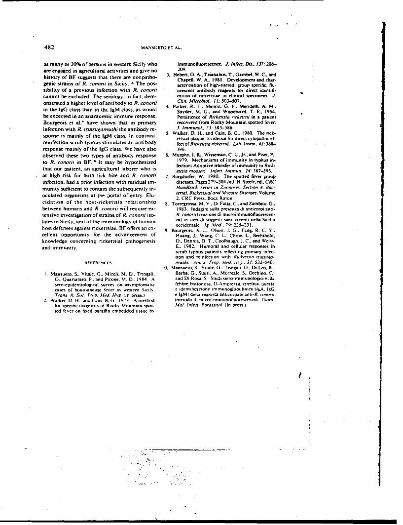
482 MANSUETO ET AL
as many as 20% of persons in western Sicily who immunolluorescence. J. Infect. Dis.. 137:206-are engaged in agricultural activities and give no 209.history of BF suggests that there are nonpatho- 3. Hebert, G. A., Tzianabos. T.. Gambel. W. C.. andChapeU, W. A.. 1980. Development and char-genic strains of R. cononi in Sicily.'. The pos- acterization of high-titered. group specific. flu-sibility of a previous infection with R. conorii orescent antibody reagents for direct identifi-cannot be excluded. The serology, in fact. dem- cation of nckettsiae in clinical specimens. J.onstrated a higher level of antibody to R. conorii Clin. Microbiot.. I1: 503-507.
4. Parker, R. T.. Menon, G. P.. Merideth, A. M.,in the IgG class than in tht IgM class, as would Snyder. M. G.. and Woodward. T. E.. 1954.be expected in an anamnestic immune response. Persistence of Rickettsia ncketisiu in a patientBourgeois et al." have shown that in primary recovered from Rocky Mountain spotted fever.infection with R. Isutsugamushi the antibody re- J. Immunol., 73: 383-386.5. Walker. D. H.. and Cain. B. G.. [980. The rick-sponse is mainly of the IgM class. In contrast,. Wttkir. p. Evince for drc y ice-
ettsial plaque. Evidence for direct cytopathic ef-reinfection scrub typhus stimulates an antibody feet ofRickettsia rickettsti. Lab. Inwst.. 43:388-response mainly of the IgG class. We have also 396.observed these two types of antibody response 6. Murphy, J. R.. Wisseman, C. L.. Jr.. and Fiset. P..to R. conorii in BF.'0 It may be hypothesized 1979. Mechanisms of immunity in typhus in-
fection: Adoptive transfer of immunity to Rick-that our patient, an agricultural laborer who is ettsia mooset. Infect. Immun.. 24: 387-393.at high risk for both tick bite and R. conorii 7. Burgdorfer, W.. 1980. The spotted fever groupinfection, had a prior infection with residual im- diseases. Pages 279-301 inJ. H. Steele.ed.. CRCmunity sufficient to contain the subsequently in- Handbook Seres in Zoonoses. Section .4. Bac-oculated organisms at ih- p•ortal of entry. Elu- treal. Rickettsial and .tfcoac Diseases. Volume2. CRC Press, Boca Raton.cidation of the host-rickettsia relationship 8. Torregrossa. M. V., Di Fatta. C.. and Zambito, G..between humans and R. conorii will require ex- 1983. Indagini sulla presenza di anticorpi anti-tensive investigation of strains of R. conorti iso- R. cnnorit (reazione di microimmunofluorescen-lates in Sicily. and of the immunology of human za) in sien di soggetti sani viventi nefia Sicilia
occidentale. Ig. Mod. 79: 225-231.host defenses against rickettsiae. BF offers an ex- 9. Bourgeois, A. L., Olson. J. G., Fang. R. C. Y..cellent opportunity for the advancement of Huang. J., Wang. C. L.. Chow. L. Bechthold.knowledge concerning rickettsial pathogenesis D., Dennis, D. T., Coolbaugh. J. C.. and Weiwtand immunity. E.. 1982. Humoral and cellular responses in
scrub typhus patients reflecting primary infec-tion and reinfection with Rwkettsia tsutsuga.
REFERENCES mush$. .4m. J Trop. Mied. Iv.q.. 31: 532-540.10. Mansucto, S., Vitale. G.. Tringali. G.. Di Leo. R.,
I. Mansueto. S.. Vitale. G.. Miceli. M. D., Tnngali. Barba. G.. Staiti, A.. Morreale. S.. Occhino. C..G.. Quanrararo. P.. and Picone. M. D.. 1984. A and Di Rosa. S. Studi siero-immunologici n.J[gsero-epidemiological survey on asymptomaiic febbre botionosa. ll-Ampierna, cinctica. ,juratacases of boutonneuse fever in western Sicily. e identilficayione immunoglobulinica (I&A. [gGTrans. R. Soc. Trop. Mied. Hyg (!n press.) e IgM) della nsposta anticorpale anti-R. conorit
2. Walker, D. H.. and Cain. B. G.. 1978, A method (metodo di micro-immunofluoresceszal, Giorn.for specific diagnosis of Rocks Mountain spot- Mal. Infert. Parassitol (In press.)ted fever on fixed paraffin embedded tissue by
',.I
AwrinscwaoisL Awarr ANOD CtahboisaaAry. May 111111. p. -582-3 Vol. 25. NO. 5
Copyrilpht 0 1984. Anierrae Society for Microbiolog
Effect of Synthetic Protease Inhibitors of the Amnidine Type on CellInjury by Rickettsia rickettsii
DAVID'H. WALKER.* RICHARD ft. TID WELL TIM M. RECTOR. Amo J. DIETER CERATZ
Deparrnwitt of Pathology. Univenrsiy of North Carolina or Chapel Hill. Chapel HUIl. Normh Carolina 27514
Received 21 Decemlier 19BYcete I match 19"
To evaluate the uiportance of proteodytic actvity, in the pathogenesis of cell injury by Rkckettsioricketriii. a series of four aromatic amidine, inhibitors of trypsin-like promiases were introduced into theplaque model. The compounds were shown to be active toward plaque reduction with their order ofeffectiveness parallel to their antitr si activi .ty. One of the compounds. biA-amsdino-2-benzirnidazolyl).methane. at a concentration of 10 14n demonstrated complete inhibition of plaque formation on day 6.Jas(S-amsidi io-2-benzinssdazolylhrmethane at the same concentration reduced cell injury even when added tothe system 'after 72 It of ricketisial infection. The reduction in morbidity in guinea pip experimentallyinfected with R. nickerruii and treated with bis(5-amidinio-2-benzintidazolylkmethane aa compared withmorbidity in infected. untreated animals. comprised delay in the ow -.t of fever and slightly fewer febrile
animals. Because b M5amidino-2-benzintidazodyl~iethane had no t' ; on phomspholipase A2, the enzyme-activity associated with penetrationinduced cell injury, it is likely thr..s a trypsin-like protease also plays anessential role esther in the physiology of R. nrwkettsu or as its pathogenic mnechani, a.s
Visions of several different genera require exposure to 43 It after the death of the chick embryos. Yolk sacs.rteodytic enzymes to achie-!e full expression of their containing rickettsiae were homogenized in a Waring blend-
biological properties (7). As a corollary to this fact. proteae er. diluted in sucrose phosphate glutamate (0.219 M sucrose.inhibitors can be expected to have potential antiviral activi- 0.0038 M KHi.P0 4. 0.00-r- M KIH-P04. 0.0049 M glutamnate.ty. This was substantiated by our recent discovery of the pH 7.0) (1)to a1% suspension, and stored frozen at - 7(Cinability of synthetic low-molecular-weight inhibitors of tryp- 1-ni samples. Samples were titrated by plaque assay in chicksin-like proteases to block respiratory syncytial virus-rn- embryo cell culture and found to contain 7 x 10' PFU./ml.duced cytopathology (2-4. 10). Although the site of action of Phl.ammied. Flasks (Corning Glass Works. Corning.these agents has not yet been determined, we presented N.Y.) with 25-cm2
t monoilavers of E6i clone Vero (African* stroog evidence that their antiviral effect may be linked to 1geen monkey kidney) cells were inoculated with 0.1 ml of a
their antiproiteolytic properte. The most potent of the suspension of R. nickerrsii diluted 10-'in brain heart infusioninhibitory agents. bisl-ainidino-2-bewszimkdaolylitnthane broth. The inoculumn was adsorbed for 30 min before the(BABIM). was showns to exert the following effects: ti) delay addition of 5 ml of minimal essential metdium (GIBCOof penetration of virus into cells. (ii) blockage of virus- Laboratories. Grand Island. N.Y.) containing 5% fetal bo-induced cell fusion. (niii reduction of muhtiple-cycle yields of vine serum (Flow Laboratories. Inc.. McLean. Va.). 0.02 Mvirus, and (iv) reduction in pathology and virus yield in HEPES (N-2-hydroxyethylpqperazine-N'-2-ethanesulfc-nicexperimsentally infected animals, acid) buffer. 2 MM L-ghttainline. ca. 0.075% NaRCO, to final
Similar to vir~st diseases. ncketssia infection of cells pH 7.3. 0.5* agarose (Sea Kemn. FMC Corp.. Marineinvolves a penetration step. This event is known to be Colloids Div.. Rockland. Maine). and different concentra-enzyme mediated. but thus far only phospholipase A?. and tions of protease inhibitors. Flasks were incubated at 35*Cnot a protease. has been shown to participate in the process for 4 days at which time 5 nil of an identical second overlay(15.-23). COur experience with respiratory syncytial virus medium constaining 0.01% neutral red was added. AfterSuV666ted application of the amidine inhibitors so the rickets- further incubation, plaques were counted on various combi-sial syszac in search of evidence of proteolysis in the nations of days 5. 6. and 7(414. 16. 20ý-22).pathogenetic sequeticc. Such evidence was readily found in Pitntesie Ithlbiitars. Inhibitors of trypsin-like proteasesa study of the nickettsial plaque assay. and the results are the used in these experiments were BABIM. 1.2-bislS-arnidino-subject of this communication. 2-benzimidazolyl~ethane. 1.5-bislS-amidino-2-benzimtidazo-
ly)pentane. and 5-smidinoindole. They were synthesized asMATERIALS AND METHODS previously reported (5. 9).
Rickettsaea. Stocks of R. rickerrsii (Sheila Smith strain) Eaperfimeatal Iealg. Each of these aromatic amidines waswere cultivated by inoculation of the yolk sac of 5-day-old incorporated into both the first and second overlay miedia ofspecific parthogen-free embryonated hen eggs (SPAFAS. three flasks at copcentrations of 10-4, 10'. and 10- 414 andNorwich. Conn.) with plaque-purified organisms provided plaque counts Nkere compared with those of monolayersby Charles L Wisseman. Jr. (University of Maryland. inoculated with the same ricketsisal suspension and overlaidBaltimore). Inoculated eggs were incubated at 35*C. and with medium containing no prosease inhibitors. Controltheir yolk sacs were harvested 5 days after inoculation. 24 to flasks includcJ u-unoiculated. untreated monolayess and also
uninfected ntonoayers treated with a 10-4 M concentration________of the aromatic amidines for evaluation of toxichy'. The
Corresponding auithor. menam plaqlue count was calculated for each concentration of
'58
. 47~,~
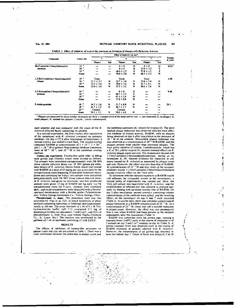
'IA
VOL,. 25. 1964 PROTEASE iNHIBITORS CLOCK RICKETI IIAL PLAQUES 583
TABLE 1. Effect of inhibitors of :rmpsrn-like pruirawes on formwiin of plaques with Rk-A~rtta rikekguauE1116 of ia"-i- say:
Ca..pouigd coma I M) 5 S 7 TiWM_
Bisl35-amidino-24*nzimadazotyl)- t0 7 - 0 t 0 0 0 = 0 0 0.017methane 10' - - 0±C 0 15.0±t3.5 S
10 ~ - - 46.0:t 1.5 S 52.0±t1.2 SNone - - 70.0 =5.0 N 69.5 =5.5 N
12-BislS-amidiio2-benzinidaolyi)- 10o Toxic Toxic Toxic 4.68ethaft 10' 21.7 ±3.7 N Z3.7 =3.9 N 23.0 =5.0 N
101 22.3±t5.8 N -29.0±-z7,6 N 31.7 =9.4 NNone 32.0±t 3.0 N 37.3±=3.2 N 38.0±= 2.6 N
1-i-istamidno-2-bnzimidozolyl)- 10-1 0 = 00 0 - - 9.46pentane 10-1 - 60.7±=5.2 S - -
10-1 67.3±=2.4 N - -None - - 71.0±=0.5 N - -
-Aniidi.ioirndole 10-4 34.7 ±2.0 N 51.7 = 0.9 N - - 29.110-1 42.7±:t1.2 N 56.7 = L7 N - -10 1 Contain. Contain. - -
None 39.5 ±5.5 N M5.0±:t6.0 N - -
Plaques are measured by mean number ofplaques per flask = standard error ofthe mean and by size: notb examiined; 0. no plaques; S.small plaques: N. normal-size plaques: Constain., cultture contaminated.
each ansidine and was compared with the count of the R. the inhibition constants (K, values) for trypsin (2). The mostrickettsiiinfeccted flasks containing nos amidinte. marked plaque reduction was observed with the most effec-
In a secend experiment. the first overlay after inoculation tive inhibitor of trypsin activity. BABIM, with no plaquesof the monoilayer with R. rickerlsii contained no aromatic being observed on day 6 after inoculation in the presence ofamidines. On day 3 172 h after inoculation of rickettsiael. a 10- Ms of the amidine. Measurable plaque reduction wansecond overlay was added to each flask. The second overlay also obs-erved at a concentration of 10- M BABIM. and thecontained BABIM at concentrations of ? x 10-'. 2 x 10- plaques present were smaller than untreated plaques. Theand 2 x 10- M to achieve final protease inhibitor concentra- least active inhibitor of trypsin. 5-amidinoindole. which hastions of 10-'. 10-1. and 10- M in the combined overlay a Kof 219.1 lnM for trypsin 45), showed minimal effects on R.medium. rickeltsji plaque count and size. The montolayers tremred with
Gulixes pig expet-itnent. Twenty-five adult 140)0- to 600-g) 1.5-bisl5-amidine-2-benzimidazolyllpentane. having an in-male guinea pigs (Hartley strain) were divided as follows. termediate K0 (19. showed evidence for reduction in cellTen animals were inoculated intrapentoneally with 330 50% injury caused by R. rickettsii as measured by plaque counttissue culture infective doses ( ca. 38 -W/, guinea pig infetc- and size. However, the etfect was less than that of BARIM.tious doses) of R. rickettsii (Sheila Smith %train) and treated At concentrations of 10- M and less, none of the aromaticwith BABIM at a dose of 15 mg/kg per day given daily by the amidines except 1.2-bis.;5-amidino-2-benzimidazolylkthaneintraperitontal moute beginning 30 mmi after rickettsial inocu- caused cytotoxic effect on the Vero cells.lation and continutng for 9 days: ten animals were inoculated To determine whether delayed exposure to BABIM wouldintra1 ,entorically with 330 W43 tissue culture infective doses still influence the cytopathic events in the monoilayer-s. aof R. ri-ketrsii and given no treatment, and five unfinfected second series of experiments was carried out. Here, theanimals were given 15 mg of I3AHIM per kg daily via the Vero cell culture was inoculated with R. rickeutstii and theintrapentoneal route for 9 days. Animals were examined establishment of infected foci was allowed to proceed nor-daily, and rectal temperatures were measured with a battery. mally by feeding with an initial overlay free of BABIM. Onoperated thermometer with a flexible probe ITelethermom- day 3 after inoculation, second overlays containing variousetcr: Yellow Springs Instrument Co.. Yellow Springs. Ohio). concentrations of the inhibitor were added, and the resulting
Phonphrgsipmge A, yul. The procedure used was that 'rd'fects on the monolayer on day 6 and 7 were recordeddcscnbed by Vigo et a]. 112), in which hydrolysis of phos- (Table 2). As can be seen, there was complete suppression ofpholipid-contlaining liposomes is followed spectrophotomet- plaque formation at a BABIM concentration of 10-4 W At arically at 340 nm. The assay mixture-, of I ml of 0. 1 IM Tris- concentration of' 10- M. there was still a notable reductionhydrochloride buffer (pH 7.21 contained 0.5 mg of in plaque count. However, the effect was less pronounrceddipal mitoyl- lecithin las liposomecs). I mM CaCl,. and 10 U of than it was when I5ABINI had been present in the mediumphospholipa~se A, from Najo naba venom (Sigma Chemical immediately after the inoculation (Table 1).Co.. St. Loui-s. M~o.. The reaction was terminated by the .BABIM was somewhat toxic for guinea pigs, causing aaddition of)I mi of methanol containing 15 mM EDTA. transient fever lŽ400 C12 early in the course of treatment 04 of
RESUTS5 animals on day 3 and 2 of 5 animals on day 4) (Table 31. ARSLSsimilar transient fever was observed early in the cours of a
The effects of inhibitors of trypsin-like proteases on BABIM treatment ot animals infected with R. nickerttii.plaque count and size are presented in Table 1. There was a However, the temperatures of all guinea pigs returned toclose correclition between the reduction in plaque count and base line before day 5. Onset of fever was delaycd: 5 of 10
Z-
TBE2. Elrect of trealmil Rickettija. nricetti plaque asode phospholipase A2 activity (from, Nag* Age venom)i. Ofwith BASIMd on day 3 a&her inoculation course, the linkage of a trypsin-fike prkw in a chain of
ISMEgreo oa dayd enzymes that includes phosphliopase: A? cas be excluded
7 (11). In fact. t. ypsin activation is necessary frall mammali-(M)an pancreatic phospholipases A? (13). Same of the other
paques size PaIWMe si possible mechanisms by which a ftypsia~lbe proiease might10 4 0±-M0 0 0±=0 0' fit into the scheme of rickettsial maiy to cells. besides
I 0- 31.3 i 3.4 N 50.0 ± 13.0 S proteoytic activation of the penetration umafnism-associ-10 67.7 ± 9.0 N 82.7 ± 9.5 N ated pliosplsolipase. include direct prlotely'ti attack on theName 76.3 = 3.5 N 82.0 t± 3.1 N host cell membrane either during entrance iwthe cell or on
standard erro of the siean and by sum: U. an plaques; N. normal function.plaque size: S. smail plaques. The reduction of plaque counts by the addition of BABINI
72 h after the establishment of rickettsinainedted foci docu-ments that the protease inhibitors are am mecrely preventinginitial rickettsial infection and stugesta thae proteas inhibi-
untreated animals had fever on day 5 aa compared with no tors may be blocking a rickettsial fuactio essential to1.febrile animals in the BABIM-treated group. On day 6. 8 of expression of the pathiogenic mechanism. The hypothesis10 untreated animals were febrile as compared with only 2 of that a protease-associated pathogenic eiecmsism is blocked10 BABIN4.treated animals. Four febrile animals in each by BABIM is also supported by the deiay of plaque farina-group died during the course of the rickettsial disease. tion by a 10', M concentration of BASIN with delayedBABIM was found to have r -) effect cn the hydrolysis of appearance of several small plaques am day 7. Rickettsaiedipalmnitoyl-lecithin liposomes by phospliolipase A, from survived the BABIM treatment and caue formation of aNajo majo venom. .. w plaques, presumably after protease activity overcame
the protease inhibitory activity. Because fatal cases ofDISCUSSION Rocky Mountain spotted fever are ohm diagnse and
The order of effectiveness of aromatic amidines in reduc- treated too late in the course of disease for rickettsiostaiucing pL.que formation by R. rickensii folows their order of antimicrobial agents to prevent the demise of the patients (6.effectiveness in inhibiting trypsin and several other trypsin- 17-19). additional inhibition of rickettsial injuy to the hostlike proteases and corresponds to their order oef efec...veness would benefit such critically ill patients. Consideration of thein blocking cell fusion by respiratory syncytial virus in viva possible use of inhibitors of trypsin-like peoteases for the(2. 5, 10). This parallelism of activities at .-w concentrations treatment of rickettsiasl c4seases would raqiire investigationargues strongly for the antirickettsial effect resulting from of these drugs in other animal models. father study of thethe action of compounds as protase inhibitors on a trypsin- mechanism of action of pratease inhibitors on the rickettsia-like enzymne. However, it was also r.:cessary to consider the host cell interaction, and infortnation on the toxicity ofthesepossibility that ansidines. might set by inhibition of phospho- compounds itt humans, especially on the laflammatasy andlipase A,. especially since a cell penetration phosphoipase coagulation mechanisms: complement. bali~rin. coagula-appears to be an important rickettsia] pathogenic mechanism tian. and fibrinolysis.and a protease inhibitor ot a different structure had previous- Data on the toxicity of BABIM incloe the acute 50%ly been shown to suppress phosphohipase A7 activity (8). We lethal dose for cotton rats of 136 tog of BABOA per kg and a 4showed this possibility to be unlikely by demonstrating that dosage, for cotton rats of 30 mg of BABIM. per kg daily for 7BABIM at a concentration of 10- MI has no effect nx days without ill effects. Current pharmacological studies of
TABLE 3. Effect of BABIMt on esperimsental infection of guinea pigs with Xwrkkema ,k**ersi
DayItwketts-fected poses pop
GABtIl trcesin No mmwaU. , p aAatal trate
Afetont FIN. D~ead Afebnle FeS. Ded Afeb* Foaem Dead3
I to 0 0 to 0 0 5 0 02 6 4(40.1) 0 1O 0 0 5 9 03 2 8(40.2) 0 to 0 0 1 4(W5.) 04 6 4(40.1) 0 10 0 0 3 !t*4.1) 0
5 0 0 0 5 5(40.6) 0 5 0 0108 2(ý40.8) 0 2 8440.7) 0 5 a0
7 6 4 43.7) 0 2 8(40.7) 05 008 5 4440.7) 1 2 8(40.7) 0 5 5
9 4 5(40.7) 1. 3. 7(40.8) 0 5 a
1 1 4 3(ý40.5) 3 4 5(:40.7) 1 5 0
12 6 3(40.7) 3 S 7(40.64 1 5 . 0
12 6 1(40.44) 3 4 3(40.4) 35 30
114 6 1(40-4) 3 4 3140.3) 3 5 1 0Final 6 0 4 6 0 4 5 1 0
'The v~alues in parentheses are the mean temperatures of kbrile guineapip.
VOL. 23. I'MI PROTEASE INHIBIORS BLLICK RICKETYIAL PLAQUES" US3
rats that received a&done of 20 mg o( BABIM per kg daily and Loewe. 1978. Dlarylamnidine dervatives miii one or both of theof mice that received a dose o( 20 tral of BABIM per kg daily aryi moieaties consisting of an indole or indolte-ike ring. Inhtibi-have not detected any toxicity. Thes~e observations of prote. tors ofarlpoine- specdIh asteioproteafses J1. Med. Chem. 2613-ase inhibitors blocking rickltisial injury to cells offer inter. 623. ,Iesting new hypilthsc.e for studying rickettsial pathogenesis. 1. idwa It 1L 1. M Cest talLJ L16.AoaiAt this point. th~y should be used as tools for -lucidating amdies icedacell ofuthior abiit to biock mpeastin. urokmas.
rickettsial ptysioogy and pathogenic mechanisms. thromnbin. and trypsin. J. Med. Ciem. 28:294--296.
ACKNOWLEDGMEN4TS 11. Vas. Own Bask. H. 1900. Intracellular ptsosplsoliposes A. Has.We ae ptefl t Rihar Lobary fo sytheis f te cns- 12.chim. Biophys. Acts 604:191-246.We a grtefl t Rihar Lobary fo sy~hcis f te crn. 12.V1M, C., G. P. Liwik mill P. J. PIWe. 1960. Mechanisms of
pounds and to Jennie Lii Hollansder for preparation of the mianu- inhibition of phosplsolipase A2. Biochens. Pisarmacod. 29.623-script. 627.
The study was supported by Public Health Service grant 13. Valimeik. 1. J.,. so G. Ht. Dei~as. 1962. Pancreatic phosplso&iHL19171-07 fronm the National Institutes of Health arid by U.S. peam A2: a model for mensbranc-bound enzymes?. p. 69-149. InsArmy Contract OAMD17-83-C-3122. P. C. Jost and 0. H. Gnffith (ed.). Uipid-vtotein interactions.
LrELTT C 14. Vol. 1. John Wiley A Sons. Inc.. New York.14.WakirD. .. ad . G. Cah. 198. The ricketssnis plaque.
1. goe re. M.. a.. .1. C. L.flee, and .5. C saad.~ 1950. The Evidence for direct cytopathic effect of Rickettsia nekiutsiFinfluence of certain Wsa.s amino ad. Is. sugar: and proteins on Lab. l10ve* 43:3ft-396.the stability of rickettsiae. 1. Bacteriol. 1550-d.c. Walker, D. k: . W. T. IbIB. .5. G. eilhort. sod Il. C. Beversly.
2. Dabh , E. J .. , J. 0. Gerstit. S. Xt. Shawe. Ad R. Rt. Tki-eil. 1963: Role of pi~osplolipase-aasociated penetration mnechanisnm1961. Inhibition of respiratory syncytial virus-host cell interac- in cell injury by Rik~eitia riitrettsu. lrfect. tIntun. *A.-40-tions by mono- and diarnidines. A,,.,microh. Agents Clsemother. 842.19:644-6%6. 16. Walker. D. HI.. W. T. Firth. mid C.-J. S. Edol 1962. Human
3. Dtsh3%i. Lt J., J. D. Gerfta. sad It. It. Tldwed. 19110. Inhibition endothelial cell culture plaques induced bry Ricikerruia ricketirru.of respiratory syncyiial viris by bissli-arudino.2-benizimidaro- Infect. Immun. 31:301-306.lyllnsethane. Virologcy 143:502-504. 17. Walker, D. H.. R. M.. Gay. siad M_4 Vaides-Iiaen. 1961. The
4. Dabovi. F_ J5.. J. D. Gerats. anO It. It. Tidwell. 1963. Enhance- occurrence ofecuhars in Rocky Mcuneain spotted fever. I . AMtsmeni of respiratory syncytial virus-induced cytopailhioogy by Acad. Dermtatod. 4:571-576.trypsin. thromnbin. and plasmin. Infrci. Immun. 40i351-358. 18. Walle. D. H.. sad W. D. Ma1.na 1979. A cute rWsa failure ini
5. Gnratz. 1. D., F. M. Steve,., K. L. "olakiald, R. F. PxrrMs. anid Rocky Mountain spottet± fever. Arch. Intern. Med. 139-443-It. R. Tidwell. 1979. Amidino-substituted aromatic heterocycles. 4.49.am probes of the specificity pocket of trypsin-like protesses. 19. Wlker. D. H_. so W. D. Manlera., 1960, RickettW Vsl vicutis.Arch. Biochem. Biopisys. 1917-.51-559. Am. Heant 1. 100-996-906.
6. Hatt*"e. MI. A. W., M~ Itetaillla., 3. J. O'Slrles. MI. Stoutker. 2M. Wilk. 0. A.. and W. Rairgihrfer. 1972. Plaue formation in3. E. Foulahsie. sad B. Hanso.. 1978. Fatal Rozky Mountain tissue cultures by Rickirtmiu ncikertsii isoLAted directly frontsporltd fever. J. Am-. Med. Assoc. 240:14913-1-503. wihole blood uind tick hemolymph. Infect. Insiun. C-736-733.
7. Korsaw, ft. 191. Proteolytic events in Viral replication. p. 171- 21. W~ae. 0. A.. It. A. Oisashee. G. Taileat. said M. G. Peacc.224. it R1. Berlin. H. Herrmnann. 1. Lcpow. and J. Tanzer (ed.). 1972. Effects of vartuss suspending media on plaue fonnatiotsMolecular basis of biological degradative processes. Academic by ricketsisie in tissue culture. Infect. Imnamu. 6050O-556.Press. Inc.. New York. 2.2. Wike. D. A.- G. Taneist. SC4 G. Peaoc. ad It. A. (nibush.
8. Kamne. H.. t. Soaba said It. Damers.. 1963. Effects of the anti- 1972. Studies of the nickettsial plaque assay technique. Infect.inflanmmatory serine esterase inhibitor. FOY, ont phoiplsolipase Iminun. 5:713-722.A7 (EC 3.1.1.4) activity in rabbit polynsorphlonuclear leuko- 23. Wlshier, H. H.. sad -T.Nigher. 1962. Phosptittipase Aand thecytes. Pttarnsacol. Res. Common. 15:869-4178. interaction zf Rickdrisia prsownzeki and mouse fibeoblasis (I.-
9. TWdseCl R. 3L. J1. D. Geasse. 0. Ois.. .6 b Dal.0 Zebi and H. "'cells). Infect. Imanun. 3L, 109-Ml.
4.
AS