Embed Size (px)
Citation preview
Sci Forschen
O p e n H U B f o r S c i e n t i f i c R e s e a r c h
Transplantation Research JournalOpen Access
Copyright: © 2017 Teraoka S, et al. This is an open-access article distributed under the terms of the Creative Commons Attribution License, which permits unrestricted use, distribution, and reproduction in any medium, provided the original author and source are credited.
Volume: 2.1Research Article
A Clinical Trial Aiming at Tolerance Induction by Adoptive Transfer of Ex Vivo-Induced, Donor-Specific Treg-Like Cells in Clinical Kidney TransplantationSatoshi Teraoka1*, Ichiro Koyama2, Hisashi Bashuda3, Koichiro Uchida3, Makoto Tonsho2, Ichiro Nakajima2, Shohei Fuchinoue2, Sonoko Habu3 and Ko Okumura4
1Department of Transplant Surgery, Mita Hospital, International University of Health and Welfare, Tokyo, Japan2Department of Surgery, Kidney Center, Tokyo Women’s Medical University, Tokyo, Japan3Department of Immunology, Juntendo University School of Medicine, Tokyo, Japan4Atopy Research Center, Juntendo University Hospital, Juntendo University School of Medicine, Tokyo, Japan
Received date: 15 Oct 2017; Accepted date: 11 Dec 2017; Published date: 18 Dec 2017.
Citation: Teraoka S, Koyama I, Bashuda H, Uchida K, Tonsho M, et al. (2017) A Clinical Trial Aiming at Tolerance Induction by Adoptive Transfer of Ex Vivo-Induced, Donor-Specific Treg-Like Cells in Clinical Kidney Transplantation. J Transplant Res 2(1): doi http://dx.doi.org/10.16966/2473-1730.115
Copyright: © 2017 Teraoka S, et al. This is an open-access article distributed under the terms of the Creative Commons Attribution License, which permits unrestricted use, distribution, and reproduction in any medium, provided the original author and source are credited.
*Corresponding author: Satoshi Teraoka, Department of Transplant Surgery, Mita Hospital, International University of Health and Welfare, Tokyo, Japan, Tel: 81-3-3475-7710; Fax: 81-3-3475-7709; E-mail: [email protected]
IntroductionThe induction of immunological tolerance is an ultimate goal in organ
transplantation. Today, the outcome of organ transplantation has been remarkably improved by the recent progress in many fields, especially in the development of potent and selective immunosuppressants. Many adverse effects of immunosuppressants, however, such as various opportunistic infections which may be fatal sometimes, nephrotoxicity which may lead to renal failure, malignancies and post-transplant lymphoproliferative disorders which may cause the fatal outcome hamper not only graft survivals but also the quality of life. Additionally hypertension, dyslipidemia, impaired glucose tolerance, cataract, glaucoma, osteoporosis and aseptic necrosis influence unfavorably on the quality of life of transplant patients. One of the greatest problems which influence long-term graft survival is a chronic rejection that cannot be successfully treated even with modern potent, selective immunosuppressants.
To overcome aforementioned problems, namely adverse effects of immunosuppressants and chronic rejection, tremendous basic experiments and clinical trials on the induction of immunological
AbstractBackground: Although the outcome of kidney transplantation has been improved with the recent progress of immunosuppression, various
adverse effects of immunosuppressants and chronic rejection remain to be solved. To overcome these problems, the induction of tolerance would be most promising and expected.
Methods: A clinical trial aiming at tolerance induction was conducted by infusing donor-specific regulatory T (Treg)-like cells induced by co-culture of the recipient and irradiated donor lymphocytes in the presence of anti-CD80/86 monoclonal antibodies. Cultured cells were infused intravenously to the recipient on 12POD. Mixed lymphocyte reaction (MLR) with donor and 3rd party lymphocytes, the subset of circulating lymphocytes and the graft function were evaluated.
Results: The phenotype of cultured cells was CD4+CD25+CTLA-4+FoxP3+, and cultured cells suppressed MLR with donor lymphocytes in a dose-dependent manner, but not with 3rd party lymphocytes. These ex vivo-induced, donor-specific Treg-like cells suppressed the immunological responsiveness of the recipient to donor antigens so as to reduce immunosuppressants to the almost half of the standard doses, but did not enable the complete withdrawal.
Conclusions: Ex vivo-induced Treg-like cells showed donor-specific hyporesponsiveness in MLR in dose-dependent manner, and induced hyporesponsiveness of the recipient to donor antigens in clinical kidney transplantation, but did not realize immunological tolerance.
Keywords: Tolerance; Regulatory T cell; CD80/86; FoxP3; Kidney transplantation
tolerance have been reported [1-12]. These trials were divided into two categories, the induction of chimerism by bone marrow transplantation or the infusion of hematopoietic stem cells and the peripheral deletion of donor-responsive clone using alemtuzumab with T cell depleting antibody or immunosuppressants. In case of bone marrow transplantation, capillary leak syndrome (engraftment syndrome) and graft versus host disease (GVHD) remain to be solved [5,6], while in case of hematopoietic stem cells, GVHD has not been observed although chimerism was introduced [8].
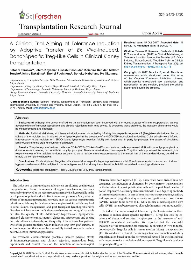
To induce the immunological tolerance by the less-invasive method, we tried to induce donor-specific regulatory T (Treg)-like cells by co-culture of donor and recipient lymphocytes in the presence of anti-CD80/86 monoclonal antibodies. We reported that the successful induction of the immunological tolerance by infusing ex vivo-induced, donor-specific Treg-like cells in rhesus monkey kidney transplantation [13]. We conducted a clinical trial aiming at tolerance induction in kidney transplantation based upon the new protocol revised for the clinical trial with respect to twice stimulation of donor-specific Treg-like cells by donor lymphocytes (Figure 1).
ISSN 2473-1730
Sci Forschen
O p e n H U B f o r S c i e n t i f i c R e s e a r c h
Citation: Teraoka S, Koyama I, Bashuda H, Uchida K, Tonsho M, et al. (2017) A Clinical Trial Aiming at Tolerance Induction by Adoptive Transfer of Ex Vivo-Induced, Donor-Specific Treg-Like Cells in Clinical Kidney Transplantation. J Transplant Res 2(1): doi http://dx.doi.org/10.16966/2473-1730.115
Open Access
2
The purposes of the study are to investigate, (1) whether Treg-like cells can be induced ex vivo also in human by the same methods, (2) whether tolerance can be induced also in clinical kidney transplantation by using ex vivo-induced, donor-specific Treg-like cells?
Materials and MethodsThis clinical trial of tolerance induction based upon the protocol
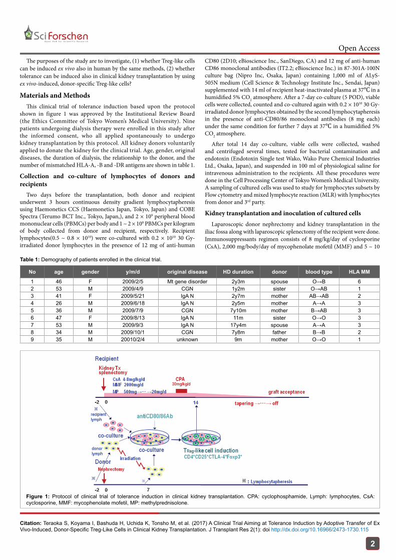
shown in figure 1 was approved by the Institutional Review Board (the Ethics Committee of Tokyo Women’s Medical University). Nine patients undergoing dialysis therapy were enrolled in this study after the informed consent, who all applied spontaneously to undergo kidney transplantation by this protocol. All kidney donors voluntarily applied to donate the kidney for the clinical trial. Age, gender, original diseases, the duration of dialysis, the relationship to the donor, and the number of mismatched HLA-A, -B and -DR antigens are shown in table 1.
Collection and co-culture of lymphocytes of donors and recipients
Two days before the transplantation, both donor and recipient underwent 3 hours continuous density gradient lymphocytapheresis using Haemonetics CCS (Haemonetics Japan, Tokyo, Japan) and COBE Spectra (Terumo BCT Inc., Tokyo, Japan,), and 2 × 10⁹ peripheral blood mononuclear cells (PBMCs) per body and 1 ~ 2 × 10⁸ PBMCs per kilogram of body collected from donor and recipient, respectively. Recipient lymphocytes(0.5 ~ 0.8 × 10¹⁰) were co-cultured with 0.2 × 10¹⁰ 30 Gy-irradiated donor lymphocytes in the presence of 12 mg of anti-human
CD80 (2D10; eBioscience Inc., SanDiego, CA) and 12 mg of anti-human CD86 monoclonal antibodies (IT2.2; eBioscience Inc.) in 87-301A-100N culture bag (Nipro Inc, Osaka, Japan) containing 1,000 ml of ALyS-505N medium (Cell Science & Technology Institute Inc., Sendai, Japan) supplemented with 14 ml of recipient heat-inactivated plasma at 37℃ in a humidified 5% CO2 atmosphere. After a 7-day co-culture (5 POD), viable cells were collected, counted and co-cultured again with 0.2 × 10¹⁰ 30 Gy-irradiated donor lymphocytes obtained by the second lymphocytapheresis in the presence of anti-CD80/86 monoclonal antibodies (8 mg each) under the same condition for further 7 days at 37℃ in a humidified 5% CO2 atmosphere.
After total 14 day co-culture, viable cells were collected, washed and centrifuged several times, tested for bacterial contamination and endotoxin (Endotoxin Single test Wako, Wako Pure Chemical Industries Ltd., Osaka, Japan), and suspended in 100 ml of physiological saline for intravenous administration to the recipients. All these procedures were done in the Cell Processing Center of Tokyo Women’s Medical University. A sampling of cultured cells was used to study for lymphocytes subsets by Flow cytometry and mixed lymphocyte reaction (MLR) with lymphocytes from donor and 3rd party.
Kidney transplantation and inoculation of cultured cellsLaparoscopic donor nephrectomy and kidney transplantation in the
iliac fossa along with laparoscopic splenectomy of the recipient were done. Immunosuppressants regimen consists of 8 mg/kg/day of cyclosporine (CsA), 2,000 mg/body/day of mycophenolate mofetil (MMF) and 5 ~ 10
No age gender y/m/d original disease HD duration donor blood type HLA MM
1 46 F 2009/2/5 Mt gene disorder 2y3m spouse O→B 6 2 53 M 2009/4/9 CGN 1y2m sister O→AB 1 3 41 F 2009/5/21 IgA N 2y7m mother AB→AB 2 4 26 M 2009/6/18 IgA N 2y5m mother A→A 3 5 36 M 2009/7/9 CGN 7y10m mother B→AB 3 6 47 F 2009/8/13 IgA N 11m sister O→O 3 7 53 M 2009/9/3 IgA N 17y4m spouse A→A 3 8 34 M 2009/10/1 CGN 7y8m father B→B 2 9 35 M 20010/2/4 unknown 9m mother O→O 1
Table 1: Demography of patients enrolled in the clinical trial.
Figure 1: Protocol of clinical trial of tolerance induction in clinical kidney transplantation. CPA: cyclophosphamide, Lymph: lymphocytes, CsA: cyclosporine, MMF: mycophenolate mofetil, MP: methylprednisolone.
Sci Forschen
O p e n H U B f o r S c i e n t i f i c R e s e a r c h
Citation: Teraoka S, Koyama I, Bashuda H, Uchida K, Tonsho M, et al. (2017) A Clinical Trial Aiming at Tolerance Induction by Adoptive Transfer of Ex Vivo-Induced, Donor-Specific Treg-Like Cells in Clinical Kidney Transplantation. J Transplant Res 2(1): doi http://dx.doi.org/10.16966/2473-1730.115
Open Access
3
mg/kg/day of methylprednisolone (MP). Basiliximab was avoided not to deplete CD25+ T cell. The doses of CsA and MMF were gradually reduced, adjusting each trough level around 150 ~ 250 ng/ml and 1.0 ~ 4.0 µg/ml, respectively for the first three months. The dose of MP was rapidly reduced to 20 mg/day on day 7, and further tapered to 8 mg/day on day 14.
On day 5 ~ 7, 25 ~ 30 mg/kg/day of cyclophosphamide (CPA) was given intravenously to deplete circulating leukocytes. Circulating leukocyte count was reduced to around 1,500/mm3 within a few days and returned to the previous value within 14 ~ 21days. On 12 POD, 0.8 ~ 1.5 × 10⁹ cultured cells were given intravenously to the recipient (Table 2). In three patients, cultured cells (0.075 ~ 0.8 × 10⁹) were given later after 12 POD (resulting in the elongated culture period), on 19, 33 and 21 POD because of ascites due to preoperative continuous ambulatory peritoneal dialysis (CAPD), delayed urination due to postoperative acute tubular necrosis (ATN), and the leakage of pancreatic juice after splenectomy, respectively. Latter two patients were given an insufficient number of cultured cells, 0.075 × 10⁹ on day 33 and 0.3 × 10⁹ on day 21, respectively, because obtained viable cultured cells tended to decrease in number when the culture period of time was extended for more than 14 days (Table 2).
Thereafter MP was tapered down gradually, and the doses of CsA and MMF were reduced based upon the value of MLR with lymphocytes from the donor and 3rd party, especially the ratio of MLR with the donor to 3rd party lymphocytes. When the doses of CsA and MMF were reduced to 25 mg/day and 250 mg/day, respectively, first MMF was discontinued followed by further reduction of CsA.
Mixed lymphocyte reactionPeripheral blood was obtained from the donor and the recipient, and
PBMCs were prepared by centrifugation over Separate-L (Muto Pure Chemicals Co., Ltd., Tokyo, Japan). Thereafter 2 × 10⁵ lymphocytes from the recipient were co-cultured with the same number of 30 Gy-irradiated donor lymphocytes in 96-well round-bottomed plates (catalog 3799; Corning Inc., New York, NY). The cells were cultured for 6 days and then pulsed with 10 µCi of [3H] thymidine for the last 18 hours. The incorporated radioactivity was measured on a 1450 MicroBeta counter (Perkin Elmer Japan Co., Ltd., Yokohama, Japan).
Flow cytometryAntibodies used for flow cytometry were CD3 PerCP (BD Biosciences,
San Jose, CA), CD4 PerCP (BD Biosciences), CTLA-4-FITC (BECKMAN COULTER, Fullerton, CA), CD4-APC (BECKMAN COULTER), CD8-APC (BECKMAN COULTER), CD25 PE (BD Biosciences), CD25 APC (BD Biosciences), CD45RA PE (2H4-RD1; BECKMAN COULTER), CD45RO PE (Human CD45RO R-PE Conjugate; invitrogen, Camarillo, CA), CD95 FITC (Human CD95 FITC Conjugate; invitrogen), CD152-PE (BECKMAN COULTER), FoxP3 FITC (Anti-Human FoxP3 FITC; eBioscience, San Diego, CA), FoxP3 PE (Anti-Human FoxP3 FITC; eBioscience), granzyme B PE (Human GranzymeB R-PE Conjugate; invitrogen), perforin FITC (FITC Mouse Anti-Human Perforin; BD Biosciences), IFN-γ FITC (Anti-Human IFN-γ FITC; BD Biosciences), and IFN-γ/IL-4 (Fastimmune IFN-γ/IL-4; BD Biosciences).
Samplings of cultured cells (1 × 10⁵) were incubated with 1 µg each of the indicated monoclonal antibodies for 30 minutes at 4℃ in PBS, washed twice, and analyzed on a FAC Scan equipped with Cellquest software version 3.3 (BD). The expression of CTLA-4 and FoxP3 was determined by intracytoplasmic staining using CD152 PE and FoxP3 FITC or PE after the cells were fixed with 2% formaldehyde and the membrane was permeabilized with BD Biosciences FoxP3 Staining Buffer Set (eBioscience, San Diego, USA).
Peripheral blood samplings of recipients were treated with the same procedures and assayed for flow cytometry.
Immunohistochemical staining of the biopsied specimen of the kidney grafts
The biopsied specimen was fixed in 10% buffered formalin and embedded in paraffin. H&E and PAS stains were performed for histological examination. The immunohistochemical staining assay using anti-CD4, -CD8, -CD20, -CD25, -FoxP3 and -granzyme B antibody was performed with iView DAB Detection Kit (Roche Tissue Diagnostics Japan Ventana, Tokyo, Japan) on a VENTANA BenchMark GX automated staining system (Roche Diagnostics Japan Ventana, Tokyo, Japan) to investigate the characteristics of infiltrated cells into kidney grafts.
Briefly, the tissue sections were deparaffinized with EZ Prep (Roche Tissue Diagnostics Japan Ventana) at 75℃, heat pretreated in Cell Conditioning 1 (CC1; Roche Tissue Diagnostics Japan Ventana) using “standard cell conditioning” for antigen retrieval at 100℃, and then incubated with anti-CD4 and -CD8 rabbit monoclonal antibody (Roche Tissue Diagnostics Japan Ventana), anti-CD20 primary antibody (Roche Tissue Diagnostics Japan Ventana), anti-CD25 mouse monoclonal antibody (NICHIREI Bioscience Inc., Tokyo, Japan), anti-human Foxp3 antibody (BioLegend Japan, Tokyo, Japan) and anti-granzyme B polyclonal antibody (Roche Tissue Diagnostics Japan Ventana) as a primary antibody, respectively, for 32 min at 37℃ after inactivation of the endogenous peroxidase with hydrogen peroxide for 4 min. They were then blocked using Endogenous Biotin Blocking Kit (Roche Tissue Diagnostics Japan Ventana), incubated with a biotinylated Ig secondary antibody for 8 min, and incubated with a streptavidin-HRP conjugate for 8 min at 37℃. The immunolocalized CD4, CD8, CD20, CD25, FoxP3 and granzyme B were visualized using a copper-enhanced DAB reaction. The slides were counterstained with Hematoxylin II (Roche Tissue Diagnostics Japan Ventana) for 4 min and Bluing Reagent (Roche Tissue Diagnostics Japan Ventana) for 4 min and coverslips were applied by an automated coverslipper (Tissue-Tek Film Automated Coverslipper; Sakura Finetek Japan, Tokyo, Japan).
ResultsThe number of viable cells obtained by co-culture of the recipient
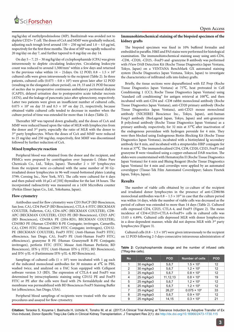
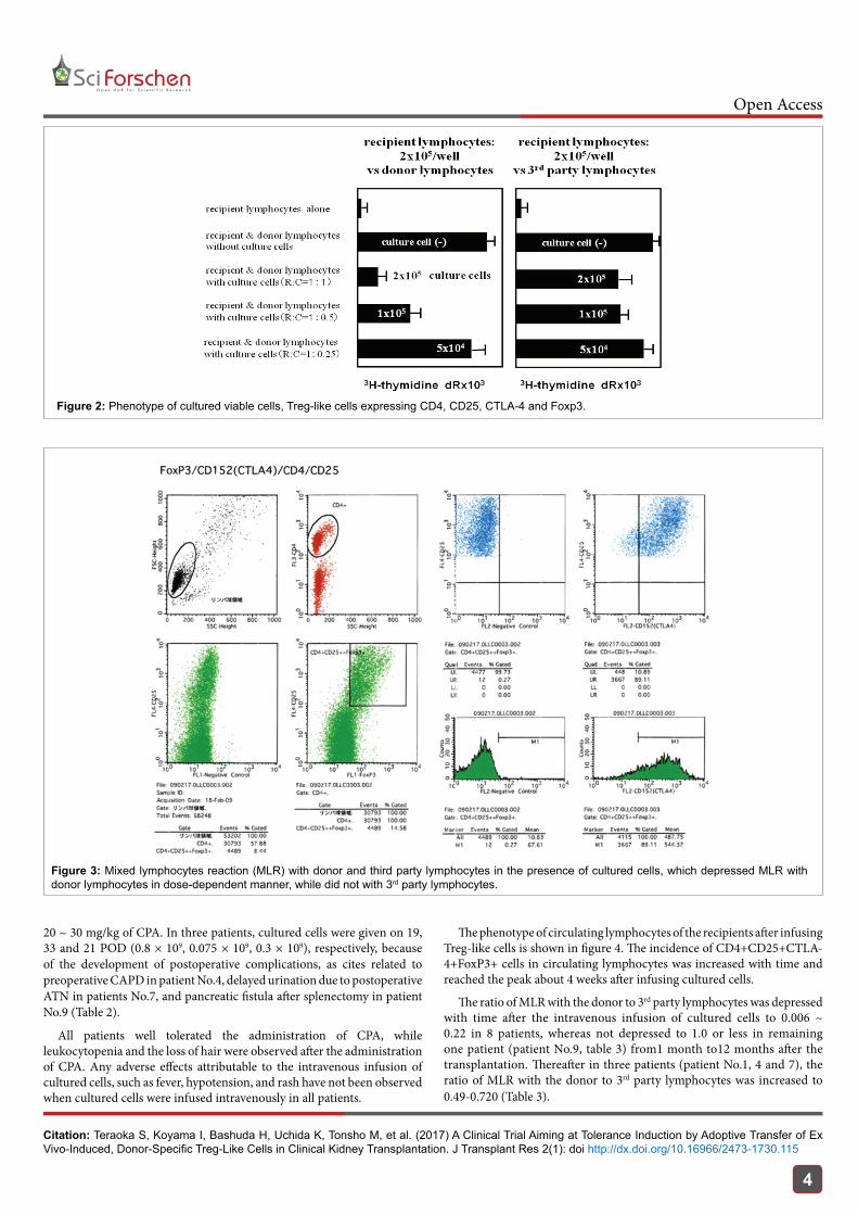
and irradiated donor lymphocytes in the presence of anti-CD80/86 monoclonal antibodies was 0.8 ~ 1.5 × 10¹⁰ in case that the culture period was within 14 days, while the number of viable cells was decreased as the period of culture was extended to more than 14 days (Table 2). Cultured cells expressed CD4, CD25, CTLA-4, and FoxP3 (Figure 2). The mean incidence of CD4+CD25+CTLA-4+FoxP3+ cells in cultured cells was 13.03 ± 6.89%. Cultured cells depressed MLR with donor lymphocytes in a dose-dependent manner, while did not depress MLR with 3rd party lymphocytes (Figure 3).
Cultured cells (0.8 ~ 1.5 × 10⁹) were given intravenously to the recipient on 12 POD following 2-3 days-consecutive intravenous administration of
No CPA POD Number of cells POD
1 30 mg/kgx3 5,6,7 1.5 × 10⁹ 12 2 30 mg/kgx3 5,6,7 1.2 × 10⁹ 12 3 25 mg/kgx3 5,6,7 0.8 × 10⁹ 12 4 25 mg/kgx3 11,12,13 0.8 × 10⁹ 19 5 25 mg/kgx3 5,6,7 1.2 × 10⁹ 12 6 25 mg/kgx3 5,6,7 1.2 × 10⁹ 12 7 25 mg/kgx2 26,27 0.075 × 10⁹ 33 8 25 mg/kgx3 5,6,7 0.9 × 10⁹ 12 9 25 mg/kgx2 14,15 0.3 × 10⁹ 21
Table 2: Cyclophosphamide dosage and the number of infused cells (TReg-like cells).
Sci Forschen
O p e n H U B f o r S c i e n t i f i c R e s e a r c h
Citation: Teraoka S, Koyama I, Bashuda H, Uchida K, Tonsho M, et al. (2017) A Clinical Trial Aiming at Tolerance Induction by Adoptive Transfer of Ex Vivo-Induced, Donor-Specific Treg-Like Cells in Clinical Kidney Transplantation. J Transplant Res 2(1): doi http://dx.doi.org/10.16966/2473-1730.115
Open Access
4
Figure 2: Phenotype of cultured viable cells, Treg-like cells expressing CD4, CD25, CTLA-4 and Foxp3.
Figure 3: Mixed lymphocytes reaction (MLR) with donor and third party lymphocytes in the presence of cultured cells, which depressed MLR with donor lymphocytes in dose-dependent manner, while did not with 3rd party lymphocytes.
20 ~ 30 mg/kg of CPA. In three patients, cultured cells were given on 19, 33 and 21 POD (0.8 × 10⁹, 0.075 × 10⁹, 0.3 × 10⁹), respectively, because of the development of postoperative complications, as cites related to preoperative CAPD in patient No.4, delayed urination due to postoperative ATN in patients No.7, and pancreatic fistula after splenectomy in patient No.9 (Table 2).
All patients well tolerated the administration of CPA, while leukocytopenia and the loss of hair were observed after the administration of CPA. Any adverse effects attributable to the intravenous infusion of cultured cells, such as fever, hypotension, and rash have not been observed when cultured cells were infused intravenously in all patients.
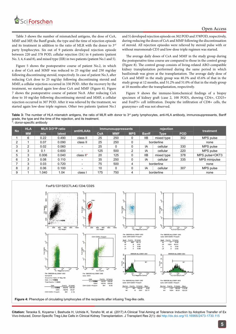
The phenotype of circulating lymphocytes of the recipients after infusing Treg-like cells is shown in figure 4. The incidence of CD4+CD25+CTLA-4+FoxP3+ cells in circulating lymphocytes was increased with time and reached the peak about 4 weeks after infusing cultured cells.
The ratio of MLR with the donor to 3rd party lymphocytes was depressed with time after the intravenous infusion of cultured cells to 0.006 ~ 0.22 in 8 patients, whereas not depressed to 1.0 or less in remaining one patient (patient No.9, table 3) from1 month to12 months after the transplantation. Thereafter in three patients (patient No.1, 4 and 7), the ratio of MLR with the donor to 3rd party lymphocytes was increased to 0.49-0.720 (Table 3).
Sci Forschen
O p e n H U B f o r S c i e n t i f i c R e s e a r c h
Citation: Teraoka S, Koyama I, Bashuda H, Uchida K, Tonsho M, et al. (2017) A Clinical Trial Aiming at Tolerance Induction by Adoptive Transfer of Ex Vivo-Induced, Donor-Specific Treg-Like Cells in Clinical Kidney Transplantation. J Transplant Res 2(1): doi http://dx.doi.org/10.16966/2473-1730.115
Open Access
5
Table 3 shows the number of mismatched antigens, the dose of CsA, MMF and MP, the Banff grade, the type and the time of rejection episode and its treatment in addition to the ratio of MLR with the donor to 3rd party lymphocytes. Six out of 9 patients developed rejection episode between 220 and 378 POD, cellular rejection (IA) in 4 patients (patient No. 3, 4, 6 and 8), and mixed type (IIB) in two patients (patient No.1 and 5).
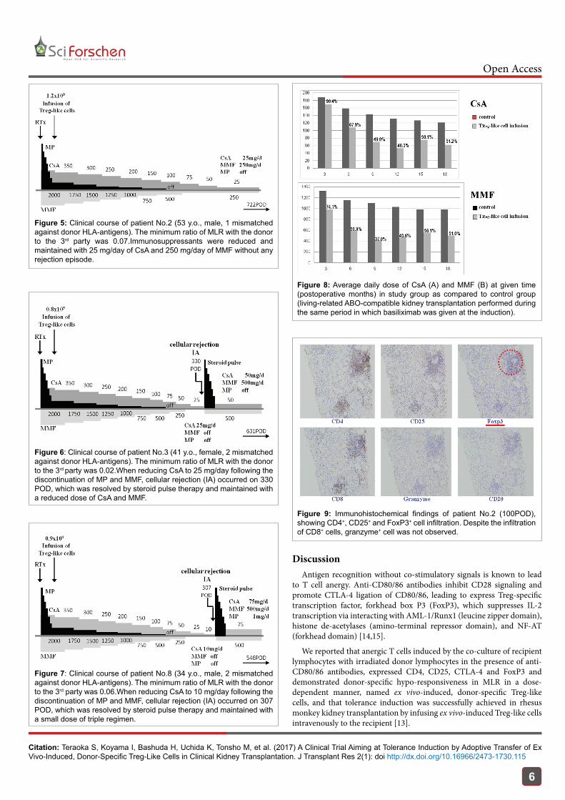
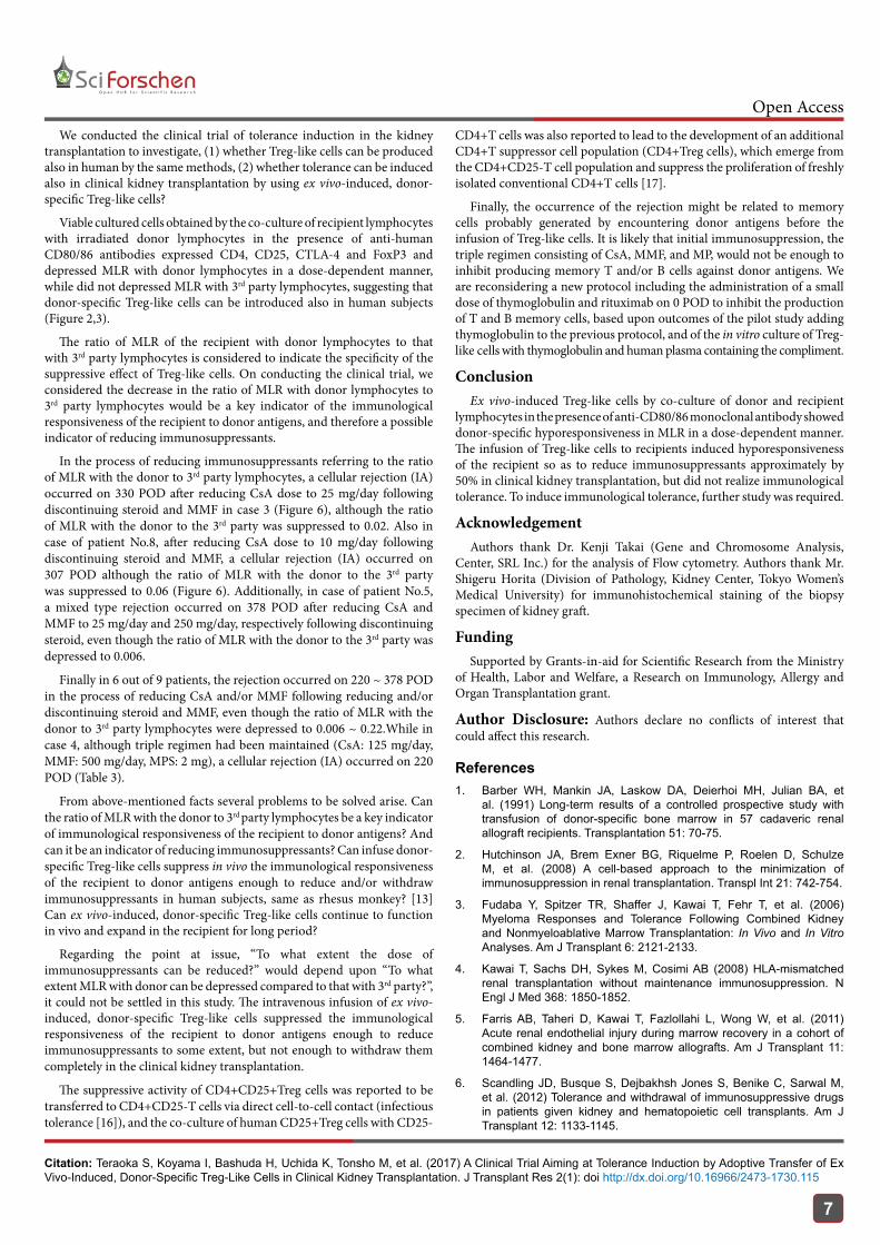
Figure 5 shows the postoperative course of patient No.2, in which a dose of CsA and MMF was reduced to 25 mg/day and 250 mg/day following discontinuing steroid, respectively. In case of patient No.3, after reducing CsA dose to 25 mg/day following discontinuing steroid and MMF, a cellular rejection occurred in 330 POD. After the recovery by the treatment, we started again low-dose CsA and MMF (Figure 6). Figure 7 shows the postoperative course of patient No.8. After reducing CsA dose to 10 mg/day following discontinuing steroid and MMF, a cellular rejection occurred in 307 POD. After it was relieved by the treatment, we started again low-dose triple regimen. Other two patients (patient No.1
Figure 4: Phenotype of circulating lymphocytes of the recipients after infusing Treg-like cells.
and 5) developed rejection episode on 302 POD and 378POD, respectively, during reducing the doses of CsA and MMF following the discontinuation of steroid. All rejection episodes were relieved by steroid pulse with or without muromonab CD3 and low-dose triple regimen was started.
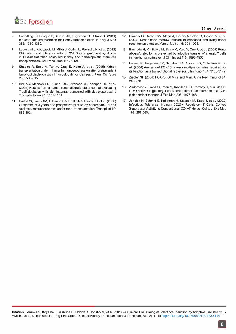
The average daily doses of CsA and MMF in the study group during the postoperative time course are compared to those in the control group (Figure 8). The control group consists of living-related ABO-compatible kidney transplantation performed during the same period in which basiliximab was given at the transplantation. The average daily dose of CsA and MMF in the study group was 40.3% and 45.6% of that in the study group at 12 months, and 51.2% and 51.0% of that in the study group at 18 months after the transplantation, respectively.
Figure 9 shows the immuno-histochemical findings of a biopsy specimen of kidney graft (case 2, 100 POD), showing CD4+, CD25+ and FoxP3+ cell infiltration. Despite the infiltration of CD8+ cells, the granzyme+ cell was not observed.
No HLA MLR D/3rdP ratio antiHLAAb Immunosuppressants rejection treatmentMM min latest CsA MMF MPS Banff Type POD
1 6 0.22 0.490 class II 25 250 0 IIB mixed type 302 MPS pulse2 1 0.07 0.090 class II 25 250 0 borderline none3 2 0.02 0.060 - 25 0 0 IA cellular 330 MPS pulse4 3 0.1 0.600 - 125 500 2 IA cellular 220 MPS pulse5 3 0.006 0.040 class II* 20 125 0 IIB mixed type 378 MPS pulse+OKT36 3 0.08 0.110 - 35 250 0 IA cellular 335 MPS minipulse7 3 0.03 0.720 - 75 500 4 borderline none8 2 0.06 0.100 - 10 0 0 IA cellular 307 MPS pulse9 1 1.040 1.04 class I 175 750 4 borderline none
Table 3: The number of HLA mismatch antigens, the ratio of MLR with donor to 3rd party lymphocytes, anti-HLA antibody, immunosuppressants, Banff grade, the type and the time of the rejection, and its treatment. *: donor-specific antibody
Sci Forschen
O p e n H U B f o r S c i e n t i f i c R e s e a r c h
Citation: Teraoka S, Koyama I, Bashuda H, Uchida K, Tonsho M, et al. (2017) A Clinical Trial Aiming at Tolerance Induction by Adoptive Transfer of Ex Vivo-Induced, Donor-Specific Treg-Like Cells in Clinical Kidney Transplantation. J Transplant Res 2(1): doi http://dx.doi.org/10.16966/2473-1730.115
Open Access
6
Figure 5: Clinical course of patient No.2 (53 y.o., male, 1 mismatched against donor HLA-antigens). The minimum ratio of MLR with the donor to the 3rd party was 0.07.Immunosuppressants were reduced and maintained with 25 mg/day of CsA and 250 mg/day of MMF without any rejection episode.
Figure 6: Clinical course of patient No.3 (41 y.o., female, 2 mismatched against donor HLA-antigens). The minimum ratio of MLR with the donor to the 3rd party was 0.02.When reducing CsA to 25 mg/day following the discontinuation of MP and MMF, cellular rejection (IA) occurred on 330 POD, which was resolved by steroid pulse therapy and maintained with a reduced dose of CsA and MMF.
Figure 7: Clinical course of patient No.8 (34 y.o., male, 2 mismatched against donor HLA-antigens). The minimum ratio of MLR with the donor to the 3rd party was 0.06.When reducing CsA to 10 mg/day following the discontinuation of MP and MMF, cellular rejection (IA) occurred on 307 POD, which was resolved by steroid pulse therapy and maintained with a small dose of triple regimen.
Figure 8: Average daily dose of CsA (A) and MMF (B) at given time (postoperative months) in study group as compared to control group (living-related ABO-compatible kidney transplantation performed during the same period in which basiliximab was given at the induction).
Figure 9: Immunohistochemical findings of patient No.2 (100POD), showing CD4+, CD25+ and FoxP3+ cell infiltration. Despite the infiltration of CD8+ cells, granzyme+ cell was not observed.
DiscussionAntigen recognition without co-stimulatory signals is known to lead
to T cell anergy. Anti-CD80/86 antibodies inhibit CD28 signaling and promote CTLA-4 ligation of CD80/86, leading to express Treg-specific transcription factor, forkhead box P3 (FoxP3), which suppresses IL-2 transcription via interacting with AML-1/Runx1 (leucine zipper domain), histone de-acetylases (amino-terminal repressor domain), and NF-AT (forkhead domain) [14,15].
We reported that anergic T cells induced by the co-culture of recipient lymphocytes with irradiated donor lymphocytes in the presence of anti-CD80/86 antibodies, expressed CD4, CD25, CTLA-4 and FoxP3 and demonstrated donor-specific hypo-responsiveness in MLR in a dose-dependent manner, named ex vivo-induced, donor-specific Treg-like cells, and that tolerance induction was successfully achieved in rhesus monkey kidney transplantation by infusing ex vivo-induced Treg-like cells intravenously to the recipient [13].
Sci Forschen
O p e n H U B f o r S c i e n t i f i c R e s e a r c h
Citation: Teraoka S, Koyama I, Bashuda H, Uchida K, Tonsho M, et al. (2017) A Clinical Trial Aiming at Tolerance Induction by Adoptive Transfer of Ex Vivo-Induced, Donor-Specific Treg-Like Cells in Clinical Kidney Transplantation. J Transplant Res 2(1): doi http://dx.doi.org/10.16966/2473-1730.115
Open Access
7
We conducted the clinical trial of tolerance induction in the kidney transplantation to investigate, (1) whether Treg-like cells can be produced also in human by the same methods, (2) whether tolerance can be induced also in clinical kidney transplantation by using ex vivo-induced, donor-specific Treg-like cells?
Viable cultured cells obtained by the co-culture of recipient lymphocytes with irradiated donor lymphocytes in the presence of anti-human CD80/86 antibodies expressed CD4, CD25, CTLA-4 and FoxP3 and depressed MLR with donor lymphocytes in a dose-dependent manner, while did not depressed MLR with 3rd party lymphocytes, suggesting that donor-specific Treg-like cells can be introduced also in human subjects (Figure 2,3).
The ratio of MLR of the recipient with donor lymphocytes to that with 3rd party lymphocytes is considered to indicate the specificity of the suppressive effect of Treg-like cells. On conducting the clinical trial, we considered the decrease in the ratio of MLR with donor lymphocytes to 3rd party lymphocytes would be a key indicator of the immunological responsiveness of the recipient to donor antigens, and therefore a possible indicator of reducing immunosuppressants.
In the process of reducing immunosuppressants referring to the ratio of MLR with the donor to 3rd party lymphocytes, a cellular rejection (IA) occurred on 330 POD after reducing CsA dose to 25 mg/day following discontinuing steroid and MMF in case 3 (Figure 6), although the ratio of MLR with the donor to the 3rd party was suppressed to 0.02. Also in case of patient No.8, after reducing CsA dose to 10 mg/day following discontinuing steroid and MMF, a cellular rejection (IA) occurred on 307 POD although the ratio of MLR with the donor to the 3rd party was suppressed to 0.06 (Figure 6). Additionally, in case of patient No.5, a mixed type rejection occurred on 378 POD after reducing CsA and MMF to 25 mg/day and 250 mg/day, respectively following discontinuing steroid, even though the ratio of MLR with the donor to the 3rd party was depressed to 0.006.
Finally in 6 out of 9 patients, the rejection occurred on 220 ~ 378 POD in the process of reducing CsA and/or MMF following reducing and/or discontinuing steroid and MMF, even though the ratio of MLR with the donor to 3rd party lymphocytes were depressed to 0.006 ~ 0.22.While in case 4, although triple regimen had been maintained (CsA: 125 mg/day, MMF: 500 mg/day, MPS: 2 mg), a cellular rejection (IA) occurred on 220 POD (Table 3).
From above-mentioned facts several problems to be solved arise. Can the ratio of MLR with the donor to 3rd party lymphocytes be a key indicator of immunological responsiveness of the recipient to donor antigens? And can it be an indicator of reducing immunosuppressants? Can infuse donor-specific Treg-like cells suppress in vivo the immunological responsiveness of the recipient to donor antigens enough to reduce and/or withdraw immunosuppressants in human subjects, same as rhesus monkey? [13] Can ex vivo-induced, donor-specific Treg-like cells continue to function in vivo and expand in the recipient for long period?
Regarding the point at issue, “To what extent the dose of immunosuppressants can be reduced?” would depend upon “To what extent MLR with donor can be depressed compared to that with 3rd party?”, it could not be settled in this study. The intravenous infusion of ex vivo-induced, donor-specific Treg-like cells suppressed the immunological responsiveness of the recipient to donor antigens enough to reduce immunosuppressants to some extent, but not enough to withdraw them completely in the clinical kidney transplantation.
The suppressive activity of CD4+CD25+Treg cells was reported to be transferred to CD4+CD25-T cells via direct cell-to-cell contact (infectious tolerance [16]), and the co-culture of human CD25+Treg cells with CD25-
CD4+T cells was also reported to lead to the development of an additional CD4+T suppressor cell population (CD4+Treg cells), which emerge from the CD4+CD25-T cell population and suppress the proliferation of freshly isolated conventional CD4+T cells [17].
Finally, the occurrence of the rejection might be related to memory cells probably generated by encountering donor antigens before the infusion of Treg-like cells. It is likely that initial immunosuppression, the triple regimen consisting of CsA, MMF, and MP, would not be enough to inhibit producing memory T and/or B cells against donor antigens. We are reconsidering a new protocol including the administration of a small dose of thymoglobulin and rituximab on 0 POD to inhibit the production of T and B memory cells, based upon outcomes of the pilot study adding thymoglobulin to the previous protocol, and of the in vitro culture of Treg-like cells with thymoglobulin and human plasma containing the compliment.
ConclusionEx vivo-induced Treg-like cells by co-culture of donor and recipient
lymphocytes in the presence of anti-CD80/86 monoclonal antibody showed donor-specific hyporesponsiveness in MLR in a dose-dependent manner. The infusion of Treg-like cells to recipients induced hyporesponsiveness of the recipient so as to reduce immunosuppressants approximately by 50% in clinical kidney transplantation, but did not realize immunological tolerance. To induce immunological tolerance, further study was required.
AcknowledgementAuthors thank Dr. Kenji Takai (Gene and Chromosome Analysis,
Center, SRL Inc.) for the analysis of Flow cytometry. Authors thank Mr. Shigeru Horita (Division of Pathology, Kidney Center, Tokyo Women’s Medical University) for immunohistochemical staining of the biopsy specimen of kidney graft.
FundingSupported by Grants-in-aid for Scientific Research from the Ministry
of Health, Labor and Welfare, a Research on Immunology, Allergy and Organ Transplantation grant.
Author Disclosure: Authors declare no conflicts of interest that could affect this research.
References1. Barber WH, Mankin JA, Laskow DA, Deierhoi MH, Julian BA, et
al. (1991) Long-term results of a controlled prospective study with transfusion of donor-specific bone marrow in 57 cadaveric renal allograft recipients. Transplantation 51: 70-75.
2. Hutchinson JA, Brem Exner BG, Riquelme P, Roelen D, Schulze M, et al. (2008) A cell-based approach to the minimization of immunosuppression in renal transplantation. Transpl Int 21: 742-754.
3. Fudaba Y, Spitzer TR, Shaffer J, Kawai T, Fehr T, et al. (2006) Myeloma Responses and Tolerance Following Combined Kidney and Nonmyeloablative Marrow Transplantation: In Vivo and In Vitro Analyses. Am J Transplant 6: 2121-2133.
4. Kawai T, Sachs DH, Sykes M, Cosimi AB (2008) HLA-mismatched renal transplantation without maintenance immunosuppression. N Engl J Med 368: 1850-1852.
5. Farris AB, Taheri D, Kawai T, Fazlollahi L, Wong W, et al. (2011) Acute renal endothelial injury during marrow recovery in a cohort of combined kidney and bone marrow allografts. Am J Transplant 11: 1464-1477.
6. Scandling JD, Busque S, Dejbakhsh Jones S, Benike C, Sarwal M, et al. (2012) Tolerance and withdrawal of immunosuppressive drugs in patients given kidney and hematopoietic cell transplants. Am J Transplant 12: 1133-1145.
Sci Forschen
O p e n H U B f o r S c i e n t i f i c R e s e a r c h
Citation: Teraoka S, Koyama I, Bashuda H, Uchida K, Tonsho M, et al. (2017) A Clinical Trial Aiming at Tolerance Induction by Adoptive Transfer of Ex Vivo-Induced, Donor-Specific Treg-Like Cells in Clinical Kidney Transplantation. J Transplant Res 2(1): doi http://dx.doi.org/10.16966/2473-1730.115
Open Access
8
7. Scandling JD, Busque S, Shizuru JA, Engleman EG, Strober S (2011) Induced immune tolerance for kidney transplantation. N Engl J Med 365: 1359-1360.
8. Leventhal J, Abecassis M, Miller J, Gallon L, Ravindra K, et al. (2012) Chimerism and tolerance without GVHD or engraftment syndrome in HLA-mismatched combined kidney and hematopoietic stem cell transplantation. Sci Transl Med 4: 124-128.
9. Shapiro R, Basu A, Tan H, Gray E, Kahn A, et al. (2005) Kidney transplantation under minimal immunosuppression after pretransplant lymphoid depletion with Thymoglobulin or Campath. J Am Coll Surg 200: 505-515.
10. Kirk AD, Mannon RB, Kleiner DE, Swanson JS, Kampen RL, et al. (2005) Results from a human renal allograft tolerance trial evaluating T-cell depletion with alemtuzumab combined with deoxyspergualin. Transplantation 80: 1051-1059.
11. Barth RN, Janus CA, Lillesand CA, Radke NA, Pirsch JD, et al. (2006) Outcomes at 3 years of a prospective pilot study of campath-1H and sirolimus immunosuppression for renal transplantation. Transpl Int 19: 885-892.
12. Ciancio G, Burke GW, Moon J, Garcia Morales R, Rosen A, et al. (2004) Donor bone marrow infusion in deceased and living donor renal transplantation. Yonsei Med J 45: 998-1003.
13. Bashuda H, Kimikawa M, Seino K, Kato Y, Ono F, et al. (2005) Renal allograft rejection is prevented by adoptive transfer of anergic T cells in non-human primates. J Clin Invest 115: 1896-1902.
14. Lopes JE, Torgerson TR, Schubert LA, Anover SD, Ocheltree EL, et al. (2006) Analysis of FOXP3 reveals multiple domains required for its function as a transcriptional repressor. J Immunol 174: 3133-3142.
15. Ziegler SF (2006) FOXP3: Of Mice and Men. Annu Rev Immunol 24: 209-226.
16. Andersson J, Tran DQ, Pesu M, Davidson TS, Ramsey H, et al. (2008) CD4+FoxP3+ regulatory T cells confer infectious tolerance in a TGF-β-dependent manner. J Exp Med 205: 1975-1981.
17. Jonuleit H, Schmitt E, Kakirman H, Stassen M, Knop J, et al. (2002) Infectious Tolerance: Human CD25+ Regulatory T Cells Convey Suppressor Activity to Conventional CD4+T Helper Cells. J Exp Med 196: 255-260.