Embed Size (px)
Citation preview
A Cohort Study of Patients Undergoing Distal TibialOsteotomy without Fibular Osteotomy for Medial
Ankle Arthritis with Mortise WideningTae-Keun Ahn, MD, Young Yi, MD, Jae-Ho Cho, MD, and Woo-Chun Lee, MD, PhD
Investigation performed at the Seoul Foot and Ankle Center, Department of Orthopaedic Surgery,Seoul Paik Hospital, Inje University, Seoul, South Korea
Background: Although the supramalleolar osteotomy can shift the weight-bearing axis laterally, it cannot reconstruct awidened ankle mortise caused by progression of medial ankle osteoarthritis. The aim of this study was to evaluateradiographic and clinical outcomes of distal tibial osteotomy without fibular osteotomy in patients with medial ankleosteoarthritis and mortise widening.
Methods: Distal tibial osteotomy without fibular osteotomy was performed in eighteen patients to treat medial ankleosteoarthritis with mortise widening. Fifteen women and three men with a mean age of fifty-seven years (range, forty-nineto sixty-four years) were followed for a mean of thirty-four months (range, twenty-four to sixty-six months). Mortise wideningwas diagnosed using valgus stress radiographs and intraoperative examination. The clinical outcome was assessed withthe American Orthopaedic Foot & Ankle Society (AOFAS) score, visual analog scale (VAS) score for pain, and the ankleosteoarthritis scale (AOS) score. The translation of the talus within the ankle mortise, talar tilt, medial distal tibial angle,and anterior distal tibial angle were evaluated on weight-bearing radiographs made preoperatively and postoperatively.
Results: The AOFAS score improved significantly from 78.4 points (95% confidence interval [CI], 74.6 to 80.5 points) to89 points (95% CI, 86.5 to 90.5 points) (p < 0.001). The VAS score for pain also decreased significantly from 6.7 points(95% CI, 6 to 7.5 points) to 2.7 points (95% CI, 2.3 to 3.3 points) (p < 0.001). The mean AOS score was 29.8 points (95%CI, 22 to 38.2 points) at the latest follow-up. The center of the talusmoved laterally within the anklemortise after the distaltibial osteotomy. Themeanmedial distal tibial angle changed from 86.6� (95% CI, 85.7� to 87.6�) to 92.9� (95% CI, 91.6�to 94.3�) (p < 0.001), and themean anterior distal tibial angle changed from 81.1� (95% CI, 78.6� to 83.6�) to 84.3� (95%CI, 81.9� to 86.4�) (p < 0.001). However, talar tilt was not corrected significantly (p = 0.916).
Conclusions: Distal tibial osteotomy without fibular osteotomy reduces pain in the short term in patients with anklearthritis, a widened mortise, and minimal talar tilt.
Level of Evidence: Therapeutic Level IV. See Instructions for Authors for a complete description of levels of evidence.
Asupramalleolar osteotomy of the tibia and fibula cre-ates angulation and translation of the ankle jointwithout changing the width of the ankle mortise.
When there is ankle osteoarthritis with mortise widening,
supramalleolar osteotomy may not achieve a stable osseousmortise. If the tibia is cut and angled toward the fibulawithout osteotomy of the fibula, a functional narrowing ofthe mortise may occur. Plafondplasty has been suggested as
Disclosure: One or more of the authors received payments or services, either directly or indirectly (i.e., via his or her institution), from a third party insupport of an aspect of this work. None of the authors, or their institution(s), have had any financial relationship, in the thirty-six months prior tosubmission of this work, with any entity in the biomedical arena that could be perceived to influence or have the potential to influence what is written in thiswork. Also, no author has had any other relationships, or has engaged in any other activities, that could be perceived to influence or have the potential toinfluence what is written in this work. The complete Disclosures of Potential Conflicts of Interest submitted by authors are always provided with theonline version of the article.
Peer Review: This article was reviewed by the Editor-in-Chief and one Deputy Editor, and it underwent blinded review by two or more outside experts. The Deputy Editorreviewed each revision of the article, and it underwent a final review by the Editor-in-Chief prior to publication. Final corrections and clarifications occurred during one ormore exchanges between the author(s) and copyeditors.
381
COPYRIGHT � 2015 BY THE JOURNAL OF BONE AND JOINT SURGERY, INCORPORATED
J Bone Joint Surg Am. 2015;97:381-8 d http://dx.doi.org/10.2106/JBJS.M.01360
a method to correct the alignment of the tibial plafond1.Plafondplasty is based on the concept that the deformity shouldbe corrected at the center of rotation and angulation2, and thecenter of rotation and angulation of the deformity is locatedin the tibial plafond when there is an intra-articular defectfrom erosion of a portion of the tibial plafond or malunionafter tibial plafond fracture. However, in ankles with mortisewidening from medial erosion into the medial malleolus (seeAppendix), angulation in the middle of the tibial plafond isnot necessary. We performed a procedure termed distal tibialosteotomy without fibular osteotomy in our patients. A similarprocedure was previously reported in the Japanese literature3.In this procedure, the osteotomized distal fragment is shiftedinferolaterally using the lateral apex of the osteotomy as ahinge.
We hypothesized that the distal tibial osteotomy withoutfibular osteotomy is a useful treatment in ankle osteoarthritiswith medial translation of the talus and widening of the anklemortise. The aim of this study was to investigate the results ofdistal tibial osteotomy without fibular osteotomy.
Materials and Methods
From January 2008 to May 2011, distal tibial osteotomy without fibularosteotomy was performed after failed conservative treatment in eighteen
consecutive patients who had symptomatic medial ankle arthritis with >3 mmof medial clear space on the valgus stress radiograph and/or intraoperatively.When ankle mortise widening was suspected, but it was not definite on thevalgus stress radiograph, the medial joint space was explored intraoperatively.
Exclusion criteria were patients with neuropathic arthropathy, end-stage os-teoarthritis, inflammatory arthritis, or ankle osteoarthritis secondary to bonedeformity from congenital abnormality, fracture, or paralysis. This study wasapproved by our institutional review board. Fifteen women and three men witha mean age of fifty-seven years (range, forty-nine to sixty-four years) werefollowed for a mean of thirty-four months (range, twenty-four to sixty-sixmonths). There was no loss of follow-up (see Appendix). The last follow-upevaluation was the most recent one when the patients were examinedradiographically.
A standard load of 150 N was applied to the ankle using the Telosdevice (METAX, Hungen, Germany) for valgus-varus stress and anteriordrawer test. Medial clear space of >3 mm on the valgus stress radiographswas indicative of mortise widening. The mortise widening, which was sus-pected on the stress radiographs but not definite, was confirmed under directvision by a gap of ‡3 mm when abduction and external rotation forces wereapplied intraoperatively.
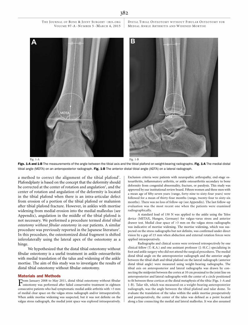
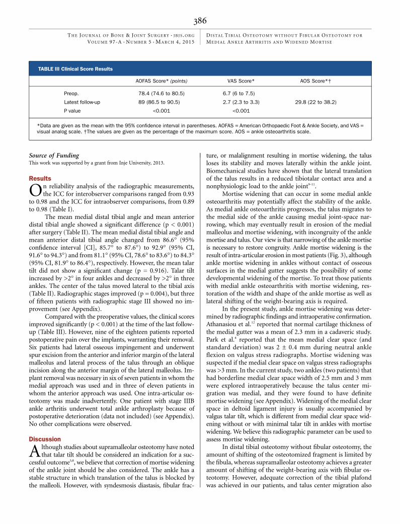
Radiographs and clinical scores were reviewed retrospectively by oneclinical fellow (T.-K.A.) and one assistant professor (J.-H.C.) specializing infoot and ankle surgery who did not attend the surgical procedures. Themedialdistal tibial angle on the anteroposterior radiograph and the anterior anglebetween the tibial shaft and tibial plafond on the lateral radiograph (anteriordistal tibial angle) were measured using weight-bearing radiographs. Thetibial axis on anteroposterior and lateral radiographs was drawn by con-necting the midpoint between the cortex at 10 cm proximal to the joint line onanteroposterior and lateral radiographs with the center of a circle positionedto fit between three cortices at the distal metaphysis of the tibia (Figs. 1-A and1-B). Talar tilt, which was measured on a weight-bearing anteroposteriorradiograph, was the angle between the tibial plafond and talar dome. Toevaluate the translation of the talus within the ankle mortise preoperativelyand postoperatively, the center of the talus was defined as a point locatedalong a line connecting the medial and lateral malleolus. It was also assumed
Fig. 1-A Fig. 1-B
Figs. 1-A and 1-B The measurements of the angle between the tibial axis and the tibial plafond on weight-bearing radiographs. Fig. 1-A The medial distal
tibial angle (MDTA) on an anteroposterior radiograph. Fig. 1-B The anterior distal tibial angle (ADTA) on a lateral radiograph.
382
THE JOURNAL OF BONE & JOINT SURGERY d J B J S .ORG
VOLUME 97-A d NUMBER 5 d MARCH 4, 2015DISTAL TIB IAL OSTEOTOMY WITHOUT FIBULAR OSTEOTOMY FOR
MEDIAL ANKLE ARTHRIT I S AND WIDENED MORTI SE
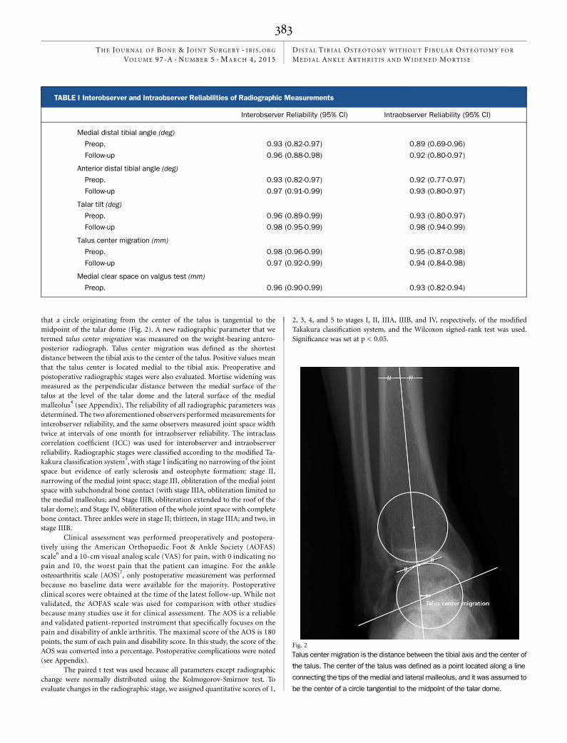
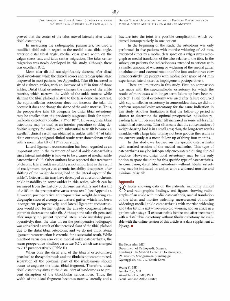
that a circle originating from the center of the talus is tangential to themidpoint of the talar dome (Fig. 2). A new radiographic parameter that wetermed talus center migration was measured on the weight-bearing antero-posterior radiograph. Talus center migration was defined as the shortestdistance between the tibial axis to the center of the talus. Positive values meanthat the talus center is located medial to the tibial axis. Preoperative andpostoperative radiographic stages were also evaluated. Mortise widening wasmeasured as the perpendicular distance between the medial surface of thetalus at the level of the talar dome and the lateral surface of the medialmalleolus
4(see Appendix). The reliability of all radiographic parameters was
determined. The two aforementioned observers performedmeasurements forinterobserver reliability, and the same observers measured joint space widthtwice at intervals of one month for intraobserver reliability. The intraclasscorrelation coefficient (ICC) was used for interobserver and intraobserverreliability. Radiographic stages were classified according to the modified Ta-kakura classification system
5, with stage I indicating no narrowing of the joint
space but evidence of early sclerosis and osteophyte formation; stage II,narrowing of the medial joint space; stage III, obliteration of the medial jointspace with subchondral bone contact (with stage IIIA, obliteration limited tothe medial malleolus; and Stage IIIB, obliteration extended to the roof of thetalar dome); and Stage IV, obliteration of the whole joint space with completebone contact. Three ankles were in stage II; thirteen, in stage IIIA; and two, instage IIIB.
Clinical assessment was performed preoperatively and postopera-tively using the American Orthopaedic Foot & Ankle Society (AOFAS)scale
6and a 10-cm visual analog scale (VAS) for pain, with 0 indicating no
pain and 10, the worst pain that the patient can imagine. For the ankleosteoarthritis scale (AOS)
7, only postoperative measurement was performed
because no baseline data were available for the majority. Postoperativeclinical scores were obtained at the time of the latest follow-up. While notvalidated, the AOFAS scale was used for comparison with other studiesbecause many studies use it for clinical assessment. The AOS is a reliableand validated patient-reported instrument that specifically focuses on thepain and disability of ankle arthritis. The maximal score of the AOS is 180points, the sum of each pain and disability score. In this study, the score of theAOS was converted into a percentage. Postoperative complications were noted(see Appendix).
The paired t test was used because all parameters except radiographicchange were normally distributed using the Kolmogorov-Smirnov test. Toevaluate changes in the radiographic stage, we assigned quantitative scores of 1,
2, 3, 4, and 5 to stages I, II, IIIA, IIIB, and IV, respectively, of the modifiedTakakura classification system, and the Wilcoxon signed-rank test was used.Significance was set at p < 0.05.
TABLE I Interobserver and Intraobserver Reliabilities of Radiographic Measurements
Interobserver Reliability (95% CI) Intraobserver Reliability (95% CI)
Medial distal tibial angle (deg)
Preop. 0.93 (0.82-0.97) 0.89 (0.69-0.96)
Follow-up 0.96 (0.88-0.98) 0.92 (0.80-0.97)
Anterior distal tibial angle (deg)
Preop. 0.93 (0.82-0.97) 0.92 (0.77-0.97)
Follow-up 0.97 (0.91-0.99) 0.93 (0.80-0.97)
Talar tilt (deg)
Preop. 0.96 (0.89-0.99) 0.93 (0.80-0.97)
Follow-up 0.98 (0.95-0.99) 0.98 (0.94-0.99)
Talus center migration (mm)
Preop. 0.98 (0.96-0.99) 0.95 (0.87-0.98)
Follow-up 0.97 (0.92-0.99) 0.94 (0.84-0.98)
Medial clear space on valgus test (mm)
Preop. 0.96 (0.90-0.99) 0.93 (0.82-0.94)
Fig. 2
Talus center migration is the distance between the tibial axis and the center of
the talus. The center of the talus was defined as a point located along a line
connecting the tips of the medial and lateral malleolus, and it was assumed to
be the center of a circle tangential to the midpoint of the talar dome.
383
THE JOURNAL OF BONE & JOINT SURGERY d J B J S .ORG
VOLUME 97-A d NUMBER 5 d MARCH 4, 2015DISTAL TIB IAL OSTEOTOMY WITHOUT FIBULAR OSTEOTOMY FOR
MEDIAL ANKLE ARTHRIT I S AND WIDENED MORTI SE
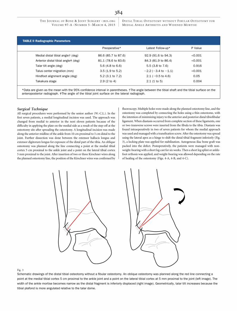
Surgical TechniqueAll surgical procedures were performed by the senior author (W.-C.L.). In thefirst seven patients, a medial longitudinal incision was used. The approach waschanged from medial to anterior in the next eleven patients because of thedifficulty in applying the plate on the medial side as a result of the step-off at theosteotomy site after spreading the osteotomy. A longitudinal incision was madealong the anterior midline of the ankle from 10 cm proximal to 5 cm distal to thejoint. Further dissection was done between the extensor hallucis longus andextensor digitorum longus for exposure of the distal part of the tibia. An obliqueosteotomy was planned along the line connecting a point at the medial tibialcortex 5 cm proximal to the ankle joint and a point on the lateral tibial cortex5 mmproximal to the joint. After insertion of two or three Kirschner wires alongthe planned osteotomy line, the position of the Kirschner wires was confirmed by
fluoroscopy.Multiple holes weremade along the planned osteotomy line, and theosteotomy was completed by connecting the holes using a thin osteotome, withthe intention ofminimizing injury to the anterior and posterior distal tibiofibularligament. When diastasis occurred from complete section of these ligaments, oneor two transverse screws were inserted from the fibula to the tibia. Diastasis wasfound intraoperatively in two of seven patients for whom the medial approachwas used andmanagedwith a transfixation screw. After the osteotomywas spreadusing the lateral apex as a hinge to shift the distal tibial fragment inferiorly (Fig.3), a locking plate was applied for stabilization. Autogenous iliac bone graft waspacked into the defect. Postoperatively, the patients were managed with non-weight-bearing with a short leg cast for six weeks. Then a short leg splint or ankle-foot orthosis was applied, and weight-bearing was allowed depending on the rateof healing of the osteotomy (Figs. 4-A, 4-B, and 4-C).
TABLE II Radiographic Parameters
Preoperative* Latest Follow-up* P Value
Medial distal tibial angle† (deg) 86.6 (85.7 to 87.6) 92.9 (91.6 to 94.3) <0.001
Anterior distal tibial angle‡ (deg) 81.1 (78.6 to 83.6) 84.3 (81.9 to 86.4) <0.001
Talar tilt angle (deg) 5.6 (4.8 to 6.6) 5.5 (3.8 to 7.6) 0.916
Talus center migration (mm) 3.5 (1.9 to 5.2) 22.2 (23.4 to 21.1) <0.001
Hindfoot alignment angle (deg) 5.2 (3.1 to 7.2) 2.1 (20.5 to 4.6) 0.05
Takakura stage 2.9 (2 to 4) 2.1 (1 to 5) 0.004
*Data are given as the mean with the 95% confidence interval in parentheses. †The angle between the tibial shaft and the tibial surface on theanteroposterior radiograph. ‡The angle of the tibial joint surface on the lateral radiograph.
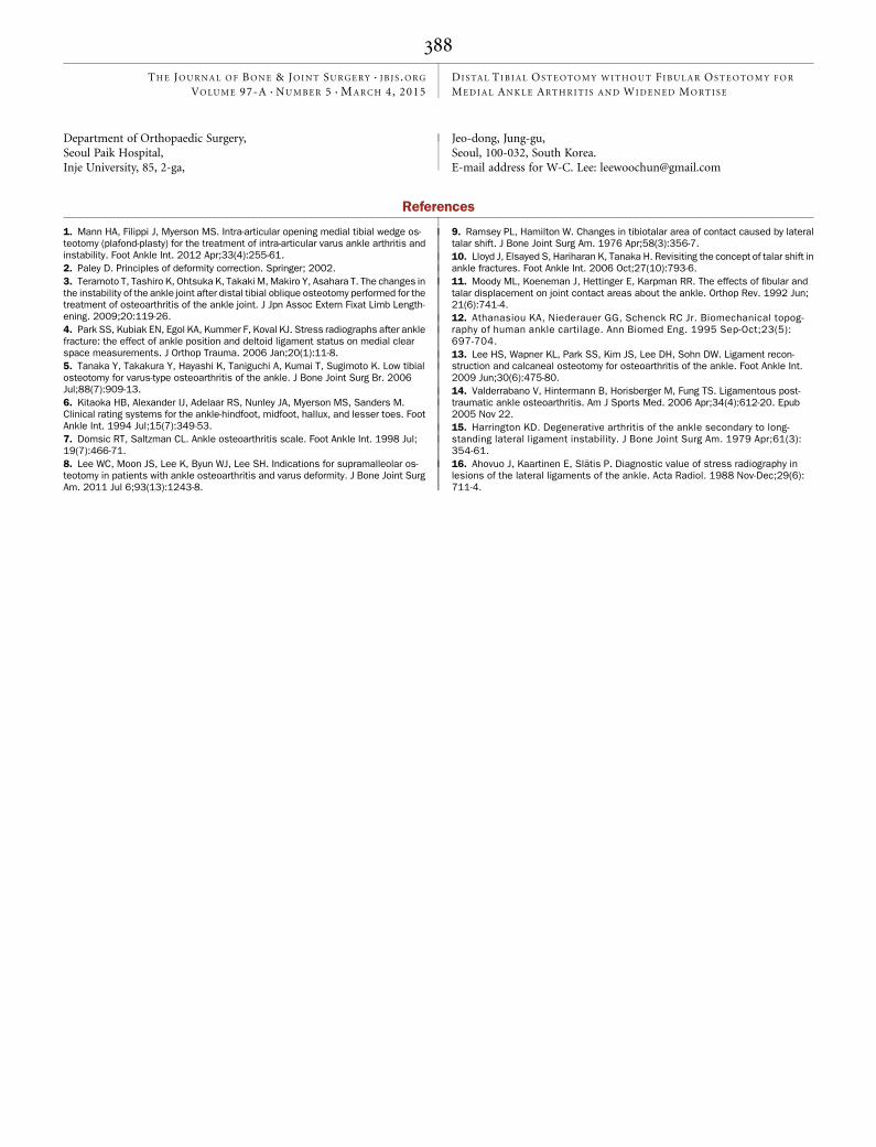
Fig. 3
Schematic drawings of the distal tibial osteotomy without a fibular osteotomy. An oblique osteotomy was planned along the red line connecting a
point at the medial tibial cortex 5 cm proximal to the ankle joint and a point on the lateral tibial cortex at 5 mm proximal to the joint (left image). The
width of the ankle mortise becomes narrow as the distal fragment is inferiorly displaced (right image). Geometrically, talar tilt increases because the
tibial plafond is more angulated relative to the talar dome.
384
THE JOURNAL OF BONE & JOINT SURGERY d J B J S .ORG
VOLUME 97-A d NUMBER 5 d MARCH 4, 2015DISTAL TIB IAL OSTEOTOMY WITHOUT FIBULAR OSTEOTOMY FOR
MEDIAL ANKLE ARTHRIT I S AND WIDENED MORTI SE
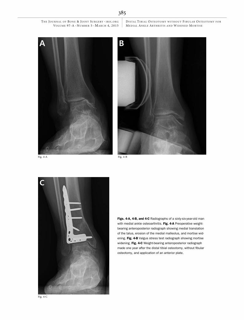
Fig. 4-A Fig. 4-B
Fig. 4-C
Figs. 4-A, 4-B, and 4-C Radiographs of a sixty-six-year-old man
with medial ankle osteoarthritis. Fig. 4-A Preoperative weight-
bearing anteroposterior radiograph showing medial translation
of the talus, erosion of the medial malleolus, and mortise wid-
ening. Fig. 4-B Valgus stress test radiograph showing mortise
widening. Fig. 4-C Weight-bearing anteroposterior radiograph
made one year after the distal tibial osteotomy, without fibular
osteotomy, and application of an anterior plate.
385
THE JOURNAL OF BONE & JOINT SURGERY d J B J S .ORG
VOLUME 97-A d NUMBER 5 d MARCH 4, 2015DISTAL TIB IAL OSTEOTOMY WITHOUT FIBULAR OSTEOTOMY FOR
MEDIAL ANKLE ARTHRIT I S AND WIDENED MORTI SE
Source of FundingThis work was supported by a grant from Inje University, 2013.
Results
On reliability analysis of the radiographic measurements,the ICC for interobserver comparisons ranged from 0.93
to 0.98 and the ICC for intraobserver comparisons, from 0.89to 0.98 (Table I).
The mean medial distal tibial angle and mean anteriordistal tibial angle showed a significant difference (p < 0.001)after surgery (Table II). The mean medial distal tibial angle andmean anterior distal tibial angle changed from 86.6� (95%confidence interval [CI], 85.7� to 87.6�) to 92.9� (95% CI,91.6� to 94.3�) and from 81.1� (95% CI, 78.6� to 83.6�) to 84.3�(95% CI, 81.9� to 86.4�), respectively. However, the mean talartilt did not show a significant change (p = 0.916). Talar tiltincreased by >2� in four ankles and decreased by >2� in threeankles. The center of the talus moved lateral to the tibial axis(Table II). Radiographic stages improved (p = 0.004), but threeof fifteen patients with radiographic stage III showed no im-provement (see Appendix).
Compared with the preoperative values, the clinical scoresimproved significantly (p < 0.001) at the time of the last follow-up (Table III). However, nine of the eighteen patients reportedpostoperative pain over the implants, warranting their removal.Six patients had lateral osseous impingement and underwentspur excision from the anterior and inferior margin of the lateralmalleolus and lateral process of the talus through an obliqueincision along the anterior margin of the lateral malleolus. Im-plant removal was necessary in six of seven patients in whom themedial approach was used and in three of eleven patients inwhom the anterior approach was used. One intra-articular os-teotomy was made inadvertently. One patient with stage IIIBankle arthritis underwent total ankle arthroplasty because ofpostoperative deterioration (data not included) (see Appendix).No other complications were observed.
Discussion
Although studies about supramalleolar osteotomy have notedthat talar tilt should be considered an indication for a suc-
cessful outcome5,8, we believe that correction of mortise wideningof the ankle joint should be also considered. The ankle has astable structure in which translation of the talus is blocked bythe malleoli. However, with syndesmosis diastasis, fibular frac-
ture, or malalignment resulting in mortise widening, the talusloses its stability and moves laterally within the ankle joint.Biomechanical studies have shown that the lateral translationof the talus results in a reduced tibiotalar contact area and anonphysiologic load to the ankle joint9-11.
Mortise widening that can occur in some medial ankleosteoarthritis may potentially affect the stability of the ankle.As medial ankle osteoarthritis progresses, the talus migrates tothe medial side of the ankle causing medial joint-space nar-rowing, which may eventually result in erosion of the medialmalleolus and mortise widening, with incongruity of the anklemortise and talus. Our view is that narrowing of the anklemortiseis necessary to restore congruity. Ankle mortise widening is theresult of intra-articular erosion inmost patients (Fig. 3), althoughankle mortise widening in ankles without contact of osseoussurfaces in the medial gutter suggests the possibility of somedevelopmental widening of the mortise. To treat those patientswith medial ankle osteoarthritis with mortise widening, res-toration of the width and shape of the ankle mortise as well aslateral shifting of the weight-bearing axis is required.
In the present study, ankle mortise widening was deter-mined by radiographic findings and intraoperative confirmation.Athanasiou et al.12 reported that normal cartilage thickness ofthe medial gutter was a mean of 2.3 mm in a cadaveric study.Park et al.4 reported that the mean medial clear space (andstandard deviation) was 2 ± 0.4 mm during neutral ankleflexion on valgus stress radiographs. Mortise widening wassuspected if the medial clear space on valgus stress radiographswas >3mm. In the current study, two ankles (two patients) thathad borderline medial clear space width of 2.5 mm and 3 mmwere explored intraoperatively because the talus center mi-gration was medial, and they were found to have definitemortise widening (see Appendix). Widening of the medial clearspace in deltoid ligament injury is usually accompanied byvalgus talar tilt, which is different from medial clear space wid-ening without or with minimal talar tilt in ankles with mortisewidening. We believe this radiographic parameter can be used toassess mortise widening.
In distal tibial osteotomy without fibular osteotomy, theamount of shifting of the osteotomized fragment is limited bythe fibula, whereas supramalleolar osteotomy achieves a greateramount of shifting of the weight-bearing axis with fibular os-teotomy. However, adequate correction of the tibial plafondwas achieved in our patients, and talus center migration also
TABLE III Clinical Score Results
AOFAS Score* (points) VAS Score* AOS Score*†
Preop. 78.4 (74.6 to 80.5) 6.7 (6 to 7.5)
Latest follow-up 89 (86.5 to 90.5) 2.7 (2.3 to 3.3) 29.8 (22 to 38.2)
P value <0.001 <0.001
*Data are given as the mean with the 95% confidence interval in parentheses. AOFAS = American Orthopaedic Foot & Ankle Society, and VAS =visual analog scale. †The values are given as the percentage of the maximum score. AOS = ankle osteoarthritis scale.
386
THE JOURNAL OF BONE & JOINT SURGERY d J B J S .ORG
VOLUME 97-A d NUMBER 5 d MARCH 4, 2015DISTAL TIB IAL OSTEOTOMY WITHOUT FIBULAR OSTEOTOMY FOR
MEDIAL ANKLE ARTHRIT I S AND WIDENED MORTI SE
proved that the center of the talus moved laterally after distaltibial osteotomy.
In measuring the radiographic parameters, we used amodified tibial axis in regard to the medial distal tibial angle,anterior distal tibial angle, medial clear space width on thevalgus stress test, and talus center migration. The talus centermigration was newly developed in this study, although therewas excellent ICC.
Mean talar tilt did not significantly decrease after distaltibial osteotomy, while the clinical scores and radiographic stageimproved in most patients (see Appendix). Talar tilt increased insix of eighteen ankles, with an increase of >2� in four of thoseankles. Distal tibial osteotomy changes the shape of the anklemortise, which narrows the width of the ankle mortise whileslanting the tibial plafond relative to the talar dome. In contrast,the supramalleolar osteotomy does not increase the talar tiltbecause it does not change the shape of the ankle mortise. Thus,the preoperative talar tilt adequate for distal tibial osteotomymay be smaller than the previously suggested limit for supra-malleolar osteotomy of either 7.3� or 10�5,8. However, distal tibialosteotomy may be used as an interim procedure to delay de-finitive surgery for ankles with substantial talar tilt because anexcellent clinical result was obtained in ankles with >7� of talartilt in our study and good clinical results were shown for an anklewith a mean talar tilt of 11� in our study.
Lateral ligament reconstruction has been regarded as animportant step in the treatment of medial ankle osteoarthritissince lateral instability is known to be a cause of medial ankleosteoarthritis1,13-15. Other authors have reported that treatmentof chronic lateral ankle instability is not important in the resultof realignment surgery as chronic instability disappears aftershifting of the weight-bearing load to the lateral aspect of theankle5,8. Osteoarthritis may have developed as a result of chronicankle instability in some ankles in this series, which can besurmised from the history of chronic instability and talar tiltof >10� on the preoperative varus stress test16 (see Appendix).However, postoperative anteroposterior weight-bearing ra-diographs showed a congruent lateral gutter, which had beenincongruent preoperatively, and lateral ligament reconstruc-tion would not further tighten the already congruent lateralgutter to decrease the talar tilt. Although the talar tilt persistedafter surgery, no patient reported lateral ankle instability post-operatively; thus, the talar tilt on the postoperative radiographwas considered a result of the increased slant of the tibial plafonddue to the distal tibial osteotomy, and we do not think lateralligament reconstruction is essential for a successful result. Whilehindfoot varus can also cause medial ankle osteoarthritis, themean preoperative hindfoot varus was 5.2�, which was changedto 2.1� postoperatively (Table II).
When only the distal end of the tibia is osteotomizedproximal to the syndesmosis and the fibula is not osteotomized,separation of the proximal part of the syndesmosis shouldoccur to angulate the distal tibial fragment. Therefore, distaltibial osteotomy aims at the distal part of syndesmosis to pre-vent disruption of the tibiofibular syndesmosis. Thus, thewidth of the distal fragment becomes narrow laterally and a
fracture into the joint is a possible complication, which oc-curred intraoperatively in one patient.
In the beginning of the study, the osteotomy was onlyperformed in five patients with mortise widening of >2 mm,evidenced either by a medial clear space on a valgus stress radio-graph or medial translation of the talus relative to the tibia. In thesubsequent patients, the indication was extended to patients witha smaller amount of widening or widening of the medial gutteron abduction and external rotation of the foot under direct viewintraoperatively. Six patients with medial clear space of <4 mmexperienced lateral osseous impingement postoperatively.
There are limitations in this study. First, no comparisonwas made with the supramalleolar osteotomy, for which theresults of more cases with longer-term follow-up have been re-ported5. Distal tibial osteotomy was used after failure occurredwith supramalleolar osteotomy in some ankles; thus, we did notperform supramalleolar osteotomy for the same indication inthis study. Another limitation is that the follow-up period isshorter to determine the optimal preoperative indication re-garding talar tilt because talar tilt increased in some ankles afterdistal tibial osteotomy. Talar tilt means that the concentration ofweight-bearing load is in a small area; thus, the long-term resultsin ankles with a large talar tilt may not be as good as the results inthe current study at a mean follow-up of thirty-four months.
In this study, we focused on the specific osteoarthritiswith marked erosion of the medial malleolus. This type ofosteoarthritis may be infrequently encountered during clinicalpractice. However, distal tibial osteotomy may be the onlyoption to spare the joint for this specific type of osteoarthritis.In conclusion, distal tibial osteotomy without fibular osteot-omy may be indicated in ankles with a widened mortise andminimal talar tilt.
AppendixTables showing data on the patients, including clinicaland radiographic findings, and figures showing radio-
graphs of an ankle with medial osteoarthritis, medial translationof the talus, and mortise widening; measurement of mortisewidening; medial ankle osteoarthritis with mortise wideningand talar tilt in a sixty-two-year-old woman; and an ankle in apatient with stage-II osteoarthritis before and after treatmentwith a distal tibial osteotomy without fibular osteotomy are avail-able with the online version of this article as a data supplement atjbjs.org. n
Tae-Keun Ahn, MDDepartment of Orthopaedic Surgery,Bundang CHA Medical Center, CHA University,59, Yatap-ro, Seongnam-si, Bundang-gu,Gyeonggi-do, 463-712, South Korea
Young Yi, MDJae-Ho Cho, MDWoo-Chun Lee, MD, PhDSeoul Foot and Ankle Center,
387
THE JOURNAL OF BONE & JOINT SURGERY d J B J S .ORG
VOLUME 97-A d NUMBER 5 d MARCH 4, 2015DISTAL TIB IAL OSTEOTOMY WITHOUT FIBULAR OSTEOTOMY FOR
MEDIAL ANKLE ARTHRIT I S AND WIDENED MORTI SE
Department of Orthopaedic Surgery,Seoul Paik Hospital,Inje University, 85, 2-ga,
Jeo-dong, Jung-gu,Seoul, 100-032, South Korea.E-mail address for W-C. Lee: [email protected]
References
1. Mann HA, Filippi J, Myerson MS. Intra-articular opening medial tibial wedge os-teotomy (plafond-plasty) for the treatment of intra-articular varus ankle arthritis andinstability. Foot Ankle Int. 2012 Apr;33(4):255-61.2. Paley D. Principles of deformity correction. Springer; 2002.3. Teramoto T, Tashiro K, Ohtsuka K, Takaki M, Makiro Y, Asahara T. The changes inthe instability of the ankle joint after distal tibial oblique osteotomy performed for thetreatment of osteoarthritis of the ankle joint. J Jpn Assoc Extern Fixat Limb Length-ening. 2009;20:119-26.4. Park SS, Kubiak EN, Egol KA, Kummer F, Koval KJ. Stress radiographs after anklefracture: the effect of ankle position and deltoid ligament status on medial clearspace measurements. J Orthop Trauma. 2006 Jan;20(1):11-8.5. Tanaka Y, Takakura Y, Hayashi K, Taniguchi A, Kumai T, Sugimoto K. Low tibialosteotomy for varus-type osteoarthritis of the ankle. J Bone Joint Surg Br. 2006Jul;88(7):909-13.6. Kitaoka HB, Alexander IJ, Adelaar RS, Nunley JA, Myerson MS, Sanders M.Clinical rating systems for the ankle-hindfoot, midfoot, hallux, and lesser toes. FootAnkle Int. 1994 Jul;15(7):349-53.7. Domsic RT, Saltzman CL. Ankle osteoarthritis scale. Foot Ankle Int. 1998 Jul;19(7):466-71.8. Lee WC, Moon JS, Lee K, Byun WJ, Lee SH. Indications for supramalleolar os-teotomy in patients with ankle osteoarthritis and varus deformity. J Bone Joint SurgAm. 2011 Jul 6;93(13):1243-8.
9. Ramsey PL, Hamilton W. Changes in tibiotalar area of contact caused by lateraltalar shift. J Bone Joint Surg Am. 1976 Apr;58(3):356-7.10. Lloyd J, Elsayed S, Hariharan K, Tanaka H. Revisiting the concept of talar shift inankle fractures. Foot Ankle Int. 2006 Oct;27(10):793-6.11. Moody ML, Koeneman J, Hettinger E, Karpman RR. The effects of fibular andtalar displacement on joint contact areas about the ankle. Orthop Rev. 1992 Jun;21(6):741-4.12. Athanasiou KA, Niederauer GG, Schenck RC Jr. Biomechanical topog-raphy of human ankle cartilage. Ann Biomed Eng. 1995 Sep-Oct;23(5):697-704.13. Lee HS, Wapner KL, Park SS, Kim JS, Lee DH, Sohn DW. Ligament recon-struction and calcaneal osteotomy for osteoarthritis of the ankle. Foot Ankle Int.2009 Jun;30(6):475-80.14. Valderrabano V, Hintermann B, Horisberger M, Fung TS. Ligamentous post-traumatic ankle osteoarthritis. Am J Sports Med. 2006 Apr;34(4):612-20. Epub2005 Nov 22.15. Harrington KD. Degenerative arthritis of the ankle secondary to long-standing lateral ligament instability. J Bone Joint Surg Am. 1979 Apr;61(3):354-61.16. Ahovuo J, Kaartinen E, Slatis P. Diagnostic value of stress radiography inlesions of the lateral ligaments of the ankle. Acta Radiol. 1988 Nov-Dec;29(6):711-4.
388
THE JOURNAL OF BONE & JOINT SURGERY d J B J S .ORG
VOLUME 97-A d NUMBER 5 d MARCH 4, 2015DISTAL TIB IAL OSTEOTOMY WITHOUT FIBULAR OSTEOTOMY FOR
MEDIAL ANKLE ARTHRIT I S AND WIDENED MORTI SE