Embed Size (px)
Citation preview
A HIGH PERFORMANCE CONTINUOUS ELECTROPORATION CHIP Zewen Wei1*, Huang Huang2*, Mengxi Wu1, Zicai Liang2, Wei Wang1 and Zhihong Li
1 National Key Laboratory of Science and Technology on Micro/Nano Fabrication, Institute of Microelectronics,Peking University, China
2Institute of Molecular Medicine, Peking University, China * Contributed Equally
ABSTRACT
Electroporation is an effective method for delivery of biological components into mammalian cells and is of great im-portance for modern life science. Here we report a novel continuous electroporation chip with great performance. Inte-gration of the microfluid channel, micromachined Au electrodes and hydrodynamic focusing enables high-throughput and high-efficiency electroporation. Using the standard expression cell line HEK-293 and expression vector pEGFP-C3, we realized excellent transfection rate (90%), cell viability (60%) and cell treating speed (1.5�105 cells per minute), all much higher than those reported in literature. KEYWORDS: Electroporation, High throughput, Hydrodynamic focusing, Microfluid.
INTRODUCTION
Electroporation, in which electric pulses are applied to create transient pores in the cell membrane, is widely used to introduce foreign impermeant molecules into cells. The previously reported electroporation microchips mainly focus on single cell manipulation and electropermeation for in situ observation. For many biological and biomedical applications such as gene therapy, however, high speed continuous transfection is highly demanded. To meet the requirements of continuous treatment, the first microfluidic electroporation chip was introduced in 2001[1]. A simple straight channel and two parallel electrodes were integrated to electroporate Huh-7 cells, but the transfection efficacy is too low for prac-tical biological application. As listed in Table 1, a number of microfabricated electroporation devices have been devel-oped to improve the electroporation efficacy and reduce side effects. By optimizing electrical parameters, Kim et al. real-ized a 75% transfection efficacy on a multi-channel chip[2]. However, a high applied voltage, up to 1300 volts, caused serious water electrolysis and corresponding bubbles and heat which were lethal for cellular flow. Some improvements, including altering the channel shape[3][6], introducing hydrodynamic focusing[4] and salt bridge[5],were made to re-duce side effects of high voltage, but essential problems in terms of low transfection efficacy and high cell mortality re-mained. Besides, the cell treating speed (1e4/min) was still lower the required number of standard biological research equipments, for example, the flow cytometer.
Kim[2] Wang [3] Zhu [4] Kim[5] Wang [6] Our Device
Channel Shape Straight Straight
(with narrow part)
Straight (with sheath
flow channel)
Straight (with salt-
bridge)
Spiral-shape
Straight(with sheath flow channel)
Electrode Position Outside of the channel
Outside of the channel
Outside of the channel
Outside of the channel
Outside of the chan-
nel
Inside of the channel
Transfection Rate on Mamalian Cells
75% on Hek293 cells
25% on CHO cells
N/A (70% on Yeast cells)
65% on K562 cells
30% on CHO cells
90% on HEK-293 cells
Applied Voltage 1300V N/A 1.5V 15V N/A 80VTreating Speed N/A 2e4/min 1e4/min 1.35e4/min N/A 1.5 e5/min
In this study, a continuous electroporation chip integrating microfluid channel, micromachined Au electrodes and hydrodynamic focusing was designed and fabricated by MEMS fabrication techniques. In contrast to reported devices, higher transfection rate, cell viability and cell treating speed were realized. This provides a potential application for high throughput cell electroporation. Furthermore, electrodes of our chip are placed in the channel, which offers convenient and repeatable operation.
DESIGN AND FABRICATION
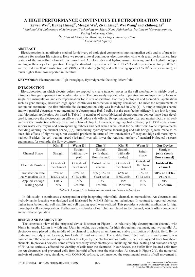
The schematic view of the proposed device is shown in Figure 1. A relatively big electroporation channel, with 30mm in length, 1.2mm in width and 75�m in height, was designed for high throughput treatment, and two parallel Au electrodes were placed in the middle of the channel to achieve an uniform and stable distribution of electric field. By in-troducing hydrodynamic focusing, two kinds of fluid were used. The middle flow, filled with cells and plasmid, was pumped into the channel and squeezed to a thin layer by the electroporation buffer, which was injected from two sheath channels. In previous devices, some effects caused by water electrolysis, including bubbles, heating and dramatic change of PH value, seriously affected the viability of cells near the electrode. In our device, the buffer flow isolated cells from the Au electrodes and prevented cells from harmful effects near the electrodes. As shown in Figure 2(a), the numerical analysis of particle trace, simulated with COMSOL software, well matched the experimental results of cell movement in
Table 1. Comparison between our work and reported devices
978-0-9798064-3-8/µTAS 2010/$20©2010 CBMS 1622 14th International Conference onMiniaturized Systems for Chemistry and Life Sciences
3 - 7 October 2010, Groningen, The Netherlands
the focusing area. Figure 2(b) showed an even distribution of 1600V/cm electric field, through which all cells were elec-troporated.
Figure 1: Schematic view of proposed electroporation chip
Figure 2: (a)The simulation results of the particle trace and corresponding experimental results, (b) the simulation shows a 1600V/cm electric field with good uniformity.
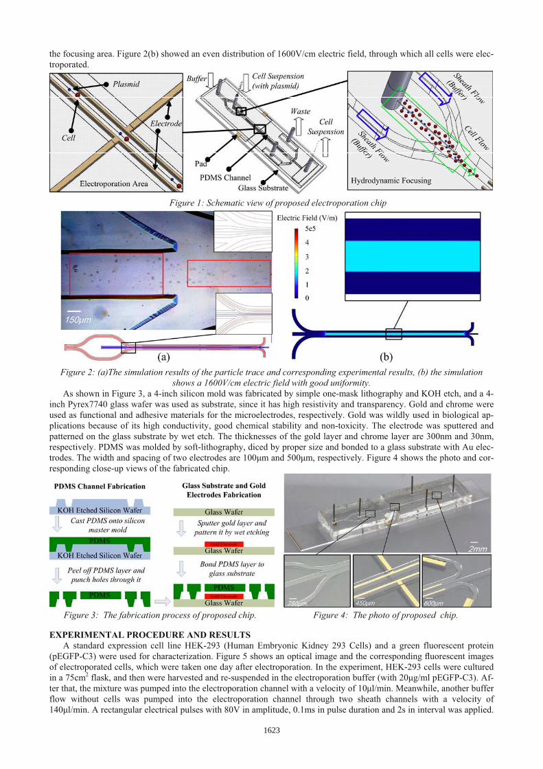
As shown in Figure 3, a 4-inch silicon mold was fabricated by simple one-mask lithography and KOH etch, and a 4-inch Pyrex7740 glass wafer was used as substrate, since it has high resistivity and transparency. Gold and chrome were used as functional and adhesive materials for the microelectrodes, respectively. Gold was wildly used in biological ap-plications because of its high conductivity, good chemical stability and non-toxicity. The electrode was sputtered and patterned on the glass substrate by wet etch. The thicknesses of the gold layer and chrome layer are 300nm and 30nm, respectively. PDMS was molded by soft-lithography, diced by proper size and bonded to a glass substrate with Au elec-trodes. The width and spacing of two electrodes are 100�m and 500�m, respectively. Figure 4 shows the photo and cor-responding close-up views of the fabricated chip.
EXPERIMENTAL PROCEDURE AND RESULTS A standard expression cell line HEK-293 (Human Embryonic Kidney 293 Cells) and a green fluorescent protein
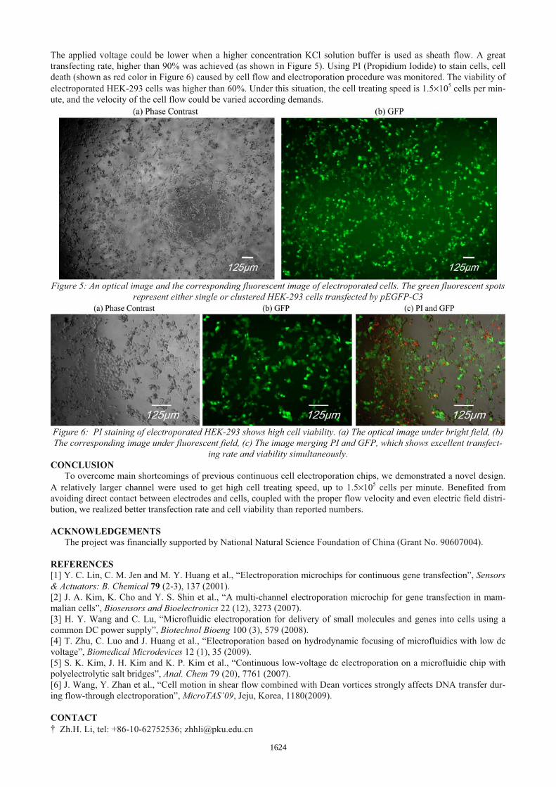
(pEGFP-C3) were used for characterization. Figure 5 shows an optical image and the corresponding �uorescent images of electroporated cells, which were taken one day after electroporation. In the experiment, HEK-293 cells were cultured in a 75cm2 flask, and then were harvested and re-suspended in the electroporation buffer (with 20�g/ml pEGFP-C3). Af-ter that, the mixture was pumped into the electroporation channel with a velocity of 10�l/min. Meanwhile, another buffer flow without cells was pumped into the electroporation channel through two sheath channels with a velocity of 140�l/min. A rectangular electrical pulses with 80V in amplitude, 0.1ms in pulse duration and 2s in interval was applied.
Figure 3: The fabrication process of proposed chip. Figure 4: The photo of proposed chip.
1623
The applied voltage could be lower when a higher concentration KCl solution buffer is used as sheath flow. A great transfecting rate, higher than 90% was achieved (as shown in Figure 5). Using PI (Propidium Iodide) to stain cells, cell death (shown as red color in Figure 6) caused by cell flow and electroporation procedure was monitored. The viability of electroporated HEK-293 cells was higher than 60%. Under this situation, the cell treating speed is 1.5�105 cells per min-ute, and the velocity of the cell flow could be varied according demands.
Figure 5: An optical image and the corresponding fluorescent image of electroporated cells. The green fluorescent spots represent either single or clustered HEK-293 cells transfected by pEGFP-C3
Figure 6: PI staining of electroporated HEK-293 shows high cell viability. (a) The optical image under bright field, (b) The corresponding image under fluorescent field, (c) The image merging PI and GFP, which shows excellent transfect-
ing rate and viability simultaneously. CONCLUSION
To overcome main shortcomings of previous continuous cell electroporation chips, we demonstrated a novel design. A relatively larger channel were used to get high cell treating speed, up to 1.5�105 cells per minute. Benefited from avoiding direct contact between electrodes and cells, coupled with the proper flow velocity and even electric field distri-bution, we realized better transfection rate and cell viability than reported numbers. ACKNOWLEDGEMENTS
The project was financially supported by National Natural Science Foundation of China (Grant No. 90607004).
REFERENCES[1] Y. C. Lin, C. M. Jen and M. Y. Huang et al., “Electroporation microchips for continuous gene transfection”, Sensors& Actuators: B. Chemical 79 (2-3), 137 (2001). [2] J. A. Kim, K. Cho and Y. S. Shin et al., “A multi-channel electroporation microchip for gene transfection in mam-malian cells”, Biosensors and Bioelectronics 22 (12), 3273 (2007). [3] H. Y. Wang and C. Lu, “Microfluidic electroporation for delivery of small molecules and genes into cells using a common DC power supply”, Biotechnol Bioeng 100 (3), 579 (2008). [4] T. Zhu, C. Luo and J. Huang et al., “Electroporation based on hydrodynamic focusing of microfluidics with low dc voltage”, Biomedical Microdevices 12 (1), 35 (2009). [5] S. K. Kim, J. H. Kim and K. P. Kim et al., “Continuous low-voltage dc electroporation on a microfluidic chip with polyelectrolytic salt bridges”, Anal. Chem 79 (20), 7761 (2007). [6] J. Wang, Y. Zhan et al., “Cell motion in shear flow combined with Dean vortices strongly affects DNA transfer dur-ing flow-through electroporation”, MicroTAS’09, Jeju, Korea, 1180(2009). CONTACT
Zh.H. Li, tel: +86-10-62752536; [email protected]
1624