Embed Size (px)
Citation preview
ORIGINAL RESEARCH ARTICLEpublished: 25 July 2012
doi: 10.3389/fphys.2012.00290
A novel member of the trehalose transporter familyfunctions as an H+-dependent trehalose transporter in thereabsorption of trehalose in Malpighian tubulesShingo Kikuta1,2,Yuka Hagiwara-Komoda1, Hiroaki Noda1,2 andTakahiro Kikawada1*1 National Institute of Agrobiological Sciences, Tsukuba, Ibaraki, Japan2 Department of Integrated Biosciences, Graduate School of Frontier Sciences, The University of Tokyo, Kashiwa, Chiba, Japan
Edited by:Neal Silverman, University ofMassachusetts Medical School, USA
Reviewed by:Dow Julian, University of Glasgow,UKJohn Crowe, University of CaliforniaDavis, USA
*Correspondence:Takahiro Kikawada, National Instituteof Agrobiological Sciences, Tsukuba,Ibaraki 305-8634, Japan.e-mail: [email protected]
In insects, Malpighian tubules are functionally analogous to mammalian kidneys in thatthey not only are essential to excrete waste molecules into the lumen but also are respon-sible for the reabsorption of indispensable molecules, such as sugars, from the lumen tothe principal cells. Among sugars, the disaccharide trehalose is highly important to insectsbecause it is the main hemolymph sugar to serve as a source of energy and carbon. Thetrehalose transporter TRET1 participates in the transfer of newly synthesized trehalosefrom the fat body across the cellular membrane into the hemolymph. Although transportproteins must play a pivotal role in the reabsorption of trehalose in Malpighian tubules,the molecular context underlying this process remains obscure. Previously, we identified aTret1 homolog (Nlst8 ) that is expressed principally in the Malpighian tubules of the brownplanthopper (BPH). Here, we used the Xenopus oocyte expression system to show thatNlST8 exerts trehalose transport activity that is elevated under low pH conditions. Thesefunctional assays indicate that Nlst8 encodes a proton-dependent trehalose transporter(H-TRET1). To examine the involvement of Nlst8 in trehalose reabsorption, we analyzedthe sugar composition of honeydew by using BPH with RNAi gene silencing. Trehalosewas detected in the honeydew as waste excreted from Nlst8 -dsRNA-injected BPH underhyperglycemic conditions. However, trehalose was not expelled from GFP -dsRNA-injectedBPH even under hyperglycemic conditions.We conclude that NlST8 could participate in tre-halose reabsorption driven by a H+ gradient from the lumen to the principal cells of theMalpighian tubules.
Keywords: trehalose, sugar reabsorption, Malpighian tubules, proton-dependent transporter
INTRODUCTIONExcretory organs, kidneys in vertebrates and the Malpighiantubules in invertebrates, are essential to discharge waste, such assmall molecules and excess salt, into the renal lumen. For smallmolecules in particular, this excretion step occurs through non-selective filtration, which means that the molecules required byliving organisms must be retrieved from the waste. Therefore,another function of excretory organs is to reabsorb indispensablemolecules, including sugars, amino acids, and water, via dedicatedtransporters located in the cellular membrane. For example, inmammals, sodium-glucose co-transporter 2 (SGLT2), which isexpressed in the apical membrane of kidney cells facing the lumen,has a pivotal role in glucose reabsorption driven by electrochemicalmembrane potentials (Wright, 2001).
In most insects, trehalose, a disaccharide composed of two glu-cose molecules linked by an α-1,1-bond, is the main hemolymph
Abbreviations: 2-DOG, 2-deoxy-d-glucose; BPH, brown planthopper; CCCP, car-bonyl cyanide m-chlorophenyl hydrazone; cRNA, capped RNA; EST, expressedsequence tag; GLUT, glucose transporter family; MBS, modified Barth’s saline; MES,2-(N -morpholino)ethanesulfonic acid; MFS, major facilitator superfamily; SLC2,solute carrier family 2; TRET1, trehalose transporter 1
sugar; it acts as a nutrient source and as a protectant against harshconditions, such as desiccation, heat, and cold (Crowe et al., 1998;Arrese and Soulages, 2010). In Locusta migratoria and Schistocercagregaria, trehalose is degraded by trehalase to be utilized as theprimary energy source for flight (Vaandrager et al., 1989; Beckeret al., 1996). In the larvae of the sleeping chironomid, Polypedilumvanderplanki, trehalose is intensively synthesized under dehydrat-ing conditions and eventually vitrified (Mitsumasu et al., 2010);it thus acts as an anhydroprotectant (Watanabe et al., 2002; Saku-rai et al., 2008). Trehalose produced in the fat body is exportedto the hemolymph (Wyatt, 1961; Mitsumasu et al., 2010), whereit may passively and non-specifically leak into the tubule lumensand be excreted as waste (Knowles, 1975). Although trehalose wasthought to be reabsorbed from the lumen to the principal cells ofthe tubules (Knowles, 1975; Jarial and Kelly-Worden, 2011), themolecular basis of this process has remained unknown.
Sugar transporters have essential roles in the appropriate distri-bution of carbohydrates throughout the body (Mueckler, 1994).They are typically categorized in two groups: (i) secondary activemembrane transporters, which promote the uphill permeationof sugars driven by electrochemical gradients of Na+ or H+
ions across the cellular membranes, and (ii) facilitative sugar
www.frontiersin.org July 2012 | Volume 3 | Article 290 | 1
Kikuta et al. Characterization of novel H+-trehalose transporter
transporters, which enable sugars to flow across membranes downconcentration gradients (Wood and Trayhurn, 2003). Until now,trehalose transporters have been identified from yeasts and insects(Stambuk et al., 1998; Kikawada et al., 2007). Saccharomyces cere-visiae possesses the α-glucoside transporter AGT1, which pro-motes uptake of disaccharides, including trehalose, sucrose, andmaltose, via an electrochemical proton gradient, which suggeststhat AGT1 belongs to the group of secondary active transporters(Han et al., 1995). Thus, AGT1 acts as an H+-dependent trehalosetransporter for the uptake of low-level trehalose as a nutrient fromculture medium under low pH conditions. Insects have a facilita-tive trehalose transporter, TRET1, which seems to be responsiblefor the regulation of trehalose levels in the hemolymph (Kikawadaet al., 2007; Kanamori et al., 2010). Secondary active transportersrather than facilitative transporters may mediate reabsorption oftrehalose in Malpighian tubules because the passively diffusedtrehalose concentration in the lumen should be lower than theconcentration in hemolymph. Yet, secondary active transportersfor trehalose in multicellular organisms, including insects, havenot been reported.
It is difficult to predict the characteristics of sugar transportersbased solely on their amino acid sequences because only subtle dif-ference exists between proton-dependent sugar transporters andfacilitative sugar transporters in the major facilitator superfam-ily (MFS; Pao et al., 1998). In plants, for instance, the sucrosetransporter LjSUT4 from Lotus japonicus shares 73% identity atthe amino acid level with PsSUF4 from Pisum sativum (Zhou
et al., 2007), although LjSUT4 is an H+ dependent transporter andPsSUF4 is a facilitative transporter for sucrose (Zhou et al., 2007;Reinders et al., 2008). These results suggest that proton-dependenttrehalose transporters may also reside in the TRET1 family ininsects. Hence, the amino acid sequence of the H+-dependenttrehalose transporter should be similar to that of the facilitativetrehalose transporter, relative to other sugar transporters.
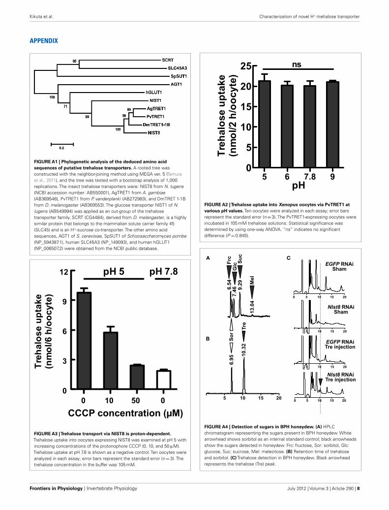
The TRET1 family constitutes a mono-clade among the insectsugar transporters (Figure A1 in Appendix; Kikuta et al., 2010).Among the TRET1 family, NlST8, which was isolated from thebrown planthopper (BPH) Nilaparvata lugens, a rice plant pest(Kikuta et al., 2010), could be a transporter in trehalose reabsorp-tion, because NlST8 is mainly expressed in Malpighian tubules(Kikuta et al., 2010). Here, we investigated the transport activityand physiological roles of NlST8 by using the Xenopus oocyteexpression system and the technique of RNAi gene silencing,respectively. Our results indicate that NlST8 is a proton-dependentTRET1 with a role in trehalose reabsorption in Malpighiantubules.
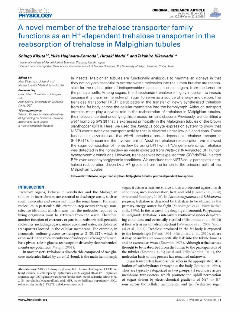
RESULTSSUBCELLULAR LOCALIZATION OF NLST8 IN XENOPUS OOCYTESTo determine whether NIST8 is a membrane-bound protein, weexamined its subcellular localization by using either a GFP-fusionprotein, NIST8::AcGFP1, or GFP alone in Xenopus oocytes. Flu-orescence of the fusion protein was principally detected in thecellular membrane (Figure 1A) but not in the cellular membrane
A B C
D
FIGURE 1 | Functional analyses of NlST8-expressing Xenopusoocyte. Localization of NlST8 in the membrane of the Xenopus oocyteas detected by use of an AcGFP1 fusion protein. (A) NlST8::AcGFP1fluorescence was observed in Xenopus oocytes. (B) AcGFP1 cRNAinjection as a control. (C) Sham as a negative control. Scale bar, 20 µm.(D) Sugar uptake analyses of NlST8 by using HPLC. Transporters wereexpressed in the cellular membrane of Xenopus oocytes by injectingthe cRNA of Nlst8. 2-DOG, 2-deoxy-glucose; Myo, myo-inositol; Suc,
sucrose; Mal, maltose; Tre, Trehalose. Sham is a negative control.Sugars were used at a concentration of 105 mM in MBS buffer. Tenoocytes were analyzed in each assay. Error bars represent the standarderror (n=3). Statistical significance was determined by using Student’st -test in each assay. “ns” indicates no significant difference; asterisksindicate a significant difference (****P < 0.0001). Myo-inositol:P =0.2626, sucrose: P =0.8057, maltose: P =0.1241, and trehalose:P < 0.0001.
Frontiers in Physiology | Invertebrate Physiology July 2012 | Volume 3 | Article 290 | 2
Kikuta et al. Characterization of novel H+-trehalose transporter
of oocytes injected with AcGFP1 capped RNA (cRNA) only(Figure 1B). Of course, no fluorescence was detected in a shamcontrol (Figure 1C). These results indicate that Nlst8 encodes amembrane-bound protein.
NLST8 IS A TREHALOSE TRANSPORTERTo explore substrates for NlST8, we conducted a sugar uptake assaywith trehalose and other carbohydrates, including myo-inositol,sucrose, maltose, and 2-deoxy-d-glucose (2-DOG; Figure 1D)using the NlST8-expressing Xenopus oocytes. Of the carbohy-drates tested, only trehalose was significantly taken up by oocytes(P < 0.0001) compared with a sham control, indicating that NlST8possessed trehalose transport activity.
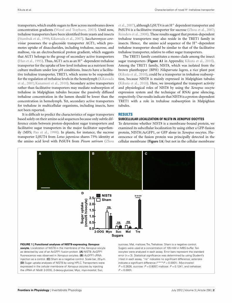
TREHALOSE UPTAKE VIA NLST8 IS DRIVEN BY A PROTON GRADIENTWe investigated the biochemical properties of NlST8’s transportactivity for trehalose. First, we analyzed trehalose uptake via NlST8under Na+-free conditions. The result indicated that trehaloseuptake by NlST8 was Na+-independent (Figure 2A). The pHdependency of NlST8 for trehalose uptake was also examinedunder various pH conditions, ranging from 5.0 to 9.0. Trehalosetransport activity of NlST8 was clearly increased at the lowerpH values (Figure 2B). At pH 5 in MES [2-(N -morpholino)ethanesulfonic acid] buffer, trehalose uptake was approximatelyfour times that at pH 7.8. In contrast, the activity of the facil-itated trehalose transporter PvTRET1 was pH-independent, asreported previously (Figure A2 in Appendix; Kikawada et al.,2007). A protonophore, carbonyl cyanide m-chlorophenyl hydra-zone (CCCP), inhibited trehalose uptake via NlST8 even at pH 5(Figure A3 in Appendix; Figure 2C). Protonophores allow protonsto selectively cross the cellular membranes, resulting in disruptionof proton gradient across the membranes. Thus, these results sug-gest that trehalose uptake by NlST8 is probably driven via a protongradient across the membrane.
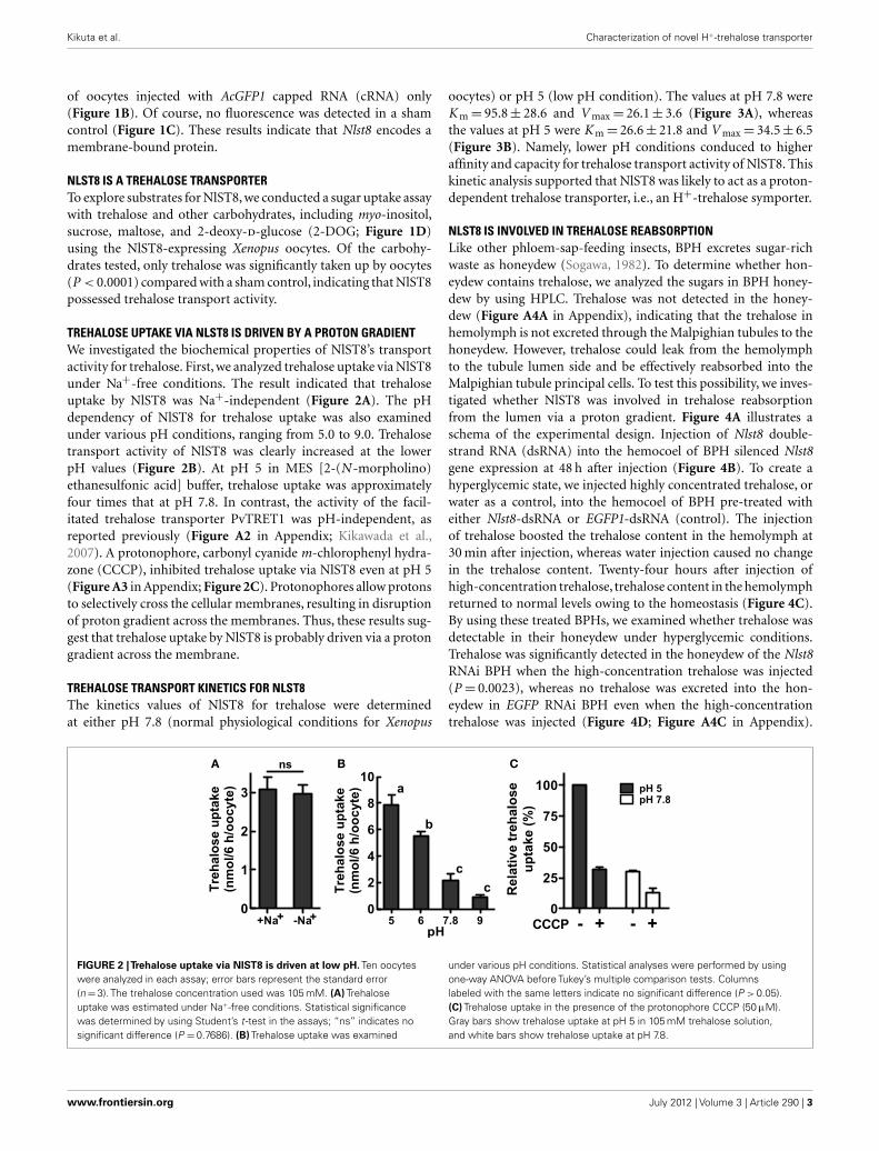
TREHALOSE TRANSPORT KINETICS FOR NLST8The kinetics values of NlST8 for trehalose were determinedat either pH 7.8 (normal physiological conditions for Xenopus
oocytes) or pH 5 (low pH condition). The values at pH 7.8 wereK m= 95.8± 28.6 and V max= 26.1± 3.6 (Figure 3A), whereasthe values at pH 5 were K m= 26.6± 21.8 and V max= 34.5± 6.5(Figure 3B). Namely, lower pH conditions conduced to higheraffinity and capacity for trehalose transport activity of NlST8. Thiskinetic analysis supported that NlST8 was likely to act as a proton-dependent trehalose transporter, i.e., an H+-trehalose symporter.
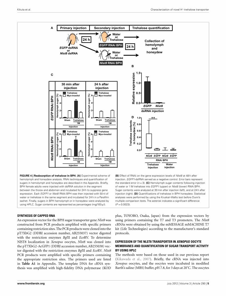
NLST8 IS INVOLVED IN TREHALOSE REABSORPTIONLike other phloem-sap-feeding insects, BPH excretes sugar-richwaste as honeydew (Sogawa, 1982). To determine whether hon-eydew contains trehalose, we analyzed the sugars in BPH honey-dew by using HPLC. Trehalose was not detected in the honey-dew (Figure A4A in Appendix), indicating that the trehalose inhemolymph is not excreted through the Malpighian tubules to thehoneydew. However, trehalose could leak from the hemolymphto the tubule lumen side and be effectively reabsorbed into theMalpighian tubule principal cells. To test this possibility, we inves-tigated whether NlST8 was involved in trehalose reabsorptionfrom the lumen via a proton gradient. Figure 4A illustrates aschema of the experimental design. Injection of Nlst8 double-strand RNA (dsRNA) into the hemocoel of BPH silenced Nlst8gene expression at 48 h after injection (Figure 4B). To create ahyperglycemic state, we injected highly concentrated trehalose, orwater as a control, into the hemocoel of BPH pre-treated witheither Nlst8-dsRNA or EGFP1-dsRNA (control). The injectionof trehalose boosted the trehalose content in the hemolymph at30 min after injection, whereas water injection caused no changein the trehalose content. Twenty-four hours after injection ofhigh-concentration trehalose, trehalose content in the hemolymphreturned to normal levels owing to the homeostasis (Figure 4C).By using these treated BPHs, we examined whether trehalose wasdetectable in their honeydew under hyperglycemic conditions.Trehalose was significantly detected in the honeydew of the Nlst8RNAi BPH when the high-concentration trehalose was injected(P = 0.0023), whereas no trehalose was excreted into the hon-eydew in EGFP RNAi BPH even when the high-concentrationtrehalose was injected (Figure 4D; Figure A4C in Appendix).
A B C
FIGURE 2 |Trehalose uptake via NlST8 is driven at low pH. Ten oocyteswere analyzed in each assay; error bars represent the standard error(n=3). The trehalose concentration used was 105 mM. (A) Trehaloseuptake was estimated under Na+-free conditions. Statistical significancewas determined by using Student’s t -test in the assays; “ns” indicates nosignificant difference (P =0.7686). (B) Trehalose uptake was examined
under various pH conditions. Statistical analyses were performed by usingone-way ANOVA before Tukey’s multiple comparison tests. Columnslabeled with the same letters indicate no significant difference (P > 0.05).(C) Trehalose uptake in the presence of the protonophore CCCP (50 µM).Gray bars show trehalose uptake at pH 5 in 105 mM trehalose solution,and white bars show trehalose uptake at pH 7.8.
www.frontiersin.org July 2012 | Volume 3 | Article 290 | 3
Kikuta et al. Characterization of novel H+-trehalose transporter
A B
FIGURE 3 | Analyses of the kinetics of NlST8 for trehalose. Ten oocyteswere analyzed in each assay; error bars represent the standard error (n=3).(A) Oocytes expressing NlST8 were incubated with various trehaloseconcentrations for 3 h at pH 7.8. (B) Oocytes expressing NlST8 wereincubated for 1 h with various trehalose concentrations at pH 5. Data werefitted to the Michaelis–Menten equation.
Trehalose was also not detectable in the honeydew of the shamcontrols (Figure 4D; Figure A4C in Appendix). These resultsshow that gene knockdown of Nlst8 by RNAi disrupts trehalosereabsorption in Malpighian tubules. Taken together, these datasuggest that NlST8 genetically regulates trehalose reabsorption inMalpighian tubules.
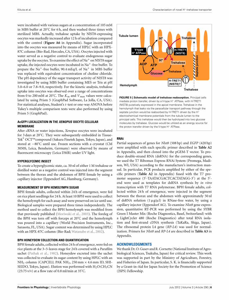
DISCUSSIONPreviously, we reported the cloning of several sugar transportergenes from the BPH N. lugens (Kikuta et al., 2010). Here, wefound that one of those genes, Nlst8, encodes a membrane proteinwith trehalose transport activity that is driven by proton (H+)electrochemical membrane potentials, indicating that NlST8 is anH+-trehalose co-transporter, H-TRET. Spatial expression analysisshowed that Nlst8 is principally expressed in Malpighian tubules(Figure A5 in Appendix), suggesting that NlST8 is involved intrehalose reabsorption in these tubules. The occurrence of sugarreabsorption from the lumen to the Malpighian tubule principalcells in insects has been observed physiologically (Knowles, 1975).However, the molecular context underlying this reabsorption hasthus far been obscure. By using an RNAi gene-silencing tech-nique, we demonstrated TRET1/NlST8’s participation in trehalosereabsorption in Malpighian tubules.
Malpighian tubules are excretory tissues in insects and com-prise a single layer of squamous epithelial cells adhered intercellu-larly with septate junctional complexes (O’Donnell, 2008; Beyen-bach et al., 2010). Excretion occurs through the tubules by tran-scellular transport and paracellular transport pathways (Beyen-bach et al., 2010). In the former pathway, molecules, absorbedvia transporters and/or channels in the basolateral membrane ofthe principal cells, are actively discharged into the tubule lumenthrough other transporters or channels situated in the apical mem-brane. In the latter pathway, molecules are sluggishly excretedalong the cleft between the septate junctions. Cations, such as Na+
and K+, are excreted transcellularly, whereas uncharged small mol-ecules, including polyethylene glycol and sucrose, are dischargedparacellularly (Beyenbach and Piermarini, 2011). Trehalose likelyseeps passively from the hemolymph into the tubule lumen via theparacellular transport pathway.
In the apical membrane of tubule principal cells, a V-typeH+ ATPase energizes proton-dependent secondary active trans-porters by forming an H+ gradient (Wieczorek et al., 2009).Cation/nH+ antiporters, which transport excess Na+ and/or K+
from the cytosol of the principal cells into the lumen, are rep-resentative examples of such transporters. Trehalose uptake byH-TRET1/NlST8 was driven by a proton gradient across the mem-brane (Figure 2B), suggesting that H-TRET1/NlST8 cooperateswith a V-type H+ ATPase that probably acts as “the trehalosepump” to promote trehalose reabsorption from the lumen. Thisidea is supported by the knockdown of Nlst8,which led to trehaloseexcretion into honeydew (Figure 4). Trehalose incorporated intothe principal cells must be utilized as an energy source to pro-mote the V-type H+ ATPase activity because in insects, the tubulesexpress high levels of soluble trehalase (Derr and Randall, 1966;Dahlman, 1970), which facilitates the degradation of trehalose intoglucose.
Recently, another disaccharide transporter, SCRT, was iden-tified from D. melanogaster. SCRT appears to be involved insucrose uptake in the intestinal tract, especially in hindgut (Meyeret al., 2011). Similarly to H-TRET1/NlST8, SCRT exerts H+ co-transport activity for disaccharides. The primary structure ofSCRT is distinct from that of the TRET1 family, including H-TRET1/NlST8 (Figure A1 in Appendix). Indeed, SCRT closelyresembles the mammalian solute carrier family 45 (SLC45; Meyeret al., 2011), whereas the insect TRET1 family belongs to the SLC2family (Kanamori et al., 2010). For SCRT, trehalose would be acompetitive inhibitor for sucrose transport activity, suggesting thattrehalose may also be a substrate for SCRT (Meyer et al., 2011). Theinvolvement of SCRT in trehalose reabsorption in the intestinaltract remains obscure.
We conclude that Malpighian tubules use a V-type H+ ATPaseto power not only the transepithelial secretion of electrolytes butalso reabsorption of passively secreted trehalose from the tubulelumen by means of H-TRET1/NlST8 (Figure 5), although the sub-cellular localization of H-TRET1/NlST8 in the principal cells ofthe Malpighian tubules has yet to be investigated. Indeed, involve-ment of tissue other than the tubules in the reabsorption oftrehalose remains controversial. Further histochemical, cytochem-ical, and genetic analyses using transgenic and knockout insectswill uncover the fine details of the molecular basis of trehalosereabsorption via H-TRET1/NlST8.
MATERIALS AND METHODSINSECTS AND PLANTSBPH (strain: Izumo) were reared and maintained on rice seedlingsat 26˚C with 16 h light: 8 h dark periods. The rice plants forhoneydew collection were cultivated at 25˚C.
RNA ISOLATION AND cDNA CLONINGTotal RNA was isolated from young female adults by usingthe RNeasy Mini kit (Qiagen, Hilden, Germany). Nlst8 cDNAsequence analysis was performed with an ABI prism 3730 anda BigDye Terminator v3.1 cycle sequecing kit (Life Technolo-gies, Carlsbad, CA, USA). Sequence data were analyzed withGENETYX-MAC ver. 16 software (GENETYX, Tokyo, Japan).
Frontiers in Physiology | Invertebrate Physiology July 2012 | Volume 3 | Article 290 | 4
Kikuta et al. Characterization of novel H+-trehalose transporter
A
C
B
D
FIGURE 4 | Reabsorption of trehalose in BPH. (A) Experimental scheme ofhemolymph and honeydew analysis. RNAi techniques and quantification ofsugars in hemolymph and honeydew are described in the Appendix. Briefly,BPH female adults were injected with dsRNA solution in the segmentbetween the thorax and abdomen and incubated for 24 h to suppress geneexpression. Each EGFP1 or Nlst8 RNAi BPH was then injected with 50 nl ofwater or trehalose in the same segment and incubated for 24 h in a Parafilmsachet. Finally, sugars in BPH hemolymph or in honeydew were analyzed byusing HPLC. Sugar contents are represented as percentages (mg/100 µl).
(B) Effect of RNAi on the gene expression levels of Nlst8 at 48 h afterinjection. EGFP1-dsRNA served as a negative control. Error bars representthe standard error (n=3). (C) Hemolymph sugar contents following injectionof water or 1 M trehalose into EGFP1 (upper) or Nlst8 (lower) RNAi BPH.Sugar contents were analyzed at 30 min after injection (left), and at 24 h afterinjection (right). (D) Quantifications of trehalose in BPH honeydew. Statisticalanalyses were performed by using the Kruskal–Wallis test before Dunn’smultiple comparison tests. The asterisk indicates a significant difference(P = 0.0023).
SYNTHESIS OF CAPPED RNAAn expression vector for the BPH sugar transporter gene Nlst8 wasconstructed from PCR products amplified with specific primerscontaining restriction sites. The PCR products were cloned into thepT7XbG2 (DDBJ accession number, AB255037) vector digestedwith the restriction enzymes BglII and EcoRV. To determineNlST8 localization in Xenopus oocytes, Nlst8 was cloned intothe pT7XbG2-AcGFP1 (DDBJ accession number, AB255038) vec-tor digested with the restriction enzymes BglII and EcoRV. Nlst8PCR products were amplified with specific primers containingthe appropriate restriction sites. The primers used are listedin Table A1 in Appendix. The template DNA for cRNA syn-thesis was amplified with high-fidelity DNA polymerase (KOD
plus, TOYOBO, Osaka, Japan) from the expression vectors byusing primers containing the T7 and T3 promoters. The Nlst8cRNAs were obtained by using the mMESSAGE mMACHINE T7kit (Life Technologies) according to the manufacturer’s standardprotocols.
EXPRESSION OF THE NLST8 TRANSPORTER IN XENOPUS OOCYTEMEMBRANES AND QUANTIFICATION OF SUGAR TRANSPORT ACTIVITYBY USING HPLCThe methods were based on those used in our previous report(Kikawada et al., 2007). Briefly, the cRNA was injected intoXenopus oocytes, and the oocytes were incubated in modifiedBarth’s saline (MBS) buffer, pH 7.8, for 3 days at 20˚C. The oocytes
www.frontiersin.org July 2012 | Volume 3 | Article 290 | 5
Kikuta et al. Characterization of novel H+-trehalose transporter
were incubated with various sugars at a concentration of 105 mMin MBS buffer at 20˚C for 6 h, and then washed three times withsterilized MBS. Actually, trehalose uptake by NlST8-expressingoocytes was markedly increased after 12 h of incubation comparedwith the control (Figure A6 in Appendix). Sugar incorporatedinto the oocytes was measured by means of HPLC with an HPX-87C column (Bio-Rad, Hercules, CA, USA). Oocytes injected withwater served as a negative control to evaluate endogenous sugaruptake by the oocytes. To examine the effect of Na+ on NlST8 sugaruptake, the injected oocytes were incubated in Na+-free buffer. Toprepare the Na+-free buffer, 90.4 mEq/L of Na+ in MBS bufferwas replaced with equivalent concentration of choline chloride.The pH dependency of the sugar transport activity of NIST8 wasinvestigated by using MBS buffer containing MES or Tris at pH5.0–6.0 or 7.8–9.0, respectively. For the kinetic analysis, trehaloseuptake into oocytes was observed over a range of concentrationsfrom 0 to 200 mM at 20˚C. The K m and V max values were calcu-lated by using Prism 5 (GraphPad Software, La Jolla, CA, USA).For statistical analyses, Student’s t -test or one-way ANOVA beforeTukey’s multiple comparison tests were also performed by usingPrism 5 (GraphPad).
AcGFP1 LOCALIZATION IN THE XENOPUS OOCYTE CELLULARMEMBRANEAfter cRNA or water injections, Xenopus oocytes were incubatedfor 3 days at 20˚C. They were subsequently embedded in Tissue-Tek® OCT™compound (Sakura Finetek Japan, Tokyo, Japan), andstored at −80˚C until use. Frozen sections with a cryostat (CM3050S, Leica, Bensheim, Germany) were observed by means offluorescent microscopy (Leica DMR) under UV light.
HYPERGLYCEMIC INSECTTo create a hyperglycemic state, ca. 50 nl of either 1 M trehalose ordistilled water as a negative control was injected into the segmentbetween the thorax and the abdomen of BPH female by using acapillary injector (Eppendorf AG, Hamburg, Germany).
MEASUREMENT OF BPH HEMOLYMPH SUGARBPH female adults, collected within 24 h of emergence, were fedon rice plant seedlings for 24 h. At least 10 BPH were used to collectthe hemolymph for each assay and were preserved on ice until use.Biological samples were prepared three times independently. Themethod used to collect the BPH hemolymph was modified fromthat previously published (Moriwaki et al., 2003). The foreleg ofthe BPH was torn off with forceps at 20˚C and the hemolymphwas poured into a capillary (World Precision Instruments, Inc.,Sarasota, FL, USA). Sugar content was determined by using HPLCwith an HPX-87C column (Bio-Rad; Watanabe et al., 2002).
BPH HONEYDEW COLLECTION AND QUANTIFICATIONBPH female adults, collected within 24 h of emergence, were fed onrice plants at the 3–5-leaves stage for 24 h covered with a Parafilmsachet (Pathak et al., 1982). Honeydew excreted into the sachetwas collected to evaluate its sugar content by using HPLC with anNH2 column (CAPCELL PAK NH2, 250 mm× 4.6 mm ID, SHI-SEIDO, Tokyo, Japan). Elution was performed with H2O:CH3CN(25:75 v/v) at a flow rate of 0.8 ml/min at 35˚C.
FIGURE 5 | Schematic model of trehalose reabsorption. Principal cellsmediate proton transfer, driven by a V-type H+ ATPase, with H-TRET1(NlST8) putatively expressed in the apical membrane. Trehalose in thehemolymph that leaks via the paracellular transport pathway through theseptate junction would be reabsorbed by H-TRET1 driven by the H+
electrochemical membrane potentials from the tubule lumen to theprincipal cells. This trehalose would then be hydrolyzed into two glucosemolecules by trehalase. Glucose would be utilized as an energy source forthe proton transfer driven by the V-type H+ ATPase.
RNAiPartial sequences of genes for Nlst8 (508 bp) and EGFP (420 bp)were amplified with each specific primer described in Table A2in Appendix, and then cloned into the pGEM-T vector. To pro-duce double-strand RNA (dsRNA) for the corresponding genes,we used the T7 Ribomax Express RNAi System (Promega, Madi-son, WI, USA) according to the manufacture’s instruction man-ual. In particular, PCR products amplified by either of the spe-cific primers (Table A2 in Appendix) fused with the T7 pro-moter sequence (5′-TAATACGACTCACTATAGG-3′) at the 5′-end were used as templates for dsRNA synthesis by in vitrotranscription with T7 RNA polymerase. BPH female adults, col-lected within 24 h of emergence, were injected in the segmentbetween the thorax and the abdomen with approximately 50 nlof dsRNA solution (1 µg/µl) in RNase-free water, by using acapillary injector (Eppendorf AG). To examine Nlst8 gene expres-sion, quantitative RT-PCR was performed by using the SYBRGreen I Master Mix (Roche Diagnostics, Basel, Switzerland) witha LightCycler 480 (Roche Diagnostics) after total RNA isola-tion and first-strand cDNA synthesis (TaKaRa, Shiga, Japan).The ribosomal protein L4 gene (RP-L4) was used for normal-ization. Primers for Nlst8 and RP-L4 are described in Table A3 inAppendix.
ACKNOWLEDGMENTSWe thank Dr. O. Gusev and R. Cornette (National Institute of Agro-biological Sciences, Tsukuba, Japan) for critical review. This workwas supported in part by the Ministry of Agriculture, Forestry,and Fisheries of Japan. In particular, S. K. is financially supportedby a Grant-in-Aid for Japan Society for the Promotion of Science(JSPS) Fellowship.
Frontiers in Physiology | Invertebrate Physiology July 2012 | Volume 3 | Article 290 | 6
Kikuta et al. Characterization of novel H+-trehalose transporter
REFERENCESArrese, E. L., and Soulages, J. L. (2010).
Insect fat body: energy, metabolism,and regulation. Annu. Rev. Entomol.55, 207–225.
Becker, A., Schloder, P., Steele, J.E., and Wegener, G. (1996). Theregulation of trehalose metabo-lism in insects. Experientia 52,433–439.
Beyenbach, K. W., and Piermarini, P.M. (2011). Transcellular and para-cellular pathways of transepithelialfluid secretion in Malpighian (renal)tubules of the yellow fever mos-quito Aedes aegypti. Acta Physiol.202, 387–407.
Beyenbach, K. W., Skaer, H., and Dow,J. A. (2010). The developmental,molecular, and transport biologyof Malpighian tubules. Annu. Rev.Entomol. 55, 351–374.
Crowe, J. H., Carpenter, J. F., and Crowe,L. M. (1998). The role of vitrificationin anhydrobiosis. Annu. Rev. Physiol.60, 73–103.
Dahlman, D. (1970). Trehalase activ-ity in the tobacco hornworm tis-sue. Ann. Entomol. Soc. Am. 63,1563–1565.
Derr, R. F., and Randall, D. D.(1966). Trehalase of the differentialgrasshopper, Melanoplus differen-tialis. J. Insect Physiol. 12, 1105–1114.
Han, E. K., Cotty, F., Sottas, C., Jiang,H., and Michels, C. A. (1995). Char-acterization of AGT1 encoding ageneral alpha-glucoside transporterfrom Saccharomyces. Mol. Microbiol.17, 1093–1107.
Jarial, M. S., and Kelly-Worden, M.(2011). Additional ultrastructuralobservations of the first segmentsof Malpighian tubules in Ceno-corixa bifida (Hemiptera: Corixi-dae) in relation to reabsorption ofsolutes. Ann. Entomol. Soc. Am. 104,768–777.
Kanamori, Y., Saito, A., Hagiwara-Komoda, Y., Tanaka, D., Mitsumasu,K., Kikuta, S., Watanabe, M., Cor-nette, R., Kikawada, T., and Okuda,
T. (2010). The trehalose transporter1 gene sequence is conserved ininsects and encodes proteins withdifferent kinetic properties involvedin trehalose import into peripheraltissues. Insect Biochem. Mol. Biol. 40,30–37.
Kikawada, T., Saito, A., Kanamori, Y.,Nakahara, Y., Iwata, K., Tanaka,D., Watanabe, M., and Okuda, T.(2007). Trehalose transporter 1, afacilitated and high-capacity tre-halose transporter, allows exoge-nous trehalose uptake into cells.Proc. Natl. Acad. Sci. U.S.A. 104,11585–11590.
Kikuta, S., Kikawada, T., Hagiwara-Komoda, Y., Nakashima, N., andNoda, H. (2010). Sugar trans-porter genes of the brown plan-thopper, Nilaparvata lugens: a facil-itated glucose/fructose transporter.Insect Biochem. Mol. Biol. 40,805–813.
Knowles, G. (1975). The reduced glu-cose permeability of the isolatedMalpighian tubules of the browflyCalliphora vomitoria. J. Exp. Biol. 62,327–340.
Meyer, H., Vitavska, O., and Wieczorek,H. (2011). Identification of an ani-mal sucrose transporter. J. Cell Sci.124, 1984–1991.
Mitsumasu, K., Kanamori, Y., Fujita,M., Iwata, K., Tanaka, D., Kikuta,S., Watanabe, M., Cornette, R.,Okuda, T., and Kikawada, T.(2010). Enzymatic control ofanhydrobiosis-related accu-mulation of trehalose in thesleeping chironomid, Polypedilumvanderplanki. FEBS J. 277,4215–4228.
Moriwaki, N., Matsushita, K., Nishina,M., Matsuda, K., and Kono, Y.(2003). High myo-inositol concen-tration in the hemolymph of plan-thoppers. Appl. Entomol. Zool. 38,359–364.
Mueckler, M. (1994). Facilitative glu-cose transporters. Eur. J. Biochem.219, 713–725.
O’Donnell, M. (2008). Insect excretorymechanisms. Adv. Insect Physiol. 35,1–122.
Pao, S. S., Paulsen, I. T., and Saier, M.H. Jr. (1998). Major facilitator super-family. Microbiol. Mol. Biol. Rev. 62,1–34.
Pathak, P. K., Saxena, R. C., and Hein-richs, E. A. (1982). Parafilm sachetfor measuring honeydew excretionby Nilaparvata lugens on rice. J.Econ. Entomol. 75, 194–195.
Reinders, A., Sivitz, A. B., Starker, C. G.,Gantt, J. S., and Ward, J. M. (2008).Functional analysis of LjSUT4, avacuolar sucrose transporter fromLotus japonicus. Plant Mol. Biol. 68,289–299.
Sakurai, M., Furuki, T., Akao, K.,Tanaka, D., Nakahara, Y., Kikawada,T., Watanabe, M., and Okuda, T.(2008). Vitrification is essential foranhydrobiosis in an African chi-ronomid, Polypedilum vanderplanki.Proc. Natl. Acad. Sci. U.S.A. 105,5093–5098.
Sogawa, K. (1982). The rice brownplanthopper: feeding physiology andhost plant interactions. Annu. Rev.Entomol. 27, 49–73.
Stambuk, B. U., Panek, A. D., Crowe, J.H., Crowe, L. M., and de Araujo, P.S. (1998). Expression of high-affinitytrehalose-H+ symport in Saccha-romyces cerevisiae. Biochim. Biophys.Acta 1379, 118–128.
Vaandrager, S. H., Haller, T. B.,Van Mar-rewijk, W. J. A., and Beenakkers, A.M. Th. (1989). Fractionation andkinetic properties of trehalase fromflight muscles and haemolymph ofthe locust, Locusta migratoria. InsectBiochem. 19, 89–94.
Watanabe, M., Kikawada, T., Mina-gawa, N., Yukuhiro, F., andOkuda, T. (2002). Mechanismallowing an insect to survive com-plete dehydration and extremetemperatures. J. Exp. Biol. 205,2799–2802.
Wieczorek, H., Beyenbach, K. W.,Huss, M., and Vitavska, O. (2009).
Vacuolar-type proton pumps ininsect epithelia. J. Exp. Biol. 212,1611–1619.
Wood, I. S., and Trayhurn, P. (2003).Glucose transporters (GLUT andSGLT): expanded families of sugartransport proteins. Br. J. Nutr. 89,3–9.
Wright, E. M. (2001). Renal Na+-glucose cotransporters. Am. J. Phys-iol. Renal Physiol. 280, F10–F18.
Wyatt, G. (1961). The biochemistry ofinsect hemolymph. Annu. Rev. Ento-mol. 6, 75–102.
Zhou, Y., Qu, H., Dibley, K. E., Offler,C. E., and Patrick, J. W. (2007).A suite of sucrose transportersexpressed in coats of developinglegume seeds includes novel pH-independent facilitators. Plant J. 49,750–764.
Conflict of Interest Statement: Theauthors declare that the research wasconducted in the absence of any com-mercial or financial relationships thatcould be construed as a potential con-flict of interest.
Received: 05 April 2012; accepted: 03 July2012; published online: 25 July 2012.Citation: Kikuta S, Hagiwara-KomodaY, Noda H and Kikawada T (2012)A novel member of the trehalose trans-porter family functions as an H+-dependent trehalose transporter in thereabsorption of trehalose in Malpighiantubules. Front. Physio. 3:290. doi:10.3389/fphys.2012.00290This article was submitted to Frontiersin Invertebrate Physiology, a specialty ofFrontiers in Physiology.Copyright © 2012 Kikuta, Hagiwara-Komoda, Noda and Kikawada. This is anopen-access article distributed under theterms of the Creative Commons Attribu-tion License, which permits use, distrib-ution and reproduction in other forums,provided the original authors and sourceare credited and subject to any copy-right notices concerning any third-partygraphics etc.
www.frontiersin.org July 2012 | Volume 3 | Article 290 | 7
Kikuta et al. Characterization of novel H+-trehalose transporter
APPENDIX
FIGURE A1 | Phylogenetic analysis of the deduced amino acidsequences of putative trehalose transporters. A rooted tree wasconstructed with the neighbor-joining method using MEGA ver. 5 (Tamuraet al., 2011), and the tree was tested with a bootstrap analysis of 1,000replications. The insect trehalose transporters were: NlST8 from N. lugens(NCBI accession number: AB550001), AgTRET1 from A. gambiae(AB369548), PvTRET1 from P. vanderplanki (AB272983), and DmTRET 1-1Bfrom D. melanogaster (AB369553). The glucose transporter NlST1 of N.lugens (AB549994) was applied as an out-group of the trehalosetransporter family. SCRT (CG4484), derived from D. melangaster, is a highlysimilar protein that belongs to the mammalian solute carrier family 45(SLC45) and is an H+-sucrose co-transporter. The other amino acidsequences, AGT1 of S. cerevisiae, SpSUT1 of Schizosaccharomyces pombe(NP_594387.1), human SLC45A3 (NP_149093), and human hGLUT1(NP_006507.2) were obtained from the NCBI public database.
5 6 7.8 90
5
10
15
20
25
Tre
ha
los
e u
pta
ke
(nm
ol/2
h/o
oc
yte
)
pH
ns
FIGURE A2 |Trehalose uptake into Xenopus oocytes via PvTRET1 atvarious pH values. Ten oocytes were analyzed in each assay; error barsrepresent the standard error (n=3). The PvTRET1-expressing oocytes wereincubated in 105 mM trehalose solutions. Statistical significance wasdetermined by using one-way ANOVA. “ns” indicates no significantdifference (P =0.845).
CCCP concentration ( M)
0 10 50 0
0
3
6
9
12
Tre
ha
los
e u
pta
ke
(nm
ol/6
h/o
oc
yte
)
pH 5 pH 7.8
FIGURE A3 |Trehalose transport via NlST8 is proton-dependent.Trehalose uptake into oocytes expressing NlST8 was examined at pH 5 withincreasing concentrations of the protonophore CCCP (0, 10, and 50 µM).Trehalose uptake at pH 7.8 is shown as a negative control. Ten oocytes wereanalyzed in each assay; error bars represent the standard error (n=3). Thetrehalose concentration in the buffer was 105 mM.
0 5 10 15 20
C
5 10 15 20
0 5 10 15 20
Nlst8 RNAiSham
EGFP RNAiSham
EGFP RNAiTre injection
5 10 15 20
Nlst8 RNAiTre injection
7.4
6G
lcS
uc
9.2
9 Me
l1
3.0
4
Frc
6.5
4S
or
A
B
6.9
5
Tre
10
.32
5 10 15 20
FIGURE A4 | Detection of sugars in BPH honeydew. (A) HPLCchromatogram representing the sugars present in BPH honeydew. Whitearrowhead shows sorbitol as an internal standard control; black arrowheadsshow the sugars detected in honeydew. Frc: fructose, Sor: sorbitol, Glc:glucose, Suc: sucrose, Mel: melezitose. (B) Retention time of trehaloseand sorbitol. (C) Trehalose detection in BPH honeydew. Black arrowheadrepresents the trehalose (Tre) peak.
Frontiers in Physiology | Invertebrate Physiology July 2012 | Volume 3 | Article 290 | 8
Kikuta et al. Characterization of novel H+-trehalose transporter
Re
lati
ve
ex
pre
ss
ion
WB HD TH AB MG OV TS SG MT FB0.0
0.5
1.0
1.5
2.0
2.5
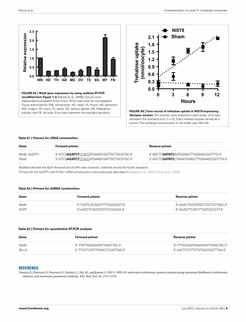
FIGURE A5 | Nlst8 gene expression by using realtime RT-PCR(modified from Figure 1 in Kikuta et al., 2010). Tissues wereindependently prepared three times. RP-L4 was used for normalization.Tissue abbreviations: WB, whole-body; HD, head; TH, thorax; AB, abdomen;MG, midgut; OV, ovary; TS, testis; SG, salivary glands; MT, Malpighiantubules, and FB, fat body. Error bars represent the standard deviation.
0 3 6 9 12
0.0
0.3
0.6
0.9
1.2
1.5
1.8
2.1
NlST8
Sham
Tre
halo
se u
pta
ke
(nm
ol/o
ocyte
)
Hours
FIGURE A6 |Time course of trehalose uptake in NlST8-expressingXenopus oocytes. Ten oocytes were analyzed in each assay; error barsrepresent the standard error (n=3). Sham-treated oocytes served as acontrol. The trehalose concentration in the buffer was 105 mM.
Table A1 | Primers for cRNA construction.
Gene Forward primer Reverse primer
Nlst8 ::AcGFP1 5′-GTCGAGATCTCCACCATGAAGCGATTGCTGCGTGC-3′ 5′-AGCTCGATATCAATGGAGCTTGGAAGCGGTTTG-3′
Nlst8 5′-GTCGAGATCTCCACCATGAAGCGATTGCTGCGTGC-3′ 5′-AGCTCGATATCCTAAATGGAGCTTGGAAGCGGTTTG-3′
Boldface denotes the BglII (forward) and EcoRV sites (reverse); underline shows the Kozak sequence.
Primers for the AcGFP1 and PvTret1 cRNA constructions were previously described (Kikawada et al., 2007; Kikuta et al., 2010).
Table A2 | Primers for dsRNA construction.
Gene Forward primer Reverse primer
Nlst8 5′-TGGTCACAGGTTTTGGCGGCT-3′ 5′-GAACTGGTATAGCTGCTCCTAGC-3′
EGFP 5′-AAGTTCAGCGTGTCCGGCGA-3′ 5′-GAAGTTCACCTTGATGCCGTT-3′
Table A3 | Primers for quantitative RT-PCR analysis.
Gene Forward primer Reverse primer
Nlst8 5′-TTATTGGAGAGATTAAGTTAC-3′ 5′-TTTGCGAATAAAATAATTAAACTAC-3′
Rp-L4 5′-TTCGTCATCTGGACCCAATCGG-3′ 5′-AACTCCCTTCTGTGGCGGTTTGA-3′
REFERENCETamura, K., Peterson, D., Peterson, N., Stecher, G., Nei, M., and Kumar, S. (2011). MEGA5: molecular evolutionary genetics analysis using maximum likelihood, evolutionary
distance, and maximum parsimony methods. Mol. Biol. Evol. 28, 2731–2739.
www.frontiersin.org July 2012 | Volume 3 | Article 290 | 9


![The Role of Trehalose 6-Phosphate in Crop Yield and … · 2020. 5. 18. · Update on Trehalose 6-Phosphate Signaling The Role of Trehalose 6-Phosphate in Crop Yield and Resilience1[OPEN]](https://img.pdfslide.net/doc/110x75/60a94aac2e9d0b10d12c4d11/the-role-of-trehalose-6-phosphate-in-crop-yield-and-2020-5-18-update-on-trehalose.jpg)