Embed Size (px)
Citation preview
Comp. Immun. Microbiol. infect. Dis., Vol. 1, pp. 269-276 0147-9571/79/0301-0269502.00/0 © Pergamon Press Ltd., 1979. Printed in Great Britain
A PROGRESS REPORT O N THE STATUS OF A NEW DISEASE OF
A M E R I C A N CATS: C Y T A U X Z O O N O S I S
D. H. FERRIS
U.S. Department of Agriculture, Science and Education Administration, Federal Research, Northeastern region, Plum Island Animal Disease Center, P.O. Box 848, Greenport,
N.Y. 11944, U.S.A.
A b s t r a c t - - A protozoon disease of domestic cats in the U.S.A. which resembled diseases of African ungulates caused by blood parasites of the family Theileridae and the genus Cytauxzoon was first described by scientists of the University of Missouri in 1976. The Plum Island Animal Disease Center of the U.S. Department of Agriculture (USDA) became involved in an effort to discover the relationship of the parasite to those of African species and to ascertain if the disease posed a threat to American livestock. More than 500 cats were infected and the disease studied experimentally by scientists of both institutions.
Passage of the parasite was accomplished by parenteral administration of fresh or deep frozen blood or tissues from infected cats. With the exception of a single cat, immunized at P1ADC, the experimental disease has terminated fatally about 20 days following exposure. Most cats died within 9 -15 days after infection. Diagnosis of the disease is accomplished by study of blood smears, stained with Giemsa and by examination of sections from major visceral organs. Piroplasms are not always found in blood smears, but large masses of schizonts are always found in at least one of the following organs: lungs, spleen, liver or lymph nodes. Since the disease almost invariably has a fatal termination in cats, it is believed that the reservoir is in another species of animal. The new disease has now been reported from Missouri, Arkansas, Texas and Georgia, with new isolations of the parasite having been made in recent months.
Key words: Cytauxzoon, cytauxzoonosis, blood parasite
U N E N O U V E L L E I N F E C T I O N F E L I N E A U X E T A T S - U N I S : L A
C Y T A U X Z O O N O S E
R e s u m e - - U n e protozoose du chat domestique ressemblant fi une maladie d'ongul~s africains causte par des parasites sanguins de la famille Theileddae et du genre Cytauxzoon a ~t6 dtcrite pour la premiere fois aux Etats-Unis par des chercheurs de l'Universite du Missouri en 1976.
Le Plum Island Animal Disease Center (P.I.A.D.C.) du Dtpar tement de l 'Agriculture des Etats- Unis s'est efforce de dtcouvrir les relations de ce parasite avec les esp&:es africains et de vtrifier si cette affection btait une menace pour le bttail amtricain. Plus de 500 chats ont 6t6 infectbs et la maladie a 6t6 6tudite exptrimentalement par des chercheurs des deux organisations.
La transmission du parasite a /~t6 rtaliste par injection parentale de sang ou de tissus frais ou congelts provenant de chats infect~s. A l'exception d 'un seul chat, immunise au P.1.A.D.C., la maladie exp~rimentale a provoqu6 la mort de tousles animaux. La plupart des chats sont morts en 9 ~i 15 jours aprts l'infection. Le diagnostic de la maladie a 6t6 effectu6 par I'~tude de frottis de sang, colores au Giemsa et par rexamen histologique de coupes des principaux organes visctraux. Les parasites ne se retrouvent pas toujours sur les frottis sanguins, mais des schizontes sont toujours prtsents en grand nombre dans au moins un des organes suivants: poumons, rate, foie ou ganglions lymphatiques. La maladie etant toujours fatale chez le chat, le rtservoir de germes se situe probablement dans une autre esptce animale. La prtsence de cette nouvelle parasitose a ~t6 constatte dans les Etats du Missouri, de l 'Arkansas, du Texas et de Gcorgie, de nouveaux isolements ayant 6t6 effectuts tout derni/~rement.
Mots clefs: Cytauxzoon, cytauxzoonose, parasite sanguin
269
270 D . H . FERRIS
INTRODUCTION
The first publication on cytauxzoonosis in the domestic cat appeared in 1976 [ 1]. Prior to this there was but a meager amount of literature on cytauxzoonosis, generally. Veterinary scientists of the University of Missouri at Columbia (UMC) have made a notable achievement in defining the disease and measuring many parameters of it. This achievement is especially noteworthy since farm cats along the Missouri-Arkansas border are not usually given much veterinary attention. We have little idea how long the disease may have been present in the Missouri-Arkansas region, although the high mortality seen encourages the concept that it is indeed a new disease in cats. The initial cases were misdiagnosed as "feline infectious anemia". Later cases were promptly and correctly diagnosed by the same practitioners as information became available in regard to the new disease.
The identity of the new disease was established by Frenkel of the Medical College of the University of Kansas on the basis of blood smears and tissue sections furnished by scientists of UMC [21. Dr. Frenkel found that the tissue forms of the parasite closely resembled Cytauxzoon species seen in tissues of the duiker [3], eland [4] and other African ruminants 151. Since Cytauxzoon parasites had been found only in African wildlife up to this time, there was an immediate response by the Animal and Plant Health Inspection Service (APHIS) and the Plum Island Animal Disease Center (PIADC) of the USDA. This response stemmed from certain basic questions:
1. Did American cats get the disease from animals transported to North America from Africa? (Was this a foreign animal disease?)
2. Did the disease in cats present a threat to domestic animals and hence our food supply? After the disease was found to be widespread in the U.S.A. and apparently not a major
threat to domestic animals, research at PIADC is being terminated; the major effort now continues at UMC. In spite of intensive efforts at both institutions, these basic questions still do not have unambiguous answers, which was a major reason for discussing the problem with scientists of the World Association of Veterinary Microbiologists, Immunologists and Specialists in Infectious Diseases at their fourth International Symposium on Feline Infectious Diseases at Alfort, France (26-28 September 1977). It is our hope that the veterinary profession will benefit from individual and collective support in solving both taxonomic and economic problems associated with feline cytauxzoonosis.
THE DISEASE AND ITS EPIZOOTIOLOGY
More than 100 serial passages of the parasite to over 500 cats were made at PIADC and UMC [6]. This status report will review briefly the following:
1. The disease syndrome, experimental and natural 2. Differential diagnosis, prognosis 3. Epizootiology (so far as it is known). The disease can be transmitted experimentally by injection of blood, splenic, hepatic,
pulmonary triturates and lymph node homogenates; amounts as low as 0.2 ml by any route were successful. The initial signs appeared in 5 -7 days after inoculation, usually in the form of anorexia and depression. There was a gradual temperature rise to as high as 40-41.1°C and usually the temperature remained high for 3 or 4 days and then dropped
c ytauxzoonosis in Americml cats 271
to normal or even sub-normal levels. Prior to death and often during the pyrexia, anemia, icterus and dehydration became apparent. Dyspnea in some cases appeared late in the course of the disease, at which time some cats cried as if in pain. The course of the disease varied from 1 to 2 weeks after the initial signs, which were quite general and resembled those of numerous other feline diseases.
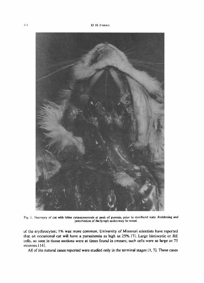
At necropsy, all animals which died or which were killed in the moribund state had a dark and enlarged spleen as well as enlarged and reddened lymph nodes [6, 7, 8]. Prior to the moribund state, some lymph nodes, particularly, the cervical lymph nodes, were petachiated (Fig. 1). The lungs were nearly always diffusely reddened, with consolidation, and some had petechia. Some animals had distention of the pericardial sac with gelantinous and icteric fluid, and with petechial and ecchymotic hemorrhages on the epicardium. The gross lesions were more severe in cats allowed to die or killed in the moribund state than cats killed earlier. The liver, in some cases which ran a longer course, was orange-brown.
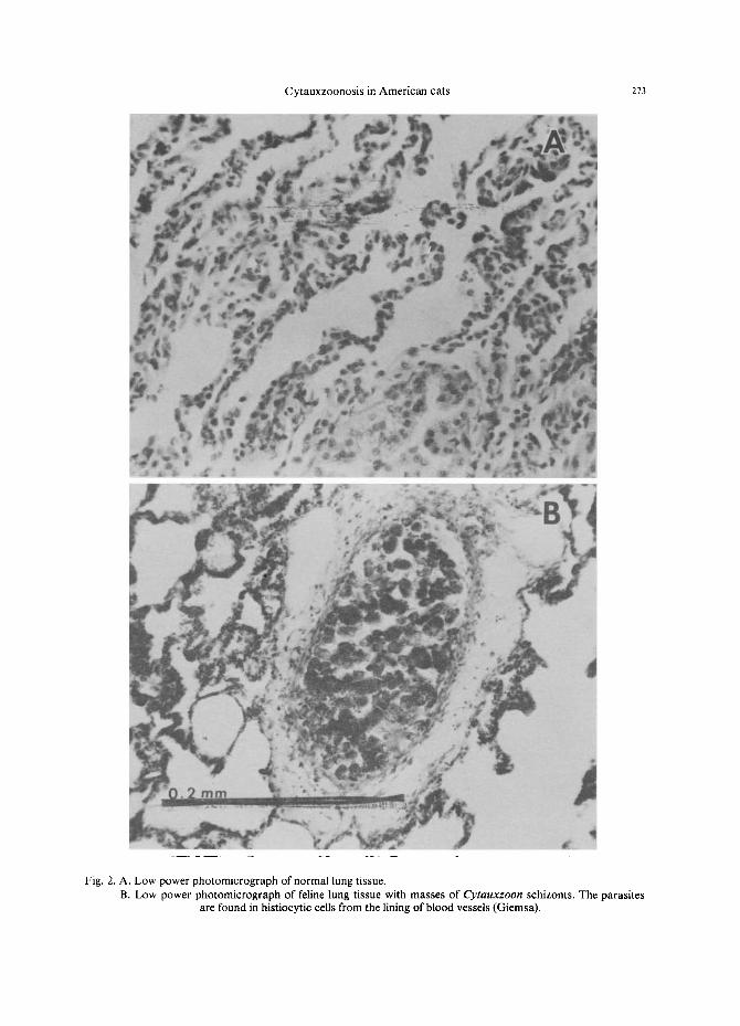
All or some of all major organs, in every case examined, had characteristic masses of parasites in venous channels [1, 6, 7, 9]. In some cases, major and minor channels were affected; in others only the minor channels. The following organs were affected: Lungs of every animal checked were found to have at least some characteristic parasite masses (Fig. 2). At low magnification these could usually be seen as masses protruding into the lumen of a vein or as an occlusion of the blood vessel; vessels cut in longitudinal section were often filled with masses of large basophilic cells. A progression of cells from slightly to enormously swollen could be found attached to the wall. At higher magnification the walls of the blood vessels were seen to consist of greatly swollen reticuloendothelial (RE) cells of the histiocytic series containing parasites. The cell nucleus was often displaced to one side and the cytoplasm filled with fine or coarse appearing granules. Broken cells with their spilled content of granules were also found.
The spleen was the next most affected organ [6, 9]. In some cats the spleen was severely affected; histologically almost every field had masses of enlarged RE cells containing the Cytauxzoon. The parasites were a little harder to distinguish with hematoxylin and eosin (H&E) than with Giemsa stain, but could be seen with either stain. Both longitudinal and cross-sectional views of venous channels had lumens partially or completely occluded with the swollen RE cells. Erythrophagocytosis was observed in the red pulp.
The liver was heavily parasitized in many instances; in other cases there were few parasites found 16, 9]. Branches of the hepatic and portal veins contained large numbers of parasitized RE cells at times, but generally with fewer occluded vessels than the spleen.
Lymph nodes usually had heavily parasitized RE cells lining the major veins and scattered along the sinusoids [6, 9]. Swollen RE cells were found in all major organs and the bone marrow, in the course of examining numerous cats.
Reproduction of the parasite takes place in histiocytes lining blood vessels, rather than in the lymphocytes, which is a major reason for placing the parasite in the genus Cytauxzoon [2, 3, 41. Lymphocytes were never invaded, nor did much if any replication take place in erythrocytes [9].
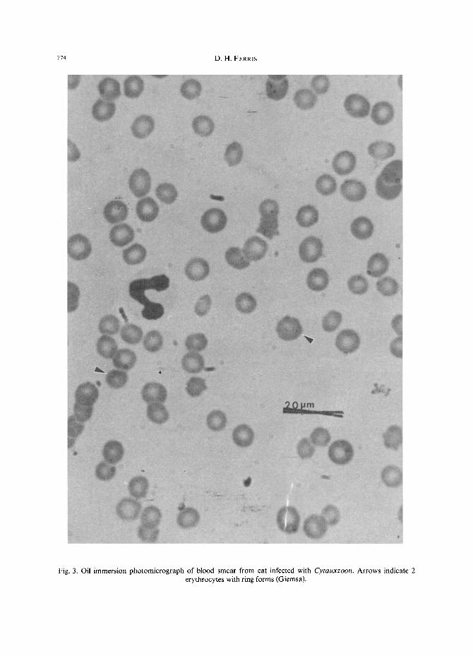
Examination of blood smears often required exacting efforts to distinguish parasites from other inclusions, e.g. Howell-Jolley or Heinz bodies [10]. For this purpose, Giemsa was the stain of choice. Since the number of parasitized erythrocytes was small at best, only the piroplasm (piro) or ring form was counted (Fig. 3). Parasitized blood cells were not found in more than half of the cats examined. The most severe parasitemia found involved about 4%
272 D . H . FERRIS
Fig. 1. Necropsy of cat with feline cytauxzoonosis at peak of pyrexia, prior to moribund state. Reddening and petechiation of the lymph nodes may be noted.
of the ery throcytes ; 1% was more c o m m o n . Univers i ty o f Missour i scientists have reported that an occas iona l cat will have a paras i t emia as high as 25% [7]. Large hist iocytic or R E cells, as seen in tissue sect ions were at t imes found in smears ; such cells were as large as 75 microns [ 14].
All of the na tu ra l cases repor ted were s tudied only in the te rminal stages [ 1, 7]. These cases
Cytauxzoonosis in American cats 273
Fig. 2. A. Low power photomicrograph of normal lung tissue. B. Low power photomicrograph of feline lung tissue with masses of Cytauxzoon schizonts. The parasites
are found in histiocytic cells from the lining of blood vessels (Giemsa).
274 D.H. FERRIS
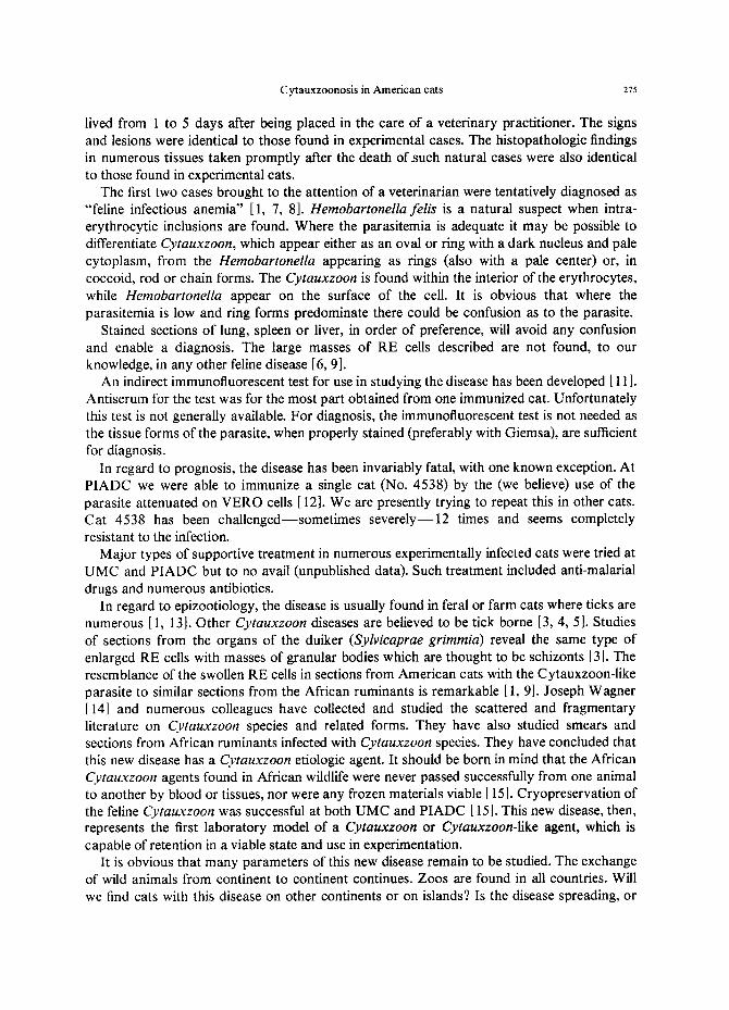
Fig. 3. Oil immersion photomicrograph of blood smear from cat infected with Cytauxzoon. Arrows indicate 2 erythrocytes with ring forms (Giemsa).
Cytauxzoonosis in American cats 275
lived from 1 to 5 days after being placed in the care of a veterinary practitioner. The signs and lesions were identical to those found in experimental cases. The histopathologic findings in numerous tissues taken promptly after the death ofauch natural cases were also identical to those found in experimental cats.
The first two cases brought to the attention of a veterinarian were tentatively diagnosed as "feline infectious anemia" [1, 7, 8]. Hemobartonellafelis is a natural suspect when intra- erythrocytic inclusions are found. Where the parasitemia is adequate it may be possible to differentiate Cytauxzoon, which appear either as an oval or ring with a dark nucleus and pale cytoplasm, from the Hemobartonella appearing as rings (also with a pale center) or, in coccoid, rod or chain forms. The Cytauxzoon is found within the interior of the erythrocytes, while Hemobartonella appear on the surface of the cell. It is obvious that where the parasitemia is low and ring forms predominate there could be confusion as to the parasite.
Stained sections of lung, spleen or liver, in order of preference, will avoid any confusion and enable a diagnosis. The large masses of RE cells described are not found, to our knowledge, in any other feline disease [6, 9].
An indirect immunofluorescent test for use in studying the disease has been developed [ 11 ]. Antiserum for the test was for the most part obtained from one immunized cat. Unfortunately this test is not generally available. For diagnosis, the immunofluorescent test is not needed as the tissue forms of the parasite, when properly stained (preferably with Giemsa), are sufficient for diagnosis.
In regard to prognosis, the disease has been invariably fatal, with one known exception. At PIADC we were able to immunize a single cat (No. 4538) by the (we believe) use of the parasite attenuated on VERO cells [ 12]. We are presently trying to repeat this in other cats. Cat 4538 has been challenged--sometimes severely--12 times and seems completely resistant to the infection.
Major types of supportive treatment in numerous experimentally infected cats were tried at UMC and PIADC but to no avail (unpublished data). Such treatment included anti-malarial drugs and numerous antibiotics.
In regard to epizootiology, the disease is usually found in feral or farm cats where ticks are numerous [1, 13]. Other Cytauxzoon diseases are believed to be tick borne [3, 4, 5]. Studies of sections from the organs of the duiker (Sylvicaprae grimmia) reveal the same type of enlarged RE cells with masses of granular bodies which are thought to be schizonts [3]. The resemblance of the swollen RE cells in sections from American cats with the Cytauxzoon-like parasite to similar sections from the African ruminants is remarkable [ 1, 9]. Joseph Wagner [14] and numerous colleagues have collected and studied the scattered and fragmentary literature on Cytauxzoon species and related forms. They have also studied smears and sections from African ruminants infected with Cytauxzoon species. They have concluded that this new disease has a Cytauxzoon etiologic agent. It should be born in mind that the African Cytauxzoon agents found in African wildlife were never passed successfully from one animal to another by blood or tissues, nor were any frozen materials viable [15]. Cryopreservation of the feline Cytauxzoon was successful at both UMC and PIADC [ 15]. This new disease, then, represents the first laboratory model of a Cytauxzoon or Cytauxzoon-like agent, which is capable of retention in a viable state and use in experimentation.
It is obvious that many parameters of this new disease remain to be studied. The exchange of wild animals from continent to continent continues. Zoos are found in all countries. Will we find cats with this disease on other continents or on islands? Is the disease spreading, or
276 D . H . FERRIS
are we mere ly recogniz ing it? W h a t are the reservoi r hos t s? It is our hope tha t in teres t has
been s t imula ted in f inding answers to these intr iguing ques t ions .
Acknowledgements--The cooperation of scientists of the University of Missouri in furnishing the original infected cat and generous assistance throughout the study is gratefully acknowledged.
The assistance of Dr. A. H. Dardiri in establishing the infection and guiding the research was indispensable. The assistance of Mr. Vincent Stopinski in the laboratory and Mr. Thomas Geiger, Frederick Moore and Clifford Schriefer in the animal room was most helpful. The assistance of Mrs. Frances Demorest in the library and Mrs. Arlene Lombardi and Mrs Dorothy Lake in preparation of the manuscript was also appreciated.
Special mention should be made of Dr. L. F. Jennings for supply of the cats and of Dr. Ralph Grusmark for their excellent conditioning prior to experimentation.
R E F E R E N C E S
1. Wagner, J. E., A fatal Cytauxzoonosis-like disease in cats J. Am. vet. med. Ass. 168, 588-598 (1976). 2. Frenkel, J. K., Department of Pathology, University of Kansas, Kansas City, KS 63103. Personal com-
munication. 3. Neitz, W. O. and Thomas, A. D., Cytauxzoon sylvicaprae gen. nov., spec. nov., a protozoan responsible for a
hitherto undescribed disease in the duiker (Sylvicaprae grimmia, Lenne). Onderstepoort J. Vet. Sci. Anim. Ind. 23, 63-73 (1948).
4. Brocklesby, D. W. Cytauxzoon taurotragi, Martin and Brocklesby, 1960. A piroplasm of the Eland (Taurotragi oryxpattersonionus Lydekker, 1906)Res. Vet. Sci. 3, 334-343 (1960).
5. Barnett, S. F., Infectious blood diseases of man and animals. II. Academic Press, New York. 269-328 (1968). 6. Wagner, J. E., Ferris, D. H., Dardiri, A. H., Kier, A. B., Wightman, S. R., Maring, E. and Morehouse, L. G.,
Experimentally induced cytauxzoon-like disease in domestic cats. In preparation. 7. Wightman, S. R., Kier, A. B. and Wagner, J. E., Feline Cytauxzoonosis: clinical features of a newly described
blood parasite disease. Feline Pract. May 1977; 24-26. 8. Wagner, J. E., Kier, A. B. and Morehouse, L. G., Feline cytauxzoonosis: a newly reported blood protozoan
disease from Southwestern Missouri. Mo. Vet. XXVI (2), 12-13 (1976). 9. Ferris, D. H., Medbus, C. A. and Dardiri, A. H., Blood and tissue forms of a new protozoan disease of the
domestic cat. In preparation. 10. Jain, N. C. and Keeton, K. S., Scanning electron microscopy of Hienz bodies in feline erythrocytes. Am. J.
vet. Res. 36 (12), 1691-1695 (1975). 11. Shindel, N., Dardiri, A. H. and Ferris, D. H., An indirect fluorescent antibody test for the diagnosis of feline
cytauxzoonosis. Can. J. comp. Med. 42 (4), 460-465 (1978). 12. Ferris, D. H. and Dardiri, A. H., Premunition and the carrier state in feline cytauxzoonosis. In preparation. 13. Bendele, R. A., Schwartz, W. L. and Jones, L. P., Cytauxzoon-like disease in Texas cats. Friskies Res. 12,
10-11 (1976). 14. Wagner, J. E., University of Missouri at Columbia, Missouri, U.S.A. Personal communication. 15. Ferris, D. H. Wagner, J. E., Hansen, R. D., Dardiri, A. H., Kier, A. B. and Wightman, S. R., Cryopreserva-
tion of feline Cytauxzoon-like blood and tissue parasites. In preparation.