Embed Size (px)
Citation preview
A rapid micro chromatin immunoprecipitation assay(lChIP)John Arne Dahl & Philippe Collas
Department of Biochemistry, Faculty of Medicine, Institute of Basic Medical Sciences, University of Oslo, Oslo 0317, Norway. Correspondence should be addressed toP.C. ([email protected]).
Published online 29 May 2008; doi:10.1038/nprot.2008.68
Interactions of proteins with DNA mediate many critical nuclear functions. Chromatin immunoprecipitation (ChIP) is a robust
technique for studying protein–DNA interactions. Current ChIP assays, however, either require large cell numbers, which prevent their
application to rare cell samples or small-tissue biopsies, or involve lengthy procedures. We describe here a 1-day micro ChIP (lChIP)
protocol suitable for up to eight parallel histone and/or transcription factor immunoprecipitations from a single batch of 1,000 cells.
lChIP technique is also suitable for monitoring the association of one protein with multiple genomic sites in 100 cells. Alterations
in cross-linking and chromatin preparation steps also make lChIP applicable to B1-mm3 fresh- or frozen-tissue biopsies. From cell
fixation to PCR-ready DNA, the procedure takes B8 h for 16 ChIPs.
INTRODUCTIONInteractions of proteins with DNA are required to maintain genomestability and to control DNA replication and repair, chromosomesegregation and gene expression. Chromatin immunoprecipitation(ChIP)1,2 is a powerful technique for studying protein–DNA inter-actions within the cell3. ChIP assays have been widely used in the pastdecade to map the location of post-translationally modified histones,transcription factors, chromatin modifiers and other nonhistoneDNA-associated proteins, either in restricted genomic regions or atthe genome-wide level1,4–15.
In a typical ChIP assay, DNA and proteins are reversibly cross-linked, usually with formaldehyde, to maintain the association ofproteins with their target DNA sequence. When analyzing theassociation of histones with DNA, however, cross-linking can beomitted, which allows for immunoprecipitation under native condi-tions (a process referred to as native ChIP2,16). Chromatin is thensheared, usually by sonication or micrococcal nuclease digestion, tofragments of B500 bp and cleared of debris by sedimentation. Thechromatin is used for immunoprecipitation of protein–DNA com-plexes using antibodies commonly coupled to agarose, sepharose ormagnetic beads. The immune complexes (i.e., the bead–antibody–protein–target DNA sequence complex) are washed under stringentconditions to remove unspecifically bound chromatin, the precipi-tated chromatin is eluted, cross-links are reversed, proteins aredigested and the ChIP DNA is purified. DNA sequences associatedwith the precipitated protein can be identified using PCR, cloningand sequencing, direct sequencing (ChIP-seq), ChIP-display or byhybridization to microarrays (ChIP-on-chip). Parameters and varia-tions of the ChIP assay and analytical tools implemented toinvestigate the profiles of DNA–protein interactions have recentlybeen addressed and reviewed3,17–24.
In spite of the versatility in the nature of DNA-bound proteinsand cell types that can be examined, conventional ChIP assays havetwo major limitations: (i) a requirement for large cell numbers(in the 106–107 range), which prevents the application of ChIP torare cell samples such as small stem cell populations, embryoniccells or small-tissue biopsies; and (ii) the length of the procedure,which can take up to 4 d. Recently developed ChIP techniques haveaimed to overcome these limitations.
� Carrier ChIP (CChIP): This method relies on a single immuno-precipitation from as few as 100 cells and has been developed toexamine histone modifications associated with developmentallyregulated genes in mouse embryos and embryonic stem cells7.CChIP includes carrier chromatin (from Drosophila cells) toreduce loss and facilitate precipitation of the target chromatin.However, the assay is tedious, it involves radioactive labeling ofPCR products for detection, and because immunoprecipitationoccurs under native conditions, it is unsuitable for precipitationof transcription factors. Moreover, the use of foreign carrierchromatin implies that fragmentation of the target cell chroma-tin cannot be assessed easily, and that primers used for detectionof bound sequences need to be highly species specific.
� microChIP: More recently, at the time our microChIP (mChIP)work was under review25, a miniaturized ChIP protocol for 10,000cells without carrier chromatin, also coincidently called microChIP,was published13. From batches of 10,000 cells, the assay enables theanalysis of histone or RNA polymerase II (RNAPII) bindingthroughout the human genome using high-density oligonucleotidearrays (ChIP-on-chip). The mChIP assay of Acevedo et al.13 takes atleast 4 d and necessitates more cells than our protocol; however, itpresents the advantage of being applicable to genome-wide studies(mChIP-chip) rather than being restricted to a few genomic regions.
� fast ChIP: The fast ChIP assay8,26 shortens two steps used inthe conventional ChIP assay thereby reducing the assay to 1 d:(i) an ultrasonic bath accelerates the rate of antibody binding totarget proteins—and thereby reduces immunoprecipitation timeto B15 min, and (ii) a resin-based (Chelex-100) DNA isolationprocedure reduces the time of cross-link reversal and DNAisolation26. In spite of these advances, however, the fast protocolis suitable only for large cell samples (in the range of 106–107).
� Quick and quantitative ChIP: In a first attempt to produce aprotocol that is both rapid and applicable to significantly fewercells, we have reported a quick and quantitative ChIP (Q2ChIP)assay suitable for up to 1,000 histone ChIPs or 100 transcriptionfactor ChIPs from 100,000 cells as starting material11. Thus,starting from 100,000 cells, many chromatin samples can beprepared in parallel and stored, and Q2ChIP can be undertaken
p
uor
G g
n ih si l
bu
P eru ta
N 800 2©
nat
ure
pro
toco
ls/
moc.er
ut an.
ww
w//:ptt
h
1032 | VOL.3 NO.6 | 2008 | NATURE PROTOCOLS
PROTOCOL
in a day. However, the starting number of cells remains limitingfor applications involving rare sorted cell populations, embryosor small-tissue biopsies.
� matrix ChIP: Very recently, a microplate-based ChIP assaywas reported, which increases throughput and simplifiesthe ChIP assay27. All steps, from immunoprecipitation toDNA purification, are done in microplate wells without sampletransfers, enabling a potential for automation. Matrix ChIPenables 96 ChIP assays for histone and various DNA-boundproteins, including transiently bound protein kinases, in asingle day.
To further address these issues, we recently reported a 1-d mChIPassay suitable for up to eight parallel ChIPs of histones and/orRNAPII from a single batch of 1,000 cells, or for a single ChIP from100 cells without carrier25. The assay has been validated by assessingpost-translational modifications of histone H3 and RNAPIIbinding to developmentally regulated promoters in embryonalcarcinoma cells and small biopsies.
Current limitations of the mChIP assay include:
1. Increased background signal from negative controls; however,because the amount of precipitated DNA relative to input isgreater with mChIP than with conventional ChIP, proteinbinding profiles are maintained25.
2. Analysis of few genomic sites without amplification of theChIP DNA.
3. Uncertainty as to how mChIP might perform for low-abundanceor transiently bound proteins.
4. mChIP is at present not recommended for sequential ChIPexperiments aimed at identifying the enrichment of morethan one histone modification in a given genomic fragment6
because of the minute amount of DNA recovered fromsequential ChIP assays.
5. Whether mChIP as described here yields enough DNA forunbiased amplification for downstream genome-wide (ChIP-on-chip or ChIP-seq) assays13,28 remains to be determined.
Downstream application of mChIP includes the analysis ofdeveloping mammalian embryos, which so far has only beenenabled by CChIP7. mChIP may also be expanded to genome-wide or whole genome applications, but this will require a DNAamplification step13. We are currently testing this application.Further, because mChIP can be used with scarce biological samples(such as small-tumor biopsies), it may enable epigenetic analysesin a clinical context29.
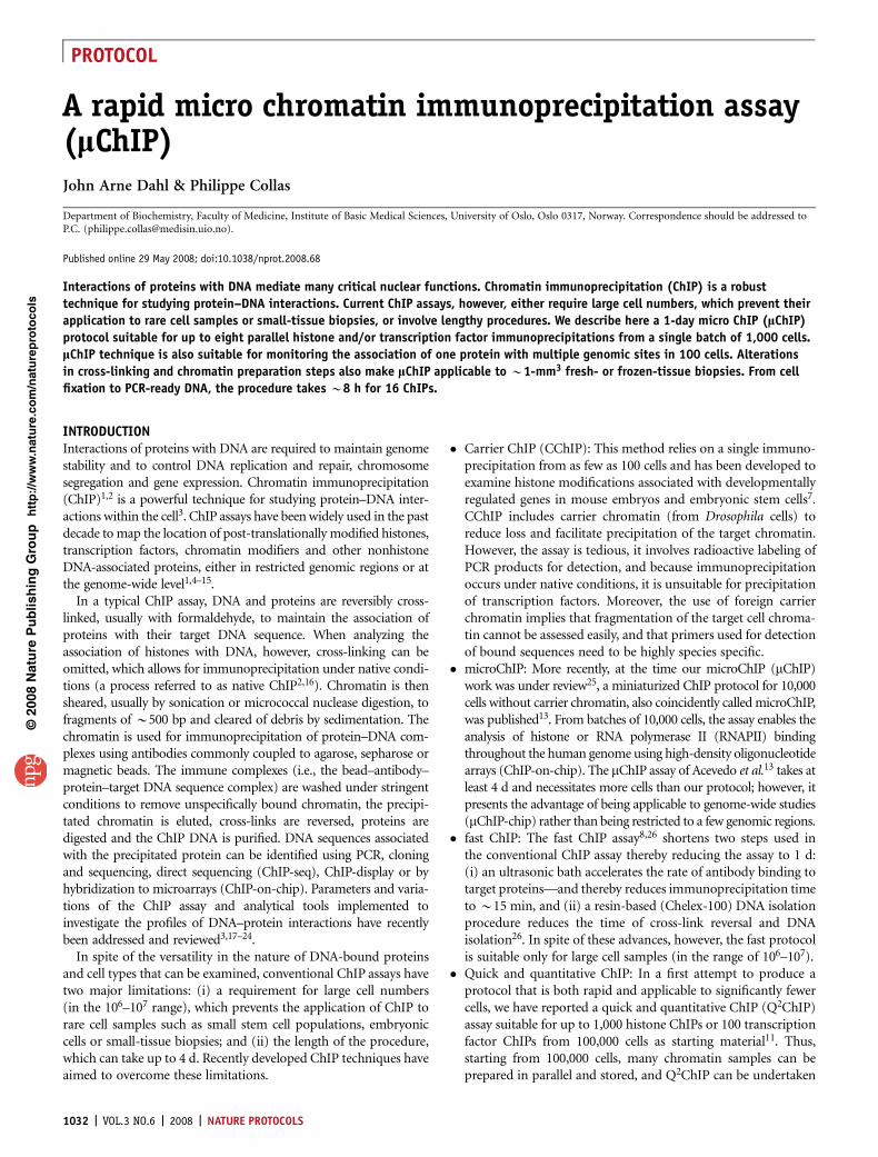
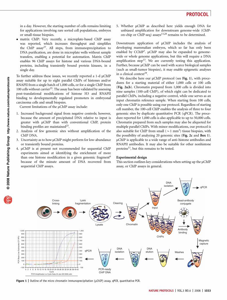
We describe here our mChIP protocol (see Fig. 1), with proce-dures for a starting material of either 1,000 cells or 100 cells(Fig. 2a,b). Chromatin prepared from 1,000 cells is divided intonine samples (100-cell ChIP), of which eight can be dedicated toparallel ChIPs, including a negative control, while one serves as aninput chromatin reference sample. When starting from 100 cells,only one ChIP is possible using our protocol. Regardless of startingcell number, the 100-cell ChIP enables the analysis of three to fourgenomic sites by duplicate quantitative PCR (qPCR). The proce-dure reported for 1,000 cells is also applicable to up to 50,000 cells.Chromatin prepared from such samples may also be aliquoted formultiple parallel ChIPs. With minor modifications, our protocol isalso suitable for ChIP from small (B1 mm3) tissue biopsies, withthe possibility of analyzing 20 genomic sites (Fig. 2c and Box 1).mChIP is applicable to a wide range of anti-histone antibodies andRNAPII antibodies. It may also be suitable for other nonhistoneproteins11, but this remains to be tested.
Experimental designThis section outlines key considerations when setting up the mChIPassay, or ChIP assays in general.
p
uor
G g
n ih si l
bu
P eru ta
N 800 2©
nat
ure
pro
toco
ls/
moc.er
ut an.
ww
w//:ptt
h
–100
100
200
300
400
500
600
PC
R B
ase
Line
Sub
trac
ted
CF
RF
U
700
800
900
1,000
0
–100
100
200
300
400
500
600
700
800
900
1,000
0
Cross-linking
Lysisfragmentation
Bead-antibodyconjugate
Magneticcapture
Antibodybinding
DNAelution
PCR-readyChIP DNA
DNAisolation
DNAisolation
Clearing
Lysate
qPCR
Chromatin
ChIPs
Washes
Input
qPCR
0 2 4 6 8 10 12 14 16 18 20 22Cycle
PCR Amplificaton vs Cycle Data 24-Jan-06 0930.odm
24 26 28 30 32 34 36 38 40 42
Figure 1 | Outline of the micro chromatin immunoprecipitation (mChIP) assay. qPCR, quantitative PCR.
NATURE PROTOCOLS | VOL.3 NO.6 | 2008 | 1033
PROTOCOL
Establishing the assay. To establish theassay, we recommend using antibodies to,for example, trimethylated lysine 4 of histoneH3 (H3K4m3; Abcam, cat. no. Ab8580) andtrimethylated H3K27 (Upstate, cat. no.05-851), because these efficiently precipitateepitopes and, therefore, generate strongPCR signals. Antibodies to acetylated H3K9(H3K9ac; e.g., Upstate, cat. no. 06-942;Diagenode, cat. no. pAb-ACHAHS-044)and to trimethylated H3K9 (H3K9m3; e.g.,Upstate, cat. no. 07-442; Diagenode, cat. no.pAb-056-050) can also be used because thesehistone modifications mark transcriptionallyactive and repressed genes, respectively, andare thus easy to discriminate. Specificity ofthe antibodies is extremely important. Inaddition, individual antibodies may performdifferently in ChIP, so testing different anti-bodies is recommended. Use, when available,ChIP-grade antibodies from several suppliers.Suggestions for assessing antibody specificityhave been published elsewhere30,31.
We recommend using a cultured cell linefor consistency of the starting material and, for PCR-based ChIPanalysis, focusing on genes with well-defined promoter sequenceinformation and expression status. For example, the humanembryonal carcinoma NCCIT cell line (American Type CultureCollection, cat. no. CRL-2073) may be good starting materialbecause large-scale and small cell number ChIP data have beenpublished with this cell line11,25.
Several ChIP protocols include an RNase treatment as partof the ChIP DNA purification step6,13,28,30,32; however, theseare designed for downstream microarray or sequencing analysis.ChIP protocols designed for PCR analysis do not include anRNase step2,7,8,11,16,26,27,31. Likewise, our protocol does notinclude any RNase step; however, it focuses on enrichmentusing sequence-specific primers, so this is not a limitation. AnRNase treatment step can easily be introduced during DNApurification (Step 21) if mChIP is to be adapted to a genome-wide analysis.
Optimizing the assay. The following aspects of the experimentmay require optimization.
� DNA–protein cross-linking: Although immunoprecipitation ofhistones is efficient under native conditions2,7,16, the mChIPprotocol includes a cross-linking step, which makes theprocedure universal for ChIPs of histone and nonhistoneproteins. Efficiency of cross-linking depends on the nature ofthe protein to cross-link, so the nature of cross-linker andduration of cross-linking should be considered. Formaldehydehas a short cross-linking spacer arm and cross-links nuclearcomponents located within 2 A of each other33, thus it can beineffective when analyzing proteins indirectly bound to DNA. Insuch instances, longer range cross-linkers are recommended,with or without formaldehyde. For more information on thesecross-linkers, we refer to reports describing alternative cross-linking reagents31,34.
Testing cross-linking efficiency is empirical. It should beremembered that extensive cross-linking may decrease solubilityof any target DNA–protein complex and cause it to be entrappedin the insoluble material removed by sedimentation31. If nocross-linking information exists for the protein of interest, werecommend starting with 1% formaldehyde for 8 min using, as apositive control, a protein known to be cross-linked under theseconditions (a histone or RNAPII). Cross-linking conditions aretested by ChIP and PCR analysis of the precipitated DNA.Failure to cross-link will result in no PCR product after ChIP;in this instance, cross-linking time may be increased and/oradditional cross-linkers should be tested alone or in combina-tion with formaldehyde34.
� Chromatin sonication: Chromatin fragments of 400–500 bp haveproven to be suitable for ChIP assays as they cover two to threenucleosomes. Longer fragments will diminish resolution andcause noise and are not recommended for microarray analysis30,while fragments that are too short may be incompatible withPCR, depending on amplicon length. For tips on sonicationconditions and tests, see Figure 3 and Box 2.
� Amount of antibody and protein A or G: The amount of anti-body and Protein A or G in the ChIP assay will affect theamount of material precipitated. Amount of antibody andProtein A or G beads to be used can be tested by qPCR followingChIP to assess the relative amounts of precipitated targetDNA sequence.
� Chromatin and reference samples: To enable the assessment of thesame pool of cells across a selection of antibodies, we favor theuse of a single chromatin preparation aliquoted into the numberof samples required for the different ChIPs, including negativecontrol(s) and reference input chromatin sample(s). Thisapproach has also been proposed by the Young laboratory30.Separate chromatin preparations within a replicate experimentmay be a cause of between-sample variation originating frominconsistencies in the number of prepared cells per batch. We
p
uor
G g
n ih si l
bu
P eru ta
N 800 2©
nat
ure
pro
toco
ls/
moc.er
ut an.
ww
w//:ptt
h
100 Cells
a c
Input
3-4 Genomic sites
20 Genomic sites
1 ChIP
Input
Lysate
Biopsy(~1 mm3)
8 ChIPs
100 Cells1,000 Cells
qPCR (×2)
qPCR (×2)
Input
3-4 Genomic sites
8 ChIPs
qPCR (×2)
b
Figure 2 | Chromatin immunoprecipitation (ChIP) from small cell samples and biopsies. Micro ChIP
(mChIP) is suitable for immunoprecipitation of histones and transcription factors in (a) eight parallel
ChIPs starting from 1,000 cells (or more) or (b) one ChIP starting from 100 cells. (c) A variation of the
mChIP assay enables eight parallel ChIPs from B1-mm3 tumor biopsies. Numbers of genomic sites that
can be examined in each approach by duplicate quantitative PCR (qPCR) are indicated.
1034 | VOL.3 NO.6 | 2008 | NATURE PROTOCOLS
PROTOCOL
emphasize the importance of constant cell numbers betweenbatches as the cell number directly affects the amount of inputchromatin, hence also the precipitation efficiency. Triplicate ChIPsshould be performed to assess variation between replicates. mChIPmeasures the association of a target genomic sequence with aspecific protein. In this instance, data are expressed as ‘percentprecipitated DNA relative to input’, with the input consisting of achromatin sample equivalent to the chromatin amount used forChIP. This formula brings out precipitation efficiency and theextent of unspecific precipitation. Some investigators expressenrichments relative to a negative control14, although this masksthe extent of unspecific precipitation, or relative to enrichment ofthe same protein to a control gene35.
Controls. The use of positive and negative controls is recom-mended. Negative controls may consist of either the use of no
antibody (beads alone) or an irrelevant antibody of the sameisotype of that used in the immunoprecipitation. The positivecontrol should be an antibody to a protein known to co-localizewith an identified locus in the cell type examined. This may includean antibody to histones, RNAPII or, as suggested by others30, theubiquitous cell cycle regulator E2F4.
Output and data analysis. ChIP data have been analyzed by slotblotting of the ChIP DNA, dot blotting36,37 and end-point PCR.Real-time PCR allows a quantification of the amount of precipi-tated DNA relative to an input sample (e.g., refs. 11,25), relative tothe unbound (nonprecipitated) material7 or relative to a negativecontrol precipitation14. Analysis of real-time PCR ChIP results isstraightforward and is described in Steps 25–27. We can recom-mend ref. 38 for a comprehensive description of real-time PCRanalysis of ChIP data.
p
uor
G g
n ih si l
bu
P eru ta
N 800 2©
nat
ure
pro
toco
ls/
moc.er
ut an.
ww
w//:ptt
h
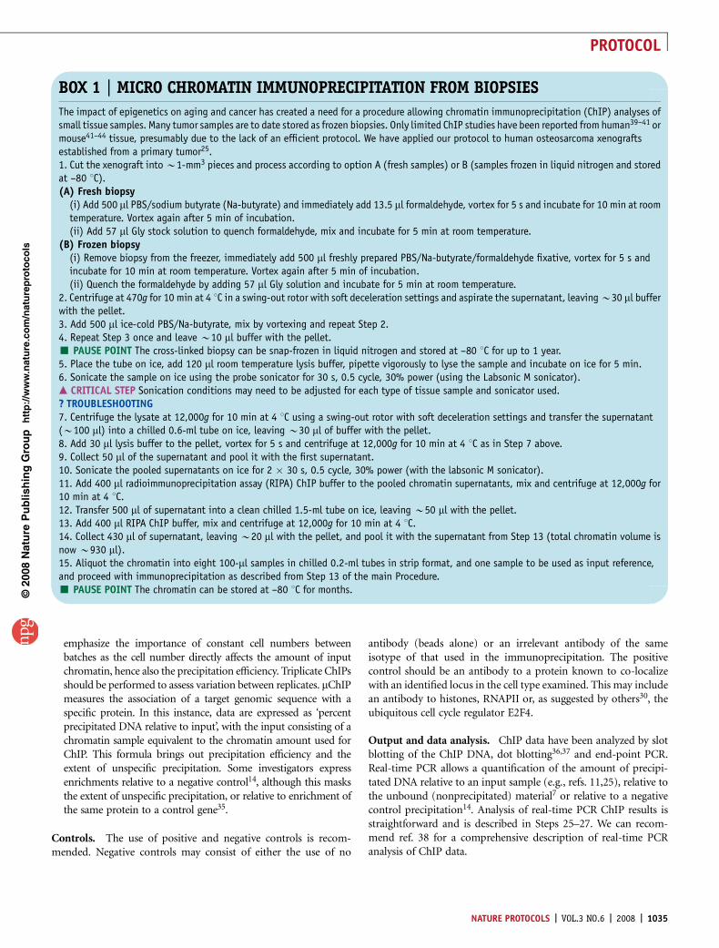
BOX 1 | MICRO CHROMATIN IMMUNOPRECIPITATION FROM BIOPSIES
The impact of epigenetics on aging and cancer has created a need for a procedure allowing chromatin immunoprecipitation (ChIP) analyses ofsmall tissue samples. Many tumor samples are to date stored as frozen biopsies. Only limited ChIP studies have been reported from human39–41 ormouse41–44 tissue, presumably due to the lack of an efficient protocol. We have applied our protocol to human osteosarcoma xenograftsestablished from a primary tumor25.1. Cut the xenograft into B1-mm3 pieces and process according to option A (fresh samples) or B (samples frozen in liquid nitrogen and storedat –80 1C).(A) Fresh biopsy
(i) Add 500 ml PBS/sodium butyrate (Na-butyrate) and immediately add 13.5 ml formaldehyde, vortex for 5 s and incubate for 10 min at roomtemperature. Vortex again after 5 min of incubation.(ii) Add 57 ml Gly stock solution to quench formaldehyde, mix and incubate for 5 min at room temperature.
(B) Frozen biopsy(i) Remove biopsy from the freezer, immediately add 500 ml freshly prepared PBS/Na-butyrate/formaldehyde fixative, vortex for 5 s andincubate for 10 min at room temperature. Vortex again after 5 min of incubation.(ii) Quench the formaldehyde by adding 57 ml Gly solution and incubate for 5 min at room temperature.
2. Centrifuge at 470g for 10 min at 4 1C in a swing-out rotor with soft deceleration settings and aspirate the supernatant, leaving B30 ml bufferwith the pellet.3. Add 500 ml ice-cold PBS/Na-butyrate, mix by vortexing and repeat Step 2.4. Repeat Step 3 once and leave B10 ml buffer with the pellet.’ PAUSE POINT The cross-linked biopsy can be snap-frozen in liquid nitrogen and stored at –80 1C for up to 1 year.5. Place the tube on ice, add 120 ml room temperature lysis buffer, pipette vigorously to lyse the sample and incubate on ice for 5 min.6. Sonicate the sample on ice using the probe sonicator for 30 s, 0.5 cycle, 30% power (using the Labsonic M sonicator).m CRITICAL STEP Sonication conditions may need to be adjusted for each type of tissue sample and sonicator used.? TROUBLESHOOTING7. Centrifuge the lysate at 12,000g for 10 min at 4 1C using a swing-out rotor with soft deceleration settings and transfer the supernatant(B100 ml) into a chilled 0.6-ml tube on ice, leaving B30 ml of buffer with the pellet.8. Add 30 ml lysis buffer to the pellet, vortex for 5 s and centrifuge at 12,000g for 10 min at 4 1C as in Step 7 above.9. Collect 50 ml of the supernatant and pool it with the first supernatant.10. Sonicate the pooled supernatants on ice for 2 � 30 s, 0.5 cycle, 30% power (with the labsonic M sonicator).11. Add 400 ml radioimmunoprecipitation assay (RIPA) ChIP buffer to the pooled chromatin supernatants, mix and centrifuge at 12,000g for10 min at 4 1C.12. Transfer 500 ml of supernatant into a clean chilled 1.5-ml tube on ice, leaving B50 ml with the pellet.13. Add 400 ml RIPA ChIP buffer, mix and centrifuge at 12,000g for 10 min at 4 1C.14. Collect 430 ml of supernatant, leaving B20 ml with the pellet, and pool it with the supernatant from Step 13 (total chromatin volume isnow B930 ml).15. Aliquot the chromatin into eight 100-ml samples in chilled 0.2-ml tubes in strip format, and one sample to be used as input reference,and proceed with immunoprecipitation as described from Step 13 of the main Procedure.’ PAUSE POINT The chromatin can be stored at –80 1C for months.
NATURE PROTOCOLS | VOL.3 NO.6 | 2008 | 1035
PROTOCOL
MATERIALSREAGENTS.Anti-histone antibodies of your choice m CRITICAL Use ChIP-grade
antibodies when available. We have successfully used with this protocol thefollowing anti-histone antibodies: anti-H3K9ac (Upstate, cat. no. 06-942),anti-H3K9m2 (Upstate, cat. no. 07-441), anti-H3K9m3 (Upstate, cat. no.07-442), anti-H3K27m3 (Upstate, cat. no. 05-851), anti-H3K9m3(Diagenode, cat. no. pAb-056-050), anti-H3K4m2 (Abcam,cat. no. Ab7766) and anti-H3K4m3 (Abcam, cat. no. Ab8580).
.Anti-RNAPII antibodies (e.g., Santa Cruz Biotechnology, cat. no. sc-899)m CRITICAL This protocol has been developed with this particular
anti-RNAPII antibody and should be tested for other antibodies,including antibodies to less abundant nuclear proteins.
.36.5% Formaldehyde (Sigma-Aldrich, cat. no. F8775) ! CAUTION Toxicby inhalation, contact with skin or if swallowed. Formaldehyde wasteshould be treated as hazardous.
.Dynabeads Protein A (Invitrogen, cat. no. 100.02D) m CRITICAL Ensure thatthe beads are well suspended before pipetting. Use Dynabeads Protein Abeads with rabbit IgGs and Dynabeads Protein G (Invitrogen, cat. no.100.04D) with mouse IgGs.
.400 mM EGTA (Sigma-Aldrich, cat. no. E0396)
p
uor
G g
n ih si l
bu
P eru ta
N 800 2©
nat
ure
pro
toco
ls/
moc.er
ut an.
ww
w//:ptt
h
1.0
10,000
a b cLa
dder
Ladd
er
Ctl 30 s
2 × 30
s
4 × 30
s
7 × 30
s
14 × 30
s
1,000850650500400300200
100
0.8
0.6
PC
R s
igna
l int
ensi
tyre
l. to
con
trol
0.4
0.2
0.0
1.096 bp298 bp
96 bp298 bp
0.8
0.6
PC
R s
igna
l int
ensi
tyre
l. to
con
trol
0.4
0.2
0.0
Ctl30
s
2 × 30
s
4 × 30
s
7 × 30
s
14 × 30
s Ctl15
s30
s
2 × 30
s
3 × 30
s
4 × 30
s
6 × 30
s
10 × 30
s
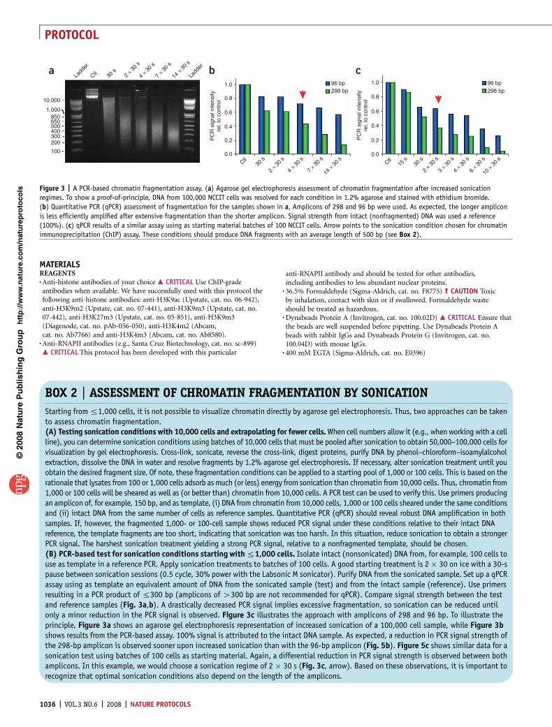
Figure 3 | A PCR-based chromatin fragmentation assay. (a) Agarose gel electrophoresis assessment of chromatin fragmentation after increased sonication
regimes. To show a proof-of-principle, DNA from 100,000 NCCIT cells was resolved for each condition in 1.2% agarose and stained with ethidium bromide.
(b) Quantitative PCR (qPCR) assessment of fragmentation for the samples shown in a. Amplicons of 298 and 96 bp were used. As expected, the longer amplicon
is less efficiently amplified after extensive fragmentation than the shorter amplicon. Signal strength from intact (nonfragmented) DNA was used a reference
(100%). (c) qPCR results of a similar assay using as starting material batches of 100 NCCIT cells. Arrow points to the sonication condition chosen for chromatin
immunoprecipitation (ChIP) assay. These conditions should produce DNA fragments with an average length of 500 bp (see Box 2).
BOX 2 | ASSESSMENT OF CHROMATIN FRAGMENTATION BY SONICATION
Starting from r1,000 cells, it is not possible to visualize chromatin directly by agarose gel electrophoresis. Thus, two approaches can be takento assess chromatin fragmentation.(A) Testing sonication conditions with 10,000 cells and extrapolating for fewer cells. When cell numbers allow it (e.g., when working with a cellline), you can determine sonication conditions using batches of 10,000 cells that must be pooled after sonication to obtain 50,000–100,000 cells forvisualization by gel electrophoresis. Cross-link, sonicate, reverse the cross-link, digest proteins, purify DNA by phenol–chloroform–isoamylalcoholextraction, dissolve the DNA in water and resolve fragments by 1.2% agarose gel electrophoresis. If necessary, alter sonication treatment until youobtain the desired fragment size. Of note, these fragmentation conditions can be applied to a starting pool of 1,000 or 100 cells. This is based on therationale that lysates from 100 or 1,000 cells adsorb as much (or less) energy from sonication than chromatin from 10,000 cells. Thus, chromatin from1,000 or 100 cells will be sheared as well as (or better than) chromatin from 10,000 cells. A PCR test can be used to verify this. Use primers producingan amplicon of, for example, 150 bp, and as template, (i) DNA from chromatin from 10,000 cells, 1,000 or 100 cells sheared under the same conditionsand (ii) intact DNA from the same number of cells as reference samples. Quantitative PCR (qPCR) should reveal robust DNA amplification in bothsamples. If, however, the fragmented 1,000- or 100-cell sample shows reduced PCR signal under these conditions relative to their intact DNAreference, the template fragments are too short, indicating that sonication was too harsh. In this situation, reduce sonication to obtain a strongerPCR signal. The harshest sonication treatment yielding a strong PCR signal, relative to a nonfragmented template, should be chosen.(B) PCR-based test for sonication conditions starting with r1,000 cells. Isolate intact (nonsonicated) DNA from, for example, 100 cells touse as template in a reference PCR. Apply sonication treatments to batches of 100 cells. A good starting treatment is 2 � 30 on ice with a 30-spause between sonication sessions (0.5 cycle, 30% power with the Labsonic M sonicator). Purify DNA from the sonicated sample. Set up a qPCRassay using as template an equivalent amount of DNA from the sonicated sample (test) and from the intact sample (reference). Use primersresulting in a PCR product of r300 bp (amplicons of 4300 bp are not recommended for qPCR). Compare signal strength between the testand reference samples (Fig. 3a,b). A drastically decreased PCR signal implies excessive fragmentation, so sonication can be reduced untilonly a minor reduction in the PCR signal is observed. Figure 3c illustrates the approach with amplicons of 298 and 96 bp. To illustrate theprinciple, Figure 3a shows an agarose gel electrophoresis representation of increased sonication of a 100,000 cell sample, while Figure 3bshows results from the PCR-based assay. 100% signal is attributed to the intact DNA sample. As expected, a reduction in PCR signal strength ofthe 298-bp amplicon is observed sooner upon increased sonication than with the 96-bp amplicon (Fig. 5b). Figure 5c shows similar data for asonication test using batches of 100 cells as starting material. Again, a differential reduction in PCR signal strength is observed between bothamplicons. In this example, we would choose a sonication regime of 2 � 30 s (Fig. 3c, arrow). Based on these observations, it is important torecognize that optimal sonication conditions also depend on the length of the amplicons.
1036 | VOL.3 NO.6 | 2008 | NATURE PROTOCOLS
PROTOCOL
.1 M Tris–HCl, pH 8.0 (Sigma-Aldrich, cat. no. T3253)
.1 M Tris–HCl, pH 7.5 (Sigma-Aldrich, cat. no. T3253)
.5 M NaCl (Sigma-Aldrich, cat. no. S5150)
.Triton X-100 (Sigma-Aldrich, cat. no. T8787)
.Na-deoxycholate (Sigma-Aldrich, cat. no. D5760)
.Gly (Sigma-Aldrich, cat. no. G8790) 1.25 M stock solution in PBS
.Chelex-100 (Bio-Rad, cat. no. 142-1253) (see REAGENT SETUP)
.Acrylamide carrier (Sigma-Aldrich, cat. no. A9099) ! CAUTION Toxic bycontact with skin or if ingested.
.Proteinase K (Sigma-Aldrich, cat. no. P2308) 20 mg ml–1 stock solution inMilliQ water
.Protease inhibitor mix (Sigma-Aldrich, cat. no. P8340)
.Phenylmethylsulphonyl fluoride (PMSF; Sigma-Aldrich, cat. no. P7626)100 mM stock solution in 100% ethanol ! CAUTION Toxic by inhalation,if absorbed through skin or if ingested.
.Sodium butyrate (Na-butyrate;Sigma-Aldrich, cat. no. B5887) 1 M stocksolution in MilliQ water. Na-butyrate is a histone deacetylase inhibitor andshould be used when investigating acetylated epitopes. Other inhibitors(e.g., phosphatase inhibitors) should be used when looking at other typesof modifications (e.g., phosphorylation)
.PBS/Na-butyrate solution: 20 mM butyrate in 1� PBS m CRITICAL Make upimmediately before use.
.PBS/Na-butyrate/formaldehyde fixative (see REAGENT SETUP) m CRITICALMake up immediately before use.
.PBS (Sigma-Aldrich, cat. no. P4417) For a 1� solution, dissolve 1 tablet in200 ml MilliQ water
.SDS (Sigma-Aldrich, cat. no. L4509) ! CAUTION Toxic by inhalation, contactwith skin or if ingested.
.3 M NaAc (Sigma-Aldrich, cat. no. S8750)
.Phenol–chloroform–isoamylalcohol (25:24:1; Invitrogen, cat. no. 15593-031)! CAUTION Toxic if absorbed through skin, inhaled or ingested.
.Chloroform–isoamylalcohol (24:1; Sigma-Aldrich, cat. no. C0549)! CAUTION Toxic if absorbed through skin, inhaled or ingested.
.96% (vol/vol) Ethanol at –20 1C
.70% (vol/vol) Ethanol at –20 1C
.IQ SYBR green (Bio-Rad, cat. no. 170-8882) ! CAUTION Use caution whenworking with DNA stains when there is no data addressing mutagenicactivity.
.Crushed ice in insulated container
.Lysis buffer (see REAGENT SETUP)
.Radioimmunoprecipitation assay (RIPA) buffer (see REAGENT SETUP)
.RIPA ChIP buffer (see REAGENT SETUP)
.Tris–EDTA (TE) buffer (see REAGENT SETUP)
.Elution buffer (see REAGENT SETUP)
.Complete elution buffer (see REAGENT SETUP)
EQUIPMENT.Siliconized pipette tips (Sigma-Aldrich, cat. no. T7656-960EA).Filter pipette tips (Universal Laboratory Plasticware, cat. no. 81241 (10 ml);
cat. no. 83241 (200 ml); cat. no. 85241 (500 ml)).0.6-ml Centrifuge tubes (Axygen, cat. no. 321-05-051).200-ml PCR tubes in 8-tube strip format (Axygen, cat. no. 321-10-051).
These tubes fit exactly in the Diagenode magnetic rack (Diagenode).Magnetic rack for 200-ml tube strips (Diagenode, cat. no. kch-816-001).Magnetic holder for 1.5-ml tubes (Invitrogen, cat. no. MPC-E).Probe sonicator (Sartorius Labsonic M sonicator (Sartorius) fitted with
3-mm diameter probe, or a similar model).Thermomixer (Eppendorf, model no. 5355-28402, or a similar model).Minicentrifuge (Merck Eurolab Galaxy Mini, model no. C1211, or a similar
model).Vortex (VWR International, model no. 444-1372, or a similar model).Rotator (Science Lab, model no. Stuart SB3, or a similar model), placed
at 4 1C.Thermal cycler with accessories (Bio-Rad MyiQ real-time PCR detection
system; BioRad or any thermal cycler with real-time assessment capacity)
REAGENT SETUPPBS/Na-butyrate/formaldehyde fixative 20 mM butyrate, 1% (vol/vol)formaldehyde, 1 mM PMSF and protease inhibitor mix in 1� PBS.m CRITICAL Make up immediately before use.Lysis buffer 50 mM Tris–HCl, pH 8.0, 10 mM EDTA, 1% (wt/vol) SDS,protease inhibitor mix (1:100 dilution from stock), 1 mM PMSF, 20 mMNa-butyrate. m CRITICAL Protease inhibitor mix, PMSF and Na-butyratemust be added just before use.RIPA buffer 10 mM Tris–HCl, pH 7.5, 140 mM NaCl, 1 mM EDTA, 0.5 mMEGTA, 1% (vol/vol) Triton X-100, 0.1% (wt/vol) SDS, 0.1% (wt/vol)Na-deoxycholate.RIPA ChIP buffer 10 mM Tris–HCl, pH 7.5, 140 mM NaCl, 1 mM EDTA,0.5 mM EGTA, 1% (vol/vol) Triton X-100, 0.1% (wt/vol) SDS, 0.1% (wt/vol)Na-deoxycholate, protease inhibitor mix (1:100 dilution from stock), 1 mMPMSF, 20 mM Na-butyrate. m CRITICAL Protease inhibitor mix, PMSF andNa-butyrate must be added just before use.Chelex solution 10% (wt/vol) Chelex in MilliQ water26.TE buffer 10 mM Tris–HCl, pH 8.0, 10 mM EDTA.Elution buffer 20 mM Tris–HCl, pH 7.5, 5 mM EDTA, 50 mM NaCl.Complete elution buffer 20 mM Tris–HCl, pH 7.5, 5 mM EDTA, 50 mMNaCl, 20 mM Na-butyrate, 1% (wt/vol) SDS, 50 mg ml–1 proteinase K.m CRITICAL Na-butyrate, SDS and proteinase K should be added just before use.
PROCEDUREAntibody–bead complexes1| Prepare a slurry of Dynabeads Protein A (if using rabbit IgGs). For 16 ChIPs, including two negative controls, place 180 mlof well-suspended Dynabeads Protein A stock solution into a 1.5-ml tube, place the tube in the magnetic holder, allow beads tobe captured, remove the buffer, remove from the magnet and add 500 ml RIPA buffer.m CRITICAL STEP Make sure the stock bead suspension is homogenous before pipetting.
2| Vortex, capture the beads, remove the buffer, add another 500 ml RIPA buffer.
3| Vortex, capture the beads, remove the buffer, add 170 ml RIPA buffer.
4| Vortex the beads and place the tube on ice.
5| Aliquot 90 ml RIPA buffer into 200-ml PCR tubes (one tube per ChIP), place on ice and add 10 ml washed DynabeadsProtein A-bead slurry from Step 4 and 2.4 mg antibody to each tube. Do not add the antibody to the negative control samplesor, alternativey, add a preimmune antibody preferably of the same isotype as the ChIP antibodies. Place at 40 r.p.m. on rotatorfor 2 h at 4 1C.m CRITICAL STEP This incubation step should be carried out during cross-linking, cell lysis and chromatin preparation (Steps 6–12)and, if necessary, can be prolonged until all chromatin samples are ready for immunoprecipitation.m CRITICAL STEP For this and subsequent steps, we recommend using 0.2-ml PCR tubes in eight-tube strip format, which easily fitin the magnetic rack, for easier handling than individual tubes.m CRITICAL STEP Throughout the rest of the protocol, handle the negative controls as the ChIP samples.
p
uor
G g
n ih si l
bu
P eru ta
N 800 2©
nat
ure
pro
toco
ls/
moc.er
ut an.
ww
w//:ptt
h
NATURE PROTOCOLS | VOL.3 NO.6 | 2008 | 1037
PROTOCOL
Cross-linking of DNA and proteins6| Add 20 mM Na-butyrate from the 1 M stock to the cell culture and mix gently. If working with cells requiring trypsinizationfor harvesting, add butyrate after Step 9. If working with biopsy samples, process as described in Box 1.m CRITICAL STEP Na-butyrate is added immediately before collecting cells and cross-linking to avoid artefactual histonehyperacetylation. For cells requiring trypsinization and/or washes before cross-linking, butyrate should be added immediately priorto adding formaldehyde (see Step 10), not while cells are in culture.
7| Discard the medium to remove dead cells (if cells are growing adherent) and add PBS/Na-butyrate (e.g., 10 ml per 175 cm2
culture flask) at room temperature (20–25 1C).
8| Harvest cells, by trypsinization or as per your standard protocol, according to cell type.
9| Count cells and resuspend 1,000 (or 100) cells in 500 ml PBS/Na-butyrate in a 0.6-ml tube at room temperature.m CRITICAL STEP Up to 50,000 cells can be used if necessary using the same protocol (more cells in the assay allow for the analysisof more loci by PCR).m CRITICAL STEP To prevent cell lysis during pipetting steps, use a 1,000-ml pipette tip or a 200-ml pipette tip with a cutoff tip toincrease the diameter of the opening.
10| Add 13.5 ml formaldehyde (1% vol/vol final concentration), mix by gentle vortexing and incubate for 8 min at roomtemperature.m CRITICAL STEP 1% Formaldehyde cross-links DNA to proteins located within 2 A of DNA33. Although cross-linking isomitted for ChIP analysis of histones in some protocols16, it makes the protocol suitable for both histone and nonhistoneChIPs from a single chromatin preparation. To simplify the cross-linking step and enhance cell recovery, we cross-link cellsin suspension regardless of whether they grow adherent or in suspension. Time of cross-linking may vary depending on theprotein to be immunoprecipitated. For most ChIP applications, 8–10-min cross-linking is sufficient (see INTRODUCTION forcross-linking tips).
11| Add 57 ml of 1.25 M Gly stock (125 mM final concentration) to quench the formaldehyde and incubate for 5 min at roomtemperature.’ PAUSE POINT Pellets of cross-linked cells can be stored at –80 1C for at least several months.
Chromatin preparation12| For preparing chromatin, two procedures are described that can be used depending on the starting cell number. Option A isfor preparing chromatin from 1,000 cells but is also suited for up to 50,000 cells with slight adjustments in sonicationconditions; see Box 2 (and Fig. 3) for details on testing chromatin fragmentation by sonication of small cell numbers. Option Bis used when starting with 100 cells, but can also be applied to up to r10,000 cells.m CRITICAL STEP When starting with 100 cells, only one immunoprecipitation can be performed per 100-cell sample. If more thanone protein is to be precipitated, use as many 100-cell samples as necessary. Prepare an additional sample for reference inputchromatin. If it is not possible to use more cells for input, the sum of PCR signals from the bound and unbound fractions can be usedas reference in lieu of the input reference sample.(A) For 1,000 cells
(i) Centrifuge formaldehyde-cross-linked cells at 470g for 10 min at 4 1C in a swing-out rotor with soft deceleration settings.Slowly aspirate and discard the supernatant, leaving B30 ml of the solution with the cell pellet to ensure that none ofthe loosely packed cells are aspirated.
(ii) Resuspend the cells in 500 ml ice-cold PBS/Na-butyrate by gentle vortexing and centrifuge at 470g for 10 min at 4 1Cas in Step 12A(i).
(iii) Repeat the washing procedure (Step 12A(ii)) once. Upon aspiration of the last wash, leave 20 ml PBS/Na-butyrate withthe cell pellet.
(iv) Add 120 ml room temperature lysis buffer, vortex for 2 � 5 s, leave on ice for 5 min and resuspend cells by vortexing.Ensure that no liquid is trapped in the lid.
(v) Sonicate on ice for 3 � 30 s, with 30 s pauses on ice between each 30-s session, using the probe sonicator. With theLabsonic M sonicator, use the following pulse settings: cycle 0.5, 30% power. Repeat for each tube while leaving thesonicated samples on ice.m CRITICAL STEP Sonication should produce chromatin fragments of B500 bp (range may be 200–1,200 bp). The sonica-tion regime indicated is suitable for a variety of cultured cell lines (e.g., NCCIT, Jurkat, 293T) but must be optimized foreach cell type, particularly for primary cells. Do not allow samples to foam as foaming reduces sonication efficiency.
(vi) Add 400 ml RIPA ChIP buffer to the tube (which contains B140 ml lysate) and mix by vortexing.
p
uor
G g
n ih si l
bu
P eru ta
N 800 2©
nat
ure
pro
toco
ls/
moc.er
ut an.
ww
w//:ptt
h
1038 | VOL.3 NO.6 | 2008 | NATURE PROTOCOLS
PROTOCOL
(vii) Centrifuge at 12,000g for 10 min at 4 1C, carefully aspirate the supernatant (chromatin) and transfer it into a clean 1.5-mltube chilled on ice.m CRITICAL STEP To avoid aspirating the sedimented material, which is practically invisible, leave B50 ml supernatantin the tube after aspiration. The sediment contains SDS-insoluble cellular debris which may stick to the beads and causeunspecific background or, in the worst case, mask any specific enrichment caused by the subsequent immunoprecipitation(ChIP).
(viii) Add 410 ml RIPA ChIP buffer to the remaining pellet, mix by vortexing and centrifuge at 12,000g for 10 min at 4 1C.(ix) Aspirate the supernatant, leaving B20 ml with the (invisible) pellet and pool it with the first supernatant. This yields
B920 ml of chromatin suitable for eight parallel ChIPs and one input reference. Discard the pellets.m CRITICAL STEP Diluting the chromatin reduces SDS concentration to B0.1% (wt/vol), which is suitable for immuno-precipitation with most antibodies.’ PAUSE POINT Chromatin from Z100,000 cells can be stored at �80 1C for up to 1 year; chromatin from smaller cellnumbers can be stored at �80 1C for at least two weeks. Prolonged storage has not been tested.
(x) Aliquot 100 ml chromatin each into, e.g., eight chilled 0.2-ml tubes (in strip form) containing antibody–bead complexesheld to the wall in the magnetic rack (on ice), and from which the RIPA buffer has been pipetted out (prepared inSteps 1–5 from the main PROCEDURE). For less than eight ChIPs, store leftover chromatin at –80 1C.
(xi) Add 100 ml chromatin to a 0.6- or 1.5-ml tube chilled on ice. This will be used as input chromatin and processed asdescribed in either Box 3 or 4.m CRITICAL STEP A 1.5-ml tube is used in this step if DNA is to be purified by phenol–chloroform–isoamylalcoholextraction (Step 21A and Box 3). For DNA purification with Chelex-100 (Step 21B and Box 4), a 0.6-ml tube is preferred.
(B) For 100 cells(i) Centrifuge formaldehyde-cross-linked cells at 470g for 10 min at 4 1C in a swing-out rotor with soft deceleration settings.
Aspirate the supernatant; leave B30 ml of the solution with the pellet.(ii) Add 500 ml ice-cold PBS/Na-butyrate, resuspend the cells by gentle vortexing and centrifuge at 470g for 10 min at
4 1C using a swing-out rotor with soft deceleration settings.(iii) Repeat the washing procedure (Step 12B(ii)) once. Leave B20 ml PBS/Na-butyrate with the pellet (invisible) after
removing the last wash.(iv) Add 120 ml lysis buffer, vortex for 2 � 5 s and incubate for 3 min on ice.(v) Centrifuge the nuclei at 860g for 10 min at 4 1C using a swing-out rotor with soft deceleration settings and discard the
supernatant; leave 20–30 ml of lysis buffer in the tube.m CRITICAL STEP Keeping cells in lysis buffer for over 3 min prior to centrifugation increases the chance of SDSprecipitating. If the SDS precipitates during centrifugation, remove the lysis buffer, add 200 ml RIPA ChIP buffer, dissolvethe SDS by vortexing and centrifuge the nuclei as in Step 12B(v).
(vi) Add 120 ml RIPA ChIP buffer and vortex for 10 s.(vii) Sonicate each tube on ice for 2 � 30 s, with 30-s pauses on ice between each 30-s session, using the probe sonicator
(cycle 0.5 and 30% power with the Labsonic M sonicator). Repeat for each tube while leaving the sonicated samples on ice.m CRITICAL STEP When starting with 100 cells, it is impossible to visualize chromatin fragmentation by agarose gelelectrophoresis. Instead, we use a PCR assay (see Box 2).
p
uor
G g
n ih si l
bu
P eru ta
N 800 2©
nat
ure
pro
toco
ls/
moc.er
ut an.
ww
w//:ptt
h
BOX 3 | PURIFICATION OF DNA FROM INPUT CHROMATIN SAMPLES BYPHENOL–CHLOROFORM–ISOAMYLALCOHOL EXTRACTION
1. To input chromatin samples from Step 12 of the main Procedure, add 200 ml elution buffer and 7.5 ml of a 10� dilution (2 mg ml–1) of theproteinase K solution, vortex and incubate on a heating block for 2 h at 68 1C.2. Remove samples from the heating block and add 200 ml elution buffer.3. Continue with the main Procedure from Step 21A(viii), processing the input samples and the ChIP samples in parallel.
BOX 4 | PURIFICATION OF DNA FROM INPUT CHROMATIN SAMPLES USING CHELEX-100
1. To input chromatin samples from Step 12, add 10 ml acrylamide carrier and 250 ml of 96% ethanol at –20 1C. Vortex thoroughly and placeat –80 1C for 30 min.2. Thaw, immediately centrifuge at 20,000g for 15 min at 4 1C and wash the pellet in 500 ml of 70% ethanol. Dry the pellet.3. Add 40 ml of 10% (wt/vol) Chelex-100 to the dried pellet and vortex for 10 s.4. Continue with the main Procedure from Step 21B(ii), processing the input samples and the ChIP samples in parallel.
NATURE PROTOCOLS | VOL.3 NO.6 | 2008 | 1039
PROTOCOL
(viii) For ChIP samples, pipette the lysate several times using a siliconized pipette tip and transfer into a 0.2-ml PCR tubecontaining antibody-coated beads prepared in Steps 1–5 and from which the RIPA buffer has been removed. For inputsamples, transfer into a 1.5-ml or 0.6-ml tube and process as described in Box 3 or 4, respectively.
Immunoprecipitation and washes13| Remove the tube strips (from Steps 12A or 12B) from the magnetic rack to release the antibody–bead complexes into thechromatin suspension and place the tubes on a rotator at 40 r.p.m. for 2 h at 4 1C.’ PAUSE POINT This step can be carried out overnight at 4 1C if necessary, but prolonged incubation may lead to enhancedbackground.
14| Centrifuge the tubes in a minicentrifuge for 1 s to bring down any solution trapped in the lid during the incubation onthe rotator, and capture the immune complexes (the ChIP material) on the tube wall by placing the tubes in the chilledmagnetic rack.
15| Discard the supernatant, add 100 ml ice-cold RIPA buffer and remove the tubes from the magnetic rack to release immunecomplexes into the buffer. Resuspend the complexes by gentle manual agitation and place the tubes on a rotator at 40 r.p.m.for 4 min at 4 1C.
16| Repeat Steps 14 and 15 two more times.m CRITICAL STEP Always briefly spin the tubes in a minicentrifuge for 1 s to bring down any liquid trapped in the lid prior topositioning the tubes in the magnetic rack.
17| Centrifuge the tubes in a minicentrifuge for 1 s.
18| Remove the supernatant, add 100 ml TE buffer and incubate on a rotator at 4 1C for 4 min at 40 r.p.m.
19| Centrifuge the tubes in a minicentrifuge for 1 s.
20| Place the tubes on ice (not in the magnetic rack), transfer the content of each tube into separate clean 0.2-ml tubes onice, capture the complexes in the magnetic rack and remove the TE buffer.m CRITICAL STEP A drawback of most ChIP procedures is unspecific chromatin background. Background becomes significantwhen reducing the amount of input material in ChIP due to reduction in the signal and, with a reduction of sample volumes, dueto an increase in surface-to-volume ratio. The tube surface is a source of unspecific binding of chromatin. Transferring theChIP material in TE buffer to a clean tube has in our hands proven to be critical to enhance specificity of the ChIP assay11.
Isolation of DNA21| Two protocols are presented for recovery of DNA from ChIP material and from input chromatin. Option A describes DNAelution, cross-link reversal and protein digestion steps, followed by a phenol–chloroform–isoamylalcohol DNA purification.Option B describes a Chelex-100-mediated DNA purification reported previously26, with modifications for small samples and toaccelerate handling. Both procedures can be used regardless of the starting cell number in the ChIP assay without affectingthe result25,26. Protocols for DNA isolation from input chromatin are given in Box 3 (phenol–chloroform–isoamylalcohol) andBox 4 (Chelex-100).(A) DNA elution, cross-link reversal, proteinase K digestion and DNA purification from ChIP samples byphenol–chloroform–isoamyalcohol extraction
(i) Place the tubes from Step 20 in a rack and add 150 ml complete elution buffer to each tube.m CRITICAL STEP Step 21A(i–xiii) are performed at room temperature unless indicated otherwise.
(ii) Incubate for 2 h on the thermomixer at 68 1C, 1,300 r.p.m. Meanwhile, prepare the input sample (from Step 12)as described in Box 3.m CRITICAL STEP In contrast to conventional ChIP protocols, DNA elution from immune complexes, cross-link reversaland protein digestion are combined into a single 2-h step11.
(iii) Remove tubes (Step 21A(ii)) from the thermomixer and centrifuge for 3 s with a minicentrifuge.(iv) Capture the beads using the magnetic rack, collect the supernatant and place it into a clean 1.5-ml tube.(v) Add 150 ml elution buffer to the remaining ChIP material and incubate on the thermomixer for 5 min at 68 1C,
1,300 r.p.m.(vi) Remove the tubes from the thermomixer, capture the beads using the magnetic rack, collect the supernatant and combine
it with the first supernatant.(vii) Add 200 ml elution buffer to the eluted ChIP material.
p
uor
G g
n ih si l
bu
P eru ta
N 800 2©
nat
ure
pro
toco
ls/
moc.er
ut an.
ww
w//:ptt
h
1040 | VOL.3 NO.6 | 2008 | NATURE PROTOCOLS
PROTOCOL
(viii) Extract DNA once (from ChIP samples and from input samples prepared as described in Box 3) with an equal volumeof phenol–chloroform–isoamylalcohol, centrifuge at 15,000g for 5 min to separate the phases and transfer 460 ml of theaqueous (top) phase to a clean tube.
(ix) Extract once with an equal volume of chloroform–isoamylalcohol, centrifuge at 15,000g for 5 min and transfer 400 ml ofthe aqueous (top) phase to a clean tube.m CRITICAL STEP Use filtered tips when adding phenol–chloroform–isoamylalcohol and chloroform–isoamylalcohol toprevent the dripping of reagents during transfer.
(x) Add 44 ml of 3 M NaAc (pH 7.0), 10 ml of 0.25% (wt/vol) acrylamide carrier and 1 ml of 96% ethanol at –20 1C. Mixthoroughly and incubate for at least 1 h at –80 1C.’ PAUSE POINT DNA can be left at –80 1C for several hours or days if more convenient.
(xi) Thaw the tubes and centrifuge at 20,000g for 15 min at 4 1C.(xii) Remove the supernatant, add 1 ml of 70% ethanol at –20 1C and vortex briefly to wash the DNA pellet. Centrifuge at
20,000g for 10 min at 4 1C. Repeat this step one more time.(xiii) Remove the supernatant and dissolve the DNA in 30 ml TE for ChIPs from chromatin from 100 cells or 60 ml for a ChIP
from chromatin from 1,000 cells.m CRITICAL STEP The volume of TE depends on the number of cells in the ChIP. Note that low DNA concentration leadsto degradation of the DNA more rapidly than at high concentrations. Thus, we recommend immediately to use DNA for PCRfor ChIPs from r1,000 cells. We have, however, stored DNA from ChIPs from 50,000 cells at –20 1C for months withoutsigns of degradation.’ PAUSE POINT DNA can be immediately used for PCR or stored at –20 1C for up to 1 week if necessary.
(B) Purification using Chelex-100(i) To the washed ChIP samples (from Step 20), add 40 ml of 10% (wt/vol) Chelex-100, release immune complexes and vortex
for 10 s.m CRITICAL STEP Make sure the Chelex-100 beads are in suspension while pipetting and that the opening of the pipettetip is large enough not to hinder the beads.
(ii) Boil ChIP samples and input samples (prepared as described in Box 4) for 10 min using, for example, a thermal cycler, andcool to room temperature.
(iii) Add 1 ml proteinase K solution, vortex and incubate at 55 1C, 30 min, at 1,300 r.p.m. in the Thermomixer.(iv) Boil for 10 min, centrifuge for 10 s in a minicentrifuge and keep tubes upright for B1 min on the bench, with no magnet,
to allow beads to settle.(v) Using a siliconized tip, transfer 30 ml of the supernatant into a clean 0.6-ml tube chilled on ice.
m CRITICAL STEP When aspirating, be careful to avoid transferring beads as they may interfere with the PCR. Tilting ofthe tube when pipetting may facilitate the procedure.
(vi) Add 10 ml MilliQ H2O to the remaining beads, vortex and centrifuge for 10 s in a minicentrifuge.(vii) After the beads settle, collect 12 ml of the supernatant, pool with the first supernatant and vortex.
m CRITICAL STEP The volumes collected must be identical between samples if ChIP results are to be compared.Chelex-100 enhances DNA recovery but yields larger volumes than phenol–chloroform–isoamylalcohol extraction andethanol precipitation. With the latter method, DNA can be eluted in as little as 10 ml; however, with the Chelex-100method, it is difficult to elute in o40 ml. The final ChIP results, however, are similar when using either DNA isolationmethod25,26.’ PAUSE POINT The DNA can be immediately used for PCR or stored at –20 1C for up to 1 week.
Real-time PCR22| Prepare individual 25 ml qPCRs, as tabulated below, for all ChIP samples and input samples with each primer pair. Alsoinclude suitable standard curve samples using genomic DNA, preferably obtained through a similar preparation as input DNA.
Component Amount per reaction (ll) Final amount
MilliQ water 6.5SYBR green master mix (2�) 12.5 1�Forward primer (20 mM) 0.5 400 nMReverse primer (20 mM) 0.5 400 nMDNA template 5
m CRITICAL STEP Prepare a standard curve with genomic DNA. Make sure to include a wide range of DNA concentrationsto cover the range of your ChIP DNA samples and generate a reliable standard curve. We routinely use seven concentrations
p
uor
G g
n ih si l
bu
P eru ta
N 800 2©
nat
ure
pro
toco
ls/
moc.er
ut an.
ww
w//:ptt
h
NATURE PROTOCOLS | VOL.3 NO.6 | 2008 | 1041
PROTOCOL
ranging from 0.005 to 20 ng ml–1 DNA.Use 5 ml DNA in each PCR. Determine onestandard curve for each primer pair andfor each PCR plate.
23| Set up a real-time PCR programwith 40 cycles.
24| Acquire the data using yourreal-time PCR data acquisition program(e.g., Bio-Rad MyIQ).? TROUBLESHOOTING
Data analysis25| Calculate the amount of DNA ineach sample using the standard curve.
26| Export the data into Excelspreadsheets.
27| Determine the amount of precipi-tated DNA relative to input as ((Amountof ChIP DNA)/(Amount of input DNA))� 100 and plot data into a histogram(Figs. 4 and 5).m CRITICAL STEP We analyze at leastthree independent ChIPs, each induplicate qPCRs and express the dataas percent (±s.d.) precipitated DNArelative to input DNA.? TROUBLESHOOTING
� TIMINGTimings indicated are for eight parallelChIPs from one cell type.Steps 1–5, preparation of antibody–bead complexes: 2.5 h (takes placeduring cross-linking, cell lysis andchromatin preparation steps)Steps 6–9, harvesting of cells andcounting: 5–10 minSteps 10 and 11, cross-link andquenching: 15 minSteps 12A/12B, chromatin preparation(lysis, shearing, dilution): 90 minSteps 13–15, immunoprecipitation: 2 hSteps 16–20, wash of immunecomplexes: 30 minSteps 21A and Box 3, DNA isolationfor ChIP and input samples (phenol/chloroform procedure): 6 hSteps 21B and Box 4, DNA isolation for ChIP and input samples (Chelex-100 procedure): 1 hSteps 22–24, qPCR: 3.5 hSteps 25–27, Data analysis: 30 min
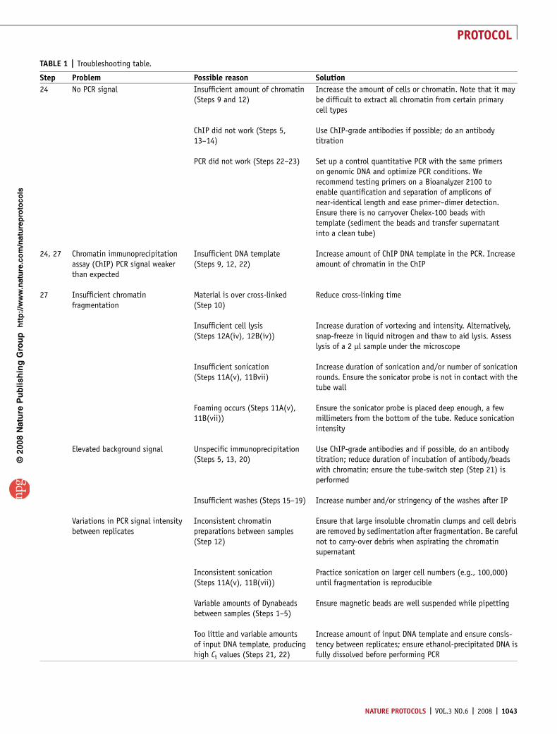
? TROUBLESHOOTINGTroubleshooting advice can be found in Table 1.
p
uor
G g
n ih si l
bu
P eru ta
N 800 2©
nat
ure
pro
toco
ls/
moc.er
ut an.
ww
w//:ptt
h
100a b
c
GAPDH No Ab
RNAP IINANOGOCT4
100,000-Cell ChIP
10,000 cells
1,000 cells
1,000-Cell ChIP
100-Cell ChIP
80
60
40
20
0
100
80
60
40
Pre
cipi
tatio
n re
lativ
e to
inpu
t (%
)
Pre
cipi
tatio
n re
lativ
e to
inpu
t (%
)P
reci
pita
tion
rela
tive
toin
put (
%)
20
0
100
80
60
40
No Ab
H3K9a
c
H3K4m
2
H3K9m
2
H3K9m
3
H3K27
m3
H3K4m
3No
Ab
H3K9a
c
H3K4m
2
H3K9m
2
RNAPII
20
0
100 GAPDH
NANOG
SLC10A6
OCT480
6050403020100
02468
101214
5
4
3
2
1
GAPDH OCT4 SLC10A6
GAPDH OCT4 SLC10A6
GAPDH OCT4 SLC10A6
100,000-Cell ChIP
10,000 Cells
1,000-Cell ChIP
1,000 Cells
100-Cell ChIP
0
60
40
20
0
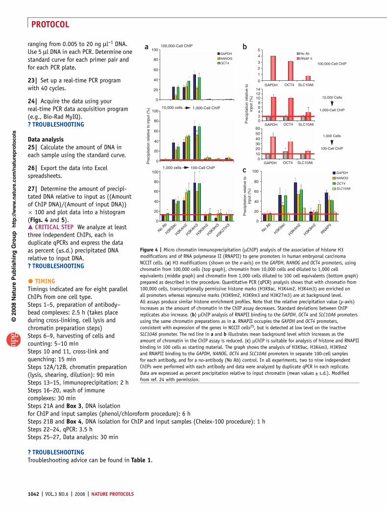
Figure 4 | Micro chromatin immunoprecipitation (mChIP) analysis of the association of histone H3
modifications and of RNA polymerase II (RNAPII) to gene promoters in human embryonal carcinoma
NCCIT cells. (a) H3 modifications (shown on the x-axis) on the GAPDH, NANOG and OCT4 promoters, using
chromatin from 100,000 cells (top graph), chromatin from 10,000 cells and diluted to 1,000 cell
equivalents (middle graph) and chromatin from 1,000 cells diluted to 100 cell equivalents (bottom graph)
prepared as described in the procedure. Quantitative PCR (qPCR) analysis shows that with chromatin from
100,000 cells, transcriptionally permissive histone marks (H3K9ac, H3K4m2, H3K4m3) are enriched on
all promoters whereas repressive marks (H3K9m2, H3K9m3 and H3K27m3) are at background level.
All assays produce similar histone enrichment profiles. Note that the relative precipitation value (y-axis)
increases as the amount of chromatin in the ChIP assay decreases. Standard deviations between ChIP
replicates also increase. (b) mChIP analysis of RNAPII binding to the GAPDH, OCT4 and SLC10A6 promoters
using the same chromatin preparations as in a. RNAPII occupies the GAPDH and OCT4 promoters,
consistent with expression of the genes in NCCIT cells25, but is detected at low level on the inactive
SLC10A6 promoter. The red line in a and b illustrates mean background level which increases as the
amount of chromatin in the ChIP assay is reduced. (c) mChIP is suitable for analysis of histone and RNAPII
binding in 100 cells as starting material. The graph shows the analysis of H3K9ac, H3K4m3, H3K9m2
and RNAPII binding to the GAPDH, NANOG, OCT4 and SLC10A6 promoters in separate 100-cell samples
for each antibody, and for a no-antibody (No Ab) control. In all experiments, two to nine independent
ChIPs were performed with each antibody and data were analyzed by duplicate qPCR in each replicate.
Data are expressed as percent precipitation relative to input chromatin (mean values ± s.d.). Modified
from ref. 24 with permission.
1042 | VOL.3 NO.6 | 2008 | NATURE PROTOCOLS
PROTOCOL
p
uor
G g
n ih si l
bu
P eru ta
N 800 2©
nat
ure
pro
toco
ls/
moc.er
ut an.
ww
w//:ptt
h
TABLE 1 | Troubleshooting table.
Step Problem Possible reason Solution
24 No PCR signal Insufficient amount of chromatin(Steps 9 and 12)
Increase the amount of cells or chromatin. Note that it maybe difficult to extract all chromatin from certain primarycell types
ChIP did not work (Steps 5,13–14)
Use ChIP-grade antibodies if possible; do an antibodytitration
PCR did not work (Steps 22–23) Set up a control quantitative PCR with the same primerson genomic DNA and optimize PCR conditions. Werecommend testing primers on a Bioanalyzer 2100 toenable quantification and separation of amplicons ofnear-identical length and ease primer–dimer detection.Ensure there is no carryover Chelex-100 beads withtemplate (sediment the beads and transfer supernatantinto a clean tube)
24, 27 Chromatin immunoprecipitationassay (ChIP) PCR signal weakerthan expected
Insufficient DNA template(Steps 9, 12, 22)
Increase amount of ChIP DNA template in the PCR. Increaseamount of chromatin in the ChIP
27 Insufficient chromatinfragmentation
Material is over cross-linked(Step 10)
Reduce cross-linking time
Insufficient cell lysis(Steps 12A(iv), 12B(iv))
Increase duration of vortexing and intensity. Alternatively,snap-freeze in liquid nitrogen and thaw to aid lysis. Assesslysis of a 2 ml sample under the microscope
Insufficient sonication(Steps 11A(v), 11Bvii)
Increase duration of sonication and/or number of sonicationrounds. Ensure the sonicator probe is not in contact with thetube wall
Foaming occurs (Steps 11A(v),11B(vii))
Ensure the sonicator probe is placed deep enough, a fewmillimeters from the bottom of the tube. Reduce sonicationintensity
Elevated background signal Unspecific immunoprecipitation(Steps 5, 13, 20)
Use ChIP-grade antibodies and if possible, do an antibodytitration; reduce duration of incubation of antibody/beadswith chromatin; ensure the tube-switch step (Step 21) isperformed
Insufficient washes (Steps 15–19) Increase number and/or stringency of the washes after IP
Variations in PCR signal intensitybetween replicates
Inconsistent chromatinpreparations between samples(Step 12)
Ensure that large insoluble chromatin clumps and cell debrisare removed by sedimentation after fragmentation. Be carefulnot to carry-over debris when aspirating the chromatinsupernatant
Inconsistent sonication(Steps 11A(v), 11B(vii))
Practice sonication on larger cell numbers (e.g., 100,000)until fragmentation is reproducible
Variable amounts of Dynabeadsbetween samples (Steps 1–5)
Ensure magnetic beads are well suspended while pipetting
Too little and variable amountsof input DNA template, producinghigh Ct values (Steps 21, 22)
Increase amount of input DNA template and ensure consis-tency between replicates; ensure ethanol-precipitated DNA isfully dissolved before performing PCR
NATURE PROTOCOLS | VOL.3 NO.6 | 2008 | 1043
PROTOCOL
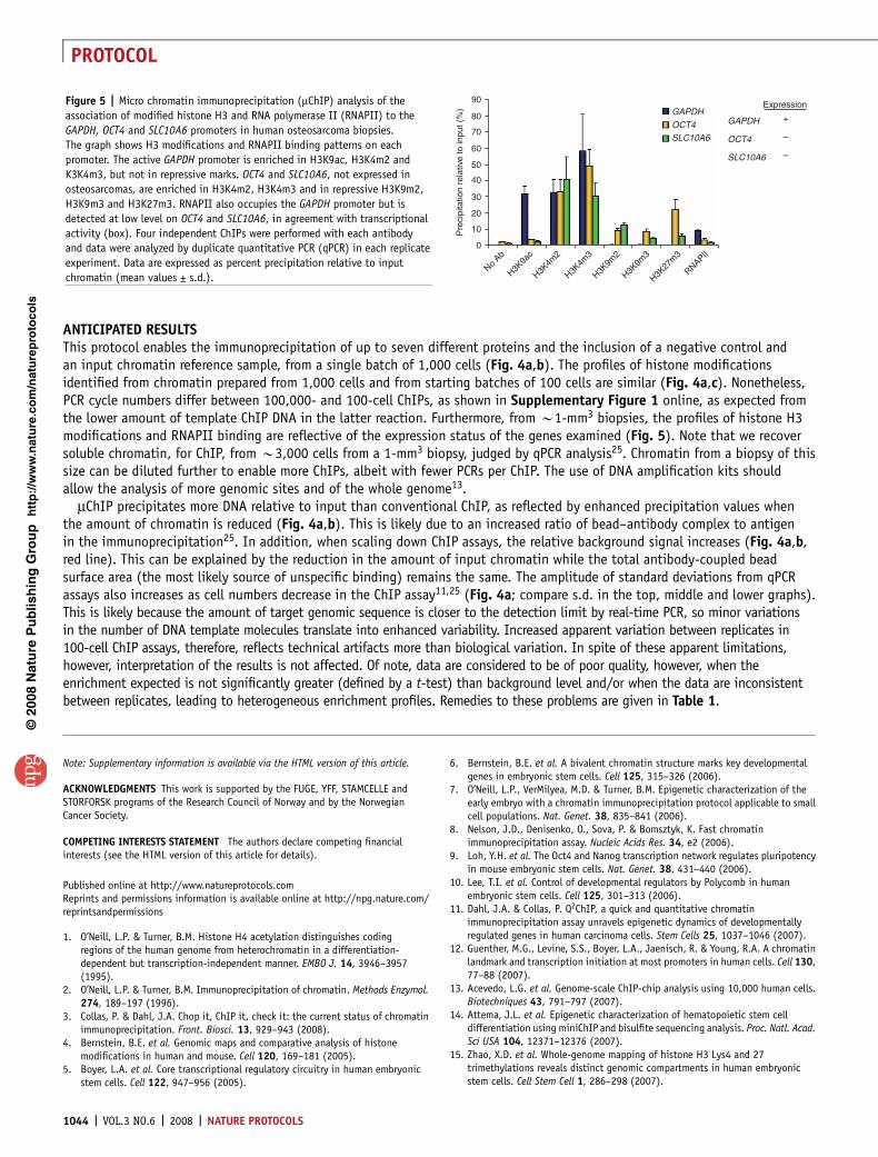
ANTICIPATED RESULTSThis protocol enables the immunoprecipitation of up to seven different proteins and the inclusion of a negative control andan input chromatin reference sample, from a single batch of 1,000 cells (Fig. 4a,b). The profiles of histone modificationsidentified from chromatin prepared from 1,000 cells and from starting batches of 100 cells are similar (Fig. 4a,c). Nonetheless,PCR cycle numbers differ between 100,000- and 100-cell ChIPs, as shown in Supplementary Figure 1 online, as expected fromthe lower amount of template ChIP DNA in the latter reaction. Furthermore, from B1-mm3 biopsies, the profiles of histone H3modifications and RNAPII binding are reflective of the expression status of the genes examined (Fig. 5). Note that we recoversoluble chromatin, for ChIP, from B3,000 cells from a 1-mm3 biopsy, judged by qPCR analysis25. Chromatin from a biopsy of thissize can be diluted further to enable more ChIPs, albeit with fewer PCRs per ChIP. The use of DNA amplification kits shouldallow the analysis of more genomic sites and of the whole genome13.
mChIP precipitates more DNA relative to input than conventional ChIP, as reflected by enhanced precipitation values whenthe amount of chromatin is reduced (Fig. 4a,b). This is likely due to an increased ratio of bead–antibody complex to antigenin the immunoprecipitation25. In addition, when scaling down ChIP assays, the relative background signal increases (Fig. 4a,b,red line). This can be explained by the reduction in the amount of input chromatin while the total antibody-coupled beadsurface area (the most likely source of unspecific binding) remains the same. The amplitude of standard deviations from qPCRassays also increases as cell numbers decrease in the ChIP assay11,25 (Fig. 4a; compare s.d. in the top, middle and lower graphs).This is likely because the amount of target genomic sequence is closer to the detection limit by real-time PCR, so minor variationsin the number of DNA template molecules translate into enhanced variability. Increased apparent variation between replicates in100-cell ChIP assays, therefore, reflects technical artifacts more than biological variation. In spite of these apparent limitations,however, interpretation of the results is not affected. Of note, data are considered to be of poor quality, however, when theenrichment expected is not significantly greater (defined by a t-test) than background level and/or when the data are inconsistentbetween replicates, leading to heterogeneous enrichment profiles. Remedies to these problems are given in Table 1.
Note: Supplementary information is available via the HTML version of this article.
ACKNOWLEDGMENTS This work is supported by the FUGE, YFF, STAMCELLE andSTORFORSK programs of the Research Council of Norway and by the NorwegianCancer Society.
COMPETING INTERESTS STATEMENT The authors declare competing financialinterests (see the HTML version of this article for details).
Published online at http://www.natureprotocols.comReprints and permissions information is available online at http://npg.nature.com/reprintsandpermissions
1. O’Neill, L.P. & Turner, B.M. Histone H4 acetylation distinguishes codingregions of the human genome from heterochromatin in a differentiation-dependent but transcription-independent manner. EMBO J. 14, 3946–3957(1995).
2. O’Neill, L.P. & Turner, B.M. Immunoprecipitation of chromatin. Methods Enzymol.274, 189–197 (1996).
3. Collas, P. & Dahl, J.A. Chop it, ChIP it, check it: the current status of chromatinimmunoprecipitation. Front. Biosci. 13, 929–943 (2008).
4. Bernstein, B.E. et al. Genomic maps and comparative analysis of histonemodifications in human and mouse. Cell 120, 169–181 (2005).
5. Boyer, L.A. et al. Core transcriptional regulatory circuitry in human embryonicstem cells. Cell 122, 947–956 (2005).
6. Bernstein, B.E. et al. A bivalent chromatin structure marks key developmentalgenes in embryonic stem cells. Cell 125, 315–326 (2006).
7. O’Neill, L.P., VerMilyea, M.D. & Turner, B.M. Epigenetic characterization of theearly embryo with a chromatin immunoprecipitation protocol applicable to smallcell populations. Nat. Genet. 38, 835–841 (2006).
8. Nelson, J.D., Denisenko, O., Sova, P. & Bomsztyk, K. Fast chromatinimmunoprecipitation assay. Nucleic Acids Res. 34, e2 (2006).
9. Loh, Y.H. et al. The Oct4 and Nanog transcription network regulates pluripotencyin mouse embryonic stem cells. Nat. Genet. 38, 431–440 (2006).
10. Lee, T.I. et al. Control of developmental regulators by Polycomb in humanembryonic stem cells. Cell 125, 301–313 (2006).
11. Dahl, J.A. & Collas, P. Q2ChIP, a quick and quantitative chromatinimmunoprecipitation assay unravels epigenetic dynamics of developmentallyregulated genes in human carcinoma cells. Stem Cells 25, 1037–1046 (2007).
12. Guenther, M.G., Levine, S.S., Boyer, L.A., Jaenisch, R. & Young, R.A. A chromatinlandmark and transcription initiation at most promoters in human cells. Cell 130,77–88 (2007).
13. Acevedo, L.G. et al. Genome-scale ChIP-chip analysis using 10,000 human cells.Biotechniques 43, 791–797 (2007).
14. Attema, J.L. et al. Epigenetic characterization of hematopoietic stem celldifferentiation using miniChIP and bisulfite sequencing analysis. Proc. Natl. Acad.Sci USA 104, 12371–12376 (2007).
15. Zhao, X.D. et al. Whole-genome mapping of histone H3 Lys4 and 27trimethylations reveals distinct genomic compartments in human embryonicstem cells. Cell Stem Cell 1, 286–298 (2007).
p
uor
G g
n ih si l
bu
P eru ta
N 800 2©
nat
ure
pro
toco
ls/
moc.er
ut an.
ww
w//:ptt
h
0
RNAPII
H3K9m
3
H3K9m
2
H3K4m
3
H3K4m
2
H3K9a
c
No Ab
10
20
30
40
50
60
70
Pre
cipi
tatio
n re
lativ
e to
inpu
t (%
)
80
SLC10A6
SLC10A6 OCT4
OCT4 GAPDHGAPDH
–
–
+
Expression90
H3K27
m3
Figure 5 | Micro chromatin immunoprecipitation (mChIP) analysis of the
association of modified histone H3 and RNA polymerase II (RNAPII) to the
GAPDH, OCT4 and SLC10A6 promoters in human osteosarcoma biopsies.
The graph shows H3 modifications and RNAPII binding patterns on each
promoter. The active GAPDH promoter is enriched in H3K9ac, H3K4m2 and
K3K4m3, but not in repressive marks. OCT4 and SLC10A6, not expressed in
osteosarcomas, are enriched in H3K4m2, H3K4m3 and in repressive H3K9m2,
H3K9m3 and H3K27m3. RNAPII also occupies the GAPDH promoter but is
detected at low level on OCT4 and SLC10A6, in agreement with transcriptional
activity (box). Four independent ChIPs were performed with each antibody
and data were analyzed by duplicate quantitative PCR (qPCR) in each replicate
experiment. Data are expressed as percent precipitation relative to input
chromatin (mean values ± s.d.).
1044 | VOL.3 NO.6 | 2008 | NATURE PROTOCOLS
PROTOCOL
16. O’Neill, L.P. & Turner, B.M. Immunoprecipitation of native chromatin: NChIP.Methods 31, 76–82 (2003).
17. Hudson, M.E. & Snyder, M. High-throughput methods of regulatory elementdiscovery. Biotechniques 41, 673 675, 677 passim (2006).
18. Dunn, J.J., McCorkle, S.R., Everett, L. & Anderson, C.W. Paired-end genomicsignature tags: a method for the functional analysis of genomes and epigenomes.Genet. Eng. (NY) 28, 159–173 (2007).
19. Aiba, K., Carter, M.G., Matoba, R. & Ko, M.S. Genomic approaches to earlyembryogenesis and stem cell biology. Semin. Reprod. Med. 24, 330–339 (2006).
20. Clark, D.J. & Shen, C.H. Mapping histone modifications by nucleosomeimmunoprecipitation. Methods Enzymol. 410, 416–430 (2006).
21. Negre, N., Lavrov, S., Hennetin, J., Bellis, M. & Cavalli, G. Mapping the distributionof chromatin proteins by ChIP on chip. Methods Enzymol. 410, 316–341 (2006).
22. Wu, J., Smith, L.T., Plass, C. & Huang, T.H. ChIP-chip comes of age for genome-wide functional analysis. Cancer Res. 66, 6899–6902 (2006).
23. Bulyk, M.L. DNA microarray technologies for measuring protein-DNA interactions.Curr. Opin. Biotechnol. 17, 422–430 (2006).
24. O’Geen, H., Nicolet, C.M., Blahnik, K., Green, R. & Farnham, P.J. Comparison of samplepreparation methods for ChIP-chip assays. Biotechniques 41, 577–580 (2006).
25. Dahl, J.A. & Collas, P. MicroChIP—a rapid micro chromatin immunoprecipitationassay for small cell samples and biopsies. Nucleic Acids Res. 36, e15 (2008).
26. Nelson, J.D., Denisenko, O. & Bomsztyk, K. Protocol for the fast chromatinimmunoprecipitation (ChIP) method. Nat. Protoc. 1, 179–185 (2006).
27. Flanagin, S., Nelson, J.D., Castner, D.G., Denisenko, O. & Bomsztyk, K. Microplate-based chromatin immunoprecipitation method, Matrix ChIP: a platform to studysignaling of complex genomic events. Nucleic Acids Res. 36, e17 (2008).
28. Mikkelsen, T.S. et al. Genome-wide maps of chromatin state in pluripotent andlineage-committed cells. Nature 448, 553–560 (2007).
29. Kiermer, V. Embryos and biopsies on the ChIP-ing forecast. Nat. Methods 3, 583(2006).
30. Lee, T.I., Johnstone, S.E. & Young, R.A. Chromatin immunoprecipitation andmicroarray-based analysis of protein location. Nat. Protoc. 1, 729–748 (2006).
31. Spencer, V.A., Sun, J.M., Li, L. & Davie, J.R. Chromatin immunoprecipitation: atool for studying histone acetylation and transcription factor binding. Methods31, 67–75 (2003).
32. Barski, A. et al. High-resolution profiling of histone methylations in the humangenome. Cell 129, 823–837 (2007).
33. Orlando, V. Mapping chromosomal proteins in vivo by formaldehyde-crosslinked-chromatin immunoprecipitation. Trends Biochem. Sci. 25, 99–104 (2000).
34. Zeng, P.Y., Vakoc, C.R., Chen, Z.C., Blobel, G.A. & Berger, S.L. In vivodual cross-linking for identification of indirect DNA-associated proteinsby chromatin immunoprecipitation. Biotechniques 41, 694 696, 698(2006).
35. Roh, T.Y., Cuddapah, S., Cui, K. & Zhao, K. The genomic landscape of histonemodifications in human T cells. Proc. Natl. Acad. Sci USA 103, 15782–15787(2006).
36. Hakelien, A.M., Landsverk, H.B., Robl, J.M., Skalhegg, B.S. & Collas, P.Reprogramming fibroblasts to express T-cell functions using cell extracts.Nat. Biotechnol. 20, 460–466 (2002).
37. Landsverk, H.B. et al. Reprogrammed gene expression in a somatic cell-freeextract. EMBO Rep. 3, 384–389 (2002).
38. Vandesompele, J., De, P.A. & Speleman, F. Elimination of primer-dimer artifactsand genomic coamplification using a two-step SYBR green I real-time RT-PCR.Anal. Biochem. 303, 95–98 (2002).
39. Ito, K. et al. Decreased histone deacetylase activity in chronic obstructivepulmonary disease. N. Engl. J. Med. 352, 1967–1976 (2005).
40. Pollicino, T. et al. Hepatitis B virus replication is regulated by the acetylationstatus of hepatitis B virus cccDNA-bound H3 and H4 histones. Gastroenterology130, 823–837 (2006).
41. Zuccato, C. et al. Widespread disruption of repressor element-1 silencingtranscription factor/neuron-restrictive silencer factor occupancy at its targetgenes in Huntington’s disease. J. Neurosci. 27, 6972–6983 (2007).
42. Ge, W. et al. Coupling of cell migration with neurogenesis by proneural bHLHfactors. Proc. Natl. Acad. Sci USA 103, 1319–1324 (2006).
43. Huang, Y., Doherty, J.J. & Dingledine, R. Altered histone acetylation at glutamatereceptor 2 and brain-derived neurotrophic factor genes is an early event triggeredby status epilepticus. J. Neurosci. 22, 8422–8428 (2002).
44. Le, T.N. et al. Dlx homeobox genes promote cortical interneuron migration fromthe basal forebrain by direct repression of the semaphorin receptor neuropilin-2.J. Biol. Chem. 282, 19071–19081 (2007).
p
uor
G g
n ih si l
bu
P eru ta
N 800 2©
nat
ure
pro
toco
ls/
moc.er
ut an.
ww
w//:ptt
h
NATURE PROTOCOLS | VOL.3 NO.6 | 2008 | 1045
PROTOCOL


![Research Paper JNK/AP1 Pathway Regulates MYC ...Chromatin immunoprecipitation assays (ChIP) ChIP analysis was performed as previously described [11]. Chromatin solutions were precipitated](https://img.pdfslide.net/doc/110x75/608625bcea8a6a2e9165f1fb/research-paper-jnkap1-pathway-regulates-myc-chromatin-immunoprecipitation-assays.jpg)