Embed Size (px)
Citation preview
257
Acta Neurologica Taiwanica Vol 20 No 4 December 2011
From the Department of Neurology, National NeuroscienceInstitute Singapore General Hospital campus, Outram Road,Singapore 169608.Received May 13, 2011. Revised June 21, 2011.Accepted December 5, 2011.
Correspondence to: Simon Kang-Seng Ting, MD. Departmentof Neurology, National Neuroscience Institute SingaporeGeneral Hospital campus, Outram Road, Singapore 169608.E-mail: [email protected]
A Rare Cause of Cerebellar Ataxia Syndrome: Superficial Siderosis of Central Nervous System
Simon Kang Seng Ting, MRCP, Kumar M Prakash, FRCP
INTRODUCTION
Superficial siderosis (SS) of the central nervous sys-tem (CNS) is a rare neurological condition and the etiol-ogy is still not clear. It results from chronic depositionof hemosiderin in parts of the CNS that are adjacent tothe cerebrospinal fluid (CSF). This in turn causes thebrownish discoloration of the leptomeninges and the
adjacent brain parenchyma. The pigmentation has apredilection for the superior vermis, crests of the cere-bellar folia, basal frontal lobe, temporal lobe cortex,brainstem, cranial nerves I and VIII as well as the spinalcord and nerve roots. It is postulated that SS is sec-ondary to recurrent subarachnoid hemorrhage (SAH)which induce intracellular uptake of iron and ultimatelyleading to destruction of the neural tissues. SS can also
Abstract-Purpose: To describe and emphasize importance of recognizing superficial siderosis (SS) of the central ner-
vous system (CNS) when assessing cerebellar ataxia syndromeCase Report: Superficial siderosis (SS) of the central nervous system (CNS) is a rare disorder that results
from chronic hemosiderin deposition in the subpial layers of the brain and the spinal cord. Althoughrecurrent bleeding in the subarachnoid space is the most likely explanation, a definite history of sub-arachnoid hemorrhage (SAH) is often lacking. Among the clinical presentations described in the litera-ture include sensorineural deafness, dementia, anosmia, pyramidal tract signs and cerebellar ataxia.However, due to its rarity, SS remains one of the least considered differential diagnosis in patients withsporadic ataxia syndrome. We describe a case of progressive gait imbalance that was initially misdiag-nosed for several years until a brain MRI study showed evidence of diffuse hemosiderin deposition sug-gestive of SS of CNS.
Conclusion: MR brain with gradient-echo T2-weightd images should be included in all MR studies carriedout to investigate the etiology of cerebellar ataxia to allow early diagnosis and prompt intervention forSS.
Key Words: cerebellar ataxia, superficial siderosis
Acta Neurol Taiwan 2011;20:257-261
occur as a late complication of neurosurgical procedures,or as a genetic disease caused by primary ceruloplasmindeficiency. The most common clinical presentation isslowly progressive bilateral hearing impairment, whichis often associated with gait imbalance. Less commonlyit has also been reported to cause anosmia, cognitiveimpairment, seizures, pyramidal tract signs, as well assensory and autonomic symptoms(1).
We report a 60-year-old man who had gradual onsetof severe sensorineural deafness and gait ataxia for 6years who was eventually diagnosed with SS of CNS.
CASE REPORT
A 60-year-old Indonesian gentleman presented witha 6-year history of gradual progressive bilateral deaf-ness. One year after the onset of hearing loss, he startedexperiencing unsteadiness of gait. These symptoms pro-gressed over the years and resulted in frequent falls. Hedenied history of loss of smell, chronic recurrentheadaches, head trauma, head surgery or excessive alco-hol consumption. He had no positive family history ofsimilar hearing or gait disturbances. He had sought med-ical opinion in many hospitals in different parts of the
world and series of neuroimaging as well as laboratorystudies were conducted. He was told to have a signifi-cant amount of cerebellar atrophy but no definite causewas found.
Neurological examination showed evidence ofdysarthria with scanning speech, bilateral sensorineuraldeafness, dysmetria, abnormal heel shin test and broad-based ataxic gait. The extraocular eye movements werenormal and there was no clinical involvement of thepyramidal tracts, extrapyramidal or autonomic system.Routine laboratory investigations as well as serum ceru-loplasmin (232mg/l) were within normal limits.
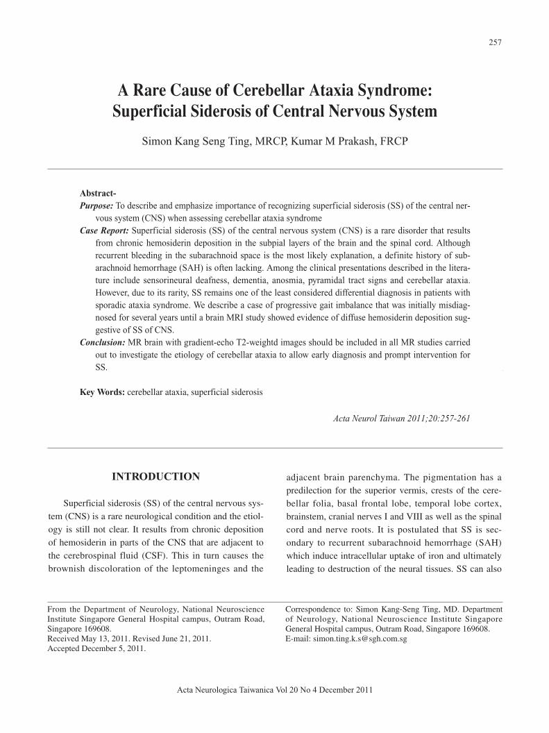
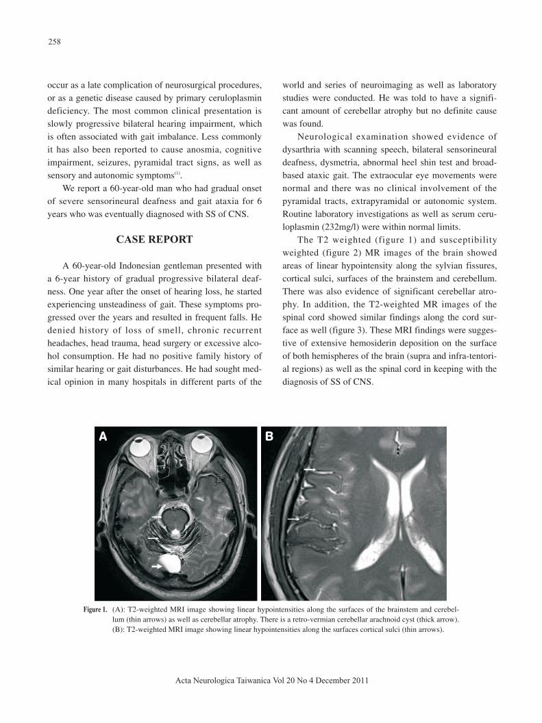
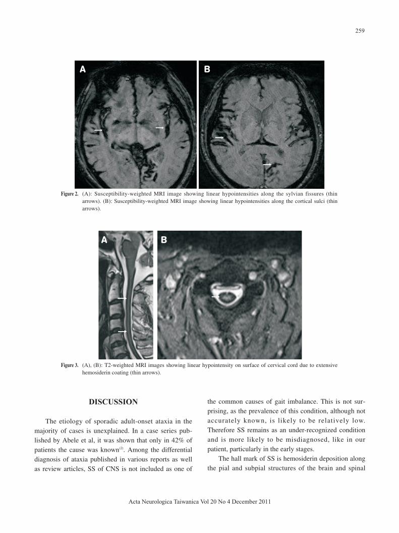
The T2 weighted (figure 1) and susceptibilityweighted (figure 2) MR images of the brain showedareas of linear hypointensity along the sylvian fissures,cortical sulci, surfaces of the brainstem and cerebellum.There was also evidence of significant cerebellar atro-phy. In addition, the T2-weighted MR images of thespinal cord showed similar findings along the cord sur-face as well (figure 3). These MRI findings were sugges-tive of extensive hemosiderin deposition on the surfaceof both hemispheres of the brain (supra and infra-tentori-al regions) as well as the spinal cord in keeping with thediagnosis of SS of CNS.
258
Acta Neurologica Taiwanica Vol 20 No 4 December 2011
Figure 1. (A): T2-weighted MRI image showing linear hypointensities along the surfaces of the brainstem and cerebel-lum (thin arrows) as well as cerebellar atrophy. There is a retro-vermian cerebellar arachnoid cyst (thick arrow).(B): T2-weighted MRI image showing linear hypointensities along the surfaces cortical sulci (thin arrows).
A B
DISCUSSION
The etiology of sporadic adult-onset ataxia in themajority of cases is unexplained. In a case series pub-lished by Abele et al, it was shown that only in 42% ofpatients the cause was known(2). Among the differentialdiagnosis of ataxia published in various reports as wellas review articles, SS of CNS is not included as one of
the common causes of gait imbalance. This is not sur-prising, as the prevalence of this condition, although notaccurately known, is likely to be relatively low.Therefore SS remains as an under-recognized conditionand is more likely to be misdiagnosed, like in ourpatient, particularly in the early stages.
The hall mark of SS is hemosiderin deposition alongthe pial and subpial structures of the brain and spinal
259
Acta Neurologica Taiwanica Vol 20 No 4 December 2011
A B
Figure 3. (A), (B): T2-weighted MRI images showing linear hypointensity on surface of cervical cord due to extensivehemosiderin coating (thin arrows).
A B
Figure 2. (A): Susceptibility-weighted MRI image showing linear hypointensities along the sylvian fissures (thinarrows). (B): Susceptibility-weighted MRI image showing linear hypointensities along the cortical sulci (thinarrows).
cord, as a result of recurrent bleeding in the subarach-noid space, resulting in damage to the cerebellar cortex,cochlear nerves, cerebral cortex, and spinal cord. Theclinical presentation of SS closely mimics a degenerativecerebellar disorder and usually is associated with bilater-al sensorineural hearing loss. Other manifestations mayinclude and pyramidal tract signs, dementia, bladderincontinence, anosmia, anisocoria, and sensory deficits.Our patient also complained of progressive deafness andgait ataxia for 6 years. However, despite extensive inves-tigations including neuroimaging studies in multiplemedical institutions, no etiological cause was establisheduntil recently.
MR imaging (particularly T2-weighted imaging) isthe investigation of choice for the diagnosis of SS. Thecharacteristic linear hypointensity seen on T2-weightedin vivo MR imaging correlated with the hemosiderindeposition around the surface of the central nervous sys-tem seen at postmortem. However, in the early stages ofSS, the findings may be subtle and the T2 hypointensityfollowing the contours of the brain and spinal cord maybe easily missed. Gradient-echo T2-weighted imageshave a higher sensitivity for hemosiderin deposition. Themagnetic susceptibility effects of blood-degradationproducts such as ferritin and hemosiderin are also morepronounced at higher field strengths.
T2-weighted MR imaging typically shows a rim ofhypointensity around the cerebellum and brain stem. Thesuperior vermis, quadrigeminal plate, and basal cerebralsurface are preferentially affected. Cerebellar atrophy isoften present, and the superior vermis and anterior cere-bellar hemispheres may be preferentially involved by theatrophy. The ability of the brain to biosynthesize ferritinin response to prolonged contact with hemoglobin iron isimportant in the pathogenesis of superficial siderosis.Accelerated ferritin synthesis in the Bergmann glia ofthe cerebellum may account for the preferential cerebel-lar involvement(4). The linear marginal T2 hypointensitymay also involve the Sylvian fissure, interhemisphericfissure, and cortical sulci(3). These MR changes wereseen in our patient.
The pathologic changes of SS are characterizedwell(4). Macroscopically, there is dark brown discol-
oration of the leptomeninges and superficial CNSparenchyma as well as the subependymal lining through-out the neuroaxis. Microscopically, there is extensivehemosiderin deposition in the leptomeninges, subpialand subependymal regions. The leptomeninges are thick-ened, and there are varying degrees of neuronal loss,reactive gliosis, and demyelination. The superficial foliaof the cerebellum almost always are involved with lossof Purkinje cells and Bergmann gliosis. In addition, cra-nial nerve VIII and, to a lesser extent, cranial nerves Iexhibit dense accumulation of hemosiderin, that is oftenassociated with demyelination and atrophy. The exactreason for the differential cranial nerve involvement isnot known. However Fearnley et al has suggested thatthe differential surface contact of the cranial nerves withthe CSF hemosiderin may account for this(5).
An important follow on step, after diagnosing SS,would be to identify and correct the potential source ofrecurrent subarachnoid hemorrhage in order to arrest theclinical deterioration. In the absence of an intracranialabnormality, further evaluation should include MRimaging of the spine. If no source is identified, furtherinvestigation with catheter angiography may be warrant-ed to find the potential bleeders. However, in somecases, the source of bleeding remains obscure even withextensive investigations.
SS is known to be associated with a spectrum oflesions, including CSF cavity lesions (meningoceles,pseudomeningoceles, pseudoencephaloceles, cavityremaining after a hemispherectomy), trauma (such ascervical nerve root avulsions), neoplasms (ependymo-mas, oligodendrogliomas, and astrocytomas), and vascu-lar abnormalities (arteriovenous malformations,aneurysms, and fragile capillary regrowth after brainsurgery)(5,6). In our patient a relatively large retrovermianarachnoid cyst was detected on MR causing mild masseffect on the adjacent right cerebellar hemisphere.Surgical excision of the cyst may result in symptomimprovement, as highlighted in several case reports(7,8).
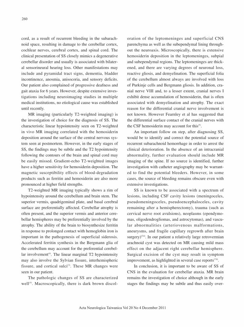
In conclusion, it is important to be aware of SS ofCNS in the evaluation for cerebellar ataxia. MR brainremains the investigation of choice although in the earlystages the findings may be subtle and thus easily over-
260
Acta Neurologica Taiwanica Vol 20 No 4 December 2011
looked. Gradient-echo T2-weightd images should beincluded in all MR studies carried out to investigate theetiology of cerebellar ataxia. This may spare patientsincurring other expensive investigations and allow earlydiagnosis and prompt intervention.
REFERENCE
1. Kumar N. Superficial siderosis: associations and therapeu-
tic implications. Arch Neurol 2007;64:491-496.
2. Abele M, Burk K et al. The aetiology of sporadic adult-
onset ataxia. Brain 2002;125:961-968.
3. Kumar N. Neuroimaging in superficial siderosis: an in-
depth look. Am J Neuroradiol 2010;31:5-14.
4. Koeppen AH, Dentinger MP. Brain hemosiderin and super-
ficial siderosis of the central nervous system. J Neuropathol
Exp Neurol 1988;47:249-270.
5. Fearnley JM, Stevens JM, Rudge P. Superficial siderosis of
the central nervous system. Brain 1995;118:1051-1066.
6. Bürk K, Skaley M, Dichgans J. High prevalence of CSF-
containing cysts in superficial hemosiderosis of the central
nervous system. J Neurol 2001;248:1005-1007.
7. Kumar N, Cohen-Gadol AA, Wright RA, et al. Superficial
siderosis. Neurology 2006;66:1144-1152.
8. Kumar N, Bledsoe JM, Davis DH. Intracranial fluid filled
collection and superficial siderosis. J Neurol Neurosurg
Psychiatry 2007;78:652-653.
261
Acta Neurologica Taiwanica Vol 20 No 4 December 2011