Embed Size (px)
Citation preview
| Journal of Clinical and Analytical Medicine1
Izole Nazofarengeal Lenfanjiom
A Rare Cause of Snoring: Isolated Nasopharyngeal Lymphangioma
Horlamanın Nadir Bir Nedeni; İzole Nazofarengeal Lenfajiom
DOI: 10.4328/JCAM.4975 Received: 13.03.2017 Accepted: 01.05.2017 Printed: 01.06.2017 J Clin Anal Med 2017;8(suppl 3): 192-5Corresponding Author: Hulya EYIGOR, Department of ENT, Antalya Research and Education Hospital, 07100, Muratpaşa, Antalya, Turkey.GSM: +905372169753 F.: +90 2422494462 E-Mail: [email protected]
Özet
Lenfanjiomalar, lenfatik sistemin nadir konjenital tümörleridir. En sık çocukluk ça-
ğında görülmekte ve sıklıkla yerleşim yeri baş ve boyun bölgesidir. Nazofarenksin
izole lenfanjioma tululumu oldukça nadir olup literatürde bildirilmiş bir kaç vaka
bulunmaktadır. Bizde bu çalışmamızda horlama ve sağ kulakta tıkanıklık yakınma-
sıyla polikliniğimize başvuran ve izole nazofarengeal lenfanjioma tanısı alan 40
yaşında bayan hastayı literatür bilgileriyle sunduk.
Anahtar Kelimeler
Lenfanjioma; Nazofarenks; Horlama; CD31; CD34
Abstract
Lymphangiomas are rare congenital tumors of the lymphatic system. They are
most often seen in childhood and frequently are located in the head and neck
region. Nasopharynx involvement of an isolated lymphangioma is extremely rare,
with very few cases reported in the literature. Together with the relevant infor-
mation in the literature, we present here a case of a 40-year-old female who
presented at our polyclinic with complaints of snoring and obstruction in the right
ear and was diagnosed with isolated nasopharyngeal lymphangioma.
Keywords
Lymphangioma; Nasopharynx; Snoring; CD31; CD34
Hulya Eyigor1, Oguzhan İlden1, Dinc Suren2, Dondu Nergis2, Levent Renda1
1Department of ENT Head and Neck Surgery, 2Department of Pathology,Antalya Education and Research Hospital, Antalya, Turkey
38. Türk Ulusal Kulak Burun Boğaz ve Baş Boyun Cerrahisi Kongresi 2016’da Poster olarak sunulmuştur.
I Journal of Clinical and Analytical Medicine192
| Journal of Clinical and Analytical Medicine
Izole Nazofarengeal Lenfanjiom
2
IntroductionLymphangiomas are rarely seen congenital tumors of the lym-phatic system. They are most frequently seen in childhood and generally diagnosed in infancy; approximately 90% of cases are seen by the age of 2 years [1]. According to the size of the cavi-ty, lymphangioma can be classified as microcytic, macrocytic, or cystic hygroma. These tumors are seen most often in the head and neck region and more rarely may be observed in the axilla and abdomen. As isolated nasopharyngeal lymphangiomas are extremely rare, few cases have been reported in the literature since the first case in 1966 [1,2]. In the light of information in literature, we present the case of a 40-year-old patient diagnosed with isolated nasopharyngeal lymphangioma, who underwent surgery and had no complaints during a 1-year follow-up period.
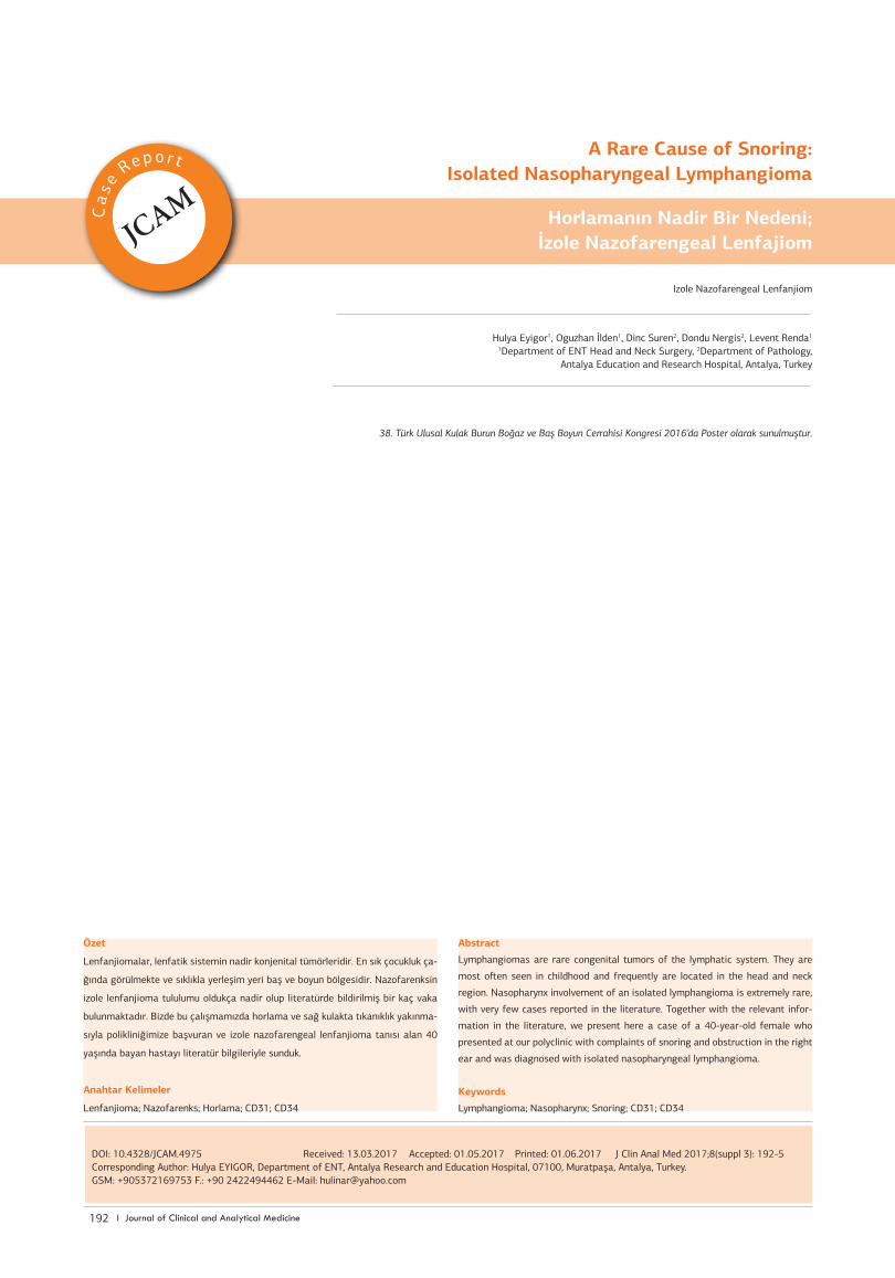
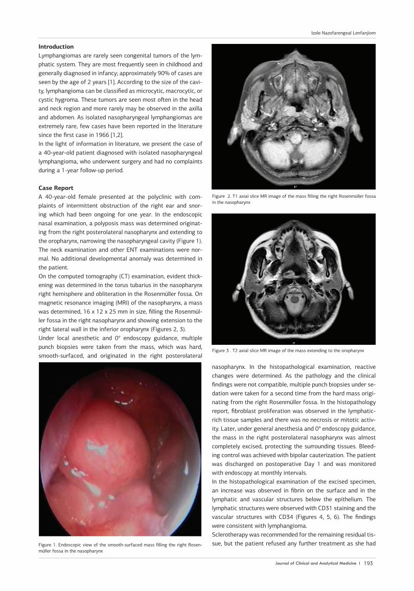
Case ReportA 40-year-old female presented at the polyclinic with com-plaints of intermittent obstruction of the right ear and snor-ing which had been ongoing for one year. In the endoscopic nasal examination, a polyposis mass was determined originat-ing from the right posterolateral nasopharynx and extending to the oropharynx, narrowing the nasopharyngeal cavity (Figure 1). The neck examination and other ENT examinations were nor-mal. No additional developmental anomaly was determined in the patient. On the computed tomography (CT) examination, evident thick-ening was determined in the torus tubarius in the nasopharynx right hemisphere and obliteration in the Rosenmüller fossa. On magnetic resonance imaging (MRI) of the nasopharynx, a mass was determined, 16 x 12 x 25 mm in size, filling the Rosenmül-ler fossa in the right nasopharynx and showing extension to the right lateral wall in the inferior oropharynx (Figures 2, 3). Under local anesthetic and 0° endoscopy guidance, multiple punch biopsies were taken from the mass, which was hard, smooth-surfaced, and originated in the right posterolateral
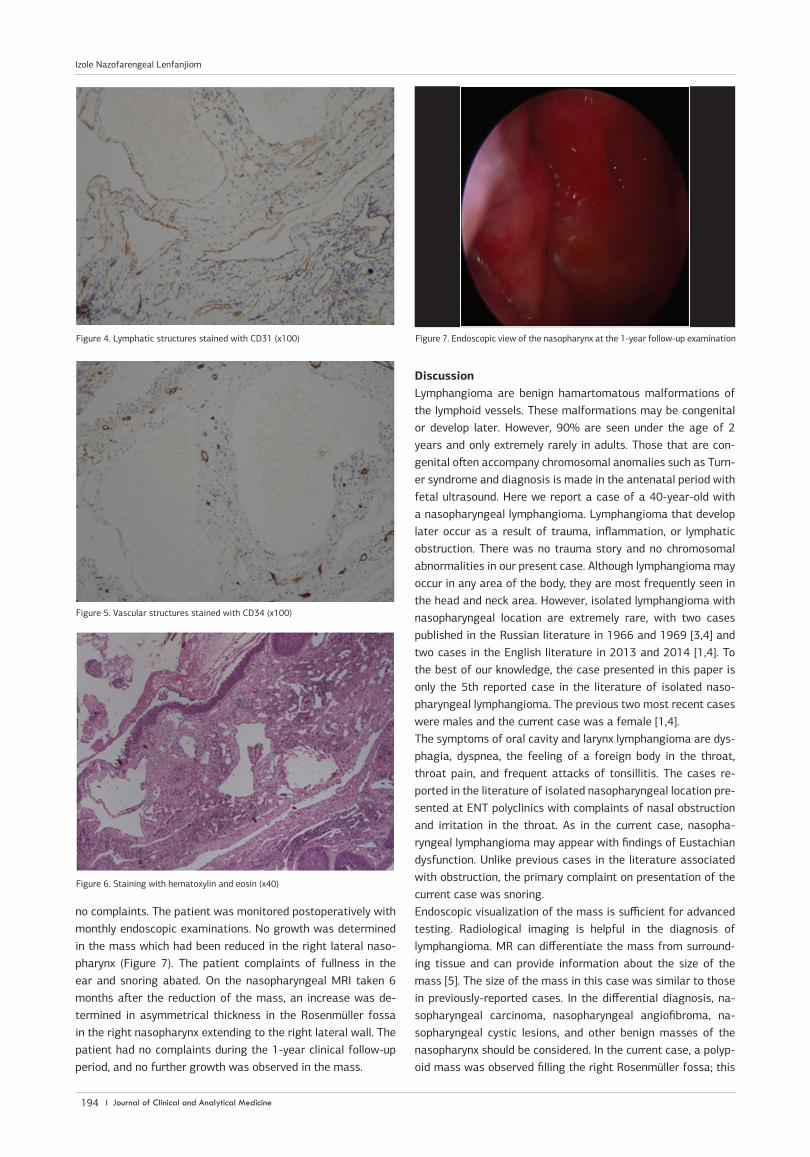
nasopharynx. In the histopathological examination, reactive changes were determined. As the pathology and the clinical findings were not compatible, multiple punch biopsies under se-dation were taken for a second time from the hard mass origi-nating from the right Rosenmüller fossa. In the histopathology report, fibroblast proliferation was observed in the lymphatic-rich tissue samples and there was no necrosis or mitotic activ-ity. Later, under general anesthesia and 0° endoscopy guidance, the mass in the right posterolateral nasopharynx was almost completely excised, protecting the surrounding tissues. Bleed-ing control was achieved with bipolar cauterization. The patient was discharged on postoperative Day 1 and was monitored with endoscopy at monthly intervals. In the histopathological examination of the excised specimen, an increase was observed in fibrin on the surface and in the lymphatic and vascular structures below the epithelium. The lymphatic structures were observed with CD31 staining and the vascular structures with CD34 (Figures 4, 5, 6). The findings were consistent with lymphangioma. Sclerotherapy was recommended for the remaining residual tis-sue, but the patient refused any further treatment as she had Figure 1. Endoscopic view of the smooth-surfaced mass filling the right Rosen-
müller fossa in the nasopharynx
Figure 2. T1 axial slice MR image of the mass filling the right Rosenmüller fossa in the nasopharynx
Figure 3 . T2 axial slice MR image of the mass extending to the oropharynx
Journal of Clinical and Analytical Medicine I 193
Izole Nazofarengeal Lenfanjiom
| Journal of Clinical and Analytical Medicine
Izole Nazofarengeal Lenfanjiom
3
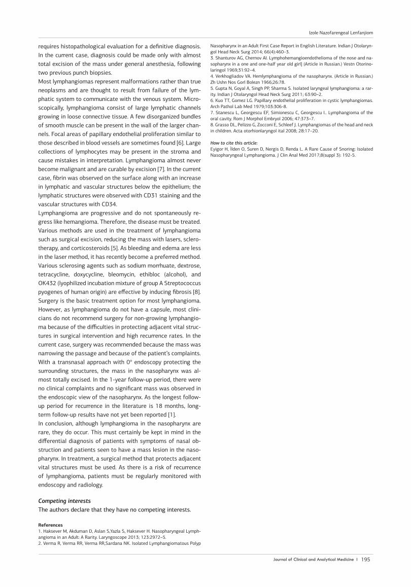
no complaints. The patient was monitored postoperatively with monthly endoscopic examinations. No growth was determined in the mass which had been reduced in the right lateral naso-pharynx (Figure 7). The patient complaints of fullness in the ear and snoring abated. On the nasopharyngeal MRI taken 6 months after the reduction of the mass, an increase was de-termined in asymmetrical thickness in the Rosenmüller fossa in the right nasopharynx extending to the right lateral wall. The patient had no complaints during the 1-year clinical follow-up period, and no further growth was observed in the mass.
DiscussionLymphangioma are benign hamartomatous malformations of the lymphoid vessels. These malformations may be congenital or develop later. However, 90% are seen under the age of 2 years and only extremely rarely in adults. Those that are con-genital often accompany chromosomal anomalies such as Turn-er syndrome and diagnosis is made in the antenatal period with fetal ultrasound. Here we report a case of a 40-year-old with a nasopharyngeal lymphangioma. Lymphangioma that develop later occur as a result of trauma, inflammation, or lymphatic obstruction. There was no trauma story and no chromosomal abnormalities in our present case. Although lymphangioma may occur in any area of the body, they are most frequently seen in the head and neck area. However, isolated lymphangioma with nasopharyngeal location are extremely rare, with two cases published in the Russian literature in 1966 and 1969 [3,4] and two cases in the English literature in 2013 and 2014 [1,4]. To the best of our knowledge, the case presented in this paper is only the 5th reported case in the literature of isolated naso-pharyngeal lymphangioma. The previous two most recent cases were males and the current case was a female [1,4]. The symptoms of oral cavity and larynx lymphangioma are dys-phagia, dyspnea, the feeling of a foreign body in the throat, throat pain, and frequent attacks of tonsillitis. The cases re-ported in the literature of isolated nasopharyngeal location pre-sented at ENT polyclinics with complaints of nasal obstruction and irritation in the throat. As in the current case, nasopha-ryngeal lymphangioma may appear with findings of Eustachian dysfunction. Unlike previous cases in the literature associated with obstruction, the primary complaint on presentation of the current case was snoring. Endoscopic visualization of the mass is sufficient for advanced testing. Radiological imaging is helpful in the diagnosis of lymphangioma. MR can differentiate the mass from surround-ing tissue and can provide information about the size of the mass [5]. The size of the mass in this case was similar to those in previously-reported cases. In the differential diagnosis, na-sopharyngeal carcinoma, nasopharyngeal angiofibroma, na-sopharyngeal cystic lesions, and other benign masses of the nasopharynx should be considered. In the current case, a polyp-oid mass was observed filling the right Rosenmüller fossa; this
Figure 4. Lymphatic structures stained with CD31 (x100) Figure 7. Endoscopic view of the nasopharynx at the 1-year follow-up examination
Figure 5. Vascular structures stained with CD34 (x100)
Figure 6. Staining with hematoxylin and eosin (x40)
I Journal of Clinical and Analytical Medicine194
Izole Nazofarengeal Lenfanjiom
| Journal of Clinical and Analytical Medicine
Izole Nazofarengeal Lenfanjiom
4
requires histopathological evaluation for a definitive diagnosis. In the current case, diagnosis could be made only with almost total excision of the mass under general anesthesia, following two previous punch biopsies. Most lymphangiomas represent malformations rather than true neoplasms and are thought to result from failure of the lym-phatic system to communicate with the venous system. Micro-scopically, lymphangioma consist of large lymphatic channels growing in loose connective tissue. A few disorganized bundles of smooth muscle can be present in the wall of the larger chan-nels. Focal areas of papillary endothelial proliferation similar to those described in blood vessels are sometimes found [6]. Large collections of lymphocytes may be present in the stroma and cause mistakes in interpretation. Lymphangioma almost never become malignant and are curable by excision [7]. In the current case, fibrin was observed on the surface along with an increase in lymphatic and vascular structures below the epithelium; the lymphatic structures were observed with CD31 staining and the vascular structures with CD34. Lymphangioma are progressive and do not spontaneously re-gress like hemangioma. Therefore, the disease must be treated. Various methods are used in the treatment of lymphangioma such as surgical excision, reducing the mass with lasers, sclero-therapy, and corticosteroids [5]. As bleeding and edema are less in the laser method, it has recently become a preferred method. Various sclerosing agents such as sodium morrhuate, dextrose, tetracycline, doxycycline, bleomycin, ethibloc (alcohol), and OK432 (lyophilized incubation mixture of group A Streptococcus pyogenes of human origin) are effective by inducing fibrosis [8]. Surgery is the basic treatment option for most lymphangioma. However, as lymphangioma do not have a capsule, most clini-cians do not recommend surgery for non-growing lymphangio-ma because of the difficulties in protecting adjacent vital struc-tures in surgical intervention and high recurrence rates. In the current case, surgery was recommended because the mass was narrowing the passage and because of the patient’s complaints. With a transnasal approach with 0° endoscopy protecting the surrounding structures, the mass in the nasopharynx was al-most totally excised. In the 1-year follow-up period, there were no clinical complaints and no significant mass was observed in the endoscopic view of the nasopharynx. As the longest follow-up period for recurrence in the literature is 18 months, long-term follow-up results have not yet been reported [1]. In conclusion, although lymphangioma in the nasopharynx are rare, they do occur. This must certainly be kept in mind in the differential diagnosis of patients with symptoms of nasal ob-struction and patients seen to have a mass lesion in the naso-pharynx. In treatment, a surgical method that protects adjacent vital structures must be used. As there is a risk of recurrence of lymphangioma, patients must be regularly monitored with endoscopy and radiology.
Competing interestsThe authors declare that they have no competing interests.
References1. Haksever M, Akduman D, Aslan S,Yazla S, Haksever H. Nasopharyngeal Lymph-angioma in an Adult: A Rarity. Laryngoscope 2013; 123:2972–5.2. Verma R, Verma RR, Verma RR,Sardana NK. Isolated Lymphangiomatous Polyp
Nasopharynx in an Adult First Case Report in English Literature. Indian J Otolaryn-gol Head Neck Surg 2014; 66(4):460–3.3. Shanturov AG, Chernov AI. Lymphohemangioendothelioma of the nose and na-sopharynx in a one and one-half year old girl] (Article in Russian.) Vestn Otorino-laringol 1969;31:92–4.4. Verkhogliadov VA. Hemlymphangioma of the nasopharynx. (Article in Russian.) Zh Ushn Nos Gorl Bolezn 1966;26:78.5. Gupta N, Goyal A, Singh PP, Sharma S. Isolated laryngeal lymphangioma: a rar-ity. Indian J Otolaryngol Head Neck Surg 2011; 63:90–2.6. Kuo TT, Gomez LG. Papillary endothelial proliferation in cystic lymphangiomas. Arch Pathol Lab Med 1979;103:306-8.7. Stanescu L, Georgescu EF, Simionescu C, Georgescu I.. Lymphangioma of the oral cavity. Rom J Morphol Embryol 2006; 47:373–7.8. Grasso DL, Pelizzo G, Zocconi E, Schleef J. Lymphangiomas of the head and neck in children. Acta otorhionlaryngol ital 2008; 28:17–20.
How to cite this article:Eyigor H, İlden O, Suren D, Nergis D, Renda L. A Rare Cause of Snoring: Isolated Nasopharyngeal Lymphangioma. J Clin Anal Med 2017;8(suppl 3): 192-5.
Journal of Clinical and Analytical Medicine I 195
Izole Nazofarengeal Lenfanjiom