Embed Size (px)
Citation preview
International Journal of Pharmaceutical Erudition
www.pharmaerudition.org May2014 , 4 (1), 39-45 39 | P a g e
ISSN 2249-3875
Review Article
A Review Article on Ectodermal Dysplasia
Bhadauria R. S., Ranjana Sharma, Gayatri Prajapat*
Shrinathji Institute of Pharmacy, Nathdwara
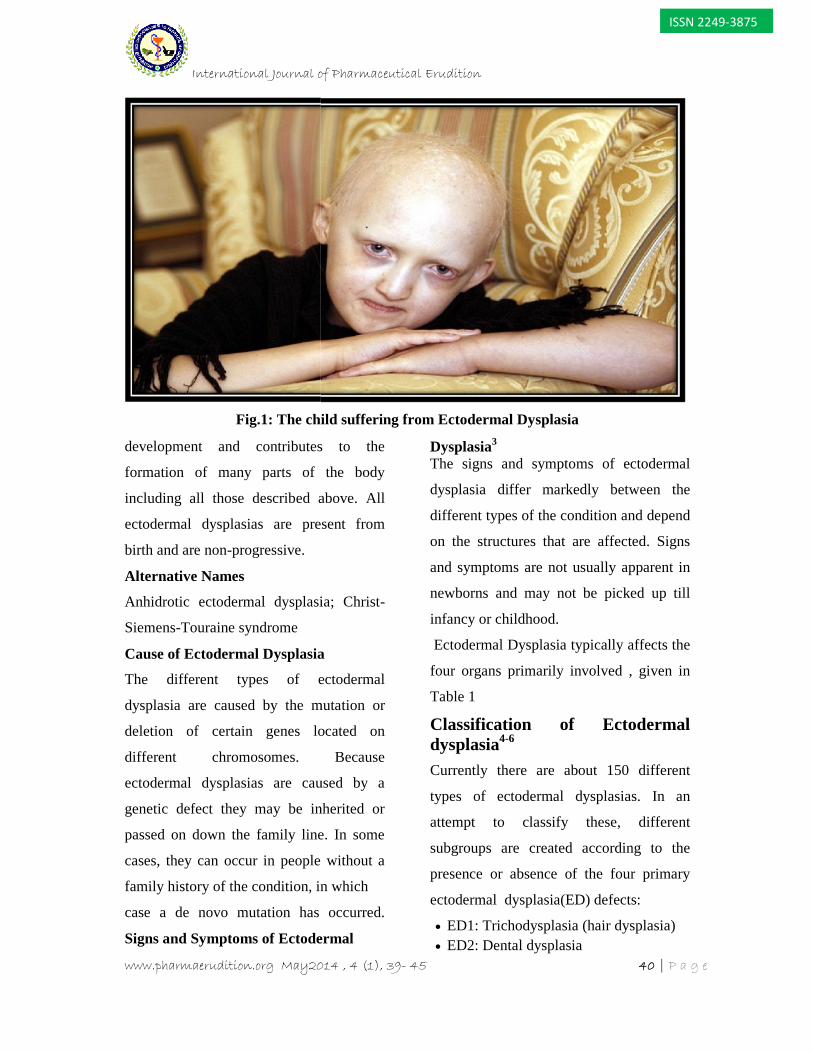
Ectodermal dysplasia is a large group of inherited disorders characterized by a primary defectin hair, teeth, nails or sweat gland function, in addition to another abnormality in a tissue ofectodermal origin, e.g. ears, eyes, lips, mucous membranes of the mouth or nose, centralnervous system. The different types of ectodermal dysplasia are caused by the mutation ordeletion of certain genes located on different chromosomes. The signs and symptoms ofectodermal dysplasia differ markedly between the different types of the condition and dependon the structures that are affected. Currently there are about 150 different types of ectodermaldysplasias. The most common ectodermal dysplasias are Hypohidrotic (anhidrotic) ED andHidrotic ED. Ectodermal dysplasia results from the abnormal morphogenesis of cutaneous ororal embryonal ectoderm (ie, hair, nails, teeth, eccrine glands). In some forms, mesodermalabnormalities are also present. The frequency of the different ectodermal dysplasias in agiven population is highly variable. Collectively, the prevalence of ectodermal dysplasia isestimated at 7 cases per 10,000 births. There is no specific treatment for EctodermalDysplasia, only disease management are available.
Key words: Renal ultrasonography, voiding cystourethrography, Genetic testing, Cloustonsyndrome, palmoplantar hyperkeratosis, Christ-Siemens-Touraine (CST) syndrome.
INTRODUCTION
Before a developing baby is large enough
to be seen, a layer of cells covers the
outside of the body. This surface layer of
cells is called the ectoderm, and from it
develops the skin, hair, nails, teeth, nerve
cells, sweat glands, parts of the eye and
ear, and parts of some other organs. Each
of the listed parts of the body is then called
an ectodermal structure. Dysplasia means
Abnormal development or growth of
tissues, organs, or cells.
The term “Ectodermal Dysplasia” was first
introduced in 1929 to describe a number of
*Address for [email protected]
conditions that are present at or shortly
after birth in which two or more of the
body’s ectodermal structures (e.g hair,
teeth, nails, sweat glands) fail to develop
or grow properly (dysplasia).
Ectodermal dysplasia is a large group of
inherited disorders characterized by a
primary defect in hair, teeth, nails or sweat
gland function, in addition to another
abnormality in a tissue of ectodermal
origin, e.g. ears, eyes, lips, mucous
membranes of the mouth or nose, central
nervous system. The ectoderm is the
outermost layer of cells in embryonic
International Journal of Pharmaceutical Erudition
www.pharmaerudition.org May2014 , 4 (1), 39- 45 40 | P a g e
ISSN 2249-3875
Fig.1: The child suffering from Ectodermal Dysplasia
development and contributes to the
formation of many parts of the body
including all those described above. All
ectodermal dysplasias are present from
birth and are non-progressive.
Alternative Names
Anhidrotic ectodermal dysplasia; Christ-
Siemens-Touraine syndrome
Cause of Ectodermal Dysplasia
The different types of ectodermal
dysplasia are caused by the mutation or
deletion of certain genes located on
different chromosomes. Because
ectodermal dysplasias are caused by a
genetic defect they may be inherited or
passed on down the family line. In some
cases, they can occur in people without a
family history of the condition, in which
case a de novo mutation has occurred.
Signs and Symptoms of Ectodermal
Dysplasia3
The signs and symptoms of ectodermal
dysplasia differ markedly between the
different types of the condition and depend
on the structures that are affected. Signs
and symptoms are not usually apparent in
newborns and may not be picked up till
infancy or childhood.
Ectodermal Dysplasia typically affects the
four organs primarily involved , given in
Table 1
Classification of Ectodermaldysplasia4-6
Currently there are about 150 different
types of ectodermal dysplasias. In an
attempt to classify these, different
subgroups are created according to the
presence or absence of the four primary
ectodermal dysplasia(ED) defects:
ED1: Trichodysplasia (hair dysplasia) ED2: Dental dysplasia
International Journal of Pharmaceutical Erudition
www.pharmaerudition.org May2014 , 4 (1), 39- 45 41 | P a g e
ISSN 2249-3875
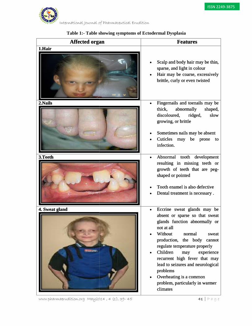
Table 1:- Table showing symptoms of Ectodermal Dysplasia
Affected organ Features1.Hair
Scalp and body hair may be thin,sparse, and light in colour
Hair may be coarse, excessivelybrittle, curly or even twisted
2.Nails Fingernails and toenails may bethick, abnormally shaped,discoloured, ridged, slowgrowing, or brittle
Sometimes nails may be absent Cuticles may be prone to
infection.
3.Teeth Abnormal tooth developmentresulting in missing teeth orgrowth of teeth that are peg-shaped or pointed
Tooth enamel is also defective Dental treatment is necessary .
4. Sweat gland Eccrine sweat glands may beabsent or sparse so that sweatglands function abnormally ornot at all
Without normal sweatproduction, the body cannotregulate temperature properly
Children may experiencerecurrent high fever that maylead to seizures and neurologicalproblems
Overheating is a commonproblem, particularly in warmerclimates
International Journal of Pharmaceutical Erudition
www.pharmaerudition.org May2014 , 4 (1), 39- 45 41 | P a g e
ISSN 2249-3875
Table 1:- Table showing symptoms of Ectodermal Dysplasia
Affected organ Features1.Hair
Scalp and body hair may be thin,sparse, and light in colour
Hair may be coarse, excessivelybrittle, curly or even twisted
2.Nails Fingernails and toenails may bethick, abnormally shaped,discoloured, ridged, slowgrowing, or brittle
Sometimes nails may be absent Cuticles may be prone to
infection.
3.Teeth Abnormal tooth developmentresulting in missing teeth orgrowth of teeth that are peg-shaped or pointed
Tooth enamel is also defective Dental treatment is necessary .
4. Sweat gland Eccrine sweat glands may beabsent or sparse so that sweatglands function abnormally ornot at all
Without normal sweatproduction, the body cannotregulate temperature properly
Children may experiencerecurrent high fever that maylead to seizures and neurologicalproblems
Overheating is a commonproblem, particularly in warmerclimates
International Journal of Pharmaceutical Erudition
www.pharmaerudition.org May2014 , 4 (1), 39- 45 42 | P a g e
ISSN 2249-3875
ED3: Onychodysplasia (nail dysplasia) ED4: Dyshidrosis (sweat gland
dysplasia)Based on the above, the 150 differenttypes of ectodermal dysplasias arecategorised into one of the followingsubgroups made up from the primary EDdefects: Subgroup 1-2-3-4 Subgroup 1-2-3 Subgroup 1-2-4 Subgroup 1-2 Subgroup 1-3 Subgroup 1-4 Subgroup 2-3-4 Subgroup 2-3 Subgroup 2-4 Subgroup 3 Subgroup 4
The most common ectodermal dysplasiasare hypohidrotic (anhidrotic) ED whichfalls under subgroup 1-2-3-4 and hydroticED which comes under subgroup 1-2-3.The three most recognised ectodermaldysplasia syndromes fall into the subgroup1-2-3-4, as they show features from allfour of the primary ED defects. They are: Ectrodactyly-ED-clefting syndrome Rapp-Hodgkin hypohidrotic ED Ankyloblepharon, ectodermal defects,
cleft lip/palate (AEC) or Hay-Wellssyndrome
Diagnosis and test of EctodermalDysplasia Sweat pore counts, pilocarpineiontophoresis, and skin biopsy maydocument hypohidrosis and a reduction inthe number of eccrine glands. Prenatal diagnosis using geneticmutation analysis may be performed forthose Ectodermal dysplasias inwhich the genetic mutation is known. Biopsy of the mucus membranes
Biopsy of the skin
Genetic testingCommonly Occurring Syndromes6-9
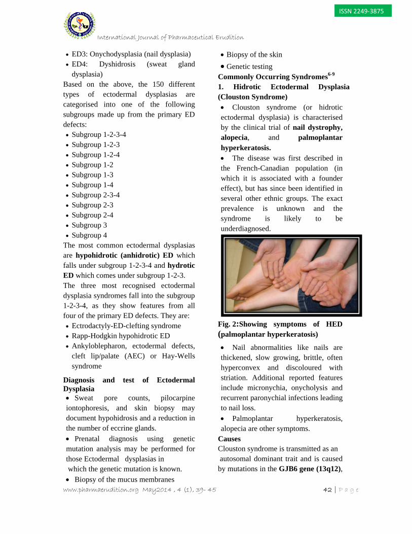
1. Hidrotic Ectodermal Dysplasia(Clouston Syndrome) Clouston syndrome (or hidroticectodermal dysplasia) is characterisedby the clinical trial of nail dystrophy,alopecia, and palmoplantarhyperkeratosis. The disease was first described inthe French-Canadian population (inwhich it is associated with a foundereffect), but has since been identified inseveral other ethnic groups. The exactprevalence is unknown and thesyndrome is likely to beunderdiagnosed.
Fig..2:Showing symptoms of HED(palmoplantar hyperkeratosis)
Nail abnormalities like nails arethickened, slow growing, brittle, oftenhyperconvex and discoloured withstriation. Additional reported featuresinclude micronychia, onycholysis andrecurrent paronychial infections leadingto nail loss. Palmoplantar hyperkeratosis,alopecia are other symptoms.
CausesClouston syndrome is transmitted as anautosomal dominant trait and is caused
by mutations in the GJB6 gene (13q12),
International Journal of Pharmaceutical Erudition
www.pharmaerudition.org May2014 , 4 (1), 39- 45 42 | P a g e
ISSN 2249-3875
ED3: Onychodysplasia (nail dysplasia) ED4: Dyshidrosis (sweat gland
dysplasia)Based on the above, the 150 differenttypes of ectodermal dysplasias arecategorised into one of the followingsubgroups made up from the primary EDdefects: Subgroup 1-2-3-4 Subgroup 1-2-3 Subgroup 1-2-4 Subgroup 1-2 Subgroup 1-3 Subgroup 1-4 Subgroup 2-3-4 Subgroup 2-3 Subgroup 2-4 Subgroup 3 Subgroup 4
The most common ectodermal dysplasiasare hypohidrotic (anhidrotic) ED whichfalls under subgroup 1-2-3-4 and hydroticED which comes under subgroup 1-2-3.The three most recognised ectodermaldysplasia syndromes fall into the subgroup1-2-3-4, as they show features from allfour of the primary ED defects. They are: Ectrodactyly-ED-clefting syndrome Rapp-Hodgkin hypohidrotic ED Ankyloblepharon, ectodermal defects,
cleft lip/palate (AEC) or Hay-Wellssyndrome
Diagnosis and test of EctodermalDysplasia Sweat pore counts, pilocarpineiontophoresis, and skin biopsy maydocument hypohidrosis and a reduction inthe number of eccrine glands. Prenatal diagnosis using geneticmutation analysis may be performed forthose Ectodermal dysplasias inwhich the genetic mutation is known. Biopsy of the mucus membranes
Biopsy of the skin
Genetic testingCommonly Occurring Syndromes6-9
1. Hidrotic Ectodermal Dysplasia(Clouston Syndrome) Clouston syndrome (or hidroticectodermal dysplasia) is characterisedby the clinical trial of nail dystrophy,alopecia, and palmoplantarhyperkeratosis. The disease was first described inthe French-Canadian population (inwhich it is associated with a foundereffect), but has since been identified inseveral other ethnic groups. The exactprevalence is unknown and thesyndrome is likely to beunderdiagnosed.
Fig..2:Showing symptoms of HED(palmoplantar hyperkeratosis)
Nail abnormalities like nails arethickened, slow growing, brittle, oftenhyperconvex and discoloured withstriation. Additional reported featuresinclude micronychia, onycholysis andrecurrent paronychial infections leadingto nail loss. Palmoplantar hyperkeratosis,alopecia are other symptoms.
CausesClouston syndrome is transmitted as anautosomal dominant trait and is caused
by mutations in the GJB6 gene (13q12),
International Journal of Pharmaceutical Erudition
www.pharmaerudition.org May2014 , 4 (1), 39- 45 43 | P a g e
ISSN 2249-3875
encoding the gap junction proteinconnexin 30 (Cx30).
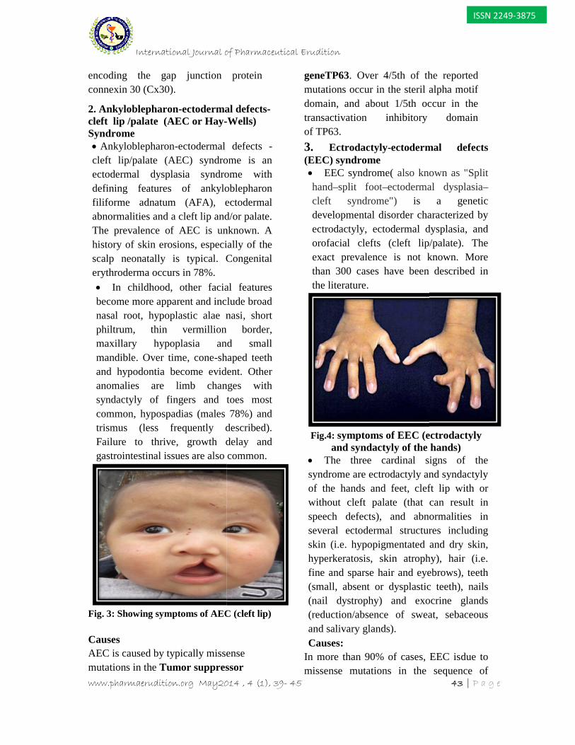
2. Ankyloblepharon-ectodermal defects-cleft lip /palate (AEC or Hay-Wells)Syndrome Ankyloblepharon-ectodermal defects -cleft lip/palate (AEC) syndrome is anectodermal dysplasia syndrome withdefining features of ankyloblepharonfiliforme adnatum (AFA), ectodermalabnormalities and a cleft lip and/or palate.The prevalence of AEC is unknown. Ahistory of skin erosions, especially of thescalp neonatally is typical. Congenitalerythroderma occurs in 78%. In childhood, other facial featuresbecome more apparent and include broadnasal root, hypoplastic alae nasi, shortphiltrum, thin vermillion border,maxillary hypoplasia and smallmandible. Over time, cone-shaped teethand hypodontia become evident. Otheranomalies are limb changes withsyndactyly of fingers and toes mostcommon, hypospadias (males 78%) andtrismus (less frequently described).Failure to thrive, growth delay andgastrointestinal issues are also common.
Fig. 3: Showing symptoms of AEC (cleft lip)
CausesAEC is caused by typically missensemutations in the Tumor suppressor
geneTP63. Over 4/5th of the reportedmutations occur in the steril alpha motifdomain, and about 1/5th occur in thetransactivation inhibitory domainof TP63.
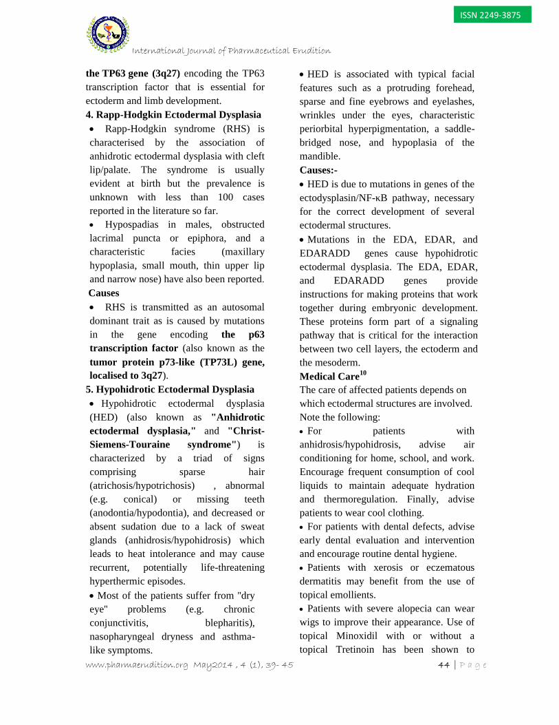
3. Ectrodactyly-ectodermal defects(EEC) syndrome EEC syndrome( also known as "Splithand–split foot–ectodermal dysplasia–cleft syndrome") is a geneticdevelopmental disorder characterized byectrodactyly, ectodermal dysplasia, andorofacial clefts (cleft lip/palate). Theexact prevalence is not known. Morethan 300 cases have been described inthe literature.
Fig.4: symptoms of EEC (ectrodactylyand syndactyly of the hands)
The three cardinal signs of thesyndrome are ectrodactyly and syndactylyof the hands and feet, cleft lip with orwithout cleft palate (that can result inspeech defects), and abnormalities inseveral ectodermal structures includingskin (i.e. hypopigmentated and dry skin,hyperkeratosis, skin atrophy), hair (i.e.fine and sparse hair and eyebrows), teeth(small, absent or dysplastic teeth), nails(nail dystrophy) and exocrine glands(reduction/absence of sweat, sebaceousand salivary glands).Causes:
In more than 90% of cases, EEC isdue tomissense mutations in the sequence of
International Journal of Pharmaceutical Erudition
www.pharmaerudition.org May2014 , 4 (1), 39- 45 43 | P a g e
ISSN 2249-3875
encoding the gap junction proteinconnexin 30 (Cx30).
2. Ankyloblepharon-ectodermal defects-cleft lip /palate (AEC or Hay-Wells)Syndrome Ankyloblepharon-ectodermal defects -cleft lip/palate (AEC) syndrome is anectodermal dysplasia syndrome withdefining features of ankyloblepharonfiliforme adnatum (AFA), ectodermalabnormalities and a cleft lip and/or palate.The prevalence of AEC is unknown. Ahistory of skin erosions, especially of thescalp neonatally is typical. Congenitalerythroderma occurs in 78%. In childhood, other facial featuresbecome more apparent and include broadnasal root, hypoplastic alae nasi, shortphiltrum, thin vermillion border,maxillary hypoplasia and smallmandible. Over time, cone-shaped teethand hypodontia become evident. Otheranomalies are limb changes withsyndactyly of fingers and toes mostcommon, hypospadias (males 78%) andtrismus (less frequently described).Failure to thrive, growth delay andgastrointestinal issues are also common.
Fig. 3: Showing symptoms of AEC (cleft lip)
CausesAEC is caused by typically missensemutations in the Tumor suppressor
geneTP63. Over 4/5th of the reportedmutations occur in the steril alpha motifdomain, and about 1/5th occur in thetransactivation inhibitory domainof TP63.
3. Ectrodactyly-ectodermal defects(EEC) syndrome EEC syndrome( also known as "Splithand–split foot–ectodermal dysplasia–cleft syndrome") is a geneticdevelopmental disorder characterized byectrodactyly, ectodermal dysplasia, andorofacial clefts (cleft lip/palate). Theexact prevalence is not known. Morethan 300 cases have been described inthe literature.
Fig.4: symptoms of EEC (ectrodactylyand syndactyly of the hands)
The three cardinal signs of thesyndrome are ectrodactyly and syndactylyof the hands and feet, cleft lip with orwithout cleft palate (that can result inspeech defects), and abnormalities inseveral ectodermal structures includingskin (i.e. hypopigmentated and dry skin,hyperkeratosis, skin atrophy), hair (i.e.fine and sparse hair and eyebrows), teeth(small, absent or dysplastic teeth), nails(nail dystrophy) and exocrine glands(reduction/absence of sweat, sebaceousand salivary glands).Causes:
In more than 90% of cases, EEC isdue tomissense mutations in the sequence of
International Journal of Pharmaceutical Erudition
www.pharmaerudition.org May2014 , 4 (1), 39- 45 44 | P a g e
ISSN 2249-3875
the TP63 gene (3q27) encoding the TP63transcription factor that is essential forectoderm and limb development.4. Rapp-Hodgkin Ectodermal Dysplasia Rapp-Hodgkin syndrome (RHS) ischaracterised by the association ofanhidrotic ectodermal dysplasia with cleftlip/palate. The syndrome is usuallyevident at birth but the prevalence isunknown with less than 100 casesreported in the literature so far. Hypospadias in males, obstructedlacrimal puncta or epiphora, and acharacteristic facies (maxillaryhypoplasia, small mouth, thin upper lipand narrow nose) have also been reported.
Causes RHS is transmitted as an autosomaldominant trait as is caused by mutationsin the gene encoding the p63transcription factor (also known as thetumor protein p73-like (TP73L) gene,localised to 3q27).
5. Hypohidrotic Ectodermal Dysplasia Hypohidrotic ectodermal dysplasia(HED) (also known as "Anhidroticectodermal dysplasia," and "Christ-Siemens-Touraine syndrome") ischaracterized by a triad of signscomprising sparse hair(atrichosis/hypotrichosis) , abnormal(e.g. conical) or missing teeth(anodontia/hypodontia), and decreased orabsent sudation due to a lack of sweatglands (anhidrosis/hypohidrosis) whichleads to heat intolerance and may causerecurrent, potentially life-threateninghyperthermic episodes. Most of the patients suffer from ''dryeye'' problems (e.g. chronicconjunctivitis, blepharitis),nasopharyngeal dryness and asthma-like symptoms.
HED is associated with typical facialfeatures such as a protruding forehead,sparse and fine eyebrows and eyelashes,wrinkles under the eyes, characteristicperiorbital hyperpigmentation, a saddle-bridged nose, and hypoplasia of themandible.Causes:- HED is due to mutations in genes of theectodysplasin/NF-κB pathway, necessaryfor the correct development of severalectodermal structures. Mutations in the EDA, EDAR, andEDARADD genes cause hypohidroticectodermal dysplasia. The EDA, EDAR,and EDARADD genes provideinstructions for making proteins that worktogether during embryonic development.These proteins form part of a signalingpathway that is critical for the interactionbetween two cell layers, the ectoderm andthe mesoderm.Medical Care10
The care of affected patients depends onwhich ectodermal structures are involved.Note the following: For patients withanhidrosis/hypohidrosis, advise airconditioning for home, school, and work.Encourage frequent consumption of coolliquids to maintain adequate hydrationand thermoregulation. Finally, advisepatients to wear cool clothing. For patients with dental defects, adviseearly dental evaluation and interventionand encourage routine dental hygiene. Patients with xerosis or eczematousdermatitis may benefit from the use oftopical emollients. Patients with severe alopecia can wearwigs to improve their appearance. Use oftopical Minoxidil with or without atopical Tretinoin has been shown to
International Journal of Pharmaceutical Erudition
www.pharmaerudition.org May2014 , 4 (1), 39- 45 45 | P a g e
ISSN 2249-3875
improve hair growth in a small number ofpatients. Patients with scalp erosions should betreated with topical and systemicantibiotics as needed. Use artificial tears to prevent damage tothe cornea in patients with reducedlacrimation. Protect nasal mucosa with saline spraysfollowed by the application of petrolatum. Allogeneic stem cell transplantation hasbeen performed in a small number ofpatients with autosomal dominantectodermal dysplasia withimmunodeficiency (EDA-ID); poorengraftment and post-transplantcomplications were common.
Surgical CareEarly repair of cleft lip or palate maylessen facial deformities and improvespeech. Other midfacial defects orhand/foot deformities may be surgicallycorrected in order to improve functionand reduce physical disfigurement.
CONCLUSIONIn this review the diseases, its causessymptoms & types of EctodermalDysplasia has been given. It has beenseen that, there is no specific treatmentfor Ectodermal Dysplasia, only diseasemanagement are available. So, we haveconcluded that further research work hasbeen needed for the treatment ofEctodermal Dysplasia.
REFERENCES
1. James, William, Berger, Timothy,Elston, Dirk Andrews' Diseases of theSkin: Clinical Dermatology. (10th ed.).Saunders. 2005
2. Freedberg, Fitzpatrick's Dermatology inGeneral Medicine. (6th ed.). McGraw-Hill,
2003 .3. First Baby Dosed in Clinical Trial forXLHED. National Foundation forEctodermal Dysplasias, 3 February 2014.4. Spfaer JA. A dental approach to carrierscreening in X linked hypohidroticectodermal dysplasia. J. Med. Genet. 1981;18 (6): 459-60.5. Kere J, Srivastava AK, Montonen O.X-linked anhidrotic (hypohidrotic)ectodermal dysplasia is caused bymutation in a novel transmembraneprotein. Nat. Genet. 1996,13 (4): 409-16.6. Okamura E, Suda N, Baba Y. Dentaland maxillofacial characteristics in six(EEC)syndrome. 2012.7. Adaimy L, Chouery E, Megarbane H,Mroueh S, Delague V, Nicolas E,Belguith H, de Mazancourt P, MegarbaneA. Mutation in WNT10A is associatedwith an autosomal recessive ectodermaldysplasia: the odonto-onycho-dermaldysplasia. Am J Hum Genet. 81:821–8,2007.8. Bal E, Baala L, Cluzeau C, El Kerch F,Ouldim K, Hadj-Rabia S, Bodemer C,Munnich A, Courtois G, Sefiani A, SmahiA. Autosomal dominant anhidroticectodermal dysplasias at the EDARADDlocus. Hum Mutat, 28:703–9, 2007.9. Bayes M, Hartung AJ, Ezer S, Pispa J,Thesleff I, Srivastava AK, Kere J. Theanhidrotic ectodermal dysplasia gene(EDA) undergoes alternative splicing andencodes ectodysplasin-A with deletionmutations in collagenous repeats. HumMol Genet, 7:1661–9, 1998.10. Bergendal B, Klar J, Stecksén-BlicksC, Norderyd J, Dahl N. Isolatedoligodontia assoc. with mutations inEDARADD, AXIN2, MSX1 and PAX9genes. Am J Med Genet A. 155A:1616–22, 2011.