Embed Size (px)
Citation preview
A C T A O P H T H A L M O L O G I C A V O L . 5 6 1 9 7 8
Department of Ophthalmology, University of Helsinki (Head: Salme Vannas), Helsinki
A SIMPLE METHOD FOR ANTERIOR SEGMENT PHOTOGRAPHY OF THE EYE WITH
STANDARD EQUIPMENT
BY
KARl KAAKINEN
A simple hand-held system for colour photography of the anterior eye structures using a standard camera and conventional incandescent slit lamp is presented for use by a general eye practitioner. No electronic flash is required. The anterior ocular pathology is well documented with the method.
Key words: macrophotography - single lens reflex camera (SRL) - slit lamp - documentation.
Photographic recordings of ocular lesions have many advantages in clinical ophthalmological work. Many changes in the eye structures can be demon- strated with repeated photographs. Photographic documents are also needed for teaching and consultation. There are also legal reasons for obtaining photographic documentation.
The high cost of photographic equipment has restricted its use to specialised clinics. But even these clinics and, not least, private eye practitioners need a quick and easy photographic method that does not require the complicated and expensive flash equipment built into the slit lamp system.
Nowadays there are quite sensitive commercial film materials. The sen- sitivity of these films can be further chemically improved during the pro- cessing. It might be possible that, as a result of this first-class sensitivity, the exposure time could be cut to be adequate for handheld photography. The
Received February 8, 1978.
902
Anterior Segment Photography
improved light pcwer of the modern camera lenses also contributes to the shortening of the exposure time.
The intention was to examine whether it is possible to obtain good pictures by using a hand-held system and a conventional camera. This could very much facilitate the documentation in clinical ophthalmological work.
Method
Photographs were taken without a flash with a conventional camera equipped with a macrophotcgraphy system, using a normal incandescent slit lamp without a microscope.
Two cameras were used for these experiments, the Olympus-Pen F and Canon EF with standard lenses, Olympus H. Zuiko Auto-S 1:1.2 f = 4 2 mm and Canon lens FD 50 mm 1:1.4 S.S.C. Both were single-lens reflex cameras with a built-in through-the-lens exposure meter system. The light measuring was done by using the cameras’ uwn system. As macrophotography equipment the Olympus Pen F had extension bellows inserted between the camera body and the lens unit. In the Canon EF the standard lens was attached in the reverse directicn to the camera body by a Canon macrophoto coupler. This maintains the performance of the lens for larger than life-size macrophoto- graphy. A special macro-objective Canon macro FD 50 mm 1:3.5 S.S.C. was
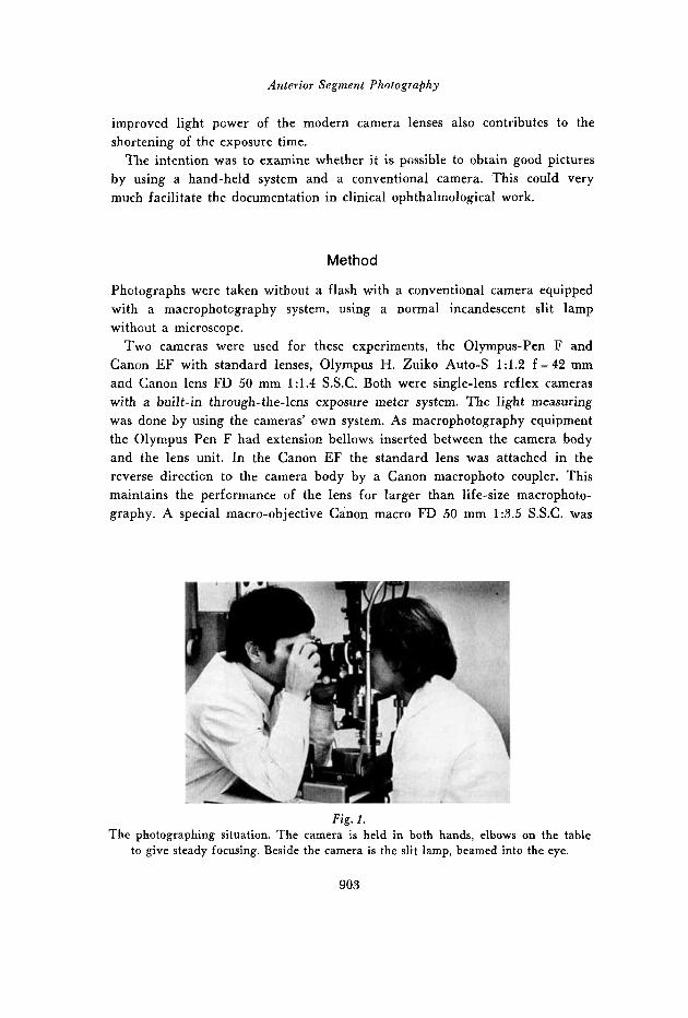
Fig. 1. The photographing situation. The camera is held in both hands, elbows on the table
to give steady focusing. Beside the camera is the slit lamp, beamed into the eye.
903
Kari Kaakinen
also used. To get life-size pictures it must be combined with an extension tube or bellows.
Different shutter speeds were tested and 1/125 sec. seemed to be the most suitable for hand-held photography. It was fast enough, but not too fast, for making the lens aperture smaller than the biggest one in order to get better depth sharpness in the picture. The width of the aperture needed is also dependent on the sensitivity of the film. Therefore, very sensitive film tungsten Kodak high-speed Ektachrome with special commercial 500 Asa (28 Din) processing was used.
A Haag-Streit slit lamp was used in these experiments. But any good slit lamp should perform equally as well. For maximum illumination, the trans- former was set to the highest voltage. The microscope was turned on its side in order to give space for the camera held in both hands. The elbows were placed on the table with the slit lamp for steady focusing (Fig. 1). The easiest way to focus is to move the camera slowly to and fro and stop when the object is sharp.
The position and width of the slit was selected to keep reflections to a minimcm and obtain ideal illumination on the object. Different types of illu- mination were tested with good results (narrow and wide slit, indirect illumi- nation, etc., see Pearce (1974) and Winning (1973).
For ideal exposure and contrast in the picture, four extra photographs were usually taken with the aperture ranges one stop and two stops over and under the light measuring values because of the different reflecting capacity of the individual parts of the eye.
Results
To illustrate the usefulness of the method in clinical work about 100 photo- graphs were taken. A selection of illustrative cases is presented.
Different changes in the cornea were quite well documented (Figs. 2, 3, 4 and 5).
Many kinds of opacities in the lens and especially pseudoexfoliation and posterior congenital, and, of course, the senile type of cataract (Fig. 6) were beautifully illustrated.
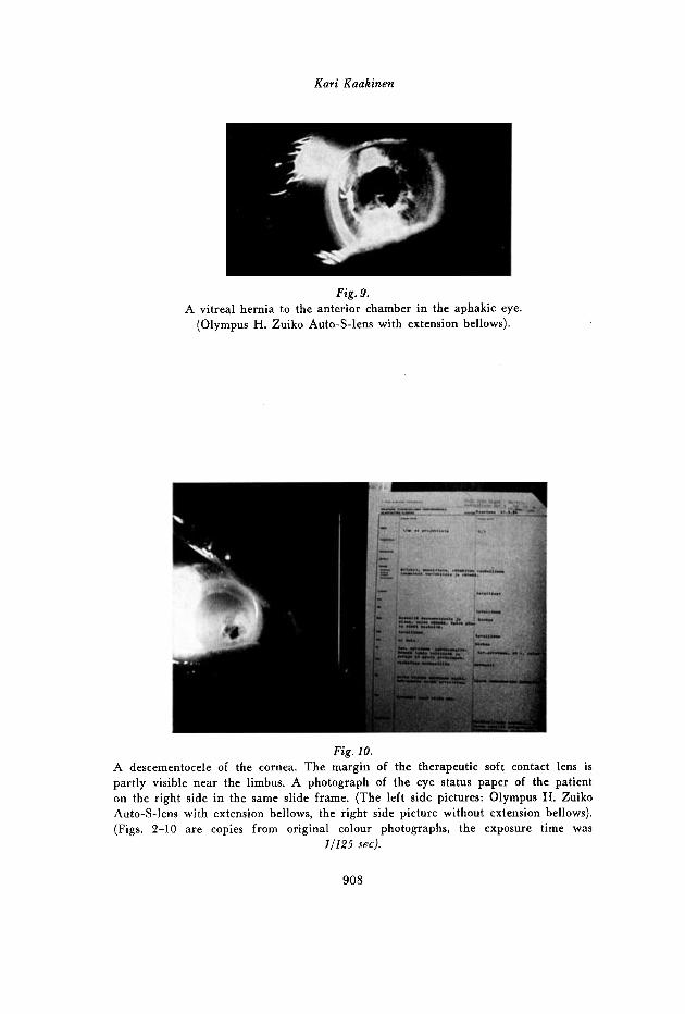
The pathology of the iris and the anterior chamber in fulminant irido- cyclitis were demonstrated. In particular, the hypopyon and the morphology and the depth of the anterior chamber could be studied from pictures taken from the side of the eye with the slit beam directed straight from the front to the eye (Fig. 7).
904
Anterior Segment Photography
Conjunctival (Fig. 8) and scleral changes were most easy to photograph and the same was true of palpebral and general face pictures using an ordinary table lamp as the light source.
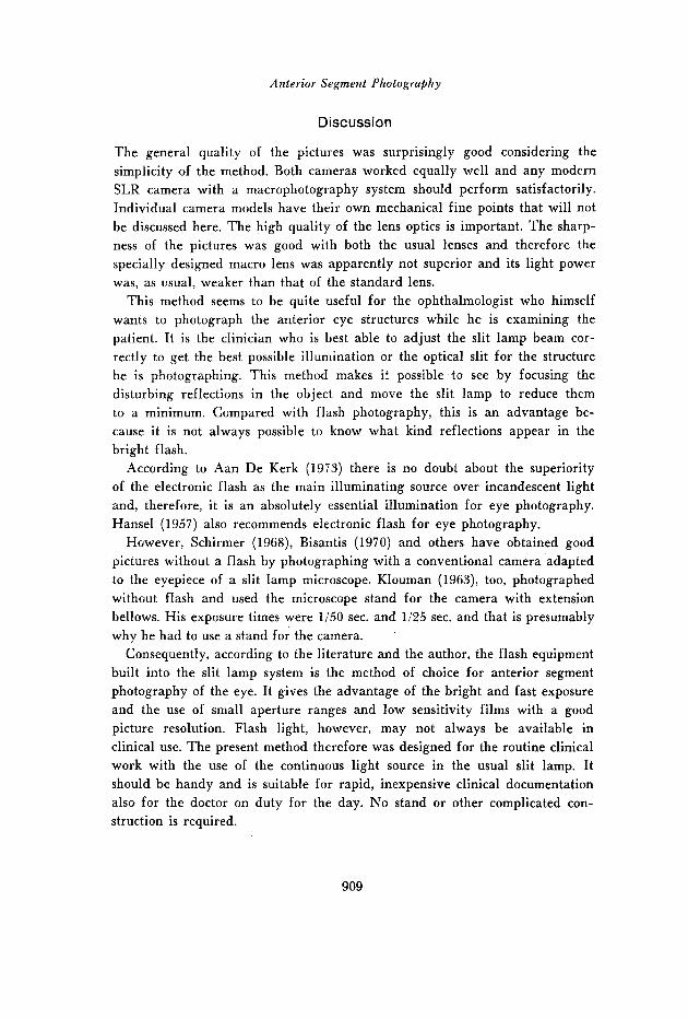
The vitreous in the normal eye was impossible to photograph with this method because the slit lamp beamed to the vitreous always comes in front of the camera lens and masks it. In the case of an aphakic eye the vitreous membrane hernia protruding into the anterior chamber was quite easy to photograph (Fig. 9).
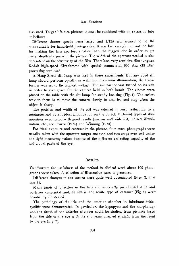
Fig. 2 and 3. Dendritic keratitis (a branch-like ulcer stained with fluorescein). Both photographs
were taken with a blue light produced by a blue filter in the slit lamp. Fig. 2. Olympus H. Zuiko Auto-S-lens with extension bellows.
F i g . 3. Cannon FD 50 mm-lens with macrophoto coupler.
905 Acta ophthal. 50, 6
Kar i Kaakinen
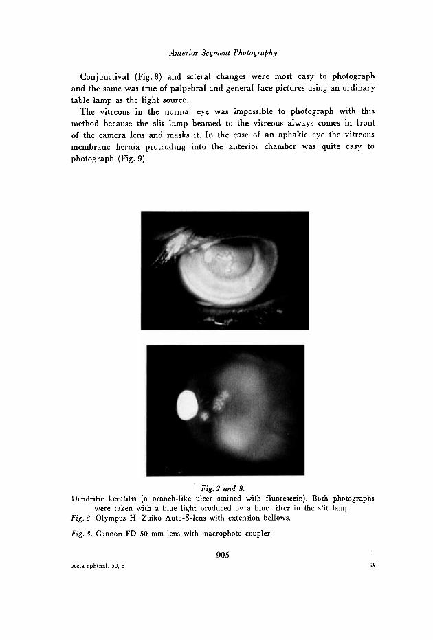
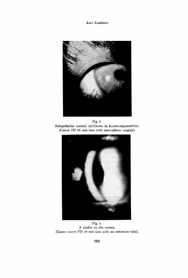
Fig . 4. Subepithelial corneal infiltrates in keratoconjunctivitis.
(Canon FD 50 mm-lens with macrophoto coupler).
Fig. 5. A cinder on the cornea.
(Canon macro FD 50 mm-lens with an extension tube).
906
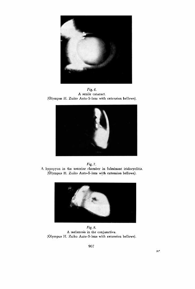
Fig. 6. A senile cataract.
(Olympus H. Zuiko Auto-S-lens with extension bellows).
Pig. 7. A hypopyon in the anterior chamber in fulminant iridocyclitis.
(Olympus H. Zuiko Auto-S-lens with extension bellows).
Fig. 8. A melanosis in the conjunctiva.
(Olympus H. Zuiko Auto-S-lens with extension bellows).
907 58*
Kari Kaakinen
Fig. 9. A vitreal hernia to the anterior chamber in the aphakic eye.
(Olympus H. Zuiko Auto-S-lens with extension bellows).
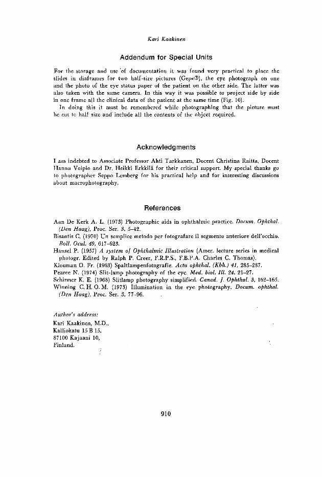
Fig. 10. A descementocele of the cornea. The margin of the therapeutic soft contact lens is partly visible near the limbus. A photograph of the eye status paper of the patient on the right side in the same slide frame. (The left side pictures: Olympus H. Zuiko Auto-S-lens with extension bellows, the right side picture without extension bellows). (Figs. 2-10 are copies from original colour photographs, the exposure time was
11125 sec).
908
Anterior Segment Photography
Discussion
The general quality of the pictures was surprisingly good considering the simplicity of the method. Both cameras worked equally well and any modern SLR camera with a macrophotography system should perform satisfactorily. Individual camera models have their own mechanical fine points that will not be discussed here. The high quality of the lens optics is important. The sharp- ness of the pictures was good with both the usual lenses and therefore the specially designed macro lens was apparently not superior and its light power was, as usual, weaker than that of the standard lens.
This method seems to be quite useful for the ophthalmologist who himself wants to photograph the anterior eye structures while he is examining the patient. It is the clinician who is best able to adjust the slit lamp beam cor- rectly to get the best possible illumination or the optical slit for the structure he is photographing. This method makes it possible to see by focusing the disturbing reflections in the object and move the slit lamp to reduce them to a minimum. Ccmpared with flash photography, this is an advantage be- cause it is not always possible to know what kind reflections appear in the bright flash.
According to Aan De Kerk (1973) there is no doubt about the superiority of the electronic flash as the main illuminating source over incandescent light and, therefore, it is an absolutely essential illumination for eye photography. Hansel (1957) also recommends electronic flash for eye photography.
However, Schirmer (1968), Bisantis (1970) and others have obtained good pictures without a flash by photographing with a conventional camera adapted to the eyepiece of a slit lamp microscope. Klouman (1963), too, photographed without flash and used the microscope stand for the camera with extension bellows. His exposure times were 1/50 sec. and 1/25 sec. and that is presumably why he had to use a stand for the camera.
Consequently, according to the literature and the author, the flash equipment built into the slit lamp system is the method of choice for anterior segment photography of the eye. I t gives the advantage of the bright and fast exposure and the use of small aperture ranges and low sensitivity films with a good picture resolution. Flash light, however, may not always be available in clinical use. The present method therefore was designed for the routine clinical work with the use of the continuous light source in the usual slit lamp. It should be handy and is suitable for rapid, inexpensive clinical documentation also for the doctor on duty for the day. No stand or other complicated con- struction is required.
909
Kari Kaakinen
Addendum for Special Units
For the storage and use ’of documentation it was found very practical to place the slides in diaframes for two half-size pictures (Gepe@), the eye photograph on one and the photo of the eye status paper of the patient on the other side. The latter was also taken with the same camera. In this way it was possible to project side by side in one frame all the clinical data of the patient at the same time (Fig. 10).
In doing this it must be remembered while photographing that the picture must be cut to half size and include all the contents of the object required.
Acknowledgments
I am indebted to Associate Professor Ahti Tarkkanen, Docent Christina Raitta, Docent Hannu Voipio and Dr. Heikki Erkkila for their critical support. My special thanks go to photographer Seppo Lemberg for his practical help and for interesting discussions about macrophotography.
References
Aan De Kerk A. L. (1973) Photographic aids in ophthalmic practice. Docum. Ophthal. (Den Haag), Proc. Ser. 3, 5-42.
Bisantis C. (1970) Un semplice metodo per fotografare il segment0 anteriore dell’occhio. Boll. Ocul. 49, 617-623.
Hansel P. (1957) A system of Ofhthalmic Zllustration (Amer. lecture series in medical photogr. Edited by Ralph P. Creer, F.R.P.S., F.B.P.A. Charles C. Thomas).
Klouman 0. Fr. (1963) Spaltlampenfotografie. Acta ophthal. (Kbh.) 41, 285-287. Pearce N. (1974) Slit-lamp photography of the eye. Med. biol. Ill. 24, 21-27. Schirmer K. E. (1968) Slitlamp photography simplified. Canad. /. Ophthal. 3, 162-165. Winning C. H. 0. M. (1973) Illumination in the eye photography. Docum. ophthal.
(Den Haag), Proc. Ser. 3, 77-96.
Author’s address: Kari Kaakinen, M.D., Kalliokatu 15 B 15, 87100 Kajaani 10, Finland.
910