Embed Size (px)
Citation preview
John E Lees
Space Research Centre University of Leicester
1 December 2014
A Small Field of View Camera for Hybrid Gamma and Optical Imaging

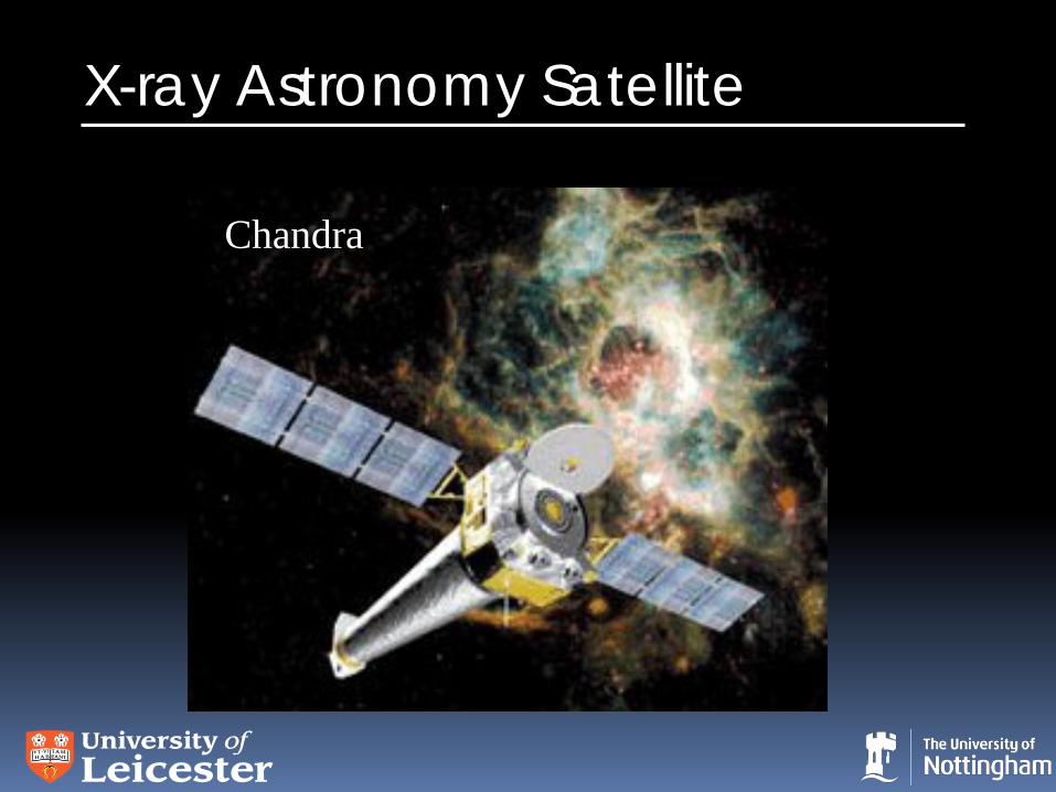
X-ray Astronomy Satellite
Chandra
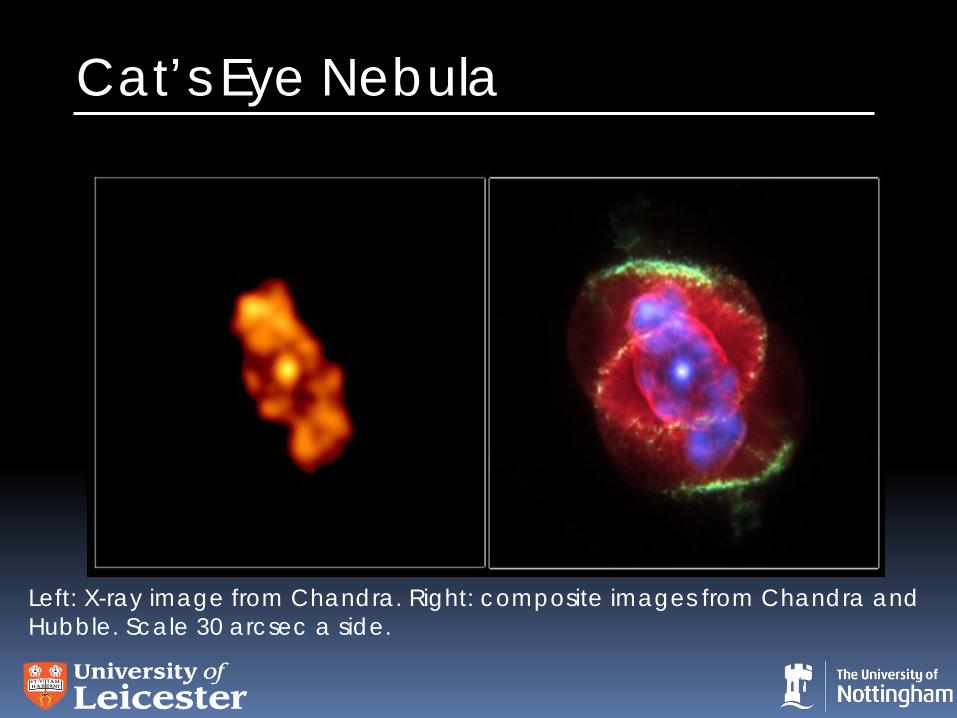
Cat’s Eye Nebula
Left: X-ray image from Chandra. Right: composite images from Chandra and Hubble. Scale 30 arcsec a side.

UoL Space Research Centre Technologies and Expertise
Optical, UV, X-rays, gamma rays
Charge Coupled Devices: CCD
XMM-Newton: EPIC
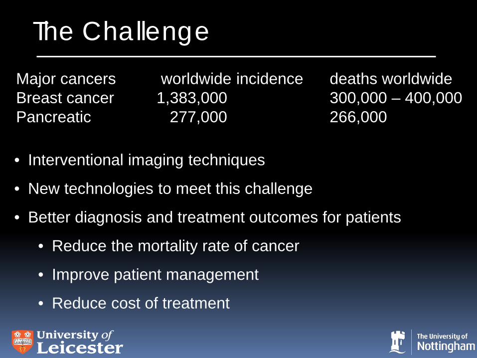
The Challenge
• Interventional imaging techniques
• New technologies to meet this challenge
• Better diagnosis and treatment outcomes for patients
• Reduce the mortality rate of cancer
• Improve patient management
• Reduce cost of treatment
Major cancers worldwide incidence deaths worldwide Breast cancer 1,383,000 300,000 – 400,000 Pancreatic 277,000 266,000
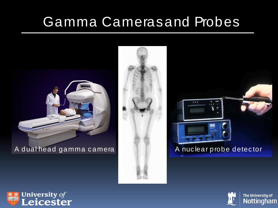
Gamma Cameras and Probes
A dual head gamma camera A nuclear probe detector
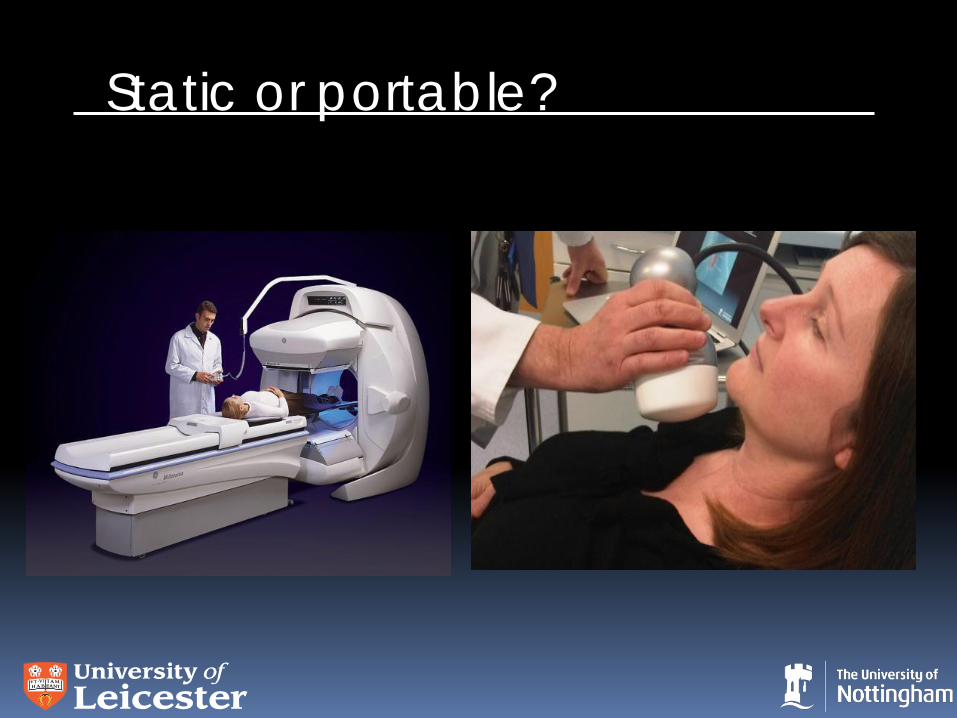
Static or portable?
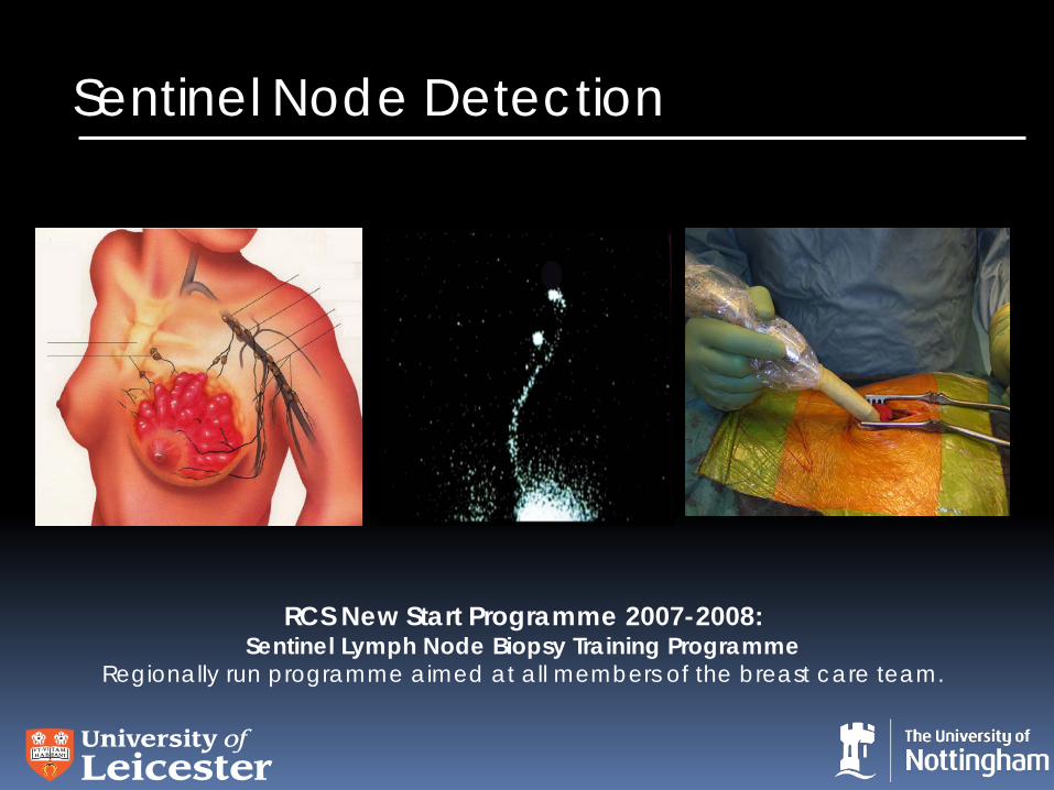
Sentinel Node Detection
RCS New Start Programme 2007-2008: Sentinel Lymph Node Biopsy Training Programme
Regionally run programme aimed at all members of the breast care team.
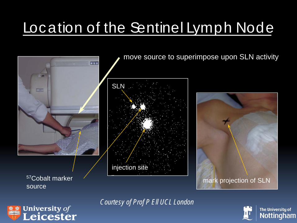
Location of the Sentinel Lymph Node
SLN
57Cobalt marker source
move source to superimpose upon SLN activity
injection site
mark projection of SLN
Courtesy of Prof P Ell UCL London
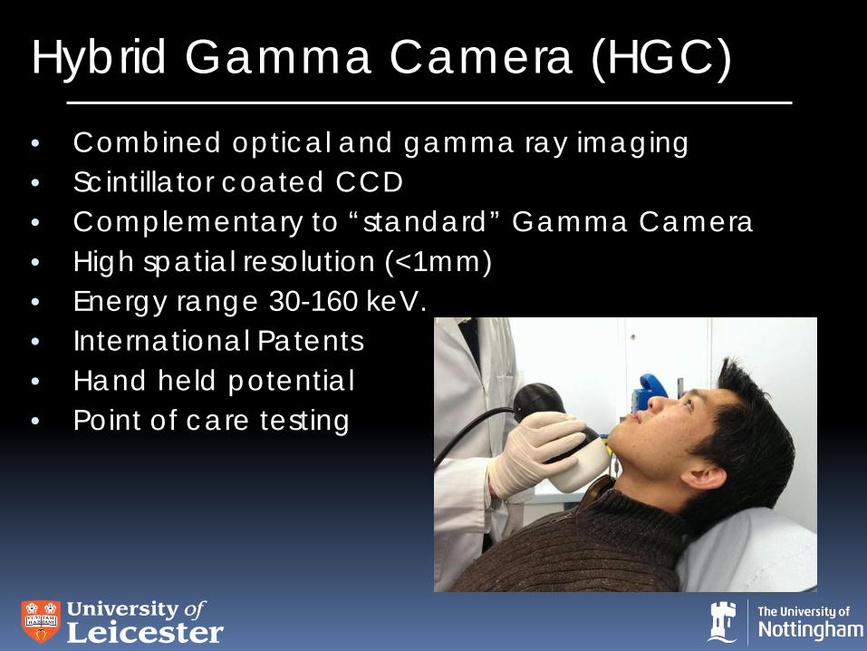
Hybrid Gamma Camera (HGC) • Combined optical and gamma ray imaging • Scintillator coated CCD • Complementary to “standard” Gamma Camera • High spatial resolution (<1mm) • Energy range 30-160 keV. • International Patents • Hand held potential • Point of care testing
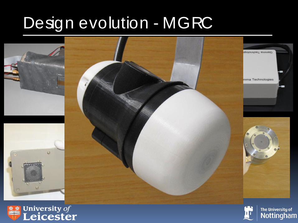
Design evolution - MGRC
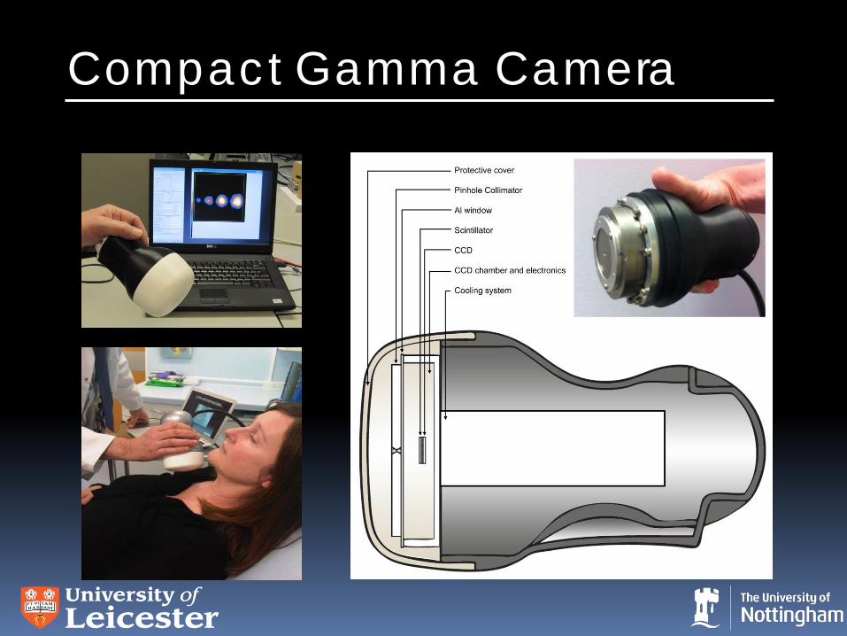
Compact Gamma Camera
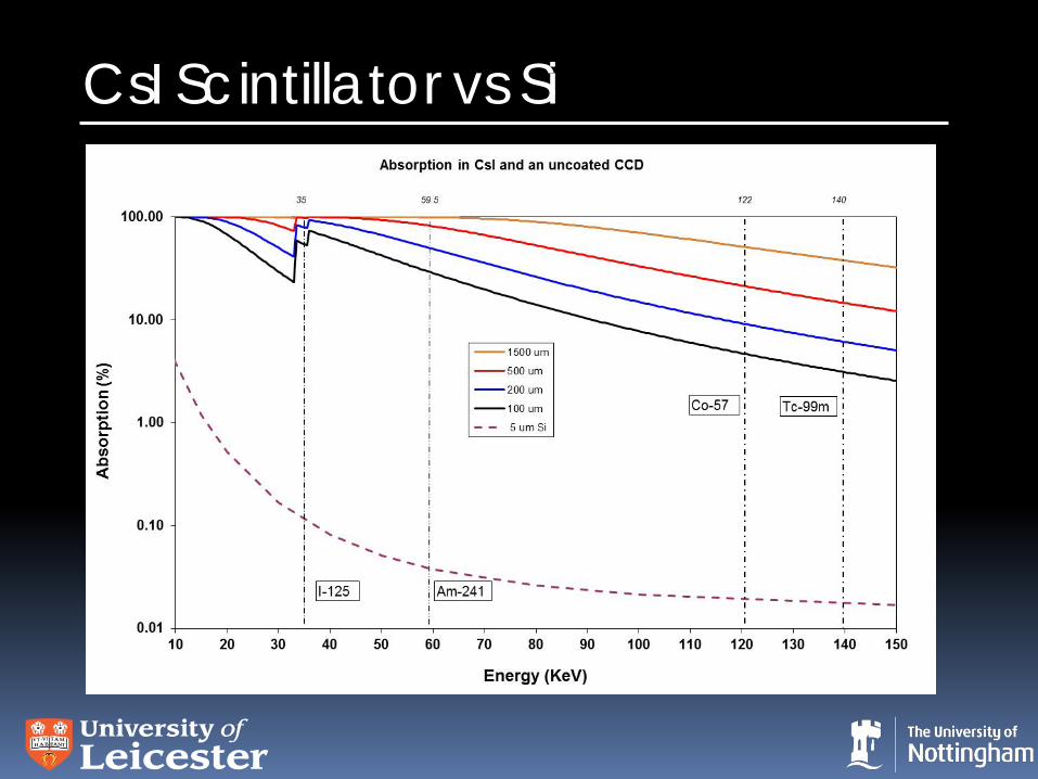
CsI Scintillator vs Si
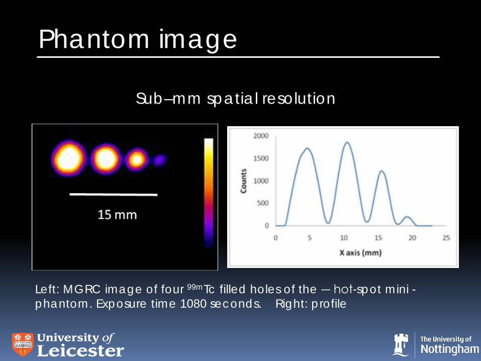
Phantom image
Left: MGRC image of four 99mTc filled holes of the ― hot-spot mini - phantom. Exposure time 1080 seconds. Right: profile
Sub–mm spatial resolution
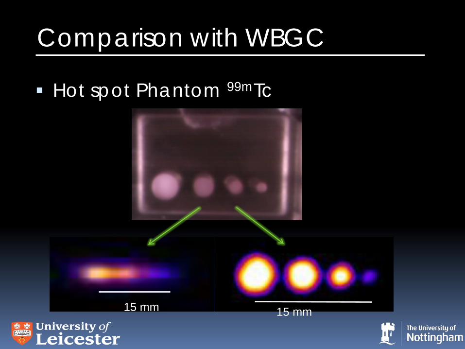
Comparison with WBGC
Hot spot Phantom 99mTc
15 mm 15 mm
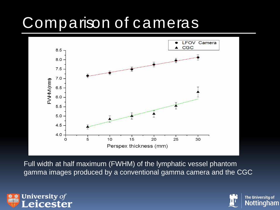
Comparison of cameras
Full width at half maximum (FWHM) of the lymphatic vessel phantom gamma images produced by a conventional gamma camera and the CGC
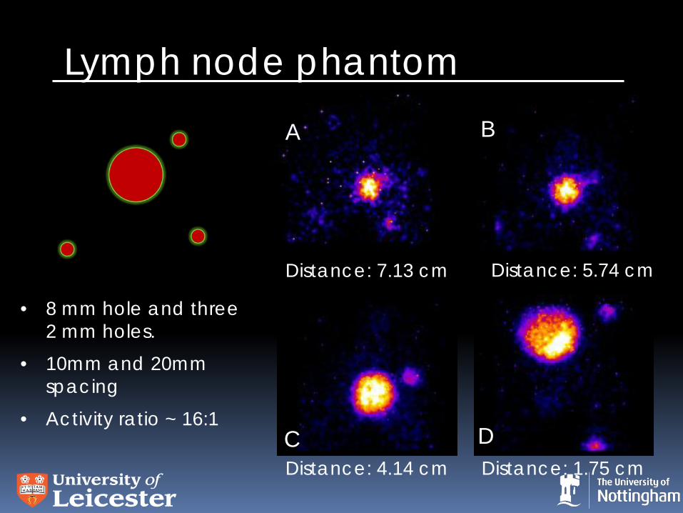
Lymph node phantom
Distance: 7.13 cm Distance: 5.74 cm
Distance: 4.14 cm Distance: 1.75 cm
• 8 mm hole and three 2 mm holes.
• 10mm and 20mm spacing
• Activity ratio ~ 16:1
A B
C D
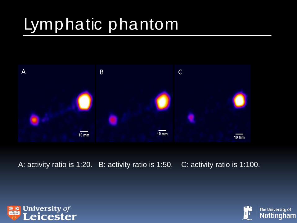
Lymphatic phantom
A B C
A: activity ratio is 1:20. B: activity ratio is 1:50. C: activity ratio is 1:100.
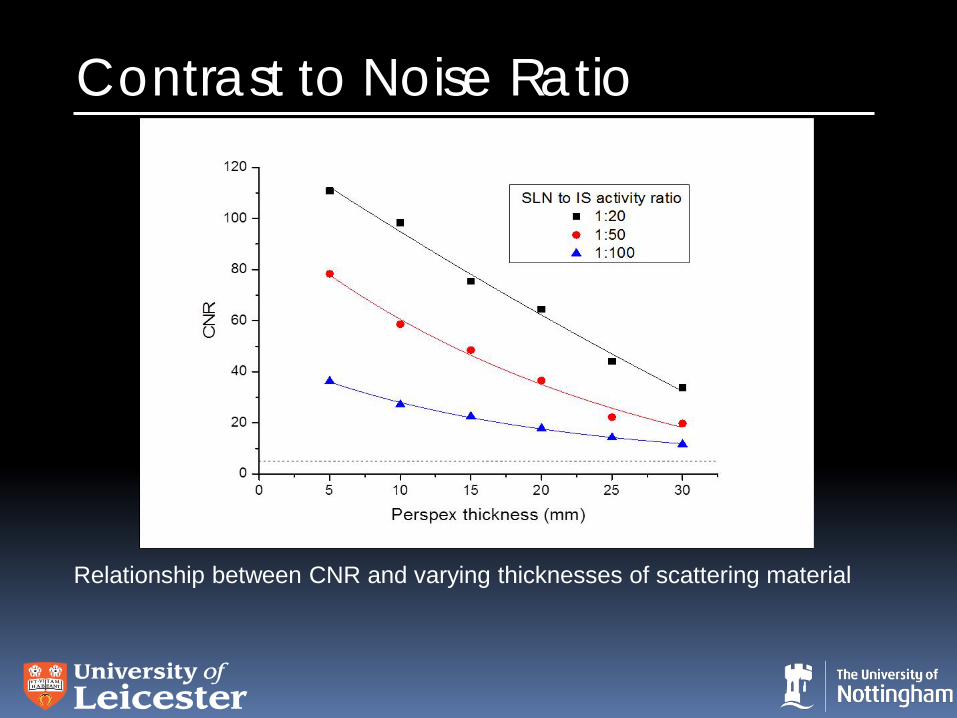
Contrast to Noise Ratio
Relationship between CNR and varying thicknesses of scattering material
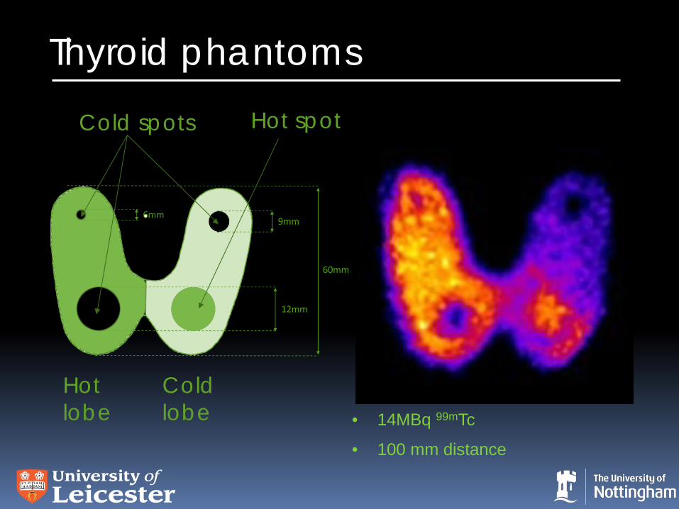
Thyroid phantoms
• 14MBq 99mTc
• 100 mm distance
Cold spots Hot spot
Hot lobe
Cold lobe
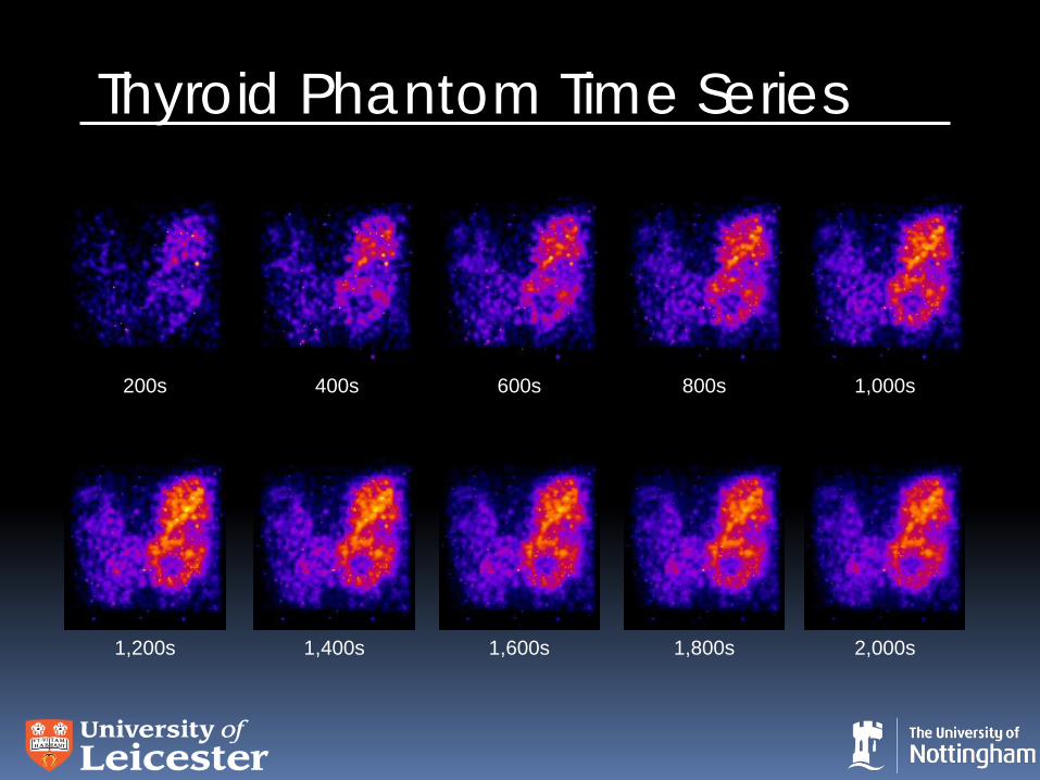
Thyroid Phantom Time Series
200s 400s 600s 800s 1,000s
1,200s 1,400s 1,600s 1,800s 2,000s
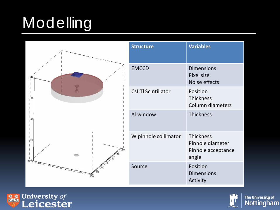
Modelling
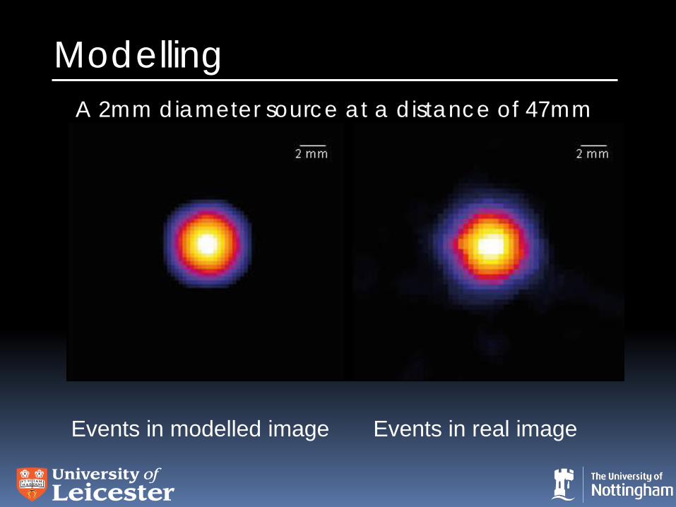
Modelling
Events in modelled image Events in real image
A 2mm diameter source at a distance of 47mm
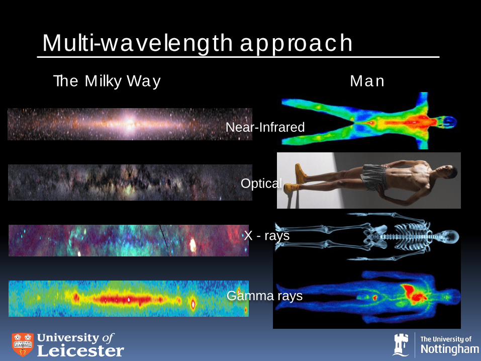
Multi-wavelength approach Man
Optical
X - rays
Gamma rays
Near-Infrared
The Milky Way
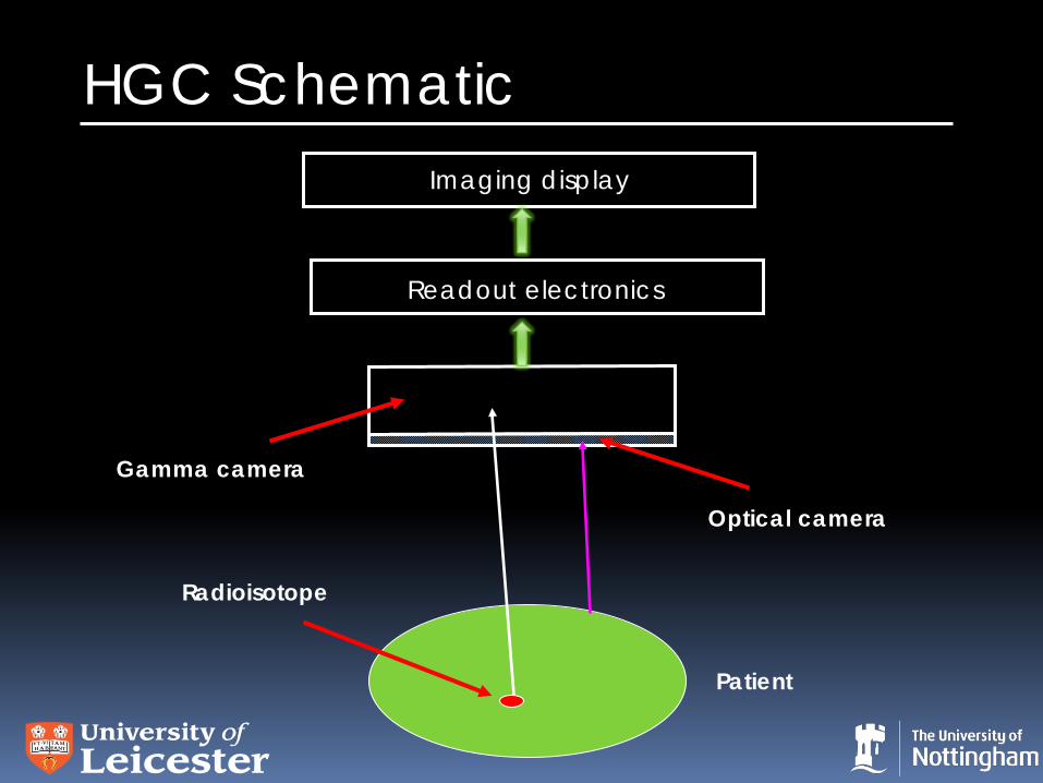
HGC Schematic
Optical camera
Gamma camera
Readout electronics
Imaging display
Patient
Radioisotope
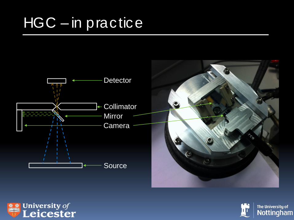
HGC – in practice
Detector
Collimator
Source
Mirror Camera
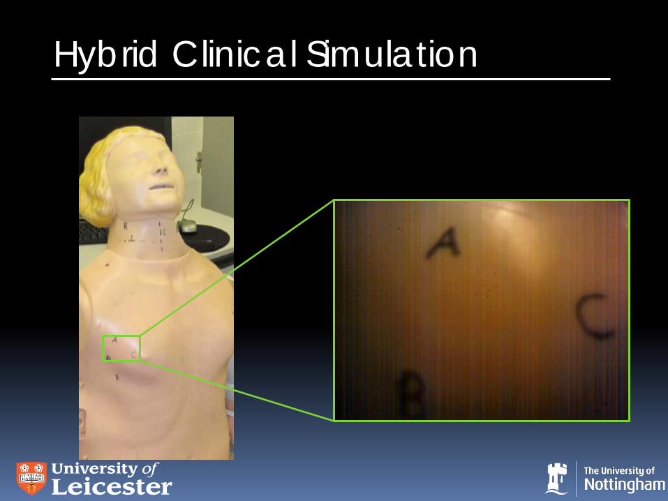
Hybrid Clinical Simulation
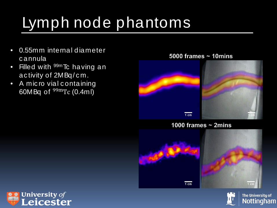
Lymph node phantoms • 0.55mm internal diameter
cannula • Filled with 99mTc having an
activity of 2MBq/cm. • A micro vial containing
60MBq of Tc99m (0.4ml)
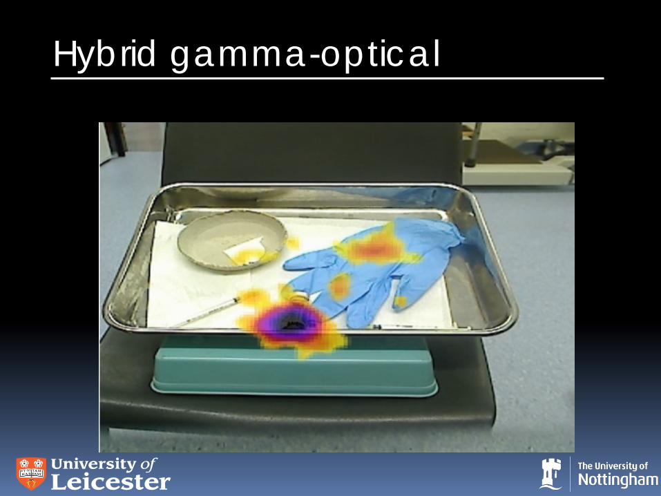
Hybrid gamma-optical
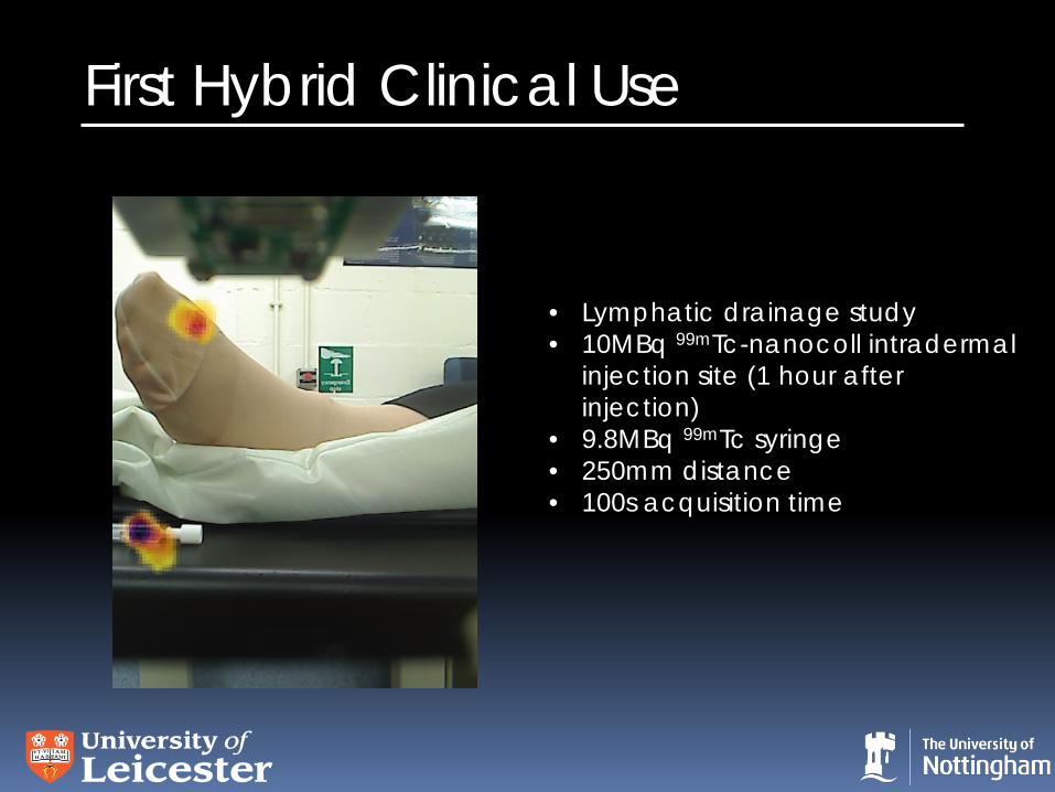
First Hybrid Clinical Use
• Lymphatic drainage study • 10MBq 99mTc-nanocoll intradermal
injection site (1 hour after injection)
• 9.8MBq 99mTc syringe • 250mm distance • 100s acquisition time
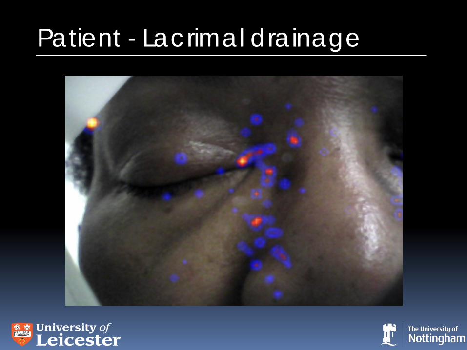
Patient - Lacrimal drainage
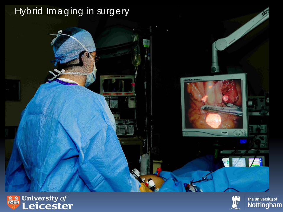
Hybrid Imaging in surgery
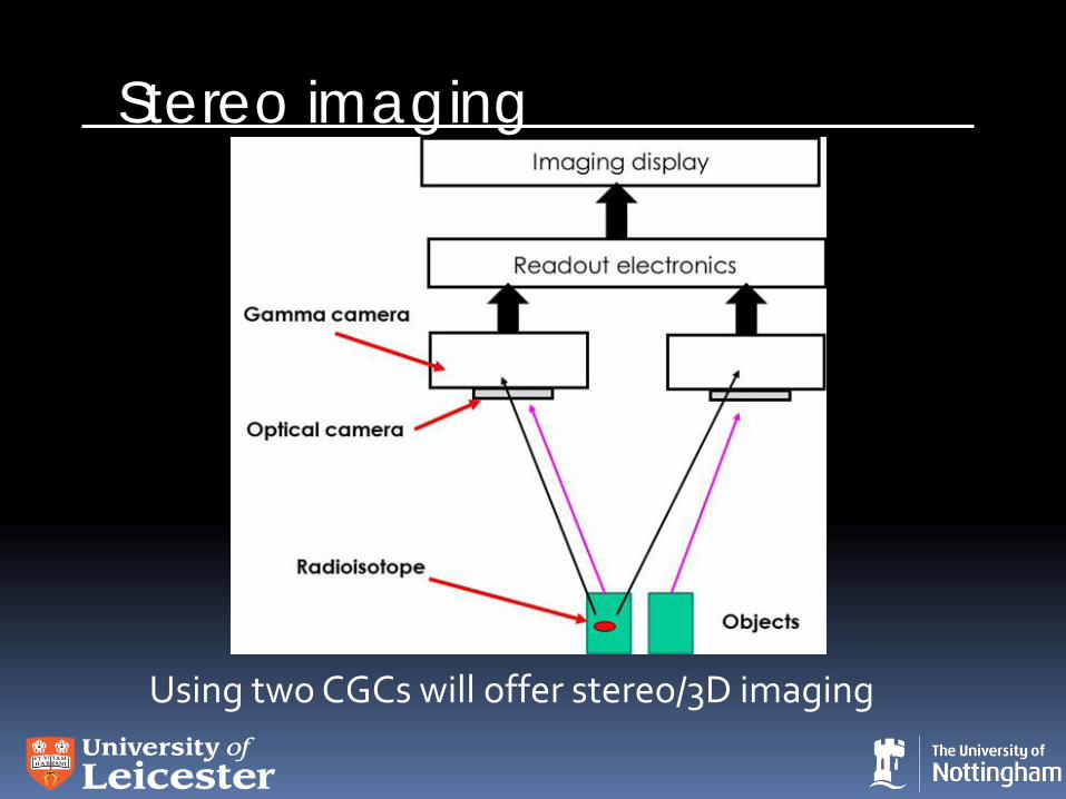
Stereo imaging
Using two CGCs will offer stereo/3D imaging
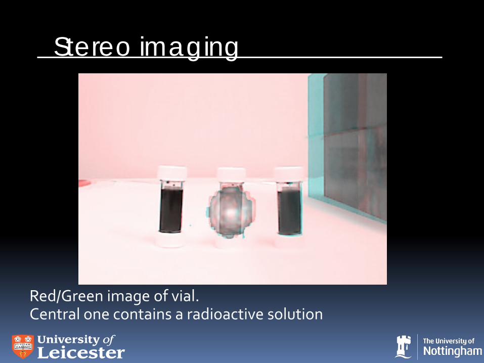
Stereo imaging
Red/Green image of vial. Central one contains a radioactive solution
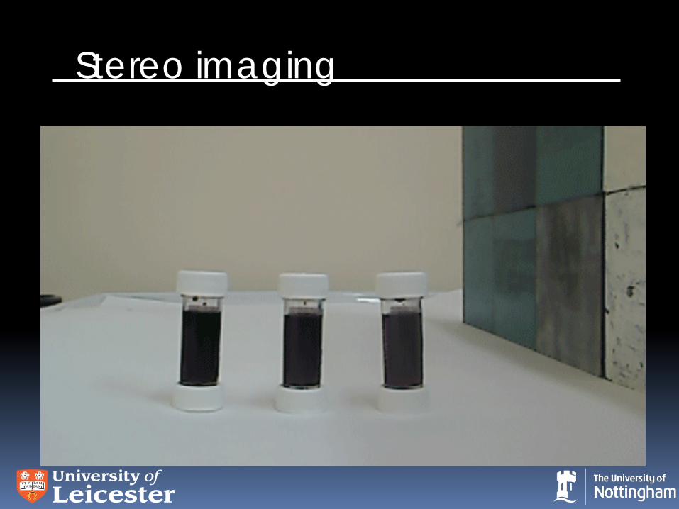
Stereo imaging
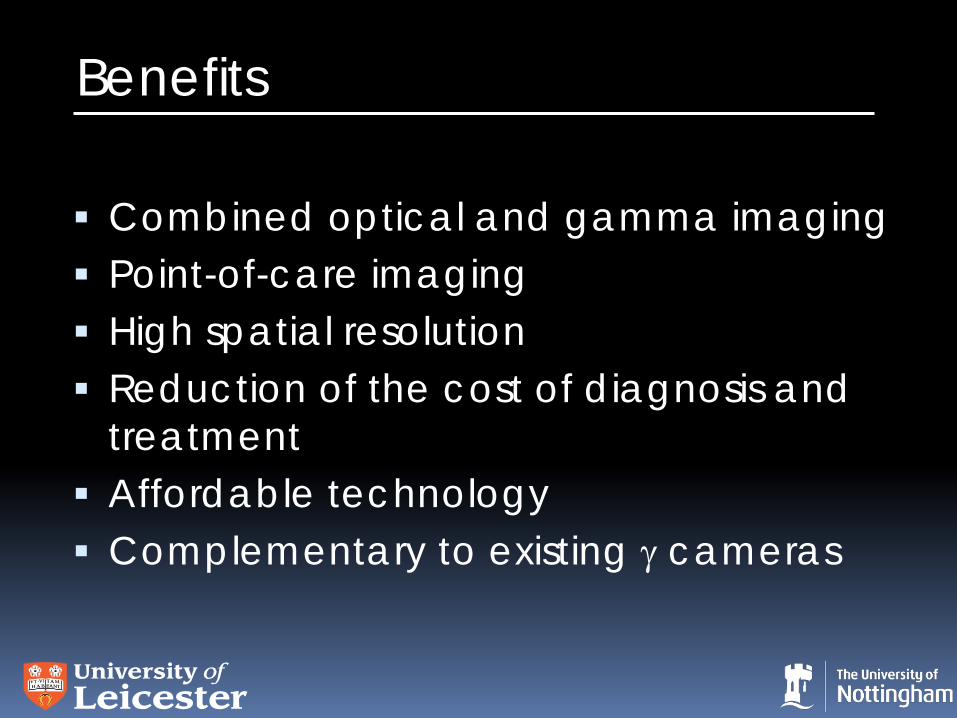
Benefits
Combined optical and gamma imaging Point-of-care imaging High spatial resolution Reduction of the cost of diagnosis and
treatment Affordable technology Complementary to existing γ cameras
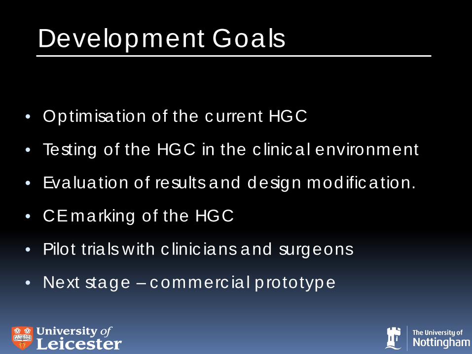
Development Goals
• Optimisation of the current HGC
• Testing of the HGC in the clinical environment
• Evaluation of results and design modification.
• CE marking of the HGC
• Pilot trials with clinicians and surgeons
• Next stage – commercial prototype
More Information John Lees [email protected] http://www.le.ac.uk/physics/bioimaging/ Papers • Bugby, S.L., J.E. Lees, B.S. Bhatia, and A.C. Perkins,
Characterisation of a high resolution small field of view portable gamma camera. Physica Medica, 30 (2014) 331-339.
• J E Lees, DJ Bassford, OE Blake, PE Blackshaw, AC Perkins, A Hybrid Camera for simultaneous imaging of gamma and optical photons, J. Inst. 7 (2012) P06009
• J E Lees, DJ Bassford, OE Blake, PE Blackshaw, AC Perkins, A high resolution Small Field Of View (SFOV) gamma camera: a columnar scintillator coated CCD imager for medical applications, J. Inst. 6 (2011) C12033
Acknowledgements University of Leicester
S L Bugby, B S Bhatia, L K Jambi, M S Alqahtani and W R McKnight
University Hospitals Nottingham
A C Perkins, A K Ng and PE Blackshaw Leicester Royal Infirmary
Helen Hill and David Monk