Embed Size (px)
Citation preview
September 22, 2016Volume 7
THEME: AXILLARY BRACHIAL PLEXUS BLOCK
ACADEMY OF REGIONAL ANAESTHESIA OF INDIA
1
Office Bearers Letter from the Editor's DeskGreetings! It is indeed a great honour for me to serve as the guest editor for this edition of the AORA newsletter.
In this issue, we look at the axillary brachial plexus block and the terminal nerves of the hand. The axillary block is an approach that has maintained its simplicity and effectiveness throughout the years. Most anaesthesiologists will reminiscence this technique being amongst their first experiences in regional anesthesia. With the advent of ultrasound, this block is now more reliable than ever. Considering that the smaller terminal nerves can now be visualized, it is possible to deliver target specific anesthesia and analgesia to the distal hand.
It is often hard to learn from people who are just like you. Too much is taken for granted.
Homogeneity is fine in a bottle of milk, but in the classroom it diminishes the curiosity that ignites
discovery. -- Vivian Gussin Paley
We are honoured to have several experts sharing their knowledge, experience and thoughts for this newsletter. Diversity in education is often unnoticed and unappreciated. Whether it is the dedicated anaesthesiologist in far reach areas trying to make a difference to the community, or the ones in our premier institutions researching newer avenues, we all have much to learn from them. We are blessed to live in a country as diverse as ours, with colleagues who approach the same problem through different eyes and share with us what they see. Nothing could be more mentally stimulating!
There is no better melting pot for diversity in regional anaesthesia than the annual AORA conference. This year's event in Hyderabad should once again be a time where we meet, share and learn from each other. Knowledge must have no boundaries and barriers. I wish all of you the best for your endeavours in regional anaesthesia.
Amjad Maniar
On behalf of Dr T.V.S Gopal, Dr Rani Sundar & Dr Guruprasad
Editorial Board
ChairmanDr. J Balavenkat Subramanian Coimbatore
Academic DirectorDr. TVS Gopal Hyderabad
PresidentDr. Sandeep DiwanPune
Vice PresidentsDr. Deep AroraGurgaonDr. M V KumarBengaluru
Secretary & TrusteeDr. Vrushali PondeMumbai
Associate SecretaryDr. Ashit MehtaSholapur
TreasurerDr. Satish KulkarniMumbai
ACADEMY OF REGIONAL ANAESTHESIA OF INDIA
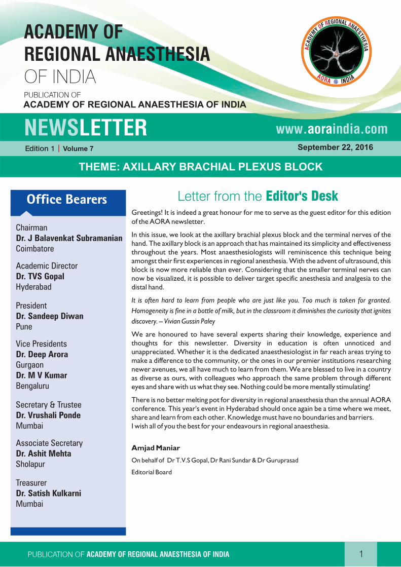
THE AESCULAP ACADEMY GUIDANCE CENTRE
Delhi Pain Management Centre GANGA HOSPITAL
A learning platform for Regional Anaesthesia
www.aesculap-academy-in.com
For more detail please visitFor Registration please contact:[email protected]
3
Dear all,
Greetings,
Individual effort when well directed can accomplish much, but the greatest good must necessarily come from the combined efforts of many. The power of combined effort knows no limitation. This is what AORA is all about.
In 2011, a few of us came together and embarked upon a project to cater to the needs of regional anesthesia in India and formed AORA. We were soon united with a myriad of regional anesthesia enthusiasts who joined us, and the numbers are increasing day by day. Your commitment has led to the growth of A O R A.
On taking over as president the last year I was in pursuit of two things. Firstly, the growth in numbers of delegates in AORA. We had an extensive membership drive over the past year and were partially successful in this endeavor.
The second was our ability to reach remote locations. The anesthesiologists from rural areas and those who could not join the mainstream conferences were the ones we targeted. We conducted live neurostimulationworkshops in Jorhat, Assam and in Bilaspur, Chattisgarh. In the future, we have proposed live workshops at Bhavnagar, Gujarat and in Bhubaneshwar, Orissa.
I request and urge all faculties to spend time and share your knowledge. We aim to build a well-defined network throughout India to ensure every anesthesiologist is trained in the basics of regional anesthesia.
In a few workshops, we were encouraged to note that the local organizations as well as hospitals in towns and cities recognized the contribution that AORA has made.
To sum it up there is no “I” in AORA it is always “WE”.
My sincere thanks to every member who has participated in our efforts.
Long Live AORA.
Sandeep Diwan
President
AORA INDIA
Message from the President
Namaste! From the Secretary of Academy of Regional Anaesthesia (AORA), INDIA.
We as AORA took a pledge of learning and teaching regional anaesthesia with all its nuances 6 years back. AORA is getting better every passing day, and with unimpeachable integrity! The eyes of the world are on us and the EDRA exams stands proof!
We have flourished to over 900 members at AORA. Every year the number of delegates AORA attracts is bigger than the previous, standing testimony to the scientific quality of AORA. As a true AORA enthusiast, every person is in the hall, contributing his or her bit by intently grasping knowledge and expressing their views. We all go home enriched, to improve patient care which is a pledge of AORA.
My dear present and future Members of AORA, we do not compromise on integrity and accountability. If you have had any difficulties, please do not hesitate to contact us.
We have yet lots of work to do, no point promising it, we will try our best to deliver.
AORA would love to take suggestions from any of you, please do not hesitate to contact us, should you have any.
With Reverence,
Yours truly,
Vrushali Ponde
Secretary & Trustee
AORA INDIA
Update from the Secretary
ACADEMY OF REGIONAL ANAESTHESIA OF INDIA
Hyderabad
Introduction
'Regional anaesthesia always works – provided you put the right dose of the right drug in the right place' wrote Denny and Harrop-Griffiths in an editorial in the British Journal of Anaesthesia. In pursuit of this simplistic goal, there was reliance on surface anatomical landmarks and paraesthesia feedback from the awake patient for successful peripheral nerve blocks. 'No paraesthesia, no anaesthesia', by Danny Moore in 1953, was the prevalent dictum. Multiple attempts to elicit paraesthesia, which was perceived as painful, failure rate of nearly 20%, and fears of neurological sequelae prompted search for safer guidance.
The advent of peripheral nerve stimulation (PNS), and later, of ultrasound guidance for regional anaesthesia (USGRA) transformed the 'art' of nerve blockade into an exact and objective science. While peripheral nerve stimulation (PNS) improved reliability by eliciting a muscle twitch to indicate correct perineural placement of the needle, ultrasound guidance offered visualization of needle, nerve and deposition of local anaesthetic solution in real time.
The past few decades witnessed a resurgence in the practice of regional anaesthesia and the clinical and economic benefits of nerve blocks have received widespread appreciation. This article provides an insight into basic physics and image interpretation of portable, high resolution ultrasound.
High Resolution Portable Ultrasound
Though the objective of eliciting a muscle twitch with electrical stimulation to denote correct perineural location of the needle was often achieved, problems with electrical stimulation did persist. Instances of partial block or failure despite a twitch, or inability to elicit a response, reinforced the credo that blocks with electrical stimulation were essentially blind procedures, and variations in human anatomy contributed to the failure. The advent of ultrasound, as a guidance tool, has redefined the practice of regional anaesthesia owing to the ability to visualize nerves and surrounding structures, the advancing needle, and the spread of local anaesthetic solution in real time.
History
La Grange and colleagues were the first to use an ultrasonographic blood flow detector to locate the subclavian artery for the performance of a supraclavicular brachial plexus block in 1978. As ultrasound technology at that time was limited, this was, however, an indirect application. The first reported series of direct visualization
INTRODUCTION TO BASICS OF ULTRASOUND FOR THE ANAESTHETIST
T V S Gopal
of the supraclavicular brachial plexus was by Stephen Kapral et al, in 1994. Thereafter, significant improvement in technology increased the popularity of ultrasound amongst anaesthesiologists for peripheral nerve blockade, and, at present, for neuraxial blocks and chronic pain procedures. Today, after more than a decade of widespread use, ultrasound guidance for regional anaesthesia is the universal 'gold standard'.
Basic Physics of Ultrasound :
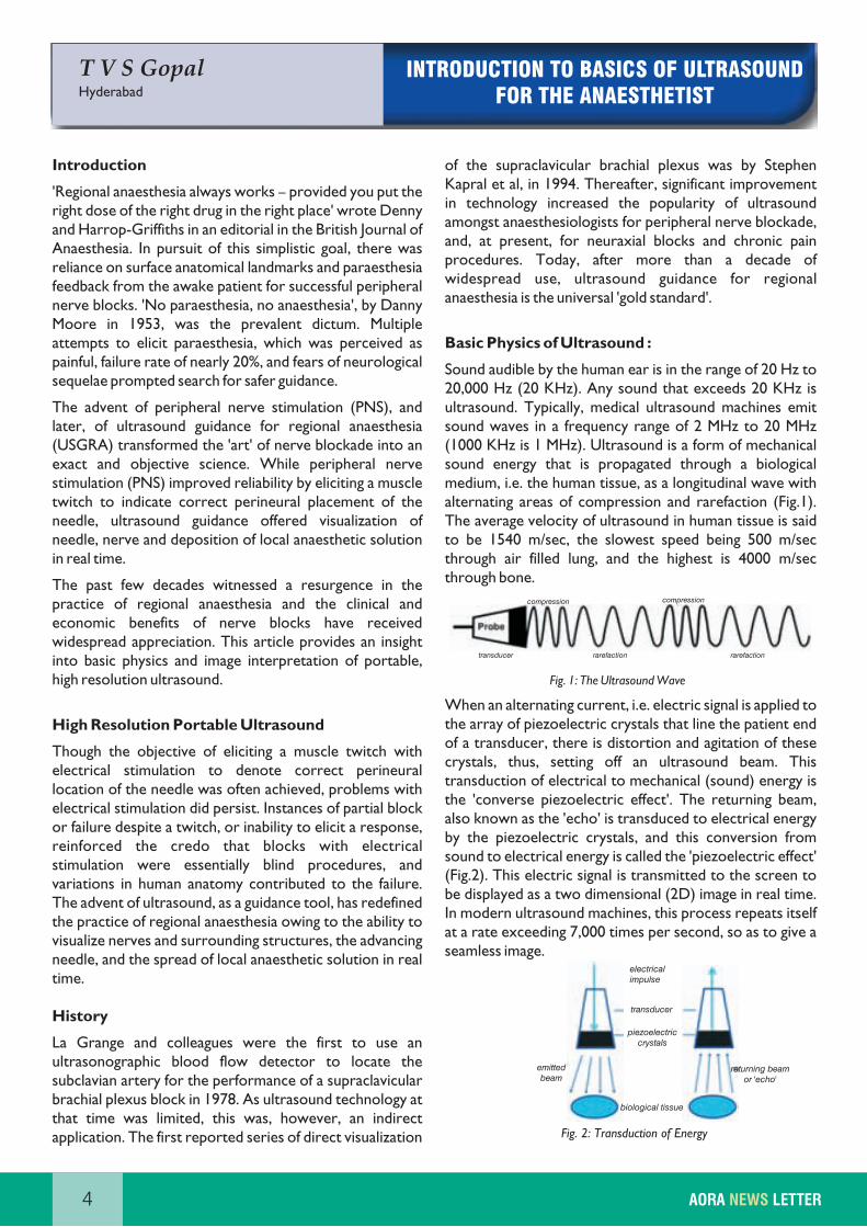
Sound audible by the human ear is in the range of 20 Hz to 20,000 Hz (20 KHz). Any sound that exceeds 20 KHz is ultrasound. Typically, medical ultrasound machines emit sound waves in a frequency range of 2 MHz to 20 MHz (1000 KHz is 1 MHz). Ultrasound is a form of mechanical sound energy that is propagated through a biological medium, i.e. the human tissue, as a longitudinal wave with alternating areas of compression and rarefaction (Fig.1). The average velocity of ultrasound in human tissue is said to be 1540 m/sec, the slowest speed being 500 m/sec through air filled lung, and the highest is 4000 m/sec through bone.
Fig. 1: The Ultrasound Wave
When an alternating current, i.e. electric signal is applied to the array of piezoelectric crystals that line the patient end of a transducer, there is distortion and agitation of these crystals, thus, setting off an ultrasound beam. This transduction of electrical to mechanical (sound) energy is the 'converse piezoelectric effect'. The returning beam, also known as the 'echo' is transduced to electrical energy by the piezoelectric crystals, and this conversion from sound to electrical energy is called the 'piezoelectric effect' (Fig.2). This electric signal is transmitted to the screen to be displayed as a two dimensional (2D) image in real time. In modern ultrasound machines, this process repeats itself at a rate exceeding 7,000 times per second, so as to give a seamless image.
Fig. 2: Transduction of Energy
4 AORA LETTERNEWS
compression compression
transducer rarefaction rarefaction
electricalimpulse
transducer
piezoelectriccrystals
emitted beam
biological tissue
returning beamor 'echo'
5
The ultrasound beam, within the biological tissue, has several interactions (Fig. 3). Any of the following are possible :1. Transmission – the beam passes through the tissue. As
it progresses, the amplitude of the original signal becomes weaker, this process is known as 'attenuation'.
2. Refraction – a change in the direction of the transmitted beam.
3. Scatter – the beam is weakened and scattered in different directions when it encounters an interface that is small or irregular.
4. Reflection – most of the incident beam is reflected back to the transducer by a structure with smooth interface.
Interaction of Ultrasound Beam at Tissue Interface
Echogenicity is the term used to describe the brightness, or darkness, of tissues relative to other structures in the field of vision. When the structure is brighter than surrounding tissues, it is said to be hyperechoic. If it is darker than surrounding tissues, it is hypoechoic. Blood vessels appear totally dark, i.e. anechoic (Fig. 4a & b).
1. Veins Anechoic (compressible with transducer)
2. Arteries Anechoic (pulsatile)
3. Muscles Hypoechoic tissue with intermittent bright striae.
4. Bone Hyperechoic curved rim with dark shadow beneath.
5. Nerves Hyperechoic/Hypoechoic
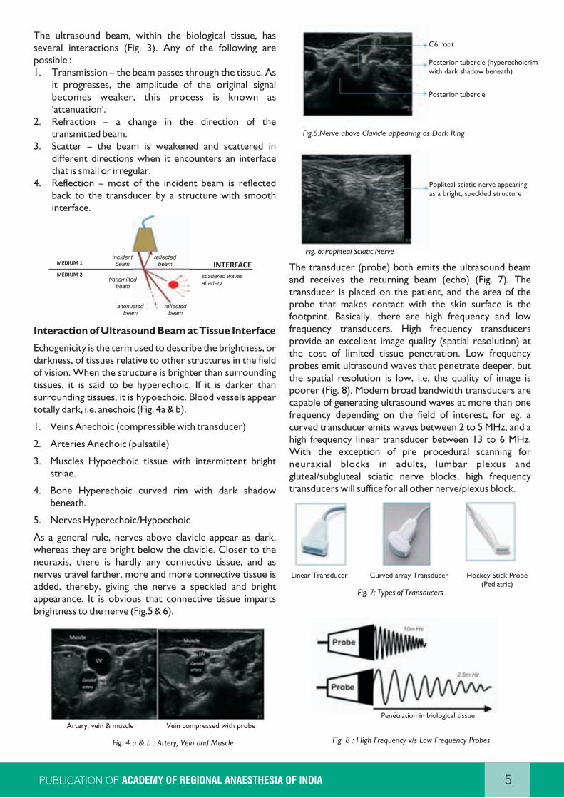
As a general rule, nerves above clavicle appear as dark, whereas they are bright below the clavicle. Closer to the neuraxis, there is hardly any connective tissue, and as nerves travel farther, more and more connective tissue is added, thereby, giving the nerve a speckled and bright appearance. It is obvious that connective tissue imparts brightness to the nerve (Fig.5 & 6).
Fig. 4 a & b : Artery, Vein and Muscle
Fig. 6: Popliteal Sciatic Nerve
The transducer (probe) both emits the ultrasound beam and receives the returning beam (echo) (Fig. 7). The transducer is placed on the patient, and the area of the probe that makes contact with the skin surface is the footprint. Basically, there are high frequency and low frequency transducers. High frequency transducers provide an excellent image quality (spatial resolution) at the cost of limited tissue penetration. Low frequency probes emit ultrasound waves that penetrate deeper, but the spatial resolution is low, i.e. the quality of image is poorer (Fig. 8). Modern broad bandwidth transducers are capable of generating ultrasound waves at more than one frequency depending on the field of interest, for eg. a curved transducer emits waves between 2 to 5 MHz, and a high frequency linear transducer between 13 to 6 MHz. With the exception of pre procedural scanning for neuraxial blocks in adults, lumbar plexus and gluteal/subgluteal sciatic nerve blocks, high frequency transducers will suffice for all other nerve/plexus block.
Fig. 7: Types of Transducers
Fig. 8 : High Frequency v/s Low Frequency Probes
Linear Transducer Curved array Transducer Hockey Stick Probe(Pediatric)
Popliteal sciatic nerve appearingas a bright, speckled structure
Artery, vein & muscle Vein compressed with probe
Penetration in biological tissue
ACADEMY OF REGIONAL ANAESTHESIA OF INDIA
Posterior tubercle (hyperechoicrim with dark shadow beneath)
C6 root
Posterior tubercle
Fig.5:Nerve above Clavicle appearing as Dark Ring
incidentbeam
reflectedbeam
transmittedbeam
attenuatedbeam
reflectedbeam
scattered wavesat artery
INTERFACEMEDIUM 1
MEDIUM 2
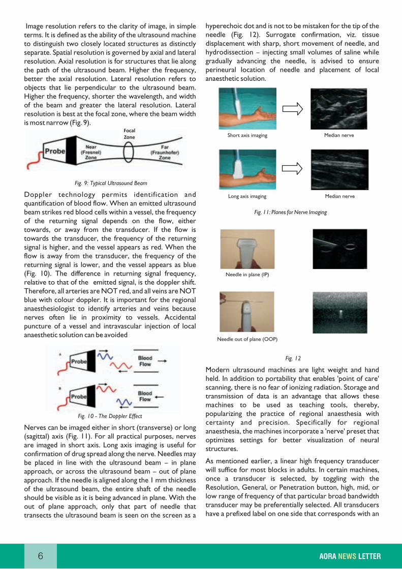
Image resolution refers to the clarity of image, in simple terms. It is defined as the ability of the ultrasound machine to distinguish two closely located structures as distinctly separate. Spatial resolution is governed by axial and lateral resolution. Axial resolution is for structures that lie along the path of the ultrasound beam. Higher the frequency, better the axial resolution. Lateral resolution refers to objects that lie perpendicular to the ultrasound beam. Higher the frequency, shorter the wavelength, and width of the beam and greater the lateral resolution. Lateral resolution is best at the focal zone, where the beam width is most narrow (Fig. 9).
Fig. 9: Typical Ultrasound Beam
Doppler technology permits identification and quantification of blood flow. When an emitted ultrasound beam strikes red blood cells within a vessel, the frequency of the returning signal depends on the flow, either towards, or away from the transducer. If the flow is towards the transducer, the frequency of the returning signal is higher, and the vessel appears as red. When the flow is away from the transducer, the frequency of the returning signal is lower, and the vessel appears as blue (Fig. 10). The difference in returning signal frequency, relative to that of the emitted signal, is the doppler shift. Therefore, all arteries are NOT red, and all veins are NOT blue with colour doppler. It is important for the regional anaesthesiologist to identify arteries and veins because nerves often lie in proximity to vessels. Accidental puncture of a vessel and intravascular injection of local anaesthetic solution can be avoided
Fig. 10 - The Doppler Effect
Nerves can be imaged either in short (transverse) or long (sagittal) axis (Fig. 11). For all practical purposes, nerves are imaged in short axis. Long axis imaging is useful for confirmation of drug spread along the nerve. Needles may be placed in line with the ultrasound beam – in plane approach, or across the ultrasound beam – out of plane approach. If the needle is aligned along the 1 mm thickness of the ultrasound beam, the entire shaft of the needle should be visible as it is being advanced in plane. With the out of plane approach, only that part of needle that transects the ultrasound beam is seen on the screen as a
hyperechoic dot and is not to be mistaken for the tip of the needle (Fig. 12). Surrogate confirmation, viz. tissue displacement with sharp, short movement of needle, and hydrodissection – injecting small volumes of saline while gradually advancing the needle, is advised to ensure perineural location of needle and placement of local anaesthetic solution.
Fig. 11: Planes for Nerve Imaging
Fig. 12
Modern ultrasound machines are light weight and hand held. In addition to portability that enables 'point of care' scanning, there is no fear of ionizing radiation. Storage and transmission of data is an advantage that allows these machines to be used as teaching tools, thereby, popularizing the practice of regional anaesthesia with certainty and precision. Specifically for regional anaesthesia, the machines incorporate a 'nerve' preset that optimizes settings for better visualization of neural structures.
As mentioned earlier, a linear high frequency transducer will suffice for most blocks in adults. In certain machines, once a transducer is selected, by toggling with the Resolution, General, or Penetration button, high, mid, or low range of frequency of that particular broad bandwidth transducer may be preferentially selected. All transducers have a prefixed label on one side that corresponds with an
6 AORA LETTERNEWS
Median nerveShort axis imaging
Median nerveLong axis imaging
Needle in plane (IP)
Needle out of plane (OOP)
FocalZone
orientation marker (a blue dot or the logo of the manufacturer) on the top left corner of the screen (Fig.13). Transducer orientation is essential to maintain uniformity such that sidedness of structures displayed on screen is constant. Some gel is applied on the transducer and a finger placed on one end to make a movement. If the movement corresponds with the orientation marker, that side of the transducer is placed to the right of the patient in short axis, and head end of the patient in long axis.
Fig. 13 - Orientation Marker at Top Left of Screen
The depth is adjusted such that the structure of interest is located at the centre of the screen. Time gain compensation (TGC) is akin to contrast function of a television set, and selectively amplifies returning echoes from greater depths, so as to present a homogenous image on the screen. An auto gain button facilitates the TGC setting with minimal effort. In machines that have focus
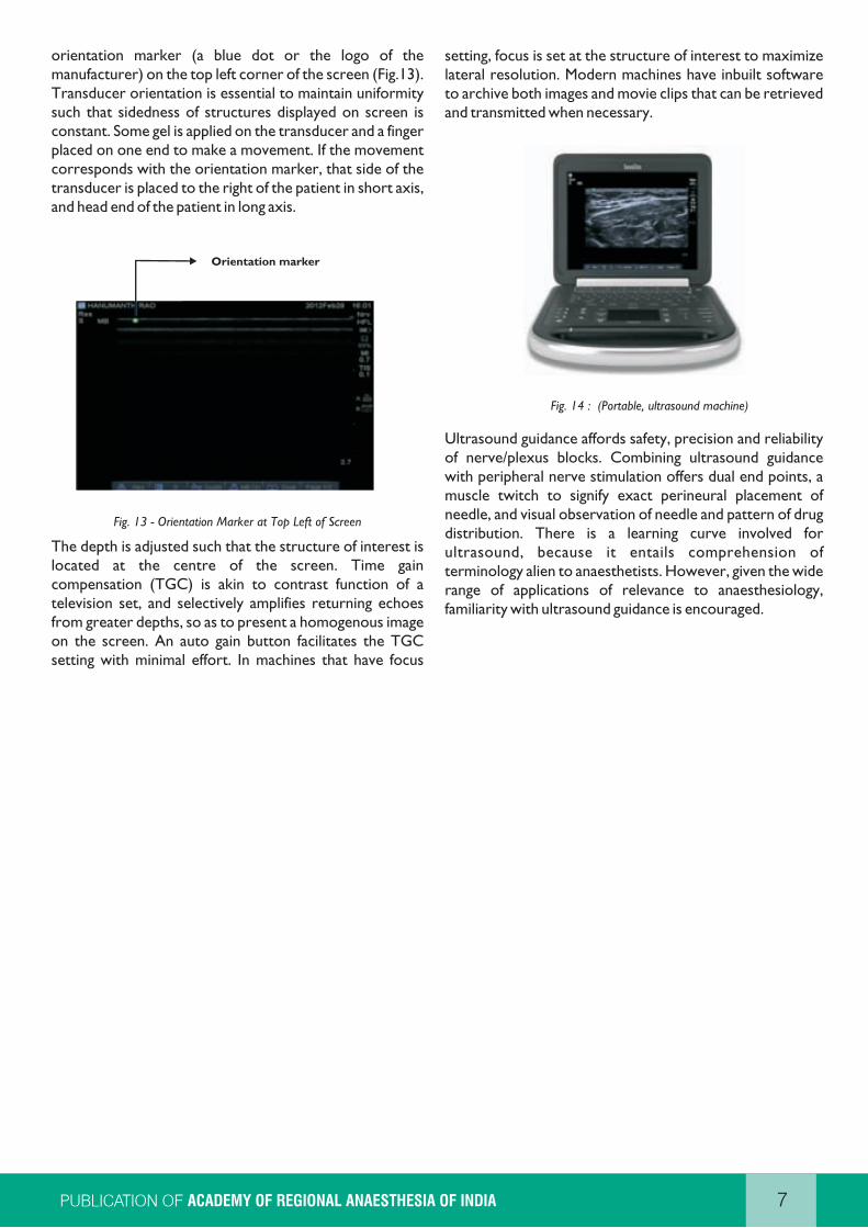
setting, focus is set at the structure of interest to maximize lateral resolution. Modern machines have inbuilt software to archive both images and movie clips that can be retrieved and transmitted when necessary.
Fig. 14 : (Portable, ultrasound machine)
Ultrasound guidance affords safety, precision and reliability of nerve/plexus blocks. Combining ultrasound guidance with peripheral nerve stimulation offers dual end points, a muscle twitch to signify exact perineural placement of needle, and visual observation of needle and pattern of drug distribution. There is a learning curve involved for ultrasound, because it entails comprehension of terminology alien to anaesthetists. However, given the wide range of applications of relevance to anaesthesiology, familiarity with ultrasound guidance is encouraged.
Orientation marker
7ACADEMY OF REGIONAL ANAESTHESIA OF INDIA
Dr Praveen Talawar,
Dr Anjolie Chhabra Anatomy of the Axillary Brachial Plexus
New Delhi
Introduction
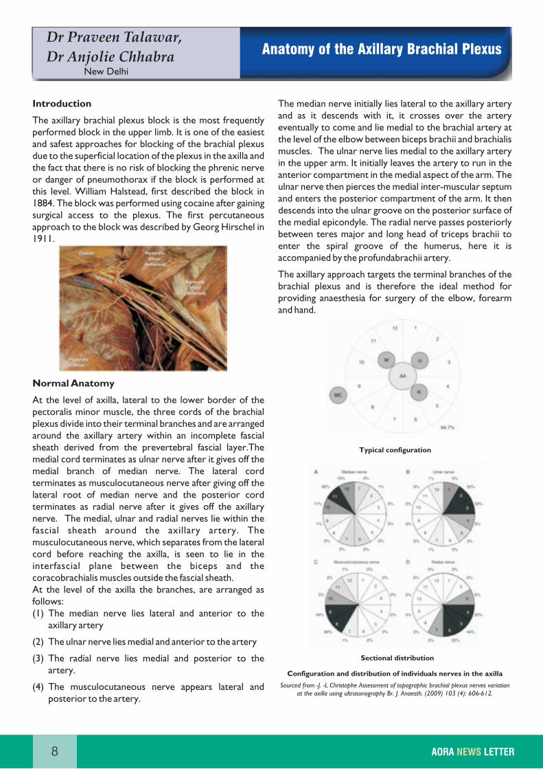
The axillary brachial plexus block is the most frequently performed block in the upper limb. It is one of the easiest and safest approaches for blocking of the brachial plexus due to the superficial location of the plexus in the axilla and the fact that there is no risk of blocking the phrenic nerve or danger of pneumothorax if the block is performed at this level. William Halstead, first described the block in 1884. The block was performed using cocaine after gaining surgical access to the plexus. The first percutaneous approach to the block was described by Georg Hirschel in 1911.
Normal Anatomy
At the level of axilla, lateral to the lower border of the pectoralis minor muscle, the three cords of the brachial plexus divide into their terminal branches and are arranged around the axillary artery within an incomplete fascial sheath derived from the prevertebral fascial layer.The medial cord terminates as ulnar nerve after it gives off the medial branch of median nerve. The lateral cord terminates as musculocutaneous nerve after giving off the lateral root of median nerve and the posterior cord terminates as radial nerve after it gives off the axillary nerve. The medial, ulnar and radial nerves lie within the fascial sheath around the axillary artery. The musculocutaneous nerve, which separates from the lateral cord before reaching the axilla, is seen to lie in the interfascial plane between the biceps and the coracobrachialis muscles outside the fascial sheath.At the level of the axilla the branches, are arranged as follows: (1) The median nerve lies lateral and anterior to the
axillary artery
(2) The ulnar nerve lies medial and anterior to the artery
(3) The radial nerve lies medial and posterior to the artery.
(4) The musculocutaneous nerve appears lateral and posterior to the artery.
The median nerve initially lies lateral to the axillary artery and as it descends with it, it crosses over the artery eventually to come and lie medial to the brachial artery at the level of the elbow between biceps brachii and brachialis muscles. The ulnar nerve lies medial to the axillary artery in the upper arm. It initially leaves the artery to run in the anterior compartment in the medial aspect of the arm. The ulnar nerve then pierces the medial inter-muscular septum and enters the posterior compartment of the arm. It then descends into the ulnar groove on the posterior surface of the medial epicondyle. The radial nerve passes posteriorly between teres major and long head of triceps brachii to enter the spiral groove of the humerus, here it is accompanied by the profundabrachii artery.
The axillary approach targets the terminal branches of the brachial plexus and is therefore the ideal method for providing anaesthesia for surgery of the elbow, forearm and hand.
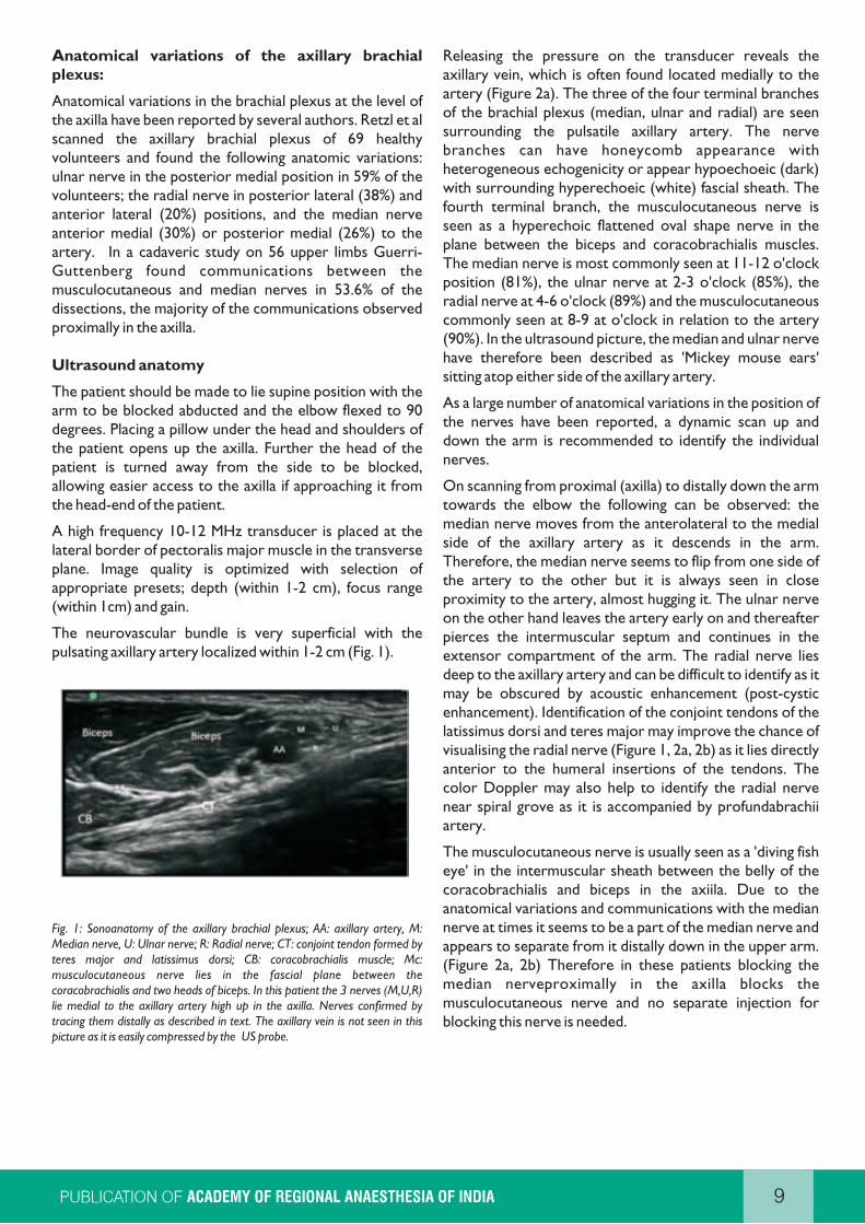
Configuration and distribution of individuals nerves in the axilla
Sourced from -J. -L Christophe Assessment of topographic brachial plexus nerves variation at the axilla using ultrasonography Br. J. Anaesth. (2009) 103 (4): 606-612.
8 AORA LETTERNEWS
Sectional distribution
Typical configuration
Anatomical variations of the axillary brachial plexus:
Anatomical variations in the brachial plexus at the level of the axilla have been reported by several authors. Retzl et al scanned the axillary brachial plexus of 69 healthy volunteers and found the following anatomic variations: ulnar nerve in the posterior medial position in 59% of the volunteers; the radial nerve in posterior lateral (38%) and anterior lateral (20%) positions, and the median nerve anterior medial (30%) or posterior medial (26%) to the artery. In a cadaveric study on 56 upper limbs Guerri-Guttenberg found communications between the musculocutaneous and median nerves in 53.6% of the dissections, the majority of the communications observed proximally in the axilla.
Ultrasound anatomy
The patient should be made to lie supine position with the arm to be blocked abducted and the elbow flexed to 90 degrees. Placing a pillow under the head and shoulders of the patient opens up the axilla. Further the head of the patient is turned away from the side to be blocked, allowing easier access to the axilla if approaching it from the head-end of the patient.
A high frequency 10-12 MHz transducer is placed at the lateral border of pectoralis major muscle in the transverse plane. Image quality is optimized with selection of appropriate presets; depth (within 1-2 cm), focus range (within 1cm) and gain.
The neurovascular bundle is very superficial with the pulsating axillary artery localized within 1-2 cm (Fig. 1).
Fig. 1: Sonoanatomy of the axillary brachial plexus; AA: axillary artery, M: Median nerve, U: Ulnar nerve; R: Radial nerve; CT: conjoint tendon formed by teres major and latissimus dorsi; CB: coracobrachialis muscle; Mc: musculocutaneous nerve lies in the fascial plane between the coracobrachialis and two heads of biceps. In this patient the 3 nerves (M,U,R) lie medial to the axillary artery high up in the axilla. Nerves confirmed by tracing them distally as described in text. The axillary vein is not seen in this picture as it is easily compressed by the US probe.
Releasing the pressure on the transducer reveals the axillary vein, which is often found located medially to the artery (Figure 2a). The three of the four terminal branches of the brachial plexus (median, ulnar and radial) are seen surrounding the pulsatile axillary artery. The nerve branches can have honeycomb appearance with heterogeneous echogenicity or appear hypoechoeic (dark) with surrounding hyperechoeic (white) fascial sheath. The fourth terminal branch, the musculocutaneous nerve is seen as a hyperechoic flattened oval shape nerve in the plane between the biceps and coracobrachialis muscles. The median nerve is most commonly seen at 11-12 o'clock position (81%), the ulnar nerve at 2-3 o'clock (85%), the radial nerve at 4-6 o'clock (89%) and the musculocutaneous commonly seen at 8-9 at o'clock in relation to the artery (90%). In the ultrasound picture, the median and ulnar nerve have therefore been described as 'Mickey mouse ears' sitting atop either side of the axillary artery.
As a large number of anatomical variations in the position of the nerves have been reported, a dynamic scan up and down the arm is recommended to identify the individual nerves.
On scanning from proximal (axilla) to distally down the arm towards the elbow the following can be observed: the median nerve moves from the anterolateral to the medial side of the axillary artery as it descends in the arm. Therefore, the median nerve seems to flip from one side of the artery to the other but it is always seen in close proximity to the artery, almost hugging it. The ulnar nerve on the other hand leaves the artery early on and thereafter pierces the intermuscular septum and continues in the extensor compartment of the arm. The radial nerve lies deep to the axillary artery and can be difficult to identify as it may be obscured by acoustic enhancement (post-cystic enhancement). Identification of the conjoint tendons of the latissimus dorsi and teres major may improve the chance of visualising the radial nerve (Figure 1, 2a, 2b) as it lies directly anterior to the humeral insertions of the tendons. The color Doppler may also help to identify the radial nerve near spiral grove as it is accompanied by profundabrachii artery.
The musculocutaneous nerve is usually seen as a 'diving fish eye' in the intermuscular sheath between the belly of the coracobrachialis and biceps in the axiila. Due to the anatomical variations and communications with the median nerve at times it seems to be a part of the median nerve and appears to separate from it distally down in the upper arm. (Figure 2a, 2b) Therefore in these patients blocking the median nerveproximally in the axilla blocks the musculocutaneous nerve and no separate injection for blocking this nerve is needed.
9ACADEMY OF REGIONAL ANAESTHESIA OF INDIA

Fig. 2a: See inset picture for position of ultrasound probe, high in the axilla at the distal end of the anterior axillary fold formed by pectoralis major and minor. Sonoanatomy reveals conjoint tendon (CT) at this level. AA: axillary artery; AV: Axillary vein; , M: Median nerve, U: Ulnar nerve; R: Radial nerve. Note that musculocutaneous nerve (Mc) is not visible in this patient at this level. It becomes visible on moving probe about 3 cm distally down the arm in Fig 2b.
Fig. 2b: Sonoanatomy on moving probe about 3 cm distally down the arm (inset picture) in the same patient, reveals the musculocutaneous nerve (Mc) appearing as if it in communication with or a branch of the Median nerve(M). Note the conjoint tendon (CT) is not seen at this level. The axillary vein is also not seen as it is easily compressed by the US probe. AA: axillary artery; M: Median nerve, U: Ulnar nerve; R: Radial nerve.
Though this pattern of the nerves on dynamic scanning is fairly constant in case of doubt the nerve stimulator can be used to confirm the individual nerves.Vascular variations are not uncommon in this region and at times an accessory axillary artery running parallel to the main axillary artery from the axilla to the elbow may be seen in 2.6% of cases.Similarly accessory axillary veins may also be present. Great caution should be taken to identify these vessels and avoid intravascular injections.
10 AORA LETTERNEWS
Dr Deep Arora,
Dr Om PrakashPeripheral nerves of the upper limb-
Anatomical reviewGurgaon
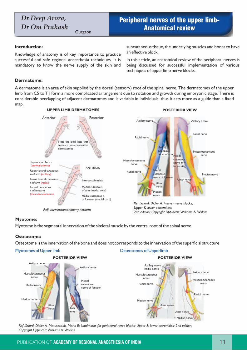
Ref: www.instantanatomy.net/arm
Ref :Sciard, Didier A. /nerves nerve blocks; Upper & lower extremities; 2nd edition; Copyright Lippincott Williams & Wilkins
Ref :Sciard, Didier A. Matuszczak, Maria E; Landmarks for peripheral nerve blocks; Upper & lower extremities; 2nd edition; Copyright Lippincott Williams & Wilkins
Anterior
UPPER LIMB DERMATOMES
Posterior
Note the axial lines thatseparate non-consecutivedermatomes
Supraclavicular ns(cervical plexus)
Upper lateral cutaneousn of arm (axillary)
Lateral cutaneousn of forearm (musculocutaneous)
Lower lateral cutaneous n of arm (radial)
ANTERIOR
Intercostobrachial
Medial cutaneous nof forearm (medial cord)
Medial cutaneousof arm (medial cord)
POSTERIOR VIEW
POSTERIOR VIEW POSTERIOR VIEW
Axillary nerveRadial nerve
Musculocutaneousnerve
Radial nerve
Median nerve
Ulnar nerve
Ulnar nerve
Median nerve
Radial nerve
Musculocutaneousnerve
Axillary nerve
11ACADEMY OF REGIONAL ANAESTHESIA OF INDIA
Axillary nerve
Radial nerve
Musculocutaneousnerve
Radial nerve
Median nerve
Ulnar nerve
Medialcutaneousnerve of arm
Medialcutaneousnerve of forearm
Ulnar nerve
Median nerve
Musculocutaneousnerve
Radial nerve
Axillary nerve
Medialcutaneousnerve of forearm
Axillary nerve
Musculocutaneousnerve
Radial nerve
Median nerve
Ulnar nerve Ulnar
nerve
Medialcutaneousnerve of forearm
Axillary nerve
12 AORA LETTERNEWS
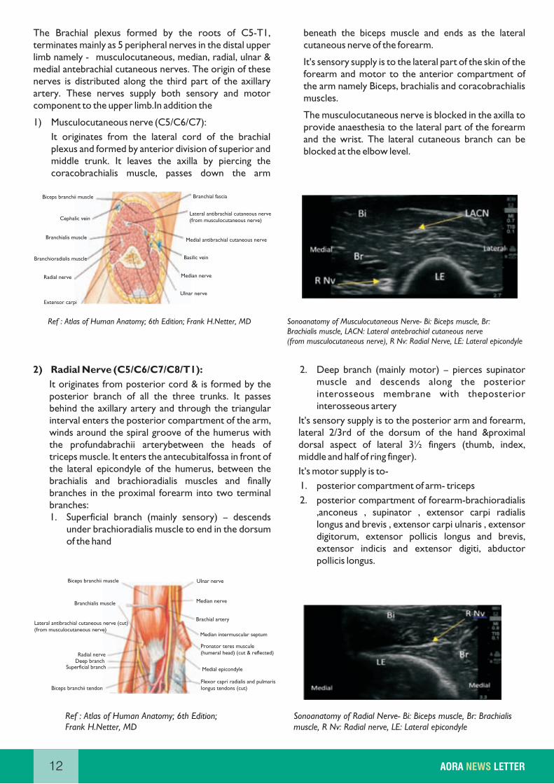
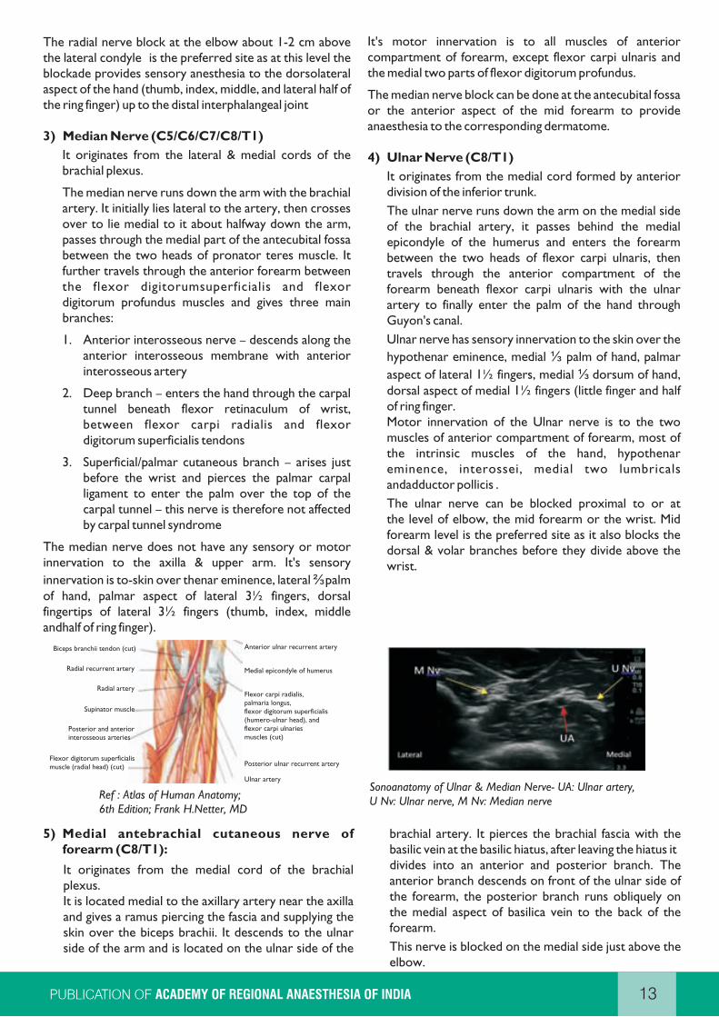
Ref : Atlas of Human Anatomy; 6th Edition; Frank H.Netter, MD Sonoanatomy of Musculocutaneous Nerve- Bi: Biceps muscle, Br: Brachialis muscle, LACN: Lateral antebrachial cutaneous nerve (from musculocutaneous nerve), R Nv: Radial Nerve, LE: Lateral epicondyle
Ref : Atlas of Human Anatomy; 6th Edition; Frank H.Netter, MD
Sonoanatomy of Radial Nerve- Bi: Biceps muscle, Br: Brachialis muscle, R Nv: Radial nerve, LE: Lateral epicondyle
Biceps branchii muscle
Cephalic vein
Branchialis muscle
Branchioradialis muscle
Radial nerve
Extensor carpi
Branchial fascia
Medial antibrachial cutaneous nerve
Basilic vein
Lateral antibrachial cutaneous nerve(from musculocutaneous nerve)
Ulnar nerve
Median nerve
Biceps branchii muscle
Branchialis muscle Median nerve
Lateral antibrachial cutaneous nerve (cut)(from musculocutaneous nerve)
Radial nerveDeep branch
Superficial branch
Biceps branchii tendon
Ulnar nerve
Brachial artery
Median intermuscular septum
Pronator teres muscule (humeral head) (cut & reflected)
Medial epicondyle
Flexor capri radialis and pulmarislongus tendons (cut)
13ACADEMY OF REGIONAL ANAESTHESIA OF INDIA
Ref : Atlas of Human Anatomy; 6th Edition; Frank H.Netter, MD
Sonoanatomy of Ulnar & Median Nerve- UA: Ulnar artery, U Nv: Ulnar nerve, M Nv: Median nerve
Biceps branchii tendon (cut)
Radial recurrent artery
Radial artery
Supinator muscle
Posterior and anteriorinterosseous arteries
Flexor digitorum superficialismuscle (radial head) (cut)
Anterior ulnar recurrent artery
Medial epicondyle of humerus
Flexor carpi radialis,palmaria longus,flexor digitorum superficialis(humero-ulnar head), andflexor carpi ulnariesmuscles (cut)
Posterior ulnar recurrent artery
Ulnar artery
Dr Hetalkumar Vadera PNS Guided Axillary Block Rajkot, Gujarat
Introduction
The Axillary brachial plexus block is safest of all approaches to brachial plexus block. Dr. Halsted first described it in 1884 at Roosevelt Hospital, New York.
You can prevent complications like phrenic nerve palsy and pneumothorax with this approach.
Indications
Elbow surgery
Forearm surgery
Hand surgery
Landmarks and patient position with trouble shooting in finding landmarks
Palpating the axillary artery in the axilla is easy even in obese patients. The nerves lie within a sheath surrounding the artery.
The Patient is positioned in supine position with arm abducted to 90? and elbow flexed.
Palpation of the axillary artery is sometimes difficult in muscular and in obese patients. We can use a nerve mapper in this situation to locate the nerve with percutaneous stimulation.
Equipment
Sterile gloves, towel, gauze squares
Syringes (10/20 ml) with LA
5 ml syringe with 2% xylocaine for skin infiltration
2.5-5 cm 22 G short bevel, insulated stimulating needle
Peripheral nerve stimulator
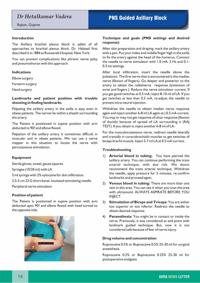
Position of patient
The Patient is positioned in supine position with arm abducted upto 90? and elbow flexed with head turned to the opposite side.
Technique and goals (PNS settings and desired response)
After skin preparation and draping, mark the axillary artery with a pen. Put your index and middle finger high in the axilla to fix the artery against the head of the humerus. Connect the needle to nerve stimulator with 1.0 mA, 2 Hz and 0.1-0.3 ms settings.
After local infiltration, insert the needle above the pulsations. The first nerve that is encountered is the median nerve (flexion of fingers). Go deeper and posterior to the artery to obtain the radialnerve response (extension of wrist and fingers ). Reduce the nerve stimulator current. If you get good twitches at 0.5 mA, inject 8-10 ml of LA. If you get twitches at less than 0.3 mA, re-adjust the needle to prevent intra-neural injection.
Withdraw the needle to obtain median nerve response again and inject another 6-8 ml LA again at ) 0.5 mA current. You may or may not get response of ulnar response (flexion of thumb) because of spread of LA surrounding it (RAJ TEST). If you obtain it, inject another 6-8 ml of LA.
For the musculocutaneous nerve, redirect needle laterally and cranially in coracobrachialis muscles to get twitches of biceps brachii muscle. Inject 5-7 ml LA at 0.5 mA current.
Troubleshooting
1) Arterial blood in tubing: You have pierced the axillary artery. You can continue performing the trans arterial technique, with due risk. We donot recommend the trans arterial technique. Withdraw the needle, apply pressure for 5 minutes, re-confirm landmarks and proceed again.
2) Venous blood in tubing: There are more than one vein in this area. You can see it when you scan the area with ultrasound. ALWAYS ASPIRATE BEFORE YOU INJECT.
3) Stimulation of Biceps and Triceps: You are either too superior or too inferior. Redirect the needle to obtain desired response.
4) Paraesthesia: You might be in contact or inside the nerve. Previously, it was considered as end point with landmark guided technique. But, now it is not considered safe because of fear of nerve injury.
Drug volume and concentration
Ropivacaine 0.5% or Bupivacaine 0.5% 25-30 ml for surgical anaesthesia.
Ropivacaine 0.2% or Bupivacaine 0.25% 25-30 ml for postoperative analgesia
14 AORA LETTERNEWS
Clinical pearls for a successful block
Because of the possibilities of numerous fascial sheath septae in axillary area (particularly true in adults), it is a good practice to stimulate individual nerves before injection. This will ensure that redirection of the needle towards the nerve pierces the sheaths. This also avoids large volume of LA and at the same time increases the success rates tremendously. Frequent failure, partial or complete sparing of individual nerves are noted even with larger volume of LA when we solely rely and deposit LA on one response. This is probably due to fascial septae preventing the adequate soakage of nerves by LA giving rise to partial blockade of individual nerves.
Possible complications
1) Vascular puncture : Apply pressure for 5 minutes.
2) Haaematoma: Avoid multiple needle insertions in patient on anticoagulants.
3) LA toxicity: Always aspirate before injecting. Calculate safe doses when you are giving multiple blocks in a single patient.
4) Nerve injury: Do not inject if you are feeling high resistance or patient complains of severe pain when you inject. When you obtain response at current of 0.3 mA or less, re-adjust the needle.
Specific Contraindications
1) Local infection
2) Patient sensitive to LA
3) Patient on anticoagulants is a relative contraindication
15ACADEMY OF REGIONAL ANAESTHESIA OF INDIA
Dr Chaithannya N S
Bangalore
Ultrasound Guided Axillary
Brachial Plexus Block
Introduction
This block is a beginner's level block. The axillary brachial plexus block offers several advantages over the other approaches to the brachial plexus. The technique is relatively simple to perform, and may be associated with a relatively lower risk of complications as compared with other brachial plexus blocks. The axillary artery is used as a landmark as it is closely associated with the nerves.
Distribution of Blockade
The axillary approach to brachial plexus blockade (including the musculocutaneous nerve) results in anesthesia of the upper limb from the midarm down to and including the hand. The axillary nerve itself is not blocked because it departs from the posterior cord high up in the axilla. As a result, the skin over the deltoid muscle is not anesthetized. When required (tourniquet use), the medial skin of the upper arm can be blocked by an additional subcutaneous injection just distal to the axilla for the intercostobrachial nerve.
Indications
lElbow, Forearm and hand surgery
Contraindications
lAbsolute contraindications- Patient refusal - Local infection - Local anaesthetic allergy
lRelative contraindications - Coagulopathy - Systemic infection
Sonoanatomy
At the level of the axilla the brachial plexus forms its main terminal branches. The patient is made comfortable in supine position with the arm abducted and the elbow extended to 90 degrees. A linear, high frequency 10-12 MHz transducer is placed in the transverse plane at delto pectoral groove to initiate scanning.
Surrounding the axillary artery are three of the four principal branches of the brachial plexus: the median (superficial and lateral to the artery), the ulnar (superficial and medial to the artery), and the radial (posterior and lateral or medial to the artery) nerves. Utilizing colour dopplermode will also aid vessel identification. Although the previously mentioned locations relative to the artery are frequently encountered, there is considerable anatomic variation from individual to individual. Three muscles surround the neurovascular bundle: the biceps
brachii (lateral and superficial), the wedge- shaped coracobrachialis (lateral and deep), and the triceps brachii (medial and posterior). The fourth principal nerve of the axillary brachial plexus, the musculocutaneous nerve, is found in the fascial layers between the biceps and coracobrachialis muscles, though its location is variable and can be seen within either muscle. It is usually seen as a hypoechoic flattened oval with a bright hyperechoic rim. Moving the transducer proximally and distally along the long axis of the arm, the musculocutaneous nerve will appear to move towards or away from the neurovascular bundle in the fascial plane between the two muscles.
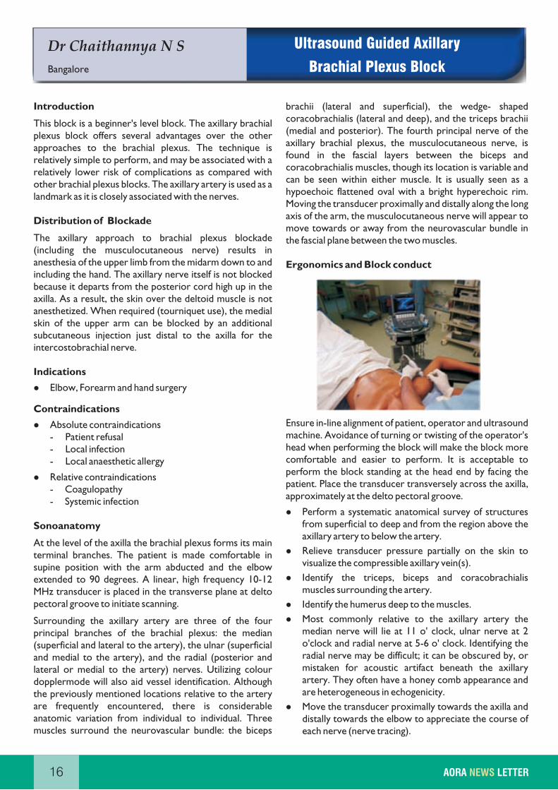
Ergonomics and Block conduct
Ensure in-line alignment of patient, operator and ultrasound machine. Avoidance of turning or twisting of the operator's head when performing the block will make the block more comfortable and easier to perform. It is acceptable to perform the block standing at the head end by facing the patient. Place the transducer transversely across the axilla, approximately at the delto pectoral groove.
lPerform a systematic anatomical survey of structures from superficial to deep and from the region above the axillary artery to below the artery.
lRelieve transducer pressure partially on the skin to visualize the compressible axillary vein(s).
lIdentify the triceps, biceps and coracobrachialis muscles surrounding the artery.
lIdentify the humerus deep to the muscles.
lMost commonly relative to the axillary artery the median nerve will lie at 11 o' clock, ulnar nerve at 2 o'clock and radial nerve at 5-6 o' clock. Identifying the radial nerve may be difficult; it can be obscured by, or mistaken for acoustic artifact beneath the axillary artery. They often have a honey comb appearance and are heterogeneous in echogenicity.
lMove the transducer proximally towards the axilla and distally towards the elbow to appreciate the course of each nerve (nerve tracing).
16 AORA LETTERNEWS
l
which lies most commonly in the plane between the
biceps and coracobrachialis muscles.
lNeedling can be either by an in plane approach or out of plane approach.
In Plane approach
lInsert a 5/10 cm 22 G insulated needle parallel to the long axis of the transducer inline with the ultrasound beam.
lThe needle should be inserted at a shallow angle because terminal branches of the brachial plexus in the axilla are superficial.
lAs the needle travels in the same plane as the ultrasound beam, the path of advancement can be visualized in real-time as the needle approaches the target nerves.
lDetermine the identity of each individual nerve by electrical stimulation if desired. It is well known that nerves can occupy variable locations around the axillary artery.
lBlock the musculocutaneous nerve separately as it branches in the coracobrachialis muscle.
lA posterior enhancement artifact deep to the artery is usually misinterpreted as the radial nerve. The radial nerve is usually located deep to the ulnar nerve in the proximal axilla. When the transducer is moved more distally, the radial nerve descends and disappears under the triceps muscle. The radial nerve can be visualized again as it travels posteriorly around the humeral shaft.
lIt is advantageous to target and anesthetize the radial nerve first posterior to the artery and then inject around the median and ulnar nerves as the needle is withdrawn.
lTwo to three redirections and injections are usually necessary for reliable blockade, as well as a separate injection to block the musculocutaneous nerve
lIn an adult patient, 20 to 25 mL of local anesthetic is usually adequate for successful blockade. Complete spread around the artery is necessary for success but
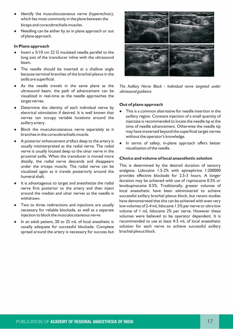
Identify the musculocutaneous nerve (hyperechoic), infrequently seen with a single injection.
The Axillary Nerve Block - Individual nerve targeted under ultrasound guidance
Out of plane approach
lThis is a common alternative for needle insertion in the axillary region. Constant injection of a small quantity of injectate is recommended to locate the needle tip at the time of needle advancement. Otherwise the needle tip may have traversed beyond the superficial target nerves without the operator's knowledge.
lIn terms of safety, in-plane approach offers better visualisation of the needle.
Choice and volume of local anaesthetic solution
This is determined by the desired duration of sensory analgesia. Lidocaine 1.5-2% with epinephrine 1:200000 provides effective blockade for 2.5-3 hours. A longer duration may be achieved with use of ropivacaine 0.5% or levobupivacaine 0.5%. Traditionally, greater volumes of local anaesthetic have been administered to achieve successful axillary brachial plexus block, but recent studies have demonstrated that this can be achieved with even very low volumes of 2-4 mL lidocaine 1.5% per nerve or ultra low volume of 1 mL lidocaine 2% per nerve. However these volumes were believed to be operator dependent. It is recommended to use at least 4-5 mL of local anaesthetic solution for each nerve to achieve successful axillary brachial plexus block.
17ACADEMY OF REGIONAL ANAESTHESIA OF INDIA
Dr Anbarasan ArdhanariDr Murali ThondebhaviBangalore
Rescue Blocks of the Upper Limb with ultrasound
18 AORA LETTERNEWS
INTRODUCTION
Ultrasound has considerably increased the efficacy and safety of peripheral nerve blocks of the distal upper extremity. It is used as a stand-alone technique or as a supplement to a patchy or failed brachial plexus block. It is important to be familiar with the anatomic distribution of the peripheral nerves in order to best meet the anaesthetic demands of the surgical procedure. We have described the peripheral nerve blocks specifically involving median, radial and ulnar nerve.
INDICATIONS
Median, radial and ulnar nerve blocks can be performed alone or combined to provide intra-operative anaesthesia or post-operative analgesia for surgical procedures of the forearm, wrist and hand.
ADVANTAGES
1. The risk of inadvertent needle trauma to pleura, major vessels like subclavian artery and axillary artery is avoided.
2. Allows preservation of proximal muscle function of the upper limb.
3. Preservation of motor strength may also allow the patients to move affected digits when instructed to do so during the surgery.
DISADVANTAGES
1. The cutaneous innervation of the upper arm is provided by the musculocutaneous nerve, medial cutaneous nerve of the arm, posterior cutaneous nerve of the arm, and intercostobrachial nerve. Distal nerve blocks will therefore not prevent tourniquet pain in a conscious patient.
2. Distal nerve blocks may not always be sufficient for surgical procedures on the forearm.
3. Multiple injections may cause more patient discomfort.
4. The peripheral nerves can be anisotropic, and scanning for them can be challenging initially.
EQUIPMENTS
1. Appropriate anaesthetic equipment and personnel for sedation, monitoring, and oxygenation.
2. Ultrasound machine with high-frequency linear array transducer (10-15 MHz).
3. Sterile ultrasound probe cover.
4. Sterile ultrasound gel.
5. Sterile skin preparation (2% chlorhexidine or other appropriate disinfectant).
6. Local anesthetic for skin infiltration (usually 0.5-1 ml 2% lignocaine).
7. A 22G, 50mm short-bevel regional anaesthesia block needle.
8. Sterile gloves.
ERGONOMICS
We recommend that the operator stand on the side being blocked, with the ultrasound machine on the opposite side of the patient. This maintains an in-line orientation between the operator, the injection site, and the ultrasound screen.
GOAL
The goal is to place the needle tip immediately adjacent to the nerve(s) of choice and to deposit 4-5 mL of local anaesthetic in the vicinity of the nerve. It is unnecessary to completely surround the entire nerve in a doughnut pattern, although this can enhance the speed of onset of the block. As with all peripheral blocks, avoidance of resistance to injection is important to decrease the risk of an intra-fascicular injection.
TECHNIQUE
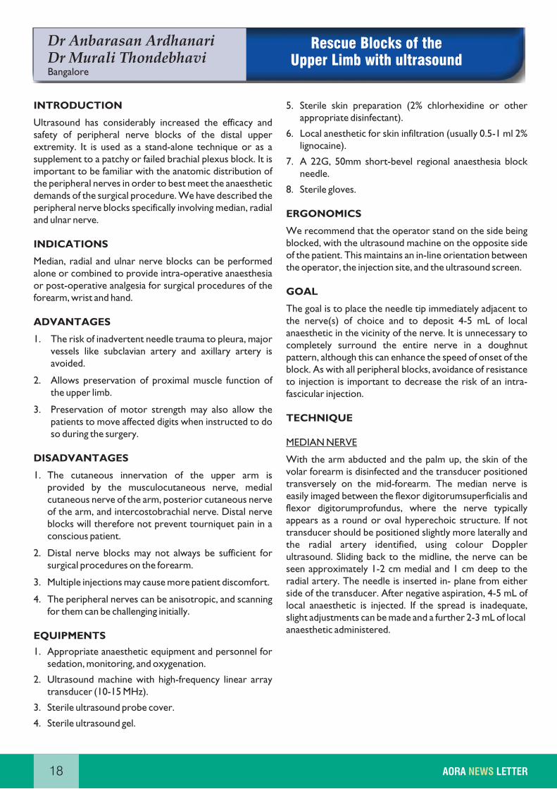
MEDIAN NERVE
With the arm abducted and the palm up, the skin of the volar forearm is disinfected and the transducer positioned transversely on the mid-forearm. The median nerve is easily imaged between the flexor digitorumsuperficialis and flexor digitorumprofundus, where the nerve typically appears as a round or oval hyperechoic structure. If not transducer should be positioned slightly more laterally and the radial artery identified, using colour Doppler ultrasound. Sliding back to the midline, the nerve can be seen approximately 1-2 cm medial and 1 cm deep to the radial artery. The needle is inserted in- plane from either side of the transducer. After negative aspiration, 4-5 mL of local anaesthetic is injected. If the spread is inadequate, slight adjustments can be made and a further 2-3 mL of localanaesthetic administered.
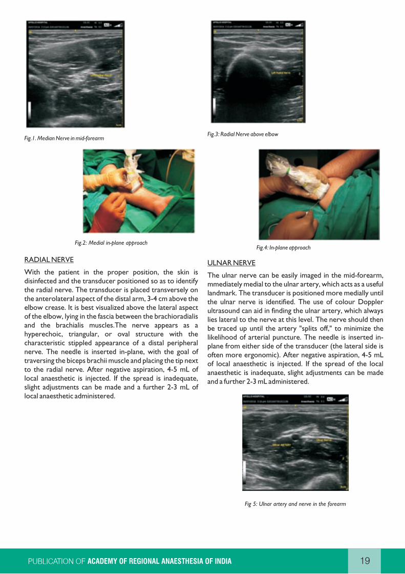
Fig.1. Median Nerve in mid-forearm
Fig.2: Medial in-plane approach
RADIAL NERVE
With the patient in the proper position, the skin is disinfected and the transducer positioned so as to identify the radial nerve. The transducer is placed transversely on the anterolateral aspect of the distal arm, 3-4 cm above the elbow crease. It is best visualized above the lateral aspect of the elbow, lying in the fascia between the brachioradialis and the brachialis muscles.The nerve appears as a hyperechoic, triangular, or oval structure with the characteristic stippled appearance of a distal peripheral nerve. The needle is inserted in-plane, with the goal of traversing the biceps brachii muscle and placing the tip next to the radial nerve. After negative aspiration, 4-5 mL of local anaesthetic is injected. If the spread is inadequate, slight adjustments can be made and a further 2-3 mL of local anaesthetic administered.
Fig.3: Radial Nerve above elbow
Fig.4: In-plane approach
ULNAR NERVE
The ulnar nerve can be easily imaged in the mid-forearm, mmediately medial to the ulnar artery, which acts as a useful landmark. The transducer is positioned more medially until the ulnar nerve is identified. The use of colour Doppler ultrasound can aid in finding the ulnar artery, which always lies lateral to the nerve at this level. The nerve should then be traced up until the artery "splits off," to minimize the likelihood of arterial puncture. The needle is inserted in-plane from either side of the transducer (the lateral side is often more ergonomic). After negative aspiration, 4-5 mL of local anaesthetic is injected. If the spread of the local anaesthetic is inadequate, slight adjustments can be made and a further 2-3 mL administered.
Fig 5: Ulnar artery and nerve in the forearm
19ACADEMY OF REGIONAL ANAESTHESIA OF INDIA
Dr Vrushali Ponde
Mumbai
20 AORA LETTERNEWS
The Axillary block:
A Paediatric perspective
Introduction
The Axillary block still remains the simplest block implied to give anesthesia for wrist and hands in neonates, infants toddlers and children.
Its simplicity is attractive because, the arterial landmark is hard to miss. The tourniquet pain which can be major concern, is easily addressed by an added intercostobrachial block or subclavian perivascular block. This can be performed under the accepted norm of concomitant sedation or light general anesthesia in this age group.
To perform any block in children, you may choose to work alone or team up with other anesthesiologists based on your comfort and expertise in handling the airway and venous access. After adequate premedication and a pleasant induction the vascular access is secure, unless there is an apparent contraindication for this sequence. The block procedure is undertaken.
Typically, the axillary block seems to act faster in kids than in adults due to supple nerves and connective tissues around, allowing for better penetration by the local anesthetic. However, the drawback is that the initial signs of local anesthetic toxicity may be masked by sedation and general anesthesia.
General considerations and Indications:
While the block is ideal for hand and wrist surgeries, the forearm too can be anesthetized by the axillary block.
The relevant anatomy and the patient position essentially remains the same as adults. Based on the size of the child, the relevant structures will appear very superficial. With the baby supine, the neck is gently turned away from the side to be blocked. A solitary anaesthesiologist depends on the continuous pulse oxymeter beeps as the block is being performed. It is preferred to have the baby breathing spontaneously as apnoea can be a sign to indicate that the local anaesthetic has been injected intravascularly. If spontaneous, the pediatric circuit bag is best placed in sight and should not be covered under the drapes.
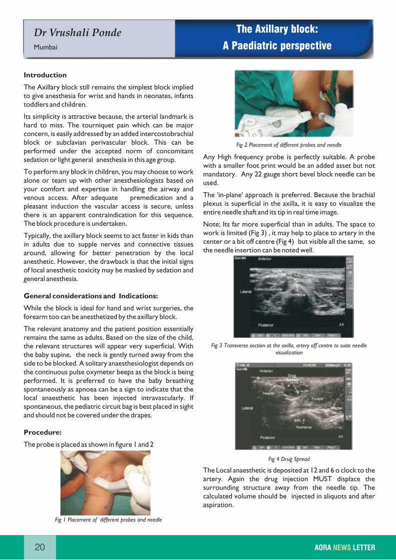
Procedure:
The probe is placed as shown in figure 1 and 2
Fig 1 Placement of different probes and needle
Fig 2 Placement of different probes and needle
Any High frequency probe is perfectly suitable. A probe with a smaller foot print would be an added asset but not mandatory. Any 22 gauge short bevel block needle can be used.
The 'in-plane' approach is preferred. Because the brachial plexus is superficial in the axilla, it is easy to visualize the entire needle shaft and its tip in real time image.
Note; Its far more superficial than in adults. The space to work is limited (Fig 3) , it may help to place to artery in the center or a bit off centre (Fig 4) but visible all the same, so the needle insertion can be noted well.
Fig 3 Transverse section at the axilla, artery off centre to suite needle visualization
Fig 4 Drug Spread
The Local anaesthetic is deposited at 12 and 6 o clock to the artery. Again the drug injection MUST displace the surrounding structure away from the needle tip. The calculated volume should be injected in aliquots and after aspiration.
LA dosages:
Dose Range in volume: 0.2–0.5 ml/kg
Midrange volume : Dose 0.3 mL/kg
Children < 8 yrs: 0.2% ropivacaine or 0.25% bupivacaine.
Children > 8 yrs: 0.5% ropivacaine or 0.5% bupivacaine.
Lignocaine 2 % with adrenaline 5 mg/kg can be used .
The syringes ideally should be kept separate.
Example:
10 kg infant.
Volume require for axillary block = 5 ml (0.5 ml/kg)
Drug :Ropivacaine 0.25 % = 3ml
Lignocaine 2 % with adrenaline =2 ml
Lignocaine 2 % with adrenaline is arguably a good indicator in paediatric regional anesthesia for intravascular injections despite concomitant GA, ST elevations is a better indication than tachycardia.
Disadvantage of axillary block :As we see it, the mobility and anatomy of axilla doesn't allow a proper catheter placement and fixation. Infraclvicular clearly scores over here.
21ACADEMY OF REGIONAL ANAESTHESIA OF INDIA
14th Conference of Asian Society ofPaediatric Anaesthesiologists
IN-HOUSE CONFERENCE
June 3rd & 4th 2017 Grand Hayat
PRECONFERENCE WORKSHOP
June 2nd 2017, Surya Children Hospital
Dr Amjad Maniar
Bangalore
22 AORA LETTERNEWS
Tips and Tricks
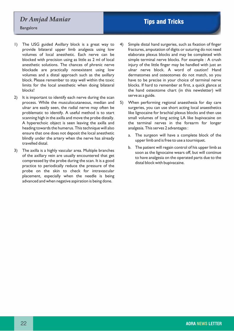
1) The USG guided Axillary block is a great way to provide bilateral upper limb analgesia using low volumes of local anesthetic. Each nerve can be blocked with precision using as little as 2 ml of local anesthetic solutions. The chances of phrenic nerve blockade are practically nonexistent using low volumes and a distal approach such as the axillary block. Please remember to stay well within the toxic limits for the local anesthetic when doing bilateral blocks!
2) It is important to identify each nerve during the scan process. While the musculocutaneous, median and ulnar are easily seen, the radial nerve may often be problematic to identify. A useful method is to start scanning high in the axilla and move the probe distally. A hyperechoic object is seen leaving the axilla and heading towards the humerus. This technique will also ensure that one does not deposit the local anesthetic blindly under the artery when the nerve has already travelled distal.
3) The axilla is a highly vascular area. Multiple branches of the axillary vein are usually encountered that get compressed by the probe during the scan. It is a good practice to periodically reduce the pressure of the probe on the skin to check for intravascular placement, especially when the needle is being advanced and when negative aspiration is being done.
4) Simple distal hand surgeries, such as fixation of finger fractures, amputation of digits or suturing do not need elaborate plexus blocks and may be completed with simple terminal nerve blocks. For example : A crush injury of the little finger may be handled with just an ulnar nerve block. A word of caution! Hand dermatomes and osteotomes do not match, so you have to be precise in your choice of terminal nerve blocks. If hard to remember at first, a quick glance at the hand osteotome chart (in this newsletter) will serve as a guide.
5) When performing regional anaesthesia for day care surgeries, you can use short acting local anaesthetics like lignocaine for brachial plexus blocks and then use small volumes of long acting LA like bupivacaine on the terminal nerves in the forearm for longer analgesia. This serves 2 advantages :
a. The surgeon will have a complete block of the upper limb and is free to use a tourniquet.
b. The patient will regain control of his upper limb as soon as the lignocaine wears off, but will continue to have analgesia on the operated parts due to the distal block with bupivacaine.
Dr Herman Sehmbi
Canada
EDRA - My Experience and Preparation
Strategies!
23ACADEMY OF REGIONAL ANAESTHESIA OF INDIA

24 AORA LETTERNEWS
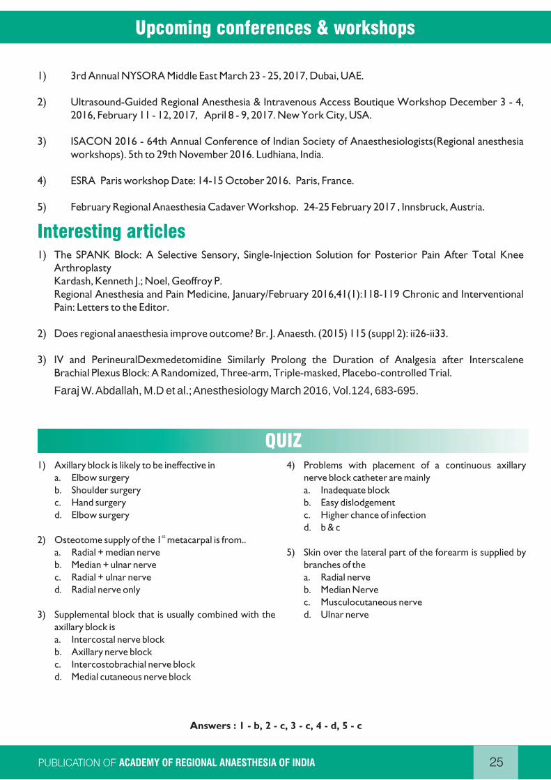
1) Axillary block is likely to be ineffective ina. Elbow surgeryb. Shoulder surgeryc. Hand surgeryd. Elbow surgery
st2) Osteotome supply of the 1 metacarpal is from..a. Radial + median nerveb. Median + ulnar nervec. Radial + ulnar nerved. Radial nerve only
3) Supplemental block that is usually combined with the axillary block isa. Intercostal nerve blockb. Axillary nerve blockc. Intercostobrachial nerve blockd. Medial cutaneous nerve block
4) Problems with placement of a continuous axillary nerve block catheter are mainlya. Inadequate blockb. Easy dislodgementc. Higher chance of infectiond. b & c
5) Skin over the lateral part of the forearm is supplied by branches of the a. Radial nerveb. Median Nervec. Musculocutaneous nerved. Ulnar nerve
Answers : 1 - b, 2 - c, 3 - c, 4 - d, 5 - c
Upcoming conferences & workshops
1) 3rd Annual NYSORA Middle East March 23 - 25, 2017, Dubai, UAE.
2) Ultrasound-Guided Regional Anesthesia & Intravenous Access Boutique Workshop December 3 - 4, 2016, February 11 - 12, 2017, April 8 - 9, 2017. New York City, USA.
3) ISACON 2016 - 64th Annual Conference of Indian Society of Anaesthesiologists(Regional anesthesia workshops). 5th to 29th November 2016. Ludhiana, India.
4) ESRA Paris workshop Date: 14-15 October 2016. Paris, France.
5) February Regional Anaesthesia Cadaver Workshop. 24-25 February 2017 , Innsbruck, Austria.
1) The SPANK Block: A Selective Sensory, Single-Injection Solution for Posterior Pain After Total Knee ArthroplastyKardash, Kenneth J.; Noel, Geoffroy P. Regional Anesthesia and Pain Medicine, January/February 2016,41(1):118-119 Chronic and Interventional Pain: Letters to the Editor.
2) Does regional anaesthesia improve outcome? Br. J. Anaesth. (2015) 115 (suppl 2): ii26-ii33.
3) IV and PerineuralDexmedetomidine Similarly Prolong the Duration of Analgesia after Interscalene Brachial Plexus Block: A Randomized, Three-arm, Triple-masked, Placebo-controlled Trial.
Faraj W. Abdallah, M.D et al.; Anesthesiology March 2016, Vol.124, 683-695.
Interesting articles
QUIZ
25ACADEMY OF REGIONAL ANAESTHESIA OF INDIA
26 AORA LETTERNEWS
Safe, easy to use, passive shielded IV catheter
Vasofix® Safety1 billion times protection
nE sures
f sirst tickssucce s
Ecoflac® Plus
The only safest standable container for closed system IV therapy
PVC-free
D -f eeEHP r Latex free
B|Braun Medical (India) Pvt. Ltd. Unit No. 601 | Wing A | 6th Floor | Boomerang | Chandivali Farm Road
Andheri (E) | Mumbai - 400 072. Tel. : (91-22) 6668 2222, Fax : (91-22) 6668 2121

Propofol-Lipuro 1%
B. Braun Regional Anaesthesia
Your First Choice for Safe Regional Anaesthesia
® Sterofundin ISO® Gelaspan
The complete balancedfluid management concept
Lipuro - Technology by B. Braun

28 AORA LETTERNEWS
SCIENTIFIC & ACADEMIC PARTNER FOR