Embed Size (px)
Citation preview
INFECTION AND IMMUNITY, Apr. 2011, p. 1458–1470 Vol. 79, No. 40019-9567/11/$12.00 doi:10.1128/IAI.01140-10Copyright © 2011, American Society for Microbiology. All Rights Reserved.
Actin Polymerization Drives Septation of Listeria monocytogenes namAHydrolase Mutants, Demonstrating Host Correction of a
Bacterial Defect�†Francis Alonzo III, P. David McMullen, and Nancy E. Freitag*
Department of Microbiology and Immunology, University of Illinois at Chicago, Chicago, Illinois
Received 26 October 2010/Returned for modification 8 January 2011/Accepted 15 January 2011
The Gram-positive bacterial cell wall presents a structural barrier that requires modification forprotein secretion and large-molecule transport as well as for bacterial growth and cell division. TheGram-positive bacterium Listeria monocytogenes adjusts cell wall architecture to promote its survival indiverse environments that include soil and the cytosol of mammalian cells. Here we provide evidence forthe enzymatic flexibility of the murein hydrolase NamA and demonstrate that bacterial septation defectsassociated with a loss of NamA are functionally complemented by physical forces associated with actinpolymerization within the host cell cytosol. L. monocytogenes �namA mutants formed long bacterial chainsduring exponential growth in broth culture; however, normal septation could be restored if mutant cellswere cocultured with wild-type L. monocytogenes bacteria or by the addition of exogenous NamA. Surpris-ingly, �namA mutants were not significantly attenuated for virulence in mice despite the pronouncedexponential growth septation defect. The physical force of L. monocytogenes-mediated actin polymerizationwithin the cytosol was sufficient to sever �namA mutant intracellular chains and thereby enable theprocess of bacterial cell-to-cell spread so critical for L. monocytogenes virulence. The inhibition of actinpolymerization by cytochalasin D resulted in extended intracellular bacterial chains for which septationwas restored following drug removal. Thus, despite the requirement for NamA for the normal septation ofexponentially growing L. monocytogenes cells, the hydrolase is essentially dispensable once L. monocyto-genes gains access to the host cell cytosol. This phenomenon represents a notable example of eukaryotichost cell complementation of a bacterial defect.
The cell wall of a Gram-positive bacterial pathogen is veryoften the first structural component of the organism to engagea host cell upon the initiation of bacterial adherence and/orhost cell invasion (70). The Gram-positive cell wall is a thickand rigid structure composed of peptidoglycan, which in turnconsists of a series of N-acetylglucosamine and N-acetylmu-ramic acid residues linked to form a lattice-like meshwork (27,64). While structurally rigid, the cell wall is a dynamic organellethat must be constantly altered and modified to accommodatebacterial cell division, protein secretion, and the assembly ortransport of large molecules such as flagella and DNA (10, 11,42, 43, 47, 64, 65). Unlike bacterial membranes, the cell wall isa permeable organelle that permits the diffusion of small mol-ecules and regularly adjusts to shifts in environmental condi-tions (27, 64). Bacteria produce a variety of enzymes dedicatedto the synthesis, degradation, and restructuring of peptidogly-can to facilitate bacterial growth and survival in diverse envi-ronments. These cell wall-specific enzymes include proteinsknown as autolysins or peptidoglycan hydrolases, which hy-drolyze the peptidoglycan bonds at specific sites to releasedistinct cell wall cleavage products (28, 58, 65).
The Gram-positive bacterium Listeria monocytogenes is an
environmental pathogen that is capable of orchestrating acomplex transition from life in the outside environment to lifewithin the cytosol of an infected host (18, 30, 51). As anenvironmental bacterium, L. monocytogenes survives in a num-ber of diverse settings, including soil, water, and silage (51, 60).As an intracellular pathogen, L. monocytogenes adheres to andinvades host cells and replicates within the cytosol, and indi-vidual bacteria are propelled into neighboring cells to spreadwithin host tissues (13, 63). L. monocytogenes survival withinhost cells is dependent on a variety of secreted virulence geneproducts, whereas bacterial survival in the outside environ-ment depends on the synthesis and assembly of bacterial fla-gella in addition to the regulated secretion of proteins andfactors that aid in nutrient acquisition (12, 62). Thus, the ap-propriate modulation of the L. monocytogenes cell wall for celldivision, bacterial organelle assembly, and protein secretion isessential for optimal bacterial fitness under a variety of dispa-rate conditions.
Seven peptidoglycan hydrolases in L. monocytogenes havebeen identified and at least partially characterized. These hy-drolases are p60 (also known as CwhA or Iap) (16, 25, 26, 31,47, 71), p45 (53), Ami (38, 39), Auto (5, 7), NamA (also knownas MurA) (10, 33, 36), lmo0327 (49), and IspC (67–69). Whilemost of these proteins contain recognizable domains linked tomurein hydrolase activity and cell wall binding and/or associ-ations (7), many display unique characteristics associated withthe regulation of catalytic activity (5) or with the anchoring ofthe proteins to the cell wall (68) as well as distinct activitiesrelating to cell wall structure and function. The hydrolases p60,
* Corresponding author. Mailing address: Department of Microbi-ology and Immunology (MC 790), 835 S. Wolcott Ave., University ofIllinois at Chicago, Chicago, IL 60612-7344. Phone: (312) 355-4903.Fax: (312) 996-6415. E-mail: [email protected].
† Supplemental material for this article may be found at http://iai.asm.org/.
� Published ahead of print on 24 January 2011.
1458
on March 26, 2018 by guest
http://iai.asm.org/
Dow
nloaded from
on March 26, 2018 by guest
http://iai.asm.org/
Dow
nloaded from
on March 26, 2018 by guest
http://iai.asm.org/
Dow
nloaded from
p45, and NamA have been shown to contribute to cell wallpeptidoglycan remodeling to enable flagellar motility, cell di-vision, cell elongation, and the secretion of bacterial proteins(10, 33, 36, 47, 53). Ami, Auto, and IspC appear to have morespecialized roles related to bacterial virulence (7, 38, 68). Autois unique in that it is present in L. monocytogenes but is notfound in the nonpathogenic species Listeria innocua. Autocontributes to the bacterial invasion of host cells, and it wassuggested previously that it functions to regulate cell wall per-meability to facilitate the secretion of virulence factors (7).Similarly, Ami and IspC have been shown to play a role inpathogenesis via their contributions to bacterial adhesion tohost cells (38, 68).
NamA was initially identified as a variably expressed immuno-genic epitope of L. monocytogenes recognized by monoclonalantibodies at the bacterial surface (19, 20, 44). NamA contributesto cell division, as mutants lacking namA form bacterial chainsduring exponential-phase growth in broth culture (10). Despitebacterial chain formation, Lenz et al. reported previously thatNamA made only modest contributions to bacterial virulencein a mouse model of infection (33, 34).
We recently identified NamA as one of a number of proteinswhose abundance in bacterial culture supernatants was in-creased in strains containing a mutationally activated form ofthe central virulence regulator PrfA (PrfA*); this mutationresults in the constitutive expression of virulence-associatedgene products (50). Given that many of the gene productsexpressed as a result of PrfA activation have been found tocontribute to bacterial virulence (1, 14, 50, 63, 72) and that theloss of NamA has thus far been shown to have only a modesteffect on L. monocytogenes virulence (33, 34), we sought tofurther explore the role of NamA in L. monocytogenes intra-cellular and extracellular growth. Here we provide additionalevidence that L. monocytogenes NamA is a temporally secretedmurein hydrolase whose flexible activity is required during theearly phases of exponential bacterial growth in broth culture.Interestingly, NamA was found to be dispensable for bacterial
septation for L. monocytogenes cells replicating within the cy-tosol of infected host cells. The physical force provided by actinpolymerization disrupted cytosolic bacterial chains and en-abled the spread of septation-defective mutants into neighbor-ing cells. Our data thus provide a mechanism to explain howNamA septation-deficient bacteria retain bacterial virulencewithin an infected eukaryotic host.
MATERIALS AND METHODS
Bacterial strains, plasmids, and media. All bacterial strains used in this studyare listed in Table 1. L. monocytogenes containing an actA-gus transcriptionalfusion (NF-L476) was used as the parent strain for all genetic manipulations (54).Escherichia coli alpha select (BioLine, Boston, MA), One Shot TOP10 (Invitro-gen Corp., Carlsbad, CA), DH5� I/q, and BH10C (29) were used as host strainsfor recombinant plasmids. All E. coli strains were grown in Luria-Broth (LB)medium (Invitrogen Corp., Carlsbad, CA), and L. monocytogenes strains weregrown in brain heart infusion (BHI) medium (Difco Laboratories, Detroit, MI).Where necessary, the medium was supplemented with antibiotics at the followingconcentrations unless otherwise specified: ampicillin at 100 �g/ml, carbenicillinat 50 �g/ml, erythromycin at 0.1 �g/ml (to induce the expression of the erm gene)and 1 �g/ml (for the selection of erythromycin-resistant bacteria), chloramphen-icol at 10 �g/ml, kanamycin at 50 �g/ml, and streptomycin at 200 �g/ml. VectorpAM401 containing a gfp3 allele (a kind gift of Daniel Portnoy) was used forexpressing green fluorescent protein (GFP) in selected strains. The temperature-sensitive shuttle vector pKSV7 (57) was used for generating deletion mutants viaallelic exchange, and the integration plasmid pIMK2 (40), a kind gift of ColinHill, was used for genetic complementation.
Construction of the �namA in-frame deletion mutant. A 1,755-bp internalin-frame deletion was generated with lmo2691 (namA) as follows: two DNAproducts containing sequences upstream and downstream of namA coding se-quences were amplified using PCR with primer pairs 2691A1/2691A2 and2691B1/2691B2 using L. monocytogenes 10403S chromosomal DNA as a tem-plate (all oligonucleotides are listed in Table 2). The resulting PCR productswere purified and used in a splice overlap extension (SOEing) PCR along withprimer pair 2691A1/2691B2, generating a 1,068-bp fragment which encompassedthe first 9 nucleotides of the lmo2691 coding region along with 519 bp ofupstream sequence and the last 9 nucleotides of the same gene with 531 bp ofdownstream sequence. The 1,068-bp fragment was cloned into the shuttle vectorpKSV7 using the BamHI and HindIII restriction sites to generate pNF1367.Plasmid pNF1367 was transformed into L. monocytogenes strain NF-L476(10403S actA-gus) (54) using electroporation as previously described (46), andthe �namA mutation was introduced into the bacterial chromosome in a single
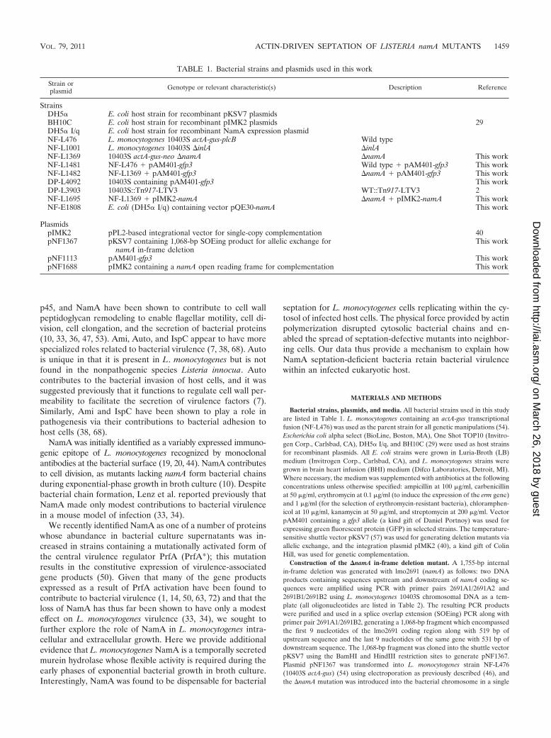
TABLE 1. Bacterial strains and plasmids used in this work
Strain orplasmid Genotype or relevant characteristic(s) Description Reference
StrainsDH5� E. coli host strain for recombinant pKSV7 plasmidsBH10C E. coli host strain for recombinant pIMK2 plasmids 29DH5� I/q E. coli host strain for recombinant NamA expression plasmidNF-L476 L. monocytogenes 10403S actA-gus-plcB Wild typeNF-L1001 L. monocytogenes 10403S �inlA �inlANF-L1369 10403S actA-gus-neo �namA �namA This workNF-L1481 NF-L476 � pAM401-gfp3 Wild type � pAM401-gfp3 This workNF-L1482 NF-L1369 � pAM401-gfp3 �namA � pAM401-gfp3 This workDP-L4092 10403S containing pAM401-gfp3 This workDP-L3903 10403S::Tn917-LTV3 WT::Tn917-LTV3 2NF-L1695 NF-L1369 � pIMK2-namA �namA � pIMK2-namA This workNF-E1808 E. coli (DH5� I/q) containing vector pQE30-namA This work
PlasmidspIMK2 pPL2-based integrational vector for single-copy complementation 40pNF1367 pKSV7 containing 1,068-bp SOEing product for allelic exchange for
namA in-frame deletionThis work
pNF1113 pAM401-gfp3 This workpNF1688 pIMK2 containing a namA open reading frame for complementation This work
VOL. 79, 2011 ACTIN-DRIVEN SEPTATION OF LISTERIA namA MUTANTS 1459
on March 26, 2018 by guest
http://iai.asm.org/
Dow
nloaded from
copy by allelic exchange (9) to generate strain NF-L1369. The presence of thein-frame deletion, which encompasses 98.9% of the structural gene, was con-firmed by the sequencing of PCR products derived from the bacterial chromo-some.
To construct a plasmid vector containing namA for complementation, primersnamApIMK2f and namApIMK2r were used to amplify the entire open readingframe (ORF) of namA (excluding the ATG start codon) from L. monocytogenes10403S genomic DNA using PCR. The product was digested with NcoI and KpnIand subcloned into appropriately digested pIMK2 to generate pNF1688. Theinsertion of namA coding sequences into pIMK2 places the expression of thegene under the control of the Phelp promoter. namA was PCR amplified withoutits native ATG because the NcoI restriction site used overlaps the ATG of Phelpand results in in-frame high-level gene expression (40). Because we encountereddifficulty in propagating the complementation vector in our standard E. coli hosts(TOP10 and DH5�) due to the apparent toxicity of the expressed gene product,we opted to use host strain BH10C (a kind gift of Nicholas Cianciotto), whichresults in the efficient restriction of plasmid copy numbers such that each cellcontains approximately 1 copy of the recombinant plasmid (29). Recombinantplasmid pNF1688 was then introduced into competent NF-L1369 (L. mono-cytogenes �namA) cells by electroporation according to a modified protocoldescribed previously by Monk et al. (40). Following electroporation, bacteriawere plated onto selective medium, and the resultant transformants wereassessed for the presence of namA by PCR amplification of namA codingsequences from L. monocytogenes chromosomal DNA (pIMK2 is a pPL2derivative and thus integrates in a single copy into the L. monocytogenesphage attachment site located within the tRNAArg gene following electropo-ration) (17, 32). The resultant pNF1688-complemented �namA strain wasdesignated NF-L1695.
Microscopic examination of bacterial morphology for monocultures andmixed cultures of wild-type and �namA strains. To facilitate the microscopicexamination and visual assessment of monocultures as well as mixed cultures,plasmid pAM401-gfp3 (pNF1113) was introduced into NF-L476 (10403S actA-gus) and NF-L1369 (�namA) by an electroporation method described previouslyby Park and Stewart (46). The GFP-expressing strains were designated NF-L1481 (wild type [WT] with pAM401-gfp3) and NF-L1482 (�namA withpAM401-gfp3). These GFP-expressing strains were used for all subsequent mi-croscopic assessments upon confirmation that plasmid pAM401-gfp3 did notalter the growth characteristics and septation associated with either the wild typeor the �namA mutant strain.
For the examination of the cell morphology of monocultures, 100 �l of eachculture (taken at the time points designated in the text) was added directly ontoindividual poly-L-lysine-treated coverslips and allowed to incubate for 5 to 10 min
at 37°C. The edges of the coverslips were then touched to a Kimwipe to removeexcess liquid and unbound bacteria. The bacteria were subsequently fixed to thecoverslips by the addition of 100 �l of 3.2% formaldehyde in phosphate-bufferedsaline (PBS) for 5 min at room temperature. Coverslips were then washed with5 dips in PBS and allowed to air dry. Each coverslip was then mounted onto aglass slide using Vectashield mounting solution (Vector Laboratories, Inc., Bur-lingame, CA) and allowed to cure in the dark overnight at room temperature.Mixed-culture coverslips were prepared similarly, with a few modifications. Inorder to distinguish wild-type cells from cells of the �namA mutant withpAM401-gfp3, bacteria were fixed (as described above), followed by the additionof 100 �l of a 1:320 dilution of Listeria O Antiserum Poly (Becton Dickinson,Sparks, MD) in PBS plus 1 mg/ml bovine serum albumin (BSA) for 30 min.Coverslips were washed by dipping five times in PBS. A 1:5,000 dilution oftetramethyl rhodamine goat anti-rabbit IgG (Molecular Probes, Eugene, OR) inPBS plus 1 mg/ml BSA was added dropwise to the coverslip, followed by incu-bation for 30 min at room temperature. The coverslip was then washed bydipping in PBS and subsequently mounted onto a glass slide with Vectashieldand allowed to cure overnight in the dark. Thus, in mixed cultures, total bacteriaappeared red, while �namA mutants appeared yellow, due to the colocalizationof rhodamine and GFP.
To determine the relative amounts of chain formation due to incompletebacterial septation in wild-type, �namA, �namA with pIMK2-namA, and mixedwild-type and �namA cultures, cells were processed for microscopy as describedabove, and the total number of cells in chains (greater than three linked cells)was counted for 10 independent fields and compared to the total number ofbacteria present in each field. A ratio of the number of cells in chains to the totalcell number was then calculated as a measure of chaining frequency. Numbersare derived from at least 3 independent experiments.
Measurement of the ability of bacterial supernatants to complement �namAmutant phenotypes. Bacterial supernatants were collected from broth cultures ofboth wild-type and �namA strains grown in BHI broth. Supernatants wereobtained from cultures diluted 1 to 100 into 200 ml of fresh BHI broth, followedby shaking at 37°C for up to 8 h. At designated time points (2, 4, 6, and 8 h), 20ml was removed from each culture, and bacteria were recovered by centrifuga-tion at 12,350 � g for 10 min. The supernatant was then filter sterilized using a0.22-�m vacuum filter (Corning, Corning, NY). These supernatants were thenadded to fresh cultures of the �namA mutant strain to determine if supernatantsderived from wild-type strains were capable of complementing the �namA chain-ing phenotype. To roughly balance for the loss of nutrients associated with theuse of filter-sterilized supernatants derived from late-exponential- and early-stationary-phase cultures, fresh BHI broth was supplemented in an approxi-mated ratio to provide normal rates of bacterial growth to stationary phase andto comparable final cell densities (optical density at 600 nm [OD600] of �1.8).The calculated ratios were as follows: for supernatants derived from culturesgrown for 2 h, no fresh BHI broth was added, since it was observed thatessentially no nutrients had been depleted by the dilute starting culture for thistime period; for supernatants derived from 4-h cultures, a 4:1 ratio of superna-tant to fresh BHI broth was used; for supernatants derived from cultures grownfor 6 h, a 3:1 ratio was used; and for supernatants derived from cultures grownfor 8 h, a 2:1 ratio was used. Growth curves were done at each of these ratiosusing wild-type L. monocytogenes to confirm that the ratio of supernatant to freshBHI medium was sufficient to allow comparable bacterial growth rates. Final celldensities obtained beginning from 1/100 culture dilutions incubated for 8 h (earlystationary phase) were between OD600 values of 1.7 and 1.8 for all cultures. Totest for the ability of each supernatant to complement the chaining phenotypeassociated with a �namA mutant, a 1-to-100 dilution of �namA cultures wassubsequently inoculated into 20 ml of the collected media from cultures grownfor 2, 4, 6, and 8 h mixed with fresh BHI medium as indicated above. Cultureswere allowed to grow for 4 h at 37°C to an OD600 of �0.5 with shaking, at whichpoint 100-�l aliquots were removed and processed for microscopy to assess thepresence and degree of filamentation as described above.
Isolation of bacterial supernatants and whole-cell lysates for protein analysis.Twenty-milliliter cultures of the wild-type and �namA strains were grown over-night in BHI medium at 37°C with shaking. The following day, bacterial cellsfrom the cultures grown overnight were washed twice in fresh BHI medium toremove any NamA protein present in the supernatant from cultures grownovernight. Bacteria were then diluted 1/20 into 200 ml of fresh BHI medium. At3 h postinoculation and every 2 h thereafter, the OD600 of each culture wasmeasured, and 20-ml aliquots were removed. The culture aliquots were centri-fuged at 12,350 � g for 10 min at 4°C to recover supernatant and bacterial cellfractions. Trichloroacetic acid (TCA) was added to the supernatant fractions toa final concentration of 10%, and the fractions were incubated on ice for 30 min.Precipitated protein was recovered by the centrifugation of the fractions at
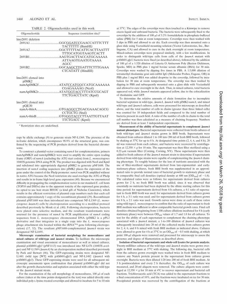
TABLE 2. Oligonucleotides used in this work
Oligonucleotide Sequence (restriction site)a
lmo2691 deletion2691A1 .....................................GGCGGATCCGAACCATTTCTTT
TACTTTTT (BamHI)2691A2 .....................................GGCTTTTTACATTCACTTAATTT
TTTGCATGGTAAGTCACTT2691B1......................................AAGTGACTTACCATGCAAAAA
ATTAAGTGAATGTAAAAAGCC
2691B2......................................GGCAAGCTTGATTTCTTTGAAACTCATATT (HindIII)
lmo2691 cloned intopIMK2
namApIMK2f ..........................ATATCCATGGCCATGCAAAAAACGAGAAAAG (NcoI)
namApIMK2r..........................ATATGGTACCTTTATCCGCAGTTTCTGACCTATC (KpnI)
lmo2691 cloned intopQE30
namA-6hisA.............................GTGAGGCCTGACGAAACAGCGCCTGCTG (StuI)
namA-6hisB.............................GTGGGTACCCTTAATTGTTAATTTCTGACC (KpnI)
a Restriction sites are underlined.
1460 ALONZO ET AL. INFECT. IMMUN.
on March 26, 2018 by guest
http://iai.asm.org/
Dow
nloaded from
12,350 � g for 10 min, and the resulting protein pellets were allowed to air dry.The pellets were subsequently washed with 4 ml of ice-cold acetone followed bycentrifugation at 12,350 � g. The washed pellets were resuspended in 250 �l ofSDS boiling buffer (5% SDS, 0.5% �-mercaptoethanol, 10% glycerol, 60 mMTris [pH 6.8]). For bacterial whole-cell lysates, the bacterial pellets (upon theremoval of the supernatant) were resuspended in 125 �l of lysis buffer (10%glycerol, 60 mM Tris [pH 6.8]), followed by the addition of lysozyme at aconcentration of 10 �g/ml and incubation for 1 h at room temperature. Sampleswere then sonicated 10 times on ice (pulse for 30 s followed by 1 min of restbetween each pulse) prior to the addition of 125 �l of 2� boiling buffer, boilingfor 5 min, and clarification by centrifugation at 11,000 rpm for 30 min at 4°C.
Western blot analysis. TCA-precipitated proteins and whole-cell lysates fromboth wild-type and �namA strains were loaded onto 10% SDS-PAGE gels.Loading volumes were altered based on the original bacterial optical density toaccount for the differences in bacterial CFU at each time point. Proteins weretransferred onto polyvinylidene difluoride (PVDF) membranes at 1.0 A (con-stant) for 1 h. All membranes were blocked for 1 h in PBS (Gibco, Carlsbad, CA)containing 0.05% Tween (PBST) and 5% milk. Membranes were incubated witha 1:2,500 dilution of monoclonal antibody C11E9 in PBST (a kind gift of ArunBhunia), which is known to specifically recognize NamA (19), or a 1:500 dilutionof monoclonal antibody against internalin A (Toxin Technologies, Sarasota, FL)at 4°C overnight with shaking, followed by three additional washes with 30 ml ofPBST for 10 min each. A 1:2,500 dilution of secondary goat anti-mouse antibodyconjugated to alkaline phosphatase (Southern Biotech, Birmingham, AL) wasincubated with shaking for 2 h at room temperature, followed by three additional10-min washes in PBST. Detection was carried out by the addition of the color-imetric substrate BCIP-NBTPlus (Southern Biotech, Birmingham, AL) for ap-proximately 10 min. Images were acquired by using an AlphaImager 2200 in-strument (Alpha Innotech, San Leandro, CA). NIH ImageJ software (http://rsbweb.nih.gov/ij/) was used to perform densitometric analyses on at least threeindependent blots. The 3-h time point was set to a value of 1.00, to which allother lanes were compared and represented as a deviation from that value. Theaverages and standard deviations (SD) were then calculated and are indicated inFig. 3. An L. monocytogenes strain with an in-frame deletion of inlA (NF-L1004)was used as a negative control for InlA Western blots.
Purification of recombinant NamA and complementation of septation defectswith purified protein. The DNA sequence corresponding to the mature form ofthe NamA enzyme was amplified from L. monocytogenes 10403S using primersNamA-6hisA and NamA-6hisB, generating a �1.6-kb PCR product as previouslydescribed (10). The product was digested with StuI and KpnI and subcloned intothe appropriately digested pQE30 N-terminal 6�-His expression vector (Qiagen,Valencia, CA), followed by transformation into E. coli DH5� I/q cells. Theresulting strain was designated NF-E1808.
The expression and purification of recombinant NamA were carried out ac-cording to the instructions provided by the supplier of the pQE30 vector(Qiagen, Valencia, CA). Briefly, a 20-ml culture of NF-E1808 grown overnight wasinoculated at a 1:50 dilution into 1 liter of LB medium containing ampicillin (100�g/ml). The culture was grown with shaking at 37°C until an approximate OD600
of 0.5 was reached, at which point isopropyl-�-D-thiogalactopyranoside (IPTG)was added at a final concentration of 0.8 mM. Cultures were allowed to continuegrowing for another 5 h, followed by centrifugation at 8,500 rpm for 15 min andsubsequent freezing of the bacterial pellet at �80°C overnight. Bacterial celllysates were prepared by resuspending the bacterial pellet in 50 ml of wash buffer(500 mM NaPO4, 300 mM NaCl, 10 mM imidazole [pH 7.4]), followed by theaddition of lysozyme (0.3 mg/ml) (Sigma, St. Louis, MO) and a protease inhibitorcocktail (Pierce, Pittsburgh, PA). The cell suspension was subsequently sonicatedon ice 10 times (10 s on and 30 s off), followed by the addition of Triton X-100(final concentration, 1%) and incubation at room temperature for 30 min. Ly-sates were clarified by centrifugation at 11,000 rpm for 30 min, followed bypassage through a 0.22-�m sterile filter. The purification of NamA from bacteriallysates was carried out by metal affinity chromatography using Cobalt resinaccording to the instructions provided by the supplier (Pierce, Pittsburgh, PA).The eluted protein fraction was dialyzed overnight against a final storage buffer(20 mM HEPES, 140 mM NaCl, 10% glycerol, 1 mM dithiothreitol [pH 7.4]),aliquoted, and frozen at �80°C. The final protein concentration was determinedby using a bicinchoninic acid assay kit (Pierce, Pittsburgh, PA).
To assess the ability of purified NamA to complement the septation defectsassociated with a �namA mutant, 10 ml of BHI broth was supplemented withpurified NamA (final concentration, 500 ng/ml), followed by the addition of aculture of the �namA strain plus pAM401-gfp (1:100 dilution) grown overnight.Cultures were allowed to grow at 37°C for 5 h (substantial chaining occurs for�namA mutants at this time), processed for microscopy as described above, andexamined for the complementation of septation defects. Nonsupplemented cul-
tures of the WT and the �namA mutant with pAM401-gfp were grown andprocessed for microscopy as controls.
Plaque assays. Plaque assays were conducted as previously described (59).Monolayers of L2 fibroblasts were grown and infected at a multiplicity of infec-tion (MOI) of 30:1. After 1 h, gentamicin was added to the medium at aconcentration of 20 �g/ml in an agarose medium overlay. After 3 days, plaquesizes and total numbers of plaques were enumerated. Results were obtained fromat least three independent experiments.
Intracellular growth assays and fluorescent microscopy of intracellular bac-teria. J774 macrophage-like and PtK2 epithelial cells were maintained as previ-ously described (4, 61), while bone marrow-derived macrophages (BMMs) werecultured from female ND4 Swiss Webster mouse femurs as previously described(21, 59). Briefly, 2 � 106 cells were placed onto glass coverslips the night beforeinfection. For gamma interferon (IFN-)-treated BMMs, the medium was sup-plemented with 1 ng/ml of IFN- (Biosource, Carlsbad, CA) 18 h prior toinfection. An MOI of 1:10 was used for all macrophages, and an MOI of 30:1 wasused for PtK2 epithelial cells. Gentamicin (20 �g/ml) was added to the mediumof infected cells at 30 min (for macrophages) or 1 h (for PtK2 cells) and at thedesignated time points, three coverslips were removed, cells were lysed in 5 ml ofwater, and serial dilutions were plated onto LB agar plates to enumerate bacte-rial CFU. Fluorescent microscopy of Listeria-infected PtK2 cells was carried outas previously described (41). Coverslips were viewed with a DeltaVision micro-scope (Applied Precision, Issaquah, WA). Images were then captured usingSoftworx image acquisition software (Applied Precision, Issaquah, WA). Forexperiments assessing the effects of the inhibition of actin polymerization onbacterial septation, cytochalasin D was added to culture dishes (final concentra-tion, 0.25 �g/ml) as previously described (56). Because cytochalasin D is dis-solved in dimethyl sulfoxide (DMSO), an equivalent volume of DMSO withoutcytochalasin D was added to all untreated dishes to control for the presence ofthis solvent in treated dishes. To remove cytochalasin D from infected cells,medium was aspirated, followed by washing 5 times with warm PBS and thesubsequent addition of minimal essential medium (MEM) containing gentamicin(20 �g/ml) only. An additional 2.5 h of infection was allowed to proceed after theremoval of cytochalasin D to allow the reestablishment of actin architecture aswell as actin-based motility prior to processing for microscopy.
Invasion and adherence assays. To measure the relative invasion efficiency of�namA mutants, PtK2 epithelial cell monolayers on glass coverslips were in-fected with the WT, the �namA mutant strain, or the �namA mutant pluspAM401-gfp at an MOI of 30:1. Infection was allowed to proceed for 1 h,followed by washing three times with warm PBS and the subsequent addition ofMEM. At this point, three coverslips were removed, placed into 5 ml of sterilewater, lysed by vortexing, and plated onto solid medium to determine the totalbacterial burden per coverslip (extracellular and intracellular). Immediatelythereafter, gentamicin (20 �g/ml) was added to culture dishes and incubated foran additional hour. Three coverslips were then removed and used to determinetotal CFU (now enumerating only intracellular bacteria). The ratio of the num-ber of bacterial CFU at hour 2 to the number of bacterial CFU at hour 1 wasdetermined and is reflected as percent invasion for each strain tested (wild-typeinvasion was set to 100%).
To measure overall bacterial adherence to PtK2 epithelial cells, monolayerswere again infected at an MOI of 30:1. Bacteria were allowed to adhere to andbegin invading cells by incubation for 1 h. Cells were subsequently washed fivetimes with warm PBS to remove any unbound bacteria, followed by the additionof warm MEM without antibiotics. Coverslips were immediately removed, andthe total number of bacterial CFU per coverslip was determined as describedabove. The total amounts of bacteria, representing bacteria that have efficientlyadhered to and begun invading cells, were compared for each strain tested.
Mouse intravenous infections. Animal procedures were approved by the IA-CUC and performed in the Biological Resources Laboratory at the University ofIllinois at Chicago. Monoculture infections were performed as previously de-scribed (1). Briefly, 2 �104 CFU (from either exponential- or stationary-phasecultures) of L. monocytogenes was inoculated into female 7-week-old ND4 SwissWebster mice via the tail vein. At 72 h postinfection, livers and spleens wereharvested and homogenized. Dilutions of homogenates were plated onto BHIagar plates containing streptomycin (200 �g/ml). Competitive index experi-ments were performed as previously described (2, 33). DP-L3903(10403S::Tn917-LTV3) was used as the reference strain for all competitive indexexperiments. After 72 h of infection, mice were sacrificed, and the livers andspleens were harvested and homogenized. Prior to plating, erythromycin wasadded to the homogenates at a final concentration of 0.1 �g/ml for 30 min toinduce erythromycin resistance. The homogenates were then spread onto platescontaining either just BHI medium or BHI medium containing erythromycin (1.0
VOL. 79, 2011 ACTIN-DRIVEN SEPTATION OF LISTERIA namA MUTANTS 1461
on March 26, 2018 by guest
http://iai.asm.org/
Dow
nloaded from
�g/ml). The following day, bacterial CFU were enumerated, and the ratio of thetest strain to the reference strain was calculated as described above.
RESULTS
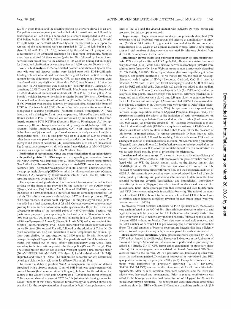
The loss of namA results in bacterial septation defects thatcan be complemented in trans. L. monocytogenes EGDe mu-tants lacking namA (33) (lmo2691, also known as murA [10])(Fig. 1A) were previously reported to exhibit bacterial septa-tion defects leading to the formation of bacterial chains (10).Consistent with these results, the L. monocytogenes 10403S-derived namA deletion strain also formed bacterial chains of 4to 12 bacteria when grown in broth culture (Fig. 1B). As NamAis a secreted peptidoglycan hydrolase that can be detected inbacterial culture supernatants (10, 33, 34), we investigatedwhether the growth of the mutant in the presence of wild-typebacteria could complement the �namA-associated septationdefect in trans. Equal numbers of L. monocytogenes wild-typeand �namA (containing a plasmid-harbored copy of the GFPgene) bacteria were mixed and grown in BHI broth for 4 h, andthe populations of bacterial cells were examined by using flu-orescence microscopy. The growth of the �namA mutant inmixed cultures with wild-type bacteria resulted in the full com-plementation of the �namA septation defect (Fig. 1C).
A comparison of �namA morphologies in mixed culturesand monocultures indicated that for mutant monocultures, theratio of the number of cells in chains to the total number of
cells visualized by Gram staining approached 1 after 4 h ofgrowth in BHI broth, indicating that nearly all cells were com-prised of chains greater than 3 bacteria in length (Fig. 1D). Incontrast, monocultures of the wild type or the complemented�namA mutant with pIMK2-namA or mixed wild-type and�namA cultures had relatively small percentages of bacteria inchains compared to the total number of cells in each field (Fig.1D). Mixed cultures did not become dominated by wild-typebacteria even after extended periods of growth, as evidencedby the maintenance of the 1:1 ratio of GFP-positive mutantbacteria to wild-type cells (Fig. 1C and data not shown). Thepresence of wild-type L. monocytogenes can thus functionallycomplement in trans the septation defect associated with the�namA mutation in broth culture.
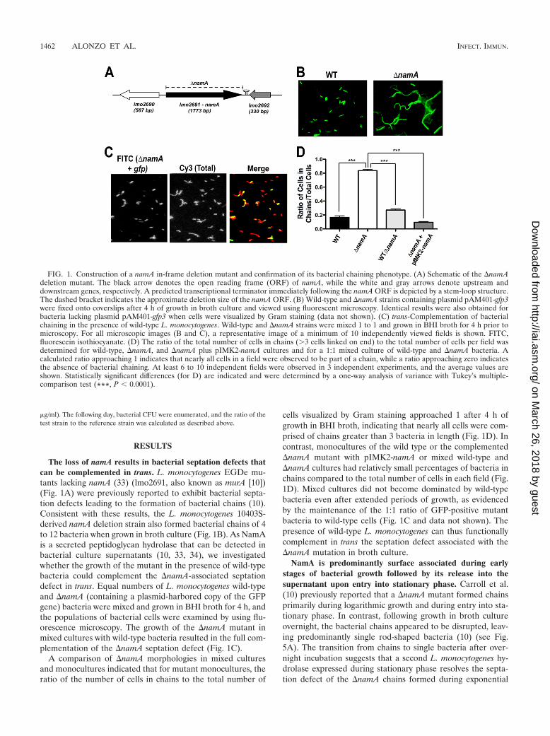
NamA is predominantly surface associated during earlystages of bacterial growth followed by its release into thesupernatant upon entry into stationary phase. Carroll et al.(10) previously reported that a �namA mutant formed chainsprimarily during logarithmic growth and during entry into sta-tionary phase. In contrast, following growth in broth cultureovernight, the bacterial chains appeared to be disrupted, leav-ing predominantly single rod-shaped bacteria (10) (see Fig.5A). The transition from chains to single bacteria after over-night incubation suggests that a second L. monocytogenes hy-drolase expressed during stationary phase resolves the septa-tion defect of the �namA chains formed during exponential
FIG. 1. Construction of a namA in-frame deletion mutant and confirmation of its bacterial chaining phenotype. (A) Schematic of the �namAdeletion mutant. The black arrow denotes the open reading frame (ORF) of namA, while the white and gray arrows denote upstream anddownstream genes, respectively. A predicted transcriptional terminator immediately following the namA ORF is depicted by a stem-loop structure.The dashed bracket indicates the approximate deletion size of the namA ORF. (B) Wild-type and �namA strains containing plasmid pAM401-gfp3were fixed onto coverslips after 4 h of growth in broth culture and viewed using fluorescent microscopy. Identical results were also obtained forbacteria lacking plasmid pAM401-gfp3 when cells were visualized by Gram staining (data not shown). (C) trans-Complementation of bacterialchaining in the presence of wild-type L. monocytogenes. Wild-type and �namA strains were mixed 1 to 1 and grown in BHI broth for 4 h prior tomicroscopy. For all microscopic images (B and C), a representative image of a minimum of 10 independently viewed fields is shown. FITC,fluorescein isothiocyanate. (D) The ratio of the total number of cells in chains (3 cells linked on end) to the total number of cells per field wasdetermined for wild-type, �namA, and �namA plus pIMK2-namA cultures and for a 1:1 mixed culture of wild-type and �namA bacteria. Acalculated ratio approaching 1 indicates that nearly all cells in a field were observed to be part of a chain, while a ratio approaching zero indicatesthe absence of bacterial chaining. At least 6 to 10 independent fields were observed in 3 independent experiments, and the average values areshown. Statistically significant differences (for D) are indicated and were determined by a one-way analysis of variance with Tukey’s multiple-comparison test (***, P � 0.0001).
1462 ALONZO ET AL. INFECT. IMMUN.
on March 26, 2018 by guest
http://iai.asm.org/
Dow
nloaded from
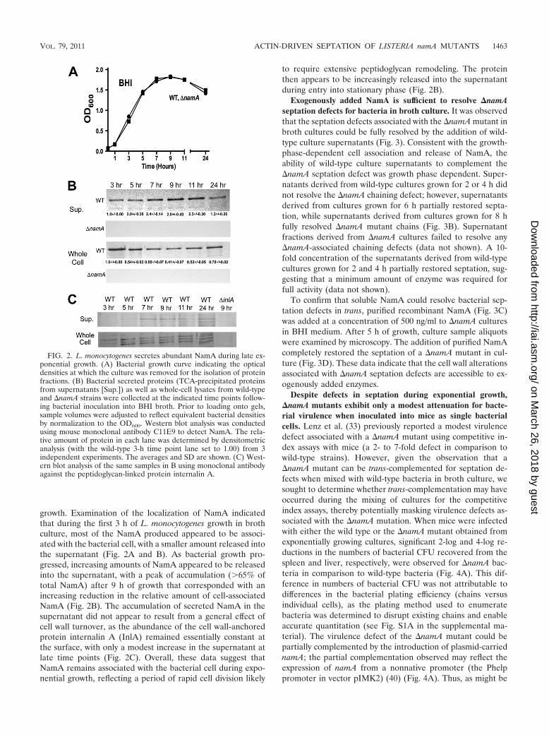
growth. Examination of the localization of NamA indicatedthat during the first 3 h of L. monocytogenes growth in brothculture, most of the NamA produced appeared to be associ-ated with the bacterial cell, with a smaller amount released intothe supernatant (Fig. 2A and B). As bacterial growth pro-gressed, increasing amounts of NamA appeared to be releasedinto the supernatant, with a peak of accumulation (65% oftotal NamA) after 9 h of growth that corresponded with anincreasing reduction in the relative amount of cell-associatedNamA (Fig. 2B). The accumulation of secreted NamA in thesupernatant did not appear to result from a general effect ofcell wall turnover, as the abundance of the cell wall-anchoredprotein internalin A (InlA) remained essentially constant atthe surface, with only a modest increase in the supernatant atlate time points (Fig. 2C). Overall, these data suggest thatNamA remains associated with the bacterial cell during expo-nential growth, reflecting a period of rapid cell division likely
to require extensive peptidoglycan remodeling. The proteinthen appears to be increasingly released into the supernatantduring entry into stationary phase (Fig. 2B).
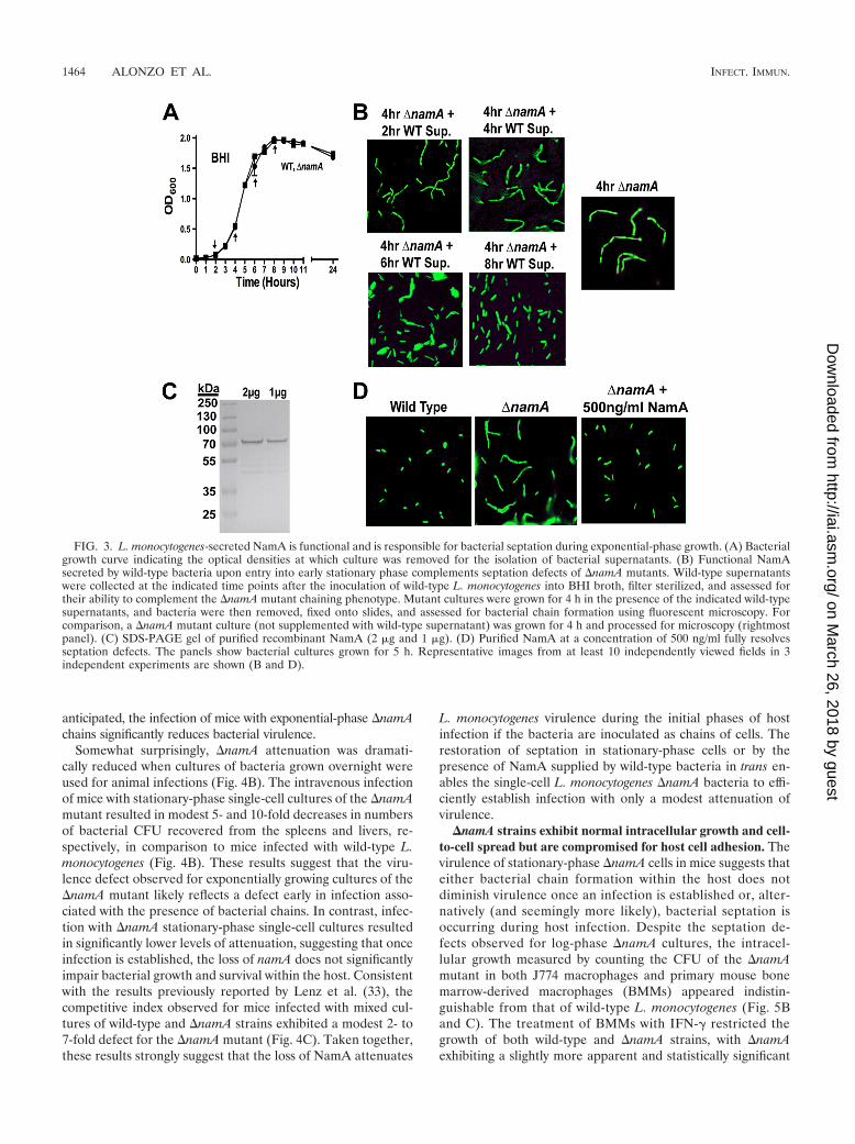
Exogenously added NamA is sufficient to resolve �namAseptation defects for bacteria in broth culture. It was observedthat the septation defects associated with the �namA mutant inbroth cultures could be fully resolved by the addition of wild-type culture supernatants (Fig. 3). Consistent with the growth-phase-dependent cell association and release of NamA, theability of wild-type culture supernatants to complement the�namA septation defect was growth phase dependent. Super-natants derived from wild-type cultures grown for 2 or 4 h didnot resolve the �namA chaining defect; however, supernatantsderived from cultures grown for 6 h partially restored septa-tion, while supernatants derived from cultures grown for 8 hfully resolved �namA mutant chains (Fig. 3B). Supernatantfractions derived from �namA cultures failed to resolve any�namA-associated chaining defects (data not shown). A 10-fold concentration of the supernatants derived from wild-typecultures grown for 2 and 4 h partially restored septation, sug-gesting that a minimum amount of enzyme was required forfull activity (data not shown).
To confirm that soluble NamA could resolve bacterial sep-tation defects in trans, purified recombinant NamA (Fig. 3C)was added at a concentration of 500 ng/ml to �namA culturesin BHI medium. After 5 h of growth, culture sample aliquotswere examined by microscopy. The addition of purified NamAcompletely restored the septation of a �namA mutant in cul-ture (Fig. 3D). These data indicate that the cell wall alterationsassociated with �namA septation defects are accessible to ex-ogenously added enzymes.
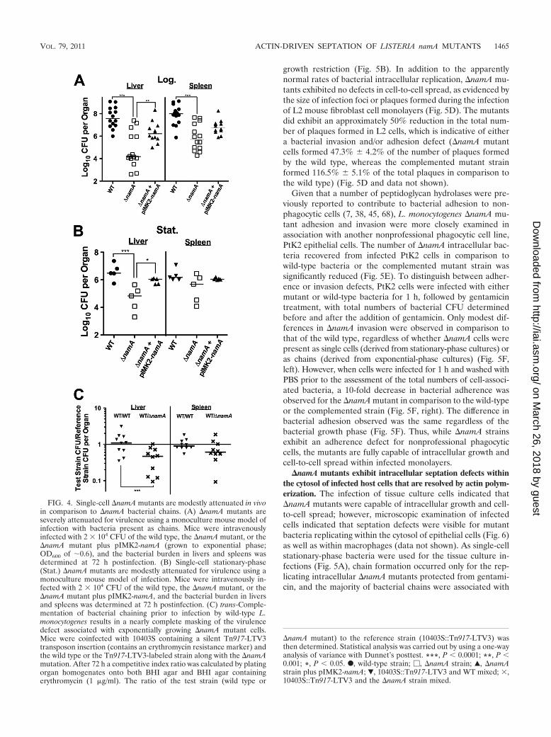
Despite defects in septation during exponential growth,�namA mutants exhibit only a modest attenuation for bacte-rial virulence when inoculated into mice as single bacterialcells. Lenz et al. (33) previously reported a modest virulencedefect associated with a �namA mutant using competitive in-dex assays with mice (a 2- to 7-fold defect in comparison towild-type strains). However, given the observation that a�namA mutant can be trans-complemented for septation de-fects when mixed with wild-type bacteria in broth culture, wesought to determine whether trans-complementation may haveoccurred during the mixing of cultures for the competitiveindex assays, thereby potentially masking virulence defects as-sociated with the �namA mutation. When mice were infectedwith either the wild type or the �namA mutant obtained fromexponentially growing cultures, significant 2-log and 4-log re-ductions in the numbers of bacterial CFU recovered from thespleen and liver, respectively, were observed for �namA bac-teria in comparison to wild-type bacteria (Fig. 4A). This dif-ference in numbers of bacterial CFU was not attributable todifferences in the bacterial plating efficiency (chains versusindividual cells), as the plating method used to enumeratebacteria was determined to disrupt existing chains and enableaccurate quantitation (see Fig. S1A in the supplemental ma-terial). The virulence defect of the �namA mutant could bepartially complemented by the introduction of plasmid-carriednamA; the partial complementation observed may reflect theexpression of namA from a nonnative promoter (the Phelppromoter in vector pIMK2) (40) (Fig. 4A). Thus, as might be
FIG. 2. L. monocytogenes secretes abundant NamA during late ex-ponential growth. (A) Bacterial growth curve indicating the opticaldensities at which the culture was removed for the isolation of proteinfractions. (B) Bacterial secreted proteins (TCA-precipitated proteinsfrom supernatants [Sup.]) as well as whole-cell lysates from wild-typeand �namA strains were collected at the indicated time points follow-ing bacterial inoculation into BHI broth. Prior to loading onto gels,sample volumes were adjusted to reflect equivalent bacterial densitiesby normalization to the OD600. Western blot analysis was conductedusing mouse monoclonal antibody C11E9 to detect NamA. The rela-tive amount of protein in each lane was determined by densitometricanalysis (with the wild-type 3-h time point lane set to 1.00) from 3independent experiments. The averages and SD are shown. (C) West-ern blot analysis of the same samples in B using monoclonal antibodyagainst the peptidoglycan-linked protein internalin A.
VOL. 79, 2011 ACTIN-DRIVEN SEPTATION OF LISTERIA namA MUTANTS 1463
on March 26, 2018 by guest
http://iai.asm.org/
Dow
nloaded from
anticipated, the infection of mice with exponential-phase �namAchains significantly reduces bacterial virulence.
Somewhat surprisingly, �namA attenuation was dramati-cally reduced when cultures of bacteria grown overnight wereused for animal infections (Fig. 4B). The intravenous infectionof mice with stationary-phase single-cell cultures of the �namAmutant resulted in modest 5- and 10-fold decreases in numbersof bacterial CFU recovered from the spleens and livers, re-spectively, in comparison to mice infected with wild-type L.monocytogenes (Fig. 4B). These results suggest that the viru-lence defect observed for exponentially growing cultures of the�namA mutant likely reflects a defect early in infection asso-ciated with the presence of bacterial chains. In contrast, infec-tion with �namA stationary-phase single-cell cultures resultedin significantly lower levels of attenuation, suggesting that onceinfection is established, the loss of namA does not significantlyimpair bacterial growth and survival within the host. Consistentwith the results previously reported by Lenz et al. (33), thecompetitive index observed for mice infected with mixed cul-tures of wild-type and �namA strains exhibited a modest 2- to7-fold defect for the �namA mutant (Fig. 4C). Taken together,these results strongly suggest that the loss of NamA attenuates
L. monocytogenes virulence during the initial phases of hostinfection if the bacteria are inoculated as chains of cells. Therestoration of septation in stationary-phase cells or by thepresence of NamA supplied by wild-type bacteria in trans en-ables the single-cell L. monocytogenes �namA bacteria to effi-ciently establish infection with only a modest attenuation ofvirulence.
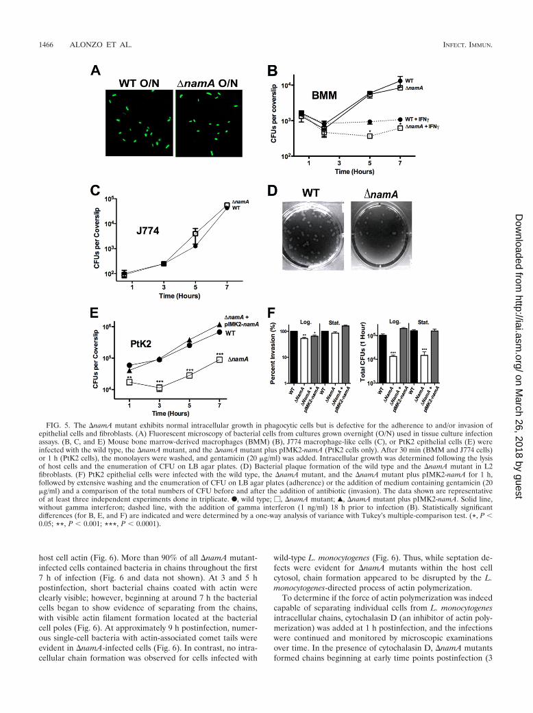
�namA strains exhibit normal intracellular growth and cell-to-cell spread but are compromised for host cell adhesion. Thevirulence of stationary-phase �namA cells in mice suggests thateither bacterial chain formation within the host does notdiminish virulence once an infection is established or, alter-natively (and seemingly more likely), bacterial septation isoccurring during host infection. Despite the septation de-fects observed for log-phase �namA cultures, the intracel-lular growth measured by counting the CFU of the �namAmutant in both J774 macrophages and primary mouse bonemarrow-derived macrophages (BMMs) appeared indistin-guishable from that of wild-type L. monocytogenes (Fig. 5Band C). The treatment of BMMs with IFN- restricted thegrowth of both wild-type and �namA strains, with �namAexhibiting a slightly more apparent and statistically significant
FIG. 3. L. monocytogenes-secreted NamA is functional and is responsible for bacterial septation during exponential-phase growth. (A) Bacterialgrowth curve indicating the optical densities at which culture was removed for the isolation of bacterial supernatants. (B) Functional NamAsecreted by wild-type bacteria upon entry into early stationary phase complements septation defects of �namA mutants. Wild-type supernatantswere collected at the indicated time points after the inoculation of wild-type L. monocytogenes into BHI broth, filter sterilized, and assessed fortheir ability to complement the �namA mutant chaining phenotype. Mutant cultures were grown for 4 h in the presence of the indicated wild-typesupernatants, and bacteria were then removed, fixed onto slides, and assessed for bacterial chain formation using fluorescent microscopy. Forcomparison, a �namA mutant culture (not supplemented with wild-type supernatant) was grown for 4 h and processed for microscopy (rightmostpanel). (C) SDS-PAGE gel of purified recombinant NamA (2 �g and 1 �g). (D) Purified NamA at a concentration of 500 ng/ml fully resolvesseptation defects. The panels show bacterial cultures grown for 5 h. Representative images from at least 10 independently viewed fields in 3independent experiments are shown (B and D).
1464 ALONZO ET AL. INFECT. IMMUN.
on March 26, 2018 by guest
http://iai.asm.org/
Dow
nloaded from
growth restriction (Fig. 5B). In addition to the apparentlynormal rates of bacterial intracellular replication, �namA mu-tants exhibited no defects in cell-to-cell spread, as evidenced bythe size of infection foci or plaques formed during the infectionof L2 mouse fibroblast cell monolayers (Fig. 5D). The mutantsdid exhibit an approximately 50% reduction in the total num-ber of plaques formed in L2 cells, which is indicative of eithera bacterial invasion and/or adhesion defect (�namA mutantcells formed 47.3% � 4.2% of the number of plaques formedby the wild type, whereas the complemented mutant strainformed 116.5% � 5.1% of the total plaques in comparison tothe wild type) (Fig. 5D and data not shown).
Given that a number of peptidoglycan hydrolases were pre-viously reported to contribute to bacterial adhesion to non-phagocytic cells (7, 38, 45, 68), L. monocytogenes �namA mu-tant adhesion and invasion were more closely examined inassociation with another nonprofessional phagocytic cell line,PtK2 epithelial cells. The number of �namA intracellular bac-teria recovered from infected PtK2 cells in comparison towild-type bacteria or the complemented mutant strain wassignificantly reduced (Fig. 5E). To distinguish between adher-ence or invasion defects, PtK2 cells were infected with eithermutant or wild-type bacteria for 1 h, followed by gentamicintreatment, with total numbers of bacterial CFU determinedbefore and after the addition of gentamicin. Only modest dif-ferences in �namA invasion were observed in comparison tothat of the wild type, regardless of whether �namA cells werepresent as single cells (derived from stationary-phase cultures) oras chains (derived from exponential-phase cultures) (Fig. 5F,left). However, when cells were infected for 1 h and washed withPBS prior to the assessment of the total numbers of cell-associ-ated bacteria, a 10-fold decrease in bacterial adherence wasobserved for the �namA mutant in comparison to the wild-typeor the complemented strain (Fig. 5F, right). The difference inbacterial adhesion observed was the same regardless of thebacterial growth phase (Fig. 5F). Thus, while �namA strainsexhibit an adherence defect for nonprofessional phagocyticcells, the mutants are fully capable of intracellular growth andcell-to-cell spread within infected monolayers.
�namA mutants exhibit intracellular septation defects withinthe cytosol of infected host cells that are resolved by actin polym-erization. The infection of tissue culture cells indicated that�namA mutants were capable of intracellular growth and cell-to-cell spread; however, microscopic examination of infectedcells indicated that septation defects were visible for mutantbacteria replicating within the cytosol of epithelial cells (Fig. 6)as well as within macrophages (data not shown). As single-cellstationary-phase bacteria were used for the tissue culture in-fections (Fig. 5A), chain formation occurred only for the rep-licating intracellular �namA mutants protected from gentami-cin, and the majority of bacterial chains were associated with
FIG. 4. Single-cell �namA mutants are modestly attenuated in vivoin comparison to �namA bacterial chains. (A) �namA mutants areseverely attenuated for virulence using a monoculture mouse model ofinfection with bacteria present as chains. Mice were intravenouslyinfected with 2 � 104 CFU of the wild type, the �namA mutant, or the�namA mutant plus pIMK2-namA (grown to exponential phase;OD600 of �0.6), and the bacterial burden in livers and spleens wasdetermined at 72 h postinfection. (B) Single-cell stationary-phase(Stat.) �namA mutants are modestly attenuated for virulence using amonoculture mouse model of infection. Mice were intravenously in-fected with 2 � 104 CFU of the wild type, the �namA mutant, or the�namA mutant plus pIMK2-namA, and the bacterial burden in liversand spleens was determined at 72 h postinfection. (C) trans-Comple-mentation of bacterial chaining prior to infection by wild-type L.monocytogenes results in a nearly complete masking of the virulencedefect associated with exponentially growing �namA mutant cells.Mice were coinfected with 10403S containing a silent Tn917-LTV3transposon insertion (contains an erythromycin resistance marker) andthe wild type or the Tn917-LTV3-labeled strain along with the �namAmutation. After 72 h a competitive index ratio was calculated by platingorgan homogenates onto both BHI agar and BHI agar containingerythromycin (1 �g/ml). The ratio of the test strain (wild type or
�namA mutant) to the reference strain (10403S::Tn917-LTV3) wasthen determined. Statistical analysis was carried out by using a one-wayanalysis of variance with Dunnet’s posttest. ***, P � 0.0001; **, P �0.001; *, P � 0.05. F, wild-type strain; �, �namA strain; Œ, �namAstrain plus pIMK2-namA; �, 10403S::Tn917-LTV3 and WT mixed; �,10403S::Tn917-LTV3 and the �namA strain mixed.
VOL. 79, 2011 ACTIN-DRIVEN SEPTATION OF LISTERIA namA MUTANTS 1465
on March 26, 2018 by guest
http://iai.asm.org/
Dow
nloaded from
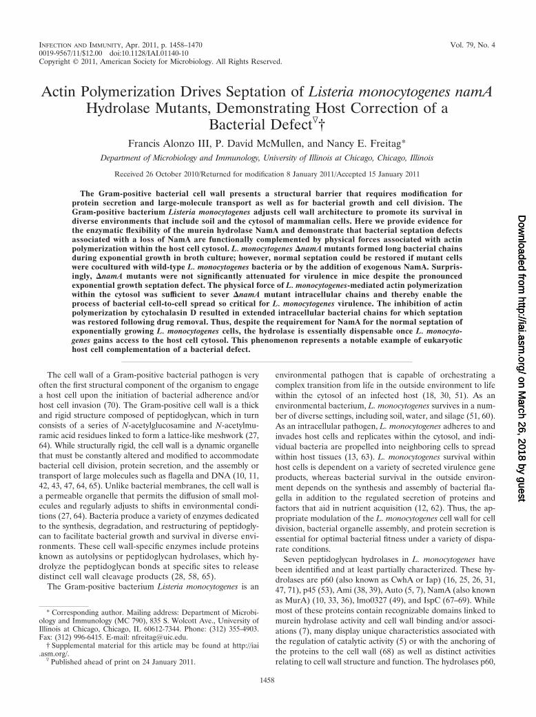
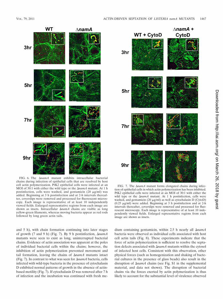
host cell actin (Fig. 6). More than 90% of all �namA mutant-infected cells contained bacteria in chains throughout the first7 h of infection (Fig. 6 and data not shown). At 3 and 5 hpostinfection, short bacterial chains coated with actin wereclearly visible; however, beginning at around 7 h the bacterialcells began to show evidence of separating from the chains,with visible actin filament formation located at the bacterialcell poles (Fig. 6). At approximately 9 h postinfection, numer-ous single-cell bacteria with actin-associated comet tails wereevident in �namA-infected cells (Fig. 6). In contrast, no intra-cellular chain formation was observed for cells infected with
wild-type L. monocytogenes (Fig. 6). Thus, while septation de-fects were evident for �namA mutants within the host cellcytosol, chain formation appeared to be disrupted by the L.monocytogenes-directed process of actin polymerization.
To determine if the force of actin polymerization was indeedcapable of separating individual cells from L. monocytogenesintracellular chains, cytochalasin D (an inhibitor of actin poly-merization) was added at 1 h postinfection, and the infectionswere continued and monitored by microscopic examinationsover time. In the presence of cytochalasin D, �namA mutantsformed chains beginning at early time points postinfection (3
FIG. 5. The �namA mutant exhibits normal intracellular growth in phagocytic cells but is defective for the adherence to and/or invasion ofepithelial cells and fibroblasts. (A) Fluorescent microscopy of bacterial cells from cultures grown overnight (O/N) used in tissue culture infectionassays. (B, C, and E) Mouse bone marrow-derived macrophages (BMM) (B), J774 macrophage-like cells (C), or PtK2 epithelial cells (E) wereinfected with the wild type, the �namA mutant, and the �namA mutant plus pIMK2-namA (PtK2 cells only). After 30 min (BMM and J774 cells)or 1 h (PtK2 cells), the monolayers were washed, and gentamicin (20 �g/ml) was added. Intracellular growth was determined following the lysisof host cells and the enumeration of CFU on LB agar plates. (D) Bacterial plaque formation of the wild type and the �namA mutant in L2fibroblasts. (F) PtK2 epithelial cells were infected with the wild type, the �namA mutant, and the �namA mutant plus pIMK2-namA for 1 h,followed by extensive washing and the enumeration of CFU on LB agar plates (adherence) or the addition of medium containing gentamicin (20�g/ml) and a comparison of the total numbers of CFU before and after the addition of antibiotic (invasion). The data shown are representativeof at least three independent experiments done in triplicate. F, wild type; �, �namA mutant; Œ, �namA mutant plus pIMK2-namA. Solid line,without gamma interferon; dashed line, with the addition of gamma interferon (1 ng/ml) 18 h prior to infection (B). Statistically significantdifferences (for B, E, and F) are indicated and were determined by a one-way analysis of variance with Tukey’s multiple-comparison test. (*, P �0.05; **, P � 0.001; ***, P � 0.0001).
1466 ALONZO ET AL. INFECT. IMMUN.
on March 26, 2018 by guest
http://iai.asm.org/
Dow
nloaded from
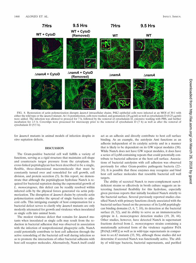
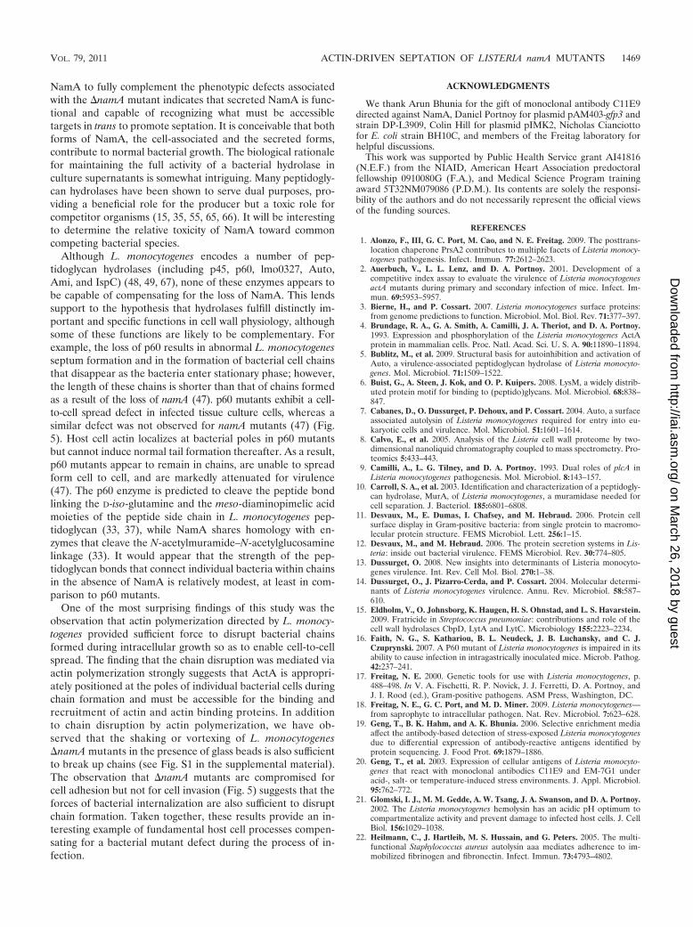
and 5 h), with chain formation continuing into later stagesof growth (7 and 9 h) (Fig. 7). By 9 h postinfection, �namAmutants were seen to exist as long, uninterrupted bacterialchains. Evidence of actin association was apparent at the polesof individual bacterial cells within the chains; however, theinhibition of actin polymerization prevented movement andtail formation, leaving the chains of �namA mutants intact(Fig. 7). In contrast to what was seen for �namA bacteria, cellsinfected with wild-type bacteria in the presence of cytochalasinD exhibited normal patterns of growth in the absence of actin-based motility (Fig. 7). If cytochalasin D was removed after 7 hof infection and the incubation was continued with fresh me-
dium containing gentamicin, within 2.5 h nearly all �namAbacteria were observed as individual cells associated with hostcell actin tails (Fig. 8). These experiments indicate that theforce of actin polymerization is sufficient to resolve the septa-tion defects associated with �namA mutants within the cytosolof infected host cells. Consistent with this observation, otherphysical forces (such as homogenization and shaking of bacte-rial cultures in the presence of glass beads) also result in thedisruption of �namA chains (see Fig. S1 in the supplementalmaterial, and data not shown). The disruption of bacterialchains via the forces exerted by actin polymerization is thuslikely to account for the substantial level of virulence observed
FIG. 6. The �namA mutant exhibits intracellular bacterialchains during infection of epithelial cells that are resolved by hostcell actin polymerization. PtK2 epithelial cells were infected at anMOI of 30:1 with either the wild type or the �namA mutant. At 1 hpostinfection, cells were washed, and gentamicin (20 �g/ml) wasadded. Beginning at 3 h postinfection and at 2-h intervals thereaf-ter, coverslips were removed and processed for fluorescent micros-copy. Each image is representative of at least 10 independentlyviewed fields. Enlarged representative regions from each image areshown as insets. Intracellular �namA chains are visible as longyellow-green filaments, whereas moving bacteria appear as red rodsfollowed by long green actin tails.
FIG. 7. The �namA mutant forms elongated chains during infec-tion of epithelial cells in which actin polymerization has been inhibited.PtK2 epithelial cells were infected at an MOI of 30:1 with either thewild type or the �namA mutant. At 1 h postinfection, cells werewashed, and gentamicin (20 �g/ml) as well as cytochalasin D (CytoD)(0.25 �g/ml) were added. Beginning at 3 h postinfection and at 2-hintervals thereafter, coverslips were removed and processed for fluo-rescent microscopy. Each image is representative of at least 10 inde-pendently viewed fields. Enlarged representative regions from eachimage are shown as insets.
VOL. 79, 2011 ACTIN-DRIVEN SEPTATION OF LISTERIA namA MUTANTS 1467
on March 26, 2018 by guest
http://iai.asm.org/
Dow
nloaded from
for �namA mutants in animal models of infection despite invitro septation defects.
DISCUSSION
The Gram-positive bacterial cell wall fulfills a variety offunctions, serving as a rigid structure that maintains cell shapeand counteracts turgor pressure from the cytoplasm. Itscross-linked peptidoglycan has been described to be a single,flexible, three-dimensional macromolecule that must beconstantly turned over and remodeled for cell growth, celldivision, and protein secretion (5). In this report, we demon-strate that although the peptidoglycan hydrolase NamA is re-quired for bacterial septation during the exponential growth ofL. monocytogenes, this defect can be readily resolved withininfected cells by the physical forces generated via actin poly-merization. The disruption of �namA chains by cytosolic actinpolymerization enables the spread of single bacteria to adja-cent cells. This intriguing example of host compensation for abacterial defect serves to clarify why �namA mutants are onlymodestly attenuated for growth and virulence when inoculatedas single cells into animal hosts.
The modest virulence defect that remains for �namA mu-tants when inoculated as single cells may result from the re-duction in bacterial adhesion that was observed in associationwith the infection of nonprofessional phagocytic cells. NamAcould potentially contribute to host cell adhesion through theactive remodeling of the bacterial cell wall during infection soas to promote the interactions of other bacterial adhesins withhost cell receptor molecules. Alternatively, NamA itself could
act as an adhesin and directly contribute to host cell surfacebinding. As an example, the autolysin Ami functions as anadhesin independent of its catalytic activity and in a mannerthat is likely to be dependent on its GW repeat modules (38).While NamA does not have GW repeat modules, it does havea series of LysM-containing repeats that could potentially con-tribute to bacterial adhesion at the host cell surface. Associa-tions of bacterial autolysins with cell adhesion was observedpreviously for other Gram-positive pathogenic bacteria (22–24). It is possible that these enzymes may recognize and bindhost cell surface molecules that resemble bacterial cell walltargets.
The ability of secreted NamA to trans-complement namA-deficient strains so effectively in broth culture suggests an in-teresting functional flexibility for this hydrolase, especiallygiven previous reports that initially localized NamA strictly tothe bacterial surface. Several previously reported studies clas-sified NamA with primary functions closely associated with thebacterial surface based on the presence of its LysM peptidogly-can binding domains (3, 6, 7, 10), its detection at the bacterialsurface (8, 52), and its ability to serve as an immunoreactiveepitope in L. monocytogenes detection studies (19, 20, 44).Other studies, however, have detected NamA in supernatantfractions derived from L. monocytogenes strains containing amutationally activated form of the virulence regulator PrfA[PrfA(L140F)] as well as in wild-type supernatants in compar-ison to secA2 mutants (33, 50), although those studies did notdetermine if secreted NamA was functionally active. The abil-ity of wild-type bacteria, bacterial supernatants, and purified
FIG. 8. Restoration of actin polymerization disrupts �namA intracellular chains. PtK2 epithelial cells were infected at an MOI of 30:1 witheither the wild type or the �namA mutant. At 1 h postinfection, cells were washed, and gentamicin (20 �g/ml) as well as cytochalasin D (0.25 �g/ml)were added. The infection was allowed to proceed for 7 h, followed by the removal of cytochalasin D, extensive washing with PBS, and furtherincubation for 2.5 h. Coverslips were processed for microscopy prior to the removal of cytochalasin D (7 h) as well as after the removal ofcytochalasin D (9.5 h).
1468 ALONZO ET AL. INFECT. IMMUN.
on March 26, 2018 by guest
http://iai.asm.org/
Dow
nloaded from
NamA to fully complement the phenotypic defects associatedwith the �namA mutant indicates that secreted NamA is func-tional and capable of recognizing what must be accessibletargets in trans to promote septation. It is conceivable that bothforms of NamA, the cell-associated and the secreted forms,contribute to normal bacterial growth. The biological rationalefor maintaining the full activity of a bacterial hydrolase inculture supernatants is somewhat intriguing. Many peptidogly-can hydrolases have been shown to serve dual purposes, pro-viding a beneficial role for the producer but a toxic role forcompetitor organisms (15, 35, 55, 65, 66). It will be interestingto determine the relative toxicity of NamA toward commoncompeting bacterial species.
Although L. monocytogenes encodes a number of pep-tidoglycan hydrolases (including p45, p60, lmo0327, Auto,Ami, and IspC) (48, 49, 67), none of these enzymes appears tobe capable of compensating for the loss of NamA. This lendssupport to the hypothesis that hydrolases fulfill distinctly im-portant and specific functions in cell wall physiology, althoughsome of these functions are likely to be complementary. Forexample, the loss of p60 results in abnormal L. monocytogenesseptum formation and in the formation of bacterial cell chainsthat disappear as the bacteria enter stationary phase; however,the length of these chains is shorter than that of chains formedas a result of the loss of namA (47). p60 mutants exhibit a cell-to-cell spread defect in infected tissue culture cells, whereas asimilar defect was not observed for namA mutants (47) (Fig.5). Host cell actin localizes at bacterial poles in p60 mutantsbut cannot induce normal tail formation thereafter. As a result,p60 mutants appear to remain in chains, are unable to spreadform cell to cell, and are markedly attenuated for virulence(47). The p60 enzyme is predicted to cleave the peptide bondlinking the D-iso-glutamine and the meso-diaminopimelic acidmoieties of the peptide side chain in L. monocytogenes pep-tidoglycan (33, 37), while NamA shares homology with en-zymes that cleave the N-acetylmuramide–N-acetylglucosaminelinkage (33). It would appear that the strength of the pep-tidoglycan bonds that connect individual bacteria within chainsin the absence of NamA is relatively modest, at least in com-parison to p60 mutants.
One of the most surprising findings of this study was theobservation that actin polymerization directed by L. monocy-togenes provided sufficient force to disrupt bacterial chainsformed during intracellular growth so as to enable cell-to-cellspread. The finding that the chain disruption was mediated viaactin polymerization strongly suggests that ActA is appropri-ately positioned at the poles of individual bacterial cells duringchain formation and must be accessible for the binding andrecruitment of actin and actin binding proteins. In additionto chain disruption by actin polymerization, we have ob-served that the shaking or vortexing of L. monocytogenes�namA mutants in the presence of glass beads is also sufficientto break up chains (see Fig. S1 in the supplemental material).The observation that �namA mutants are compromised forcell adhesion but not for cell invasion (Fig. 5) suggests that theforces of bacterial internalization are also sufficient to disruptchain formation. Taken together, these results provide an in-teresting example of fundamental host cell processes compen-sating for a bacterial mutant defect during the process of in-fection.
ACKNOWLEDGMENTS
We thank Arun Bhunia for the gift of monoclonal antibody C11E9directed against NamA, Daniel Portnoy for plasmid pAM403-gfp3 andstrain DP-L3909, Colin Hill for plasmid pIMK2, Nicholas Cianciottofor E. coli strain BH10C, and members of the Freitag laboratory forhelpful discussions.
This work was supported by Public Health Service grant AI41816(N.E.F.) from the NIAID, American Heart Association predoctoralfellowship 0910080G (F.A.), and Medical Science Program trainingaward 5T32NM079086 (P.D.M.). Its contents are solely the responsi-bility of the authors and do not necessarily represent the official viewsof the funding sources.
REFERENCES
1. Alonzo, F., III, G. C. Port, M. Cao, and N. E. Freitag. 2009. The posttrans-location chaperone PrsA2 contributes to multiple facets of Listeria monocy-togenes pathogenesis. Infect. Immun. 77:2612–2623.
2. Auerbuch, V., L. L. Lenz, and D. A. Portnoy. 2001. Development of acompetitive index assay to evaluate the virulence of Listeria monocytogenesactA mutants during primary and secondary infection of mice. Infect. Im-mun. 69:5953–5957.
3. Bierne, H., and P. Cossart. 2007. Listeria monocytogenes surface proteins:from genome predictions to function. Microbiol. Mol. Biol. Rev. 71:377–397.
4. Brundage, R. A., G. A. Smith, A. Camilli, J. A. Theriot, and D. A. Portnoy.1993. Expression and phosphorylation of the Listeria monocytogenes ActAprotein in mammalian cells. Proc. Natl. Acad. Sci. U. S. A. 90:11890–11894.
5. Bublitz, M., et al. 2009. Structural basis for autoinhibition and activation ofAuto, a virulence-associated peptidoglycan hydrolase of Listeria monocyto-genes. Mol. Microbiol. 71:1509–1522.
6. Buist, G., A. Steen, J. Kok, and O. P. Kuipers. 2008. LysM, a widely distrib-uted protein motif for binding to (peptido)glycans. Mol. Microbiol. 68:838–847.
7. Cabanes, D., O. Dussurget, P. Dehoux, and P. Cossart. 2004. Auto, a surfaceassociated autolysin of Listeria monocytogenes required for entry into eu-karyotic cells and virulence. Mol. Microbiol. 51:1601–1614.
8. Calvo, E., et al. 2005. Analysis of the Listeria cell wall proteome by two-dimensional nanoliquid chromatography coupled to mass spectrometry. Pro-teomics 5:433–443.
9. Camilli, A., L. G. Tilney, and D. A. Portnoy. 1993. Dual roles of plcA inListeria monocytogenes pathogenesis. Mol. Microbiol. 8:143–157.
10. Carroll, S. A., et al. 2003. Identification and characterization of a peptidogly-can hydrolase, MurA, of Listeria monocytogenes, a muramidase needed forcell separation. J. Bacteriol. 185:6801–6808.
11. Desvaux, M., E. Dumas, I. Chafsey, and M. Hebraud. 2006. Protein cellsurface display in Gram-positive bacteria: from single protein to macromo-lecular protein structure. FEMS Microbiol. Lett. 256:1–15.
12. Desvaux, M., and M. Hebraud. 2006. The protein secretion systems in Lis-teria: inside out bacterial virulence. FEMS Microbiol. Rev. 30:774–805.
13. Dussurget, O. 2008. New insights into determinants of Listeria monocyto-genes virulence. Int. Rev. Cell Mol. Biol. 270:1–38.
14. Dussurget, O., J. Pizarro-Cerda, and P. Cossart. 2004. Molecular determi-nants of Listeria monocytogenes virulence. Annu. Rev. Microbiol. 58:587–610.
15. Eldholm, V., O. Johnsborg, K. Haugen, H. S. Ohnstad, and L. S. Havarstein.2009. Fratricide in Streptococcus pneumoniae: contributions and role of thecell wall hydrolases CbpD, LytA and LytC. Microbiology 155:2223–2234.
16. Faith, N. G., S. Kathariou, B. L. Neudeck, J. B. Luchansky, and C. J.Czuprynski. 2007. A P60 mutant of Listeria monocytogenes is impaired in itsability to cause infection in intragastrically inoculated mice. Microb. Pathog.42:237–241.
17. Freitag, N. E. 2000. Genetic tools for use with Listeria monocytogenes, p.488–498. In V. A. Fischetti, R. P. Novick, J. J. Ferretti, D. A. Portnoy, andJ. I. Rood (ed.), Gram-positive pathogens. ASM Press, Washington, DC.
18. Freitag, N. E., G. C. Port, and M. D. Miner. 2009. Listeria monocytogenes—from saprophyte to intracellular pathogen. Nat. Rev. Microbiol. 7:623–628.
19. Geng, T., B. K. Hahm, and A. K. Bhunia. 2006. Selective enrichment mediaaffect the antibody-based detection of stress-exposed Listeria monocytogenesdue to differential expression of antibody-reactive antigens identified byprotein sequencing. J. Food Prot. 69:1879–1886.
20. Geng, T., et al. 2003. Expression of cellular antigens of Listeria monocyto-genes that react with monoclonal antibodies C11E9 and EM-7G1 underacid-, salt- or temperature-induced stress environments. J. Appl. Microbiol.95:762–772.
21. Glomski, I. J., M. M. Gedde, A. W. Tsang, J. A. Swanson, and D. A. Portnoy.2002. The Listeria monocytogenes hemolysin has an acidic pH optimum tocompartmentalize activity and prevent damage to infected host cells. J. CellBiol. 156:1029–1038.
22. Heilmann, C., J. Hartleib, M. S. Hussain, and G. Peters. 2005. The multi-functional Staphylococcus aureus autolysin aaa mediates adherence to im-mobilized fibrinogen and fibronectin. Infect. Immun. 73:4793–4802.
VOL. 79, 2011 ACTIN-DRIVEN SEPTATION OF LISTERIA namA MUTANTS 1469
on March 26, 2018 by guest
http://iai.asm.org/
Dow
nloaded from
23. Heilmann, C., M. Hussain, G. Peters, and F. Gotz. 1997. Evidence forautolysin-mediated primary attachment of Staphylococcus epidermidis to apolystyrene surface. Mol. Microbiol. 24:1013–1024.
24. Hell, W., H. G. Meyer, and S. G. Gatermann. 1998. Cloning of aas, a geneencoding a Staphylococcus saprophyticus surface protein with adhesive andautolytic properties. Mol. Microbiol. 29:871–881.
25. Hess, J., et al. 1996. Protein p60 participates in intestinal host invasion byListeria monocytogenes. Zentralbl. Bakteriol. 284:263–272.
26. Hess, J., et al. 1995. Listeria monocytogenes p60 supports host cell invasion byand in vivo survival of attenuated Salmonella typhimurium. Infect. Immun.63:2047–2053.
27. Holtje, J. V., and B. Glauner. 1990. Structure and metabolism of the mureinsacculus. Res. Microbiol. 141:75–89.
28. Holtje, J. V., and E. I. Tuomanen. 1991. The murein hydrolases of Esche-richia coli: properties, functions and impact on the course of infections invivo. J. Gen. Microbiol. 137:441–454.
29. Howell-Adams, B., and H. S. Seifert. 2000. Molecular models accounting forthe gene conversion reactions mediating gonococcal pilin antigenic variation.Mol. Microbiol. 37:1146–1158.
30. Joseph, B., and W. Goebel. 2007. Life of Listeria monocytogenes in the hostcells’ cytosol. Microbes Infect. 9:1188–1195.
31. Kuhn, M., and W. Goebel. 1989. Identification of an extracellular protein ofListeria monocytogenes possibly involved in intracellular uptake by mamma-lian cells. Infect. Immun. 57:55–61.
32. Lauer, P., M. Y. Chow, M. J. Loessner, D. A. Portnoy, and R. Calendar. 2002.Construction, characterization, and use of two Listeria monocytogenes site-specific phage integration vectors. J. Bacteriol. 184:4177–4186.
33. Lenz, L. L., S. Mohammadi, A. Geissler, and D. A. Portnoy. 2003. SecA2-dependent secretion of autolytic enzymes promotes Listeria monocytogenespathogenesis. Proc. Natl. Acad. Sci. U. S. A. 100:12432–12437.
34. Lenz, L. L., and D. A. Portnoy. 2002. Identification of a second Listeria secAgene associated with protein secretion and the rough phenotype. Mol. Mi-crobiol. 45:1043–1056.
35. Li, Z., A. J. Clarke, and T. J. Beveridge. 1998. Gram-negative bacteriaproduce membrane vesicles which are capable of killing other bacteria. J.Bacteriol. 180:5478–5483.
36. Machata, S., T. Hain, M. Rohde, and T. Chakraborty. 2005. Simultaneousdeficiency of both MurA and p60 proteins generates a rough phenotype inListeria monocytogenes. J. Bacteriol. 187:8385–8394.
37. Margot, P., M. Pagni, and D. Karamata. 1999. Bacillus subtilis 168 gene lytFencodes a gamma-D-glutamate-meso-diaminopimelate muropeptidase ex-pressed by the alternative vegetative sigma factor, sigmaD. Microbiology145(Pt. 1):57–65.
38. Milohanic, E., R. Jonquieres, P. Cossart, P. Berche, and J. L. Gaillard. 2001.The autolysin Ami contributes to the adhesion of Listeria monocytogenes toeukaryotic cells via its cell wall anchor. Mol. Microbiol. 39:1212–1224.
39. Milohanic, E., et al. 2004. Sequence and binding activity of the autolysin-adhesin Ami from epidemic Listeria monocytogenes 4b. Infect. Immun. 72:4401–4409.
40. Monk, I. R., C. G. Gahan, and C. Hill. 2008. Tools for functional postgen-omic analysis of Listeria monocytogenes. Appl. Environ. Microbiol. 74:3921–3934.
41. Mueller, K. J., and N. E. Freitag. 2005. Pleiotropic enhancement of bacterialpathogenesis resulting from the constitutive activation of the Listeria mono-cytogenes regulatory factor PrfA. Infect. Immun. 73:1917–1926.
42. Nambu, T., Y. Inagaki, and K. Kutsukake. 2006. Plasticity of the domainstructure in FlgJ, a bacterial protein involved in flagellar rod formation.Genes Genet. Syst. 81:381–389.
43. Nambu, T., T. Minamino, R. M. Macnab, and K. Kutsukake. 1999. Pep-tidoglycan-hydrolyzing activity of the FlgJ protein, essential for flagellar rodformation in Salmonella typhimurium. J. Bacteriol. 181:1555–1561.
44. Nannapaneni, R., R. Story, A. K. Bhunia, and M. G. Johnson. 1998. Reac-tivities of genus-specific monoclonal antibody EM-6E11 against Listeria spe-cies and serotypes of Listeria monocytogenes grown in nonselective and se-lective enrichment broth media. J. Food Prot. 61:1195–1198.
45. Park, J. H., et al. 2000. Specific binding of recombinant Listeria monocyto-genes p60 protein to Caco-2 cells. FEMS Microbiol. Lett. 186:35–40.
46. Park, S. F., and G. S. A. B. Stewart. 1990. High-efficiency transformation ofListeria monocytogenes by electroporation of penicillin-treated cells. Gene94:129–132.
47. Pilgrim, S., A. Kolb-Maurer, I. Gentschev, W. Goebel, and M. Kuhn. 2003.Deletion of the gene encoding p60 in Listeria monocytogenes leads to abnor-
mal cell division and loss of actin-based motility. Infect. Immun. 71:3473–3484.
48. Popowska, M. 2004. Analysis of the peptidoglycan hydrolases of Listeriamonocytogenes: multiple enzymes with multiple functions. Pol. J. Microbiol.53(Suppl.):29–34.
49. Popowska, M., and Z. Markiewicz. 2006. Characterization of Listeria mono-cytogenes protein Lmo0327 with murein hydrolase activity. Arch. Microbiol.186:69–86.
50. Port, G. C., and N. E. Freitag. 2007. Identification of novel Listeria mono-cytogenes secreted virulence factors following mutational activation of thecentral virulence regulator, PrfA. Infect. Immun. 75:5886–5897.
51. Renier, S., M. Hebraud, and M. Desvaux. 2010. Molecular biology of surfacecolonization by Listeria monocytogenes: an additional facet of an opportu-nistic Gram-positive foodborne pathogen. Environ. Microbiol. [Epub aheadof print.] doi:10.1111/j.1462-2920.2010.02378.x.
52. Schaumburg, J., et al. 2004. The cell wall subproteome of Listeria monocy-togenes. Proteomics 4:2991–3006.
53. Schubert, K., et al. 2000. P45, an extracellular 45 kDa protein of Listeriamonocytogenes with similarity to protein p60 and exhibiting peptidoglycanlytic activity. Arch. Microbiol. 173:21–28.
54. Shetron-Rama, L. M., et al. 2003. Isolation of Listeria monocytogenes mu-tants with high-level in vitro expression of host cytosol-induced gene prod-ucts. Mol. Microbiol. 48:1537–1551.
55. Singh, B. N. 1947. Myxobacteria in soils and composts; their distribution,number and lytic action on bacteria. J. Gen. Microbiol. 1:1–10.
56. Smith, G. A., et al. 1995. The two distinct phospholipases C of Listeriamonocytogenes have overlapping roles in escape from a vacuole and cell-to-cell spread. Infect. Immun. 63:4231–4237.
57. Smith, K., and P. Youngman. 1992. Use of a new integrational vector toinvestigate compartment-specific expression of the Bacillus subtilis spoIIMgene. Biochimie 74:705–711.
58. Smith, T. J., S. A. Blackman, and S. J. Foster. 2000. Autolysins of Bacillussubtilis: multiple enzymes with multiple functions. Microbiology 146(Pt. 2):249–262.
59. Sun, A. N., A. Camilli, and D. A. Portnoy. 1990. Isolation of Listeria mono-cytogenes small-plaque mutants defective for intracellular growth and cell-to-cell spread. Infect. Immun. 58:3770–3778.
60. Swaminathan, B., and P. Gerner-Smidt. 2007. The epidemiology of humanlisteriosis. Microbes Infect. 9:1236–1243.
61. Theriot, J. A., T. J. Mitchison, L. G. Tilney, and D. A. Portnoy. 1992. The rateof actin-based motility of intracellular Listeria monocytogenes equals the rateof actin polymerization. Nature 357:257–260.
62. Toledo-Arana, A., et al. 2009. The Listeria transcriptional landscape fromsaprophytism to virulence. Nature 459:950–956.
63. Vazquez-Boland, J. A., et al. 2001. Listeria pathogenesis and molecular vir-ulence determinants. Clin. Microbiol. Rev. 14:584–640.
64. Vollmer, W., D. Blanot, and M. A. de Pedro. 2008. Peptidoglycan structureand architecture. FEMS Microbiol. Rev. 32:149–167.
65. Vollmer, W., B. Joris, P. Charlier, and S. Foster. 2008. Bacterial peptidogly-can (murein) hydrolases. FEMS Microbiol. Rev. 32:259–286.
66. Vollmer, W., H. Pilsl, K. Hantke, J. V. Holtje, and V. Braun. 1997. Pesticindisplays muramidase activity. J. Bacteriol. 179:1580–1583.
67. Wang, L., and M. Lin. 2007. Identification of IspC, an 86-kilodalton proteintarget of humoral immune response to infection with Listeria monocytogenesserotype 4b, as a novel surface autolysin. J. Bacteriol. 189:2046–2054.
68. Wang, L., and M. Lin. 2008. A novel cell wall-anchored peptidoglycanhydrolase (autolysin), IspC, essential for Listeria monocytogenes virulence:genetic and proteomic analysis. Microbiology 154:1900–1913.
69. Wang, L., L. Walrond, T. D. Cyr, and M. Lin. 2007. A novel surface autolysinof Listeria monocytogenes serotype 4b, IspC, contains a 23-residue N-termi-nal signal peptide being processed in E. coli. Biochem. Biophys. Res. Com-mun. 354:403–408.
70. Weidenmaier, C., and A. Peschel. 2008. Teichoic acids and related cell-wallglycopolymers in Gram-positive physiology and host interactions. Nat. Rev.Microbiol. 6:276–287.
71. Wuenscher, M. D., S. Kohler, A. Bubert, U. Gerike, and W. Goebel. 1993.The iap gene of Listeria monocytogenes is essential for cell viability, and itsgene product, p60, has bacteriolytic activity. J. Bacteriol. 175:3491–3501.
72. Xayarath, B., H. Marquis, G. C. Port, and N. E. Freitag. 2009. Listeriamonocytogenes CtaP is a multifunctional cysteine transport-associated pro-tein required for bacterial pathogenesis. Mol. Microbiol. 74:956–973.
Editor: A. Camilli
1470 ALONZO ET AL. INFECT. IMMUN.
on March 26, 2018 by guest
http://iai.asm.org/
Dow
nloaded from
INFECTION AND IMMUNITY, May 2011, p. 2131 Vol. 79, No. 50019-9567/11/$12.00 doi:10.1128/IAI.00185-11Copyright © 2011, American Society for Microbiology. All Rights Reserved.
ERRATUM
Prime-Boost Immunization with Adenoviral and Modified Vaccinia Virus AnkaraVectors Enhances the Durability and Polyfunctionality of Protective Malaria
CD8� T-Cell ResponsesArturo Reyes-Sandoval, Tamara Berthoud, Nicola Alder, Loredana Siani, Sarah C. Gilbert,
Alfredo Nicosia, Stefano Colloca, Riccardo Cortese, and Adrian V. S. HillThe Jenner Institute, University of Oxford, Old Road Campus Research Building, Roosevelt Drive, Oxford OX3 7DQ, United
Kingdom; Centre for Statistics in Medicine, Wolfson College Annexe, Linton Road, Oxford OX2 6UD,United Kingdom; and Okairòs, Via dei Castelli Romani 22, 00040 Pomezia, Rome, Italy
Volume 78, no. 1, pages 145–153, 2010. Page 152, Acknowledgments, line 1 should read: This work was supported by theWellcome Trust and the NIHR Oxford Biomedical Research Centre program.
2131

![CYTOSKELETON NEWS - fnkprddata.blob.core.windows.net · Dynamic remodeling of the actin cytoskeleton [i.e., rapid cycling between filamentous actin (F-actin) and monomer actin (G-actin)]](https://img.pdfslide.net/doc/110x75/609edd2b88630103265d18ee/cytoskeleton-news-dynamic-remodeling-of-the-actin-cytoskeleton-ie-rapid-cycling.jpg)