Embed Size (px)
Citation preview
IntroductionMyotonia is the symptom of several skeletal muscle dis-eases that are characterized by delayed relaxation of theaffected muscle fibers after voluntary contractions (1).The primary defects in most myotonia patients areeither sodium channel mutations, as in myotonia fluc-tuans, myotonia permanens, and acetacolamide-responsive myotonia (2) or chloride channel mutationsas in myotonia congenita and myotonia levior (3,4).
Becker syndrome is a recessive nondystrophic form ofmyotonia congenita, which is caused by mutations inthe CLCN1 gene encoding the voltage-dependent chlo-ride channel CLC-1 (5). CLC-1 is expressed almostexclusively in skeletal muscles (6), and the functionalchloride channel is most probably a dimer of CLC-1proteins (7). In recessive myotonia congenita, missense,nonsense, or deletion mutations in the CLCN1 generesult in loss of function (e.g., severe truncation) orchanges in voltage-dependence of the chloride channelsuch that the open probability of the channel reduces atvoltages positive to chloride equilibrium potential (8, 9).Chloride conductance via CLC-1 is primarily responsi-ble for maintaining the negative resting membranepotential (–90 mV) of skeletal muscles (10). In myoto-nia linked to CLCN1 mutations, skeletal muscle cells arehyperexcitable due to the lack of chloride influx (11).
ADR (arrested development of righting response)myotonic mice harbor mutations in the CLCN1 gene andserve as mouse models of human Becker syndrome (12).
These mutant CLCN1 alleles arise either spontaneouslyor are induced by the 1-ethylnitrosourea mutagen, andthey do not complement each other (13). In rodents, theprototypical mutant allele CLCN1adr is due to a retropo-son insertion (12). The CLCN1adr-mto allele involves a non-sense mutation resulting in a severely truncated CLC-1product (14). Mice with one mutant CLCN1 gene arenormal, whereas by 2 weeks of age homozygous mutantsdisplay myotonic symptoms of prolonged stiff extensionposture of the limbs when they are disturbed. Homozy-gous mutants also grow more slowly and weigh about40% less than their wild-type littermates at adulthood.Interestingly, ADR myotonic mice lack type IIb glycolyt-ic skeletal muscle fibers, but show no sign of musclehypertrophy (15), which is one of the common featuresof human myotonic patients (16). Some human patientsalso show a complete absence of type IIb fibers in theirskeletal muscles (17). How chloride channel mutationscause fiber-type switching is not known.
Myocyte enhancer factor-2 (MEF2) transcription fac-tors are a family of muscle-enriched nuclear factors thatare critical for muscle differentiation and development(18). Recently, we and others identified MEF2 as animportant regulator of muscle hypertrophy and skeletal myofiber diversity, which acts downstream of calcium signaling (19). MEF2 is activated by calci-um/calmodulin-dependent enzymes, such as cal-cineurin and calcium, calmodulin-dependent proteinkinases (CaMKs), in response to increases in intracellu-
The Journal of Clinical Investigation | May 2002 | Volume 109 | Number 10 1327
Activation of the MEF2 transcription factor in skeletalmuscles from myotonic mice
Hai Wu and Eric N. Olson
Department of Molecular Biology, University of Texas Southwestern Medical Center at Dallas, Dallas, Texas, USA
Address correspondence to: Eric N. Olson, Department of Molecular Biology, University of Texas Southwestern Medical Center at Dallas, Dallas, Texas 75390-9148, USA. Phone: (214) 648-1187; Fax: (214) 648-1196; E-mail: [email protected].
Received for publication March 7, 2002, and accepted in revised form April 9, 2002.
Becker syndrome, a recessive nondystrophic myotonia caused by mutations in the chloride channel1 gene (CLCN1), is characterized by delayed muscle relaxation after contraction. The ADR (arresteddevelopment of righting response) mouse is an animal model for Becker syndrome. Skeletal musclesfrom ADR myotonic animals show an increased number of oxidative fibers with a lack of glycolyticfibers as well as signs of muscle hypertrophy. Through breeding ADR myotonic mice with mice har-boring a MEF2-dependent reporter gene, we found that the transcriptional activity of MEF2 was dra-matically enhanced in myotonic muscles. Post-translational induction of MEF2 transcriptional activ-ity correlated with the activation of p38 MAPK and did not affect MEF2 DNA-binding affinity.Expression of class II histone deacetylases (HDACs), which repress MEF2-dependent gene expression,was significantly reduced in skeletal muscles from myotonic mice. These findings suggest that thecombined effects of class II HDAC deficiency and p38 MAPK activation lead to potent upregulationof MEF2 transcriptional activity, which contributes to the long-term changes in gene expression andfiber-type transformation observed in myotonic skeletal muscles. These findings provide new molec-ular targets for potential treatment of congenital myotonia.
J. Clin. Invest. 109:1327–1333 (2002). doi:10.1172/JCI200215417.

lar calcium concentration ([Ca2+]i) (20, 21). Calcineurincan dephosphorylate MEF2 and enhance MEF2 tran-scriptional activity (22), while CaMK signaling resultsin the dissociation of histone deacetylases (HDACs)from the N-terminal DNA-binding domain of MEF2and relieves the repression imposed on MEF2 byHDACs (23, 24). Stress-responsive mitogen-activatedprotein kinases (MAPKs), such as p38 and ERK5, alsoenhance MEF2 activity by phosphorylation of the tran-scription activation domain of MEF2 (25, 26).
In this study, using “MEF2 indicator mice”, whichharbor a β-galactosidase transgene controlled by threecopies of the MEF2 DNA-binding element (27), weobserved that the in vivo transcriptional activity ofMEF2 was dramatically induced in myotonic muscles.MEF2 activation occurred through a post-translation-al mechanism and correlated with the activation of p38MAPK, a known activator of MEF2. Interestingly, theexpression level of class II HDAC (HDAC4, -5, -7) pro-teins was significantly reduced in skeletal muscles frommyotonic mice compared with wild-type mice. We pro-pose that the combined effects of class II HDAC defi-ciency and p38 MAPK activation lead to potent upreg-ulation of MEF2 transcriptional activity, whichcontributes to the long-term changes in gene expres-sion observed in myotonic skeletal muscles.
MethodsAnimals. Mice carrying the desMEF2-lacZ transgene(MEF2 indicator mice) were described previously (27).Briefly, after removal of prokaryotic sequences, β-galac-tosidase transgenes controlled by three copies of MEF2DNA-binding elements derived from the desmin pro-moter were introduced by microinjection into fertilizedoocytes of C57B6/C3H mice. The resulting transgenicoffspring were identified by Southern blot or PCRanalysis of genomic DNA. Two lines of ADR myotonicmice were purchased from The Jackson Laboratory(SWR/J-Clcn1adr-mto/+ and BALB/cByJ- Clcn1adr-mto/+; BarHarbor, Maine, USA). Both lines have mutations in theCLCN1 gene and display the same myotonic phenotype.All experiments involving animals were reviewed andapproved by the Institutional Animal Care andResearch Advisory Committee.
Gel electrophoretic mobility shift assay. A syntheticoligonucleotide representing a high-affinity MEF2-binding motif (5′GATCCTCTAAAAATAACCCT3′) fromthe muscle creatine kinase (MCK) gene (28) wasannealed to its complementary strand, labeled withpolynucleotide kinase and γ-32P-ATP (3,000 Ci/mmol;Amersham, Piscataway, New Jersey, USA), and incu-bated with muscle protein extracts (10 µg) in bindingbuffer (20 mM HEPES, pH 7.6, 50 mM KCl, 10% glyc-erol, 0.2 mM EDTA, 1 mM DTT, and 1 µg of polydI-dC per lane) for 20 minutes at room temperature.Competitive binding assays were conducted under thesame conditions, with the addition of 2 pmol (100-fold molar excess) of unlabeled competitor oligonu-cleotides that included the same sequences as the
labeled ones. DNA-protein complexes were resolved on4% polyacrylamide gels at 4°C in 45 mM Tris, 45 mMborate, 1 mM EDTA buffer, dried, and visualized usinga Phosphor-Imager system (Molecular Dynamics,Sunnyvale, California, USA).
Immunoblotting. Muscle protein was extracted fromdissected white vastus muscles by homogenization inlysis buffer (50 mM HEPES, pH 7.6, 250 mM NaCl,0.1% Nonidet P-40 [NP-40], 5 mM EDTA, 1 mM DTT,and protease inhibitor cocktail; Roche Applied Science,Indianapolis, Indiana, USA). Lysate was cleared by cen-trifugation at 15,000 g for 10 minutes. The supernatantfractions were then boiled in SDS-PAGE–loadingbuffer, separated by SDS-PAGE, and transferred tonitrocellulose membranes following standardimmunoblotting procedures. Ab’s against β-galactosi-dase (1:2,000; Promega Corp., Madison, Wisconsin,USA), MEF2A (1:2,000; Santa Cruz Biotechnology Inc.,Santa Cruz, California, USA), MEF2C (1:500; gift fromJohn Schwarz, University of Texas Medical School atHouston, Houston, Texas, USA), MEF2D (1:1,000;Transduction Laboratories, Lexington, Kentucky,USA), pan-p38 MAPK (1:1,000; Cell Signaling Tech-nology, Beverly, Massachusetts, USA), phospho-p38MAP kinase (1:1,000; Cell Signaling Technology), α-tubulin (1:3,000; Sigma-Aldrich, St. Louis, Missouri,USA), myoglobin (1:3,000; DAKO Corp., Carpinteria,California, USA), and HDAC1, -2, and -3 (1:1,000 forall; Sigma-Aldrich), HDAC4, -5, and -7 (1:2,000, 1:500,and 1:1,000, respectively; Cell Signaling Technology)were used for immunoblot analyses.
RNA isolation and RT-PCR. Total RNA was preparedfrom mouse skeletal muscles using RNA STAT-60 (Tel-Test Inc., Friendswood, Texas, USA) following themanufacturer’s instructions. RNA (1.5 µg total) wasconverted to cDNA using oligo-dT primer and Super-script II reverse transcriptase (Invitrogen Corp., SanDiego, California, USA). For PCR reactions, 2% of thecDNA pool was amplified. PCR cycles were optimizedfor each set of primers. Primer sequences are as follows:α-actin, forward (fwd): 5′TTCCTTCGTGACCACAGCTGA-ACGTG3′, reverse (rev): 5′GCAGACTCCATACCGATAAAG-GAAG3′; myoglobin, fwd: 5′GCACAACCTTGCAGAGAAA-GCCAGGAG3′, rev: 5′CAGATCTAACCCCAACCACTTT-CCACTG3′; HDAC1, fwd: 5′GGCATCTTAGCAGAGC-TCC3′, rev: 5′CTGAGCAAGGTCTGCAGC3′; HDAC2, fwd:5′AAATCAGCTCAGAAAGGCCA3′, rev: 5′ CCAAGGA-CAATAGTGGTGAG3′; HDAC3, fwd: 5′TTTGAGTTCTGC-TCGCGTTAC3′, rev: 5′CAGGTTTTCAAAGATTGTCTGGG-3′; HDAC4,fwd: 5′CTTGAGCTGCTGCAGCTTC3′, rev:5′CAGATGGACTTTCTGGCCG3′; HDAC 5, fwd: 5′GAAG-CACCTCAAGCAGCAGCAGG3′, rev: 5′CACTCTCTTTGCT-CTTCTCCTTGTT3′; HDAC6, fwd: 5′TCTCAACTGATCT-CTCCAGG3′, rev: 5′ACGCTGACTACATTGCTGCT3′;HDAC7, fwd: 5′TCACCATCAGCCTCTGAG3′, rev: 5′AG-CTGGCTGAAGTGATCC3′; HDAC8, fwd: 5′AACACGGCT-CGATGCTGG3′, rev: 5′CCAGCTGCCACTTGATGC3′;HDAC9 (MITR), fwd: 5′TCAGAGGTTCCTATGGGCCTG3′,rev: 5′TGGAGACGTTCCACTGAGGG3′; MHCIIa, fwd:
1328 The Journal of Clinical Investigation | May 2002 | Volume 109 | Number 10
5′ACAAATCTATCCAAGTTCCG3′, rev: 5′TTCGGTCATTC-CACAGCATC3′; MHCIIb, fwd: 5′CCGTGATATACAGG-ACAGTG3′, rev: 5′GTTCCGTAAGATCCAGCACG3′.
β-galactosidase staining. Dissected muscles were fixedwith 2% paraformaldehyde/0.1% glutaraldehyde in PBSon ice for 30–45 minutes followed by washing and X-galstaining (5 mM ferrocyanide, 5 mM ferricyanide, 2 mMMgCl2, 1 mg/ml X-gal, 0.01% sodium deoxycholate, and0.02% NP-40) for 1–12 hours at room temperature.
Metachromatic fiber typing of skeletal myofibers. Fiber typ-ing was performed with cryostat sections of plantaris(PLA) muscles using the metachromatic dye-ATPasemethod (29). All steps were carried out at room temper-ature. Muscle sections were treated in preincubationbuffer (50 mM potassium acetate, pH 4.4, 17 mM CaCl2)for 8 minutes, followed with three washes in Tris rinsebuffer (300 mM Tris-HCl, pH 7.8, 53 mM CaCl2). Mus-cle slides were then put into incubation buffer (53 mMglycine, pH 9.4, 28 mM CaCl2, 65 mM NaCl, 47.5mM NaOH, 4 mM ATP) for 25 minutes andrinsed three times in 1% (wt/vol) CaCl2 solution.Then, 0.1% toluidine blue was used to stain mus-cle fibers for 1 minute. The sections were rinsedin H2O and dehydrated with ethanol.
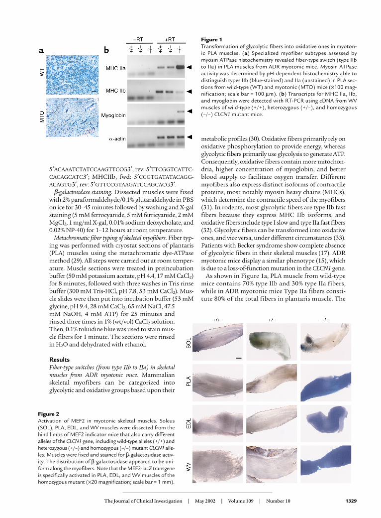
ResultsFiber-type switches (from type IIb to IIa) in skeletalmuscles from ADR myotonic mice. Mammalianskeletal myofibers can be categorized into glycolytic and oxidative groups based upon their
metabolic profiles (30). Oxidative fibers primarily rely onoxidative phosphorylation to provide energy, whereasglycolytic fibers primarily use glycolysis to generate ATP.Consequently, oxidative fibers contain more mitochon-dria, higher concentration of myoglobin, and betterblood supply to facilitate oxygen transfer. Differentmyofibers also express distinct isoforms of contractileproteins, most notably myosin heavy chains (MHCs),which determine the contractile speed of the myofibers(31). In rodents, most glycolytic fibers are type IIb fastfibers because they express MHC IIb isoforms, andoxidative fibers include type I slow and type IIa fast fibers(32). Glycolytic fibers can be transformed into oxidativeones, and vice versa, under different circumstances (33).Patients with Becker syndrome show complete absenceof glycolytic fibers in their skeletal muscles (17). ADRmyotonic mice display a similar phenotype (15), whichis due to a loss-of-function mutation in the CLCN1 gene.
As shown in Figure 1a, PLA muscle from wild-typemice contains 70% type IIb and 30% type IIa fibers,while in ADR myotonic mice Type IIa fibers consti-tute 80% of the total fibers in plantaris muscle. The
The Journal of Clinical Investigation | May 2002 | Volume 109 | Number 10 1329
Figure 1Transformation of glycolytic fibers into oxidative ones in myoton-ic PLA muscles. (a) Specialized myofiber subtypes assessed bymyosin ATPase histochemistry revealed fiber-type switch (type IIbto IIa) in PLA muscles from ADR myotonic mice. Myosin ATPaseactivity was determined by pH-dependent histochemistry able todistinguish types IIb (blue-stained) and IIa (unstained) in PLA sec-tions from wild-type (WT) and myotonic (MTO) mice (×100 mag-nification; scale bar = 100 µm). (b) Transcripts for MHC IIa, IIb,and myoglobin were detected with RT-PCR using cDNA from WVmuscles of wild-type (+/+), heterozygous (+/–), and homozygous(–/–) CLCN1 mutant mice.
Figure 2Activation of MEF2 in myotonic skeletal muscles. Soleus(SOL), PLA, EDL, and WV muscles were dissected from thehind limbs of MEF2 indicator mice that also carry differentalleles of the CLCN1 gene, including wild-type alleles (+/+) andheterozygous (+/–) and homozygous (–/–) mutant CLCN1 alle-les. Muscles were fixed and stained for β-galactosidase activ-ity. The distribution of β-galactosidase appeared to be uni-form along the myofibers. Note that the MEF2-lacZ transgeneis specifically activated in PLA, EDL, and WV muscles of thehomozygous mutant (×20 magnification; scale bar = 1 mm).
changes in fiber-type composition seen in myotonicmuscles most likely result from a switch of type IIbfibers into type IIa fibers, since there is no observablefiber atrophy or regeneration in the myotonic mus-cles (15). Fiber-type transformation was confirmed bymeasuring RNA transcript levels of MHC IIa and IIb,different isoforms of MHC expressed in fast oxidativeand fast glycolytic fibers, respectively. Comparedwith wild-type mice (+/+) and mice heterozygous forthe mutant CLCN1 allele (+/–), expression of MHCIIa significantly increased in white vastus (WV) mus-cles from ADR myotonic mice (–/–, homozygous forCLCN1 mutation), with a concomitant decrease inMHC IIb expression. Myoglobin, a marker for mus-cle oxidative capacity, also showed upregulation inmyotonic muscles.
Marked induction of MEF2 transcriptional activity inmyotonic skeletal muscles. Previously, we have shown thatMEF2 is selectively active in slow/oxidative myofibers(21). Furthermore, endurance exercise and low-fre-quency stimulation, two protocols that can triggertransformation of glycolytic fibers into oxidative ones,enhance MEF2 activity in skeletal muscles (22). MEF2-binding sites are necessary for fiber type–specific activ-ities of myoglobin and TnI slow promoters, suggestingMEF2 plays a causative role in determining slow/oxida-tive fiber identity (21).
The observation that glycolytic fibers are convertedinto oxidative fibers in myotonic muscles prompted usto investigate whether MEF2 is involved in this patho-logical process. To this end, we introduced a lacZreporter transgene into the ADR myotonic mice throughconventional breeding of ADR myotonic mice withMEF2 indicator mice, which contain a lacZ transgene
under the control of three copies of MEF2-binding ele-ments derived from the desmin promoter (desMEF2-lacZ) (27). The transgene expression is solely dependenton MEF2 transactivating function and provides readoutfor MEF2 activity that is suitable for physiological andpathological studies in intact animals.
Most skeletal muscles from adult desMEF2-lacZ micehave little lacZ expression (Figure 2), although abun-dant MEF2 proteins exist in these muscles. In mice car-rying only one allele of the mutant CLCN1 gene (+/–),MEF2 was still kept inactive (Figure 2), consistent with the notion that the mutation of the CLCN1gene in ADR myotonic mice is autosomal recessive. In mice homozygous for the CLCN1 mutation (–/–), MEF2 activity was dramatically upregulated in skeletal muscles (Figure 2). MEF2 activation was obvious in all the myotonic mice examined. Interestingly, MEF2
1330 The Journal of Clinical Investigation | May 2002 | Volume 109 | Number 10
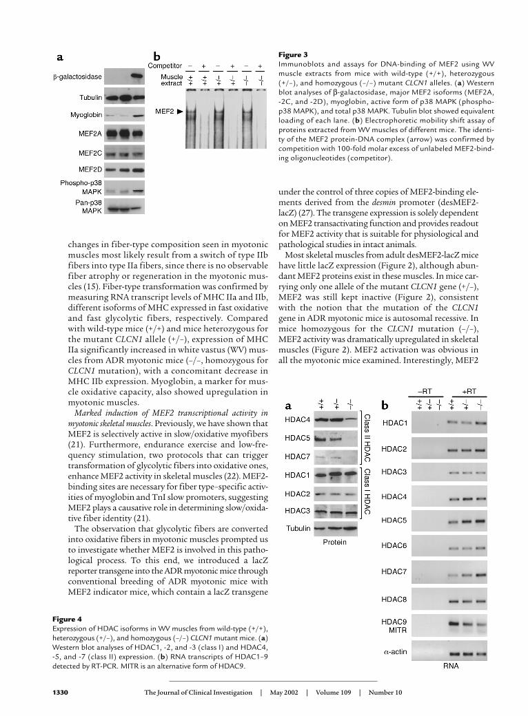
Figure 3Immunoblots and assays for DNA-binding of MEF2 using WVmuscle extracts from mice with wild-type (+/+), heterozygous(+/–), and homozygous (–/–) mutant CLCN1 alleles. (a) Westernblot analyses of β-galactosidase, major MEF2 isoforms (MEF2A,-2C, and -2D), myoglobin, active form of p38 MAPK (phospho-p38 MAPK), and total p38 MAPK. Tubulin blot showed equivalentloading of each lane. (b) Electrophoretic mobility shift assay ofproteins extracted from WV muscles of different mice. The identi-ty of the MEF2 protein-DNA complex (arrow) was confirmed bycompetition with 100-fold molar excess of unlabeled MEF2-bind-ing oligonucleotides (competitor).
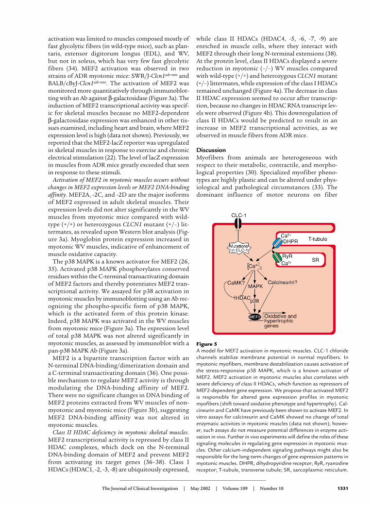
Figure 4Expression of HDAC isoforms in WV muscles from wild-type (+/+),heterozygous (+/–), and homozygous (–/–) CLCN1 mutant mice. (a)Western blot analyses of HDAC1, -2, and -3 (class I) and HDAC4, -5, and -7 (class II) expression. (b) RNA transcripts of HDAC1–9detected by RT-PCR. MITR is an alternative form of HDAC9.
activation was limited to muscles composed mostly offast glycolytic fibers (in wild-type mice), such as plan-taris, extensor digitorum longus (EDL), and WV, but not in soleus, which has very few fast glycolytic fibers (34). MEF2 activation was observed in two strains of ADR myotonic mice: SWR/J-Clcn1adr-mto andBALB/cByJ-Clcn1adr-mto. The activation of MEF2 wasmonitored more quantitatively through immunoblot-ting with an Ab against β-galactosidase (Figure 3a). Theinduction of MEF2 transcriptional activity was specif-ic for skeletal muscles because no MEF2-dependent β-galactosidase expression was enhanced in other tis-sues examined, including heart and brain, where MEF2expression level is high (data not shown). Previously, wereported that the MEF2-lacZ reporter was upregulatedin skeletal muscles in response to exercise and chronicelectrical stimulation (22). The level of lacZ expressionin muscles from ADR mice greatly exceeded that seenin response to these stimuli.
Activation of MEF2 in myotonic muscles occurs withoutchanges in MEF2 expression levels or MEF2 DNA-bindingaffinity. MEF2A, -2C, and -2D are the major isoformsof MEF2 expressed in adult skeletal muscles. Theirexpression levels did not alter significantly in the WVmuscles from myotonic mice compared with wild-type (+/+) or heterozygous CLCN1 mutant (+/–) lit-termates, as revealed upon Western blot analysis (Fig-ure 3a). Myoglobin protein expression increased inmyotonic WV muscles, indicative of enhancement ofmuscle oxidative capacity.
The p38 MAPK is a known activator for MEF2 (26,35). Activated p38 MAPK phosphorylates conservedresidues within the C-terminal transactivating domainof MEF2 factors and thereby potentiates MEF2 tran-scriptional activity. We assayed for p38 activation inmyotonic muscles by immunoblotting using an Ab rec-ognizing the phospho-specific form of p38 MAPK,which is the activated form of this protein kinase.Indeed, p38 MAPK was activated in the WV musclesfrom myotonic mice (Figure 3a). The expression levelof total p38 MAPK was not altered significantly inmyotonic muscles, as assessed by immunoblot with apan-p38 MAPK Ab (Figure 3a).
MEF2 is a bipartite transcription factor with an N-terminal DNA-binding/dimerization domain anda C-terminal transactivating domain (36). One possi-ble mechanism to regulate MEF2 activity is throughmodulating the DNA-binding affinity of MEF2.There were no significant changes in DNA binding ofMEF2 proteins extracted from WV muscles of non-myotonic and myotonic mice (Figure 3b), suggestingMEF2 DNA-binding affinity was not altered inmyotonic muscles.
Class II HDAC deficiency in myotonic skeletal muscles.MEF2 transcriptional activity is repressed by class IIHDAC complexes, which dock on the N-terminalDNA-binding domain of MEF2 and prevent MEF2from activating its target genes (36–38). Class IHDACs (HDAC1, -2, -3, -8) are ubiquitously expressed,
while class II HDACs (HDAC4, -5, -6, -7, -9) areenriched in muscle cells, where they interact withMEF2 through their long N-terminal extensions (38).At the protein level, class II HDACs displayed a severereduction in myotonic (–/–) WV muscles comparedwith wild-type (+/+) and heterozygous CLCN1 mutant(+/–) littermates, while expression of the class I HDACsremained unchanged (Figure 4a). The decrease in classII HDAC expression seemed to occur after transcrip-tion, because no changes in HDAC RNA transcript lev-els were observed (Figure 4b). This downregulation ofclass II HDACs would be predicted to result in anincrease in MEF2 transcriptional activities, as weobserved in muscle fibers from ADR mice.
DiscussionMyofibers from animals are heterogeneous withrespect to their metabolic, contractile, and morpho-logical properties (30). Specialized myofiber pheno-types are highly plastic and can be altered under phys-iological and pathological circumstances (33). Thedominant influence of motor neurons on fiber
The Journal of Clinical Investigation | May 2002 | Volume 109 | Number 10 1331
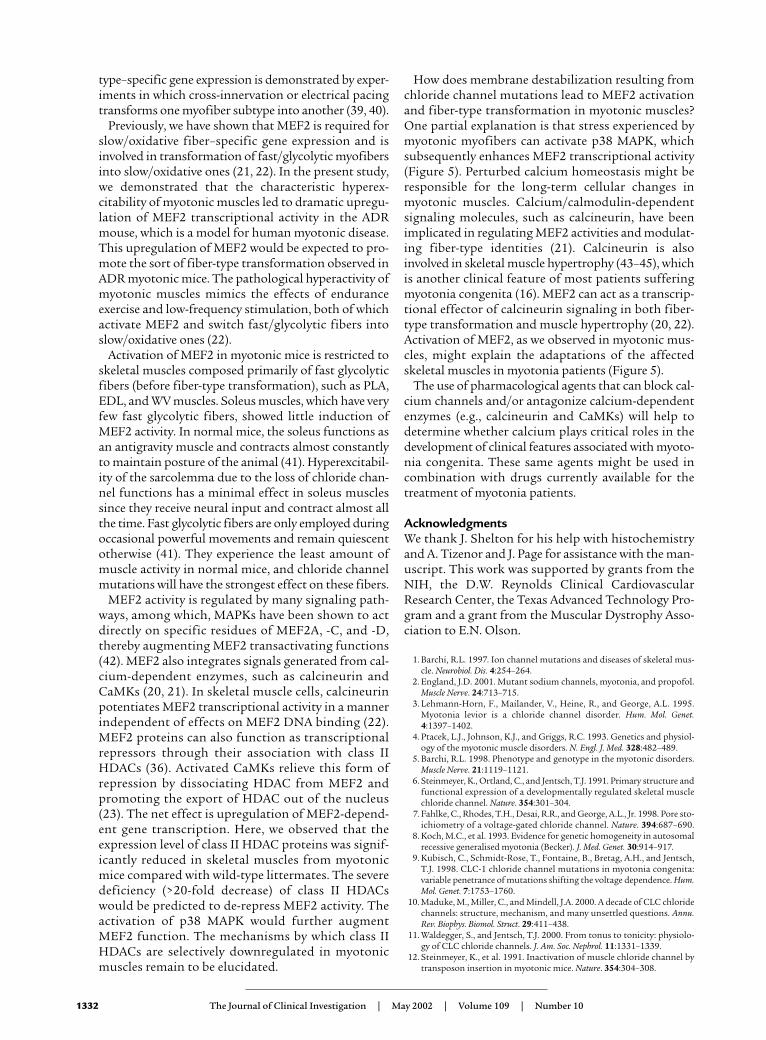
Figure 5A model for MEF2 activation in myotonic muscles. CLC-1 chloridechannels stabilize membrane potential in normal myofibers. Inmyotonic myofibers, membrane destabilization causes activation ofthe stress-responsive p38 MAPK, which is a known activator ofMEF2. MEF2 activation in myotonic muscles also correlates withsevere deficiency of class II HDACs, which function as repressors ofMEF2-dependent gene expression. We propose that activated MEF2is responsible for altered gene expression profiles in myotonicmyofibers (shift toward oxidative phenotype and hypertrophy). Cal-cineurin and CaMK have previously been shown to activate MEF2. Invitro assays for calcineurin and CaMK showed no change of totalenzymatic activities in myotonic muscles (data not shown); howev-er, such assays do not measure potential differences in enzyme acti-vation in vivo. Further in vivo experiments will define the roles of thesesignaling molecules in regulating gene expression in myotonic mus-cles. Other calcium-independent signaling pathways might also beresponsible for the long-term changes of gene expression patterns inmyotonic muscles. DHPR, dihydropyridine receptor; RyR, ryanodinereceptor; T-tubule, transverse tubule; SR, sarcoplasmic reticulum.
type–specific gene expression is demonstrated by exper-iments in which cross-innervation or electrical pacingtransforms one myofiber subtype into another (39, 40).
Previously, we have shown that MEF2 is required forslow/oxidative fiber–specific gene expression and isinvolved in transformation of fast/glycolytic myofibersinto slow/oxidative ones (21, 22). In the present study,we demonstrated that the characteristic hyperex-citability of myotonic muscles led to dramatic upregu-lation of MEF2 transcriptional activity in the ADRmouse, which is a model for human myotonic disease.This upregulation of MEF2 would be expected to pro-mote the sort of fiber-type transformation observed inADR myotonic mice. The pathological hyperactivity ofmyotonic muscles mimics the effects of enduranceexercise and low-frequency stimulation, both of whichactivate MEF2 and switch fast/glycolytic fibers intoslow/oxidative ones (22).
Activation of MEF2 in myotonic mice is restricted toskeletal muscles composed primarily of fast glycolyticfibers (before fiber-type transformation), such as PLA,EDL, and WV muscles. Soleus muscles, which have veryfew fast glycolytic fibers, showed little induction ofMEF2 activity. In normal mice, the soleus functions asan antigravity muscle and contracts almost constantlyto maintain posture of the animal (41). Hyperexcitabil-ity of the sarcolemma due to the loss of chloride chan-nel functions has a minimal effect in soleus musclessince they receive neural input and contract almost allthe time. Fast glycolytic fibers are only employed duringoccasional powerful movements and remain quiescentotherwise (41). They experience the least amount ofmuscle activity in normal mice, and chloride channelmutations will have the strongest effect on these fibers.
MEF2 activity is regulated by many signaling path-ways, among which, MAPKs have been shown to actdirectly on specific residues of MEF2A, -C, and -D,thereby augmenting MEF2 transactivating functions(42). MEF2 also integrates signals generated from cal-cium-dependent enzymes, such as calcineurin andCaMKs (20, 21). In skeletal muscle cells, calcineurinpotentiates MEF2 transcriptional activity in a mannerindependent of effects on MEF2 DNA binding (22).MEF2 proteins can also function as transcriptionalrepressors through their association with class IIHDACs (36). Activated CaMKs relieve this form ofrepression by dissociating HDAC from MEF2 andpromoting the export of HDAC out of the nucleus(23). The net effect is upregulation of MEF2-depend-ent gene transcription. Here, we observed that theexpression level of class II HDAC proteins was signif-icantly reduced in skeletal muscles from myotonicmice compared with wild-type littermates. The severedeficiency (>20-fold decrease) of class II HDACswould be predicted to de-repress MEF2 activity. Theactivation of p38 MAPK would further augmentMEF2 function. The mechanisms by which class IIHDACs are selectively downregulated in myotonicmuscles remain to be elucidated.
How does membrane destabilization resulting fromchloride channel mutations lead to MEF2 activationand fiber-type transformation in myotonic muscles?One partial explanation is that stress experienced bymyotonic myofibers can activate p38 MAPK, whichsubsequently enhances MEF2 transcriptional activity(Figure 5). Perturbed calcium homeostasis might beresponsible for the long-term cellular changes inmyotonic muscles. Calcium/calmodulin-dependentsignaling molecules, such as calcineurin, have beenimplicated in regulating MEF2 activities and modulat-ing fiber-type identities (21). Calcineurin is alsoinvolved in skeletal muscle hypertrophy (43–45), whichis another clinical feature of most patients sufferingmyotonia congenita (16). MEF2 can act as a transcrip-tional effector of calcineurin signaling in both fiber-type transformation and muscle hypertrophy (20, 22).Activation of MEF2, as we observed in myotonic mus-cles, might explain the adaptations of the affectedskeletal muscles in myotonia patients (Figure 5).
The use of pharmacological agents that can block cal-cium channels and/or antagonize calcium-dependentenzymes (e.g., calcineurin and CaMKs) will help todetermine whether calcium plays critical roles in thedevelopment of clinical features associated with myoto-nia congenita. These same agents might be used incombination with drugs currently available for thetreatment of myotonia patients.
AcknowledgmentsWe thank J. Shelton for his help with histochemistryand A. Tizenor and J. Page for assistance with the man-uscript. This work was supported by grants from theNIH, the D.W. Reynolds Clinical CardiovascularResearch Center, the Texas Advanced Technology Pro-gram and a grant from the Muscular Dystrophy Asso-ciation to E.N. Olson.
1. Barchi, R.L. 1997. Ion channel mutations and diseases of skeletal mus-cle. Neurobiol. Dis. 4:254–264.
2. England, J.D. 2001. Mutant sodium channels, myotonia, and propofol.Muscle Nerve. 24:713–715.
3. Lehmann-Horn, F., Mailander, V., Heine, R., and George, A.L. 1995.Myotonia levior is a chloride channel disorder. Hum. Mol. Genet.4:1397–1402.
4. Ptacek, L.J., Johnson, K.J., and Griggs, R.C. 1993. Genetics and physiol-ogy of the myotonic muscle disorders. N. Engl. J. Med. 328:482–489.
5. Barchi, R.L. 1998. Phenotype and genotype in the myotonic disorders.Muscle Nerve. 21:1119–1121.
6. Steinmeyer, K., Ortland, C., and Jentsch, T.J. 1991. Primary structure andfunctional expression of a developmentally regulated skeletal musclechloride channel. Nature. 354:301–304.
7. Fahlke, C., Rhodes, T.H., Desai, R.R., and George, A.L., Jr. 1998. Pore sto-ichiometry of a voltage-gated chloride channel. Nature. 394:687–690.
8. Koch, M.C., et al. 1993. Evidence for genetic homogeneity in autosomalrecessive generalised myotonia (Becker). J. Med. Genet. 30:914–917.
9. Kubisch, C., Schmidt-Rose, T., Fontaine, B., Bretag, A.H., and Jentsch,T.J. 1998. CLC-1 chloride channel mutations in myotonia congenita:variable penetrance of mutations shifting the voltage dependence. Hum.Mol. Genet. 7:1753–1760.
10. Maduke, M., Miller, C., and Mindell, J.A. 2000. A decade of CLC chloridechannels: structure, mechanism, and many unsettled questions. Annu.Rev. Biophys. Biomol. Struct. 29:411–438.
11. Waldegger, S., and Jentsch, T.J. 2000. From tonus to tonicity: physiolo-gy of CLC chloride channels. J. Am. Soc. Nephrol. 11:1331–1339.
12. Steinmeyer, K., et al. 1991. Inactivation of muscle chloride channel bytransposon insertion in myotonic mice. Nature. 354:304–308.
1332 The Journal of Clinical Investigation | May 2002 | Volume 109 | Number 10
13. Jockusch, H. 1990. Allelic mutations at the myotonia locus adr. MouseGenome. 87:72.
14. Gronemeier, M., et al. 1994. Nonsense and missense mutations in themuscular chloride channel gene Clc-1 of myotonic mice. J. Biol. Chem.269:5963–5967.
15. Reininghaus, J., Fuchtbauer, E.M., Bertram, K., and Jockusch, H. 1988.The myotonic mouse mutant ADR: physiological and histochemicalproperties of muscle. Muscle Nerve. 11:433–439.
16. Sun, C., Tranebjaerg, L., Torbergsen, T., Holmgren, G., and Van Ghelue,M. 2001. Spectrum of CLCN1 mutations in patients with myotonia con-genita in Northern Scandinavia. Eur. J. Hum. Genet. 9:903–909.
17. Crews, J., Kaiser, K.K., and Brooke, M.H. 1976. Muscle pathology ofmyotonia congenita. J. Neurol. Sci. 28:449–457.
18. Naya, F.S., and Olson, E.N. 1999. MEF2: a transcriptional target for sig-naling pathways controlling skeletal muscle growth and differentiation.Curr. Opin. Cell Biol. 11:683–688.
19. Olson, E.N., and Williams, R.S. 2000. Calcineurin signaling and muscleremodeling. Cell. 101:689–692.
20. Passier, R., et al. 2000. CaM kinase signaling induces cardiac hypertro-phy and activates the MEF2 transcription factor in vivo. J. Clin. Invest.105:1395–1406.
21. Wu, H., et al. 2000. MEF2 responds to multiple calcium-regulated sig-nals in the control of skeletal muscle fiber type. EMBO J. 19:1963–1973.
22. Wu, H., et al. 2001. Activation of MEF2 by muscle activity is mediatedthrough a calcineurin-dependent pathway. EMBO J. 20:6414–6423.
23. McKinsey, T.A., Zhang, C.L., Lu, J., and Olson, E.N. 2000. Signal-depend-ent nuclear export of a histone deacetylase regulates muscle differentia-tion. Nature. 408:106–111.
24. McKinsey, T.A., Zhang, C.L., Olson, E.N. 2000. Activation of the myocyteenhancer factor-2 transcription factor by calcium/calmodulin-depend-ent protein kinase-stimulated binding of 14-3-3 to histone deacetylase5. Proc. Natl. Acad. Sci. USA. 97:14400–14405.
25. Kato, Y., et al. 1997. BMK1/ERK5 regulates serum-induced early geneexpression through transcription factor MEF2C. EMBO J. 16:7054–7066.
26. Zhao, M., et al. 1999. Regulation of the MEF2 family of transcription fac-tors by p38. Mol. Cell. Biol. 19:21–30.
27. Naya, F.J., Wu, C., Richardson, J.A., Overbeek, P., and Olson, E.N. 1999.Transcriptional activity of MEF2 during mouse embryogenesis moni-tored with a MEF2-dependent transgene. Development. 126:2045–2052.
28. Gossett, L.A., Kelvin, D.J., Sternberg, E.A., and Olson, E.N. 1989. A newmyocyte-specific enhancer-binding factor that recognizes a conservedelement associated with multiple muscle-specific genes. Mol. Cell. Biol.9:5022–5033.
29. Ogilvie, R.W., and Feeback, D.L. 1990. A metachromatic dye-ATPasemethod for the simultaneous identification of skeletal muscle fibertypes I, IIA, IIB and IIC. Stain Technol. 65:231–241.
30. Booth, F.W., and Baldwin, K.M. 1996. Muscle plasticity: energy demandand supply processes. In The handbook of physiology: integration of motor, cir-culatory, respiratory and metabolic control during exercise. L. B. Bowell and J.T.Shepard, editors. American Physiological Society. Bethesda, Maryland,USA. 1075–1123.
31. Schiaffino, S., and Reggiani, C. 1996. Molecular diversity of myofibril-lar proteins — gene regulation and functional significance. Physiol. Rev.76:371–423.
32. Weiss, A., and Leinwand, L.A. 1996. The mammalian myosin heavy chaingene family. Annu. Rev. Cell Dev. Biol. 12:417–439.
33. Pette, D., and Staron, R.S. 2001. Transitions of muscle fiber phenotypicprofiles. Histochem. Cell. Biol. 115:359–372.
34. Ovalle, W.K., Bressler, B.H., Jasch, L.G., and Slonecker, C.E. 1983. Abnor-mal distribution of fiber types in the slow-twitch soleus muscle of theC57BL/6J dy2J/dy2J dystrophic mouse during postnatal development.Am. J. Anat. 168:291–304.
35. Yang, S.H., Galanis, A., and Sharrocks, A.D. 1999. Targeting of p38 mito-gen-activated protein kinases to MEF2 transcription factors. Mol. Cell.Biol. 19:4028–4038.
36. Lu, J., McKinsey, T.A., Nicol, R.L., and Olson, E.N. 2000. Signal-depend-ent activation of the MEF2 transcription factor by dissociation from his-tone deacetylases. Proc. Natl. Acad. Sci. USA. 97:4070–4075.
37. Miska, E.A., et al. 1999. HDAC4 deacetylase associates with and repress-es the MEF2 transcription factor. EMBO J. 18:5099–5107.
38. Lu, J., McKinsey, T.A., Zhang, C.L., and Olson, E.N. 2000. Regulation ofskeletal myogenesis by association of the MEF2 transcription factor withclass II histone deacetylases. Mol. Cell. 6:233–244.
39. Buller, A.J., Eccles, J.C., and Eccles, R.M. 1960. Differentiation of fast andslow muscles in the cat hind limb. J. Physiol. (London). 150:399–416.
40. Pette, D., and Vrbova, G. 1992. Adaptation of mammalian skeletal mus-cle fibers to chronic electrical stimulation. Rev. Physiol. Biochem. Pharma-col. 120:115–202.
41. Hennig, R., and Lomo, T. 1985. Firing patterns of motor units in normalrats. Nature. 314:164–166.
42. Han, J., and Molkentin, J.D. 2000. Regulation of MEF2 by p38 MAPKand its implication in cardiomyocyte biology. Trends Cardiovasc. Med.10:19–22.
43. McKinsey, T.A., Zhang, C.L., and Olson, E.N. 2002. MEF2: a calcium-dependent regulator of cell division, differentiation and death. TrendsBiochem. Sci. 27:40–47.
44. Musaro, A., McCullagh, K.J., Naya, F.J., Olson, E.N., and Rosenthal,N. 1999. IGF-1 induces skeletal myocyte hypertrophy through calcineurin in association with GATA-2 and NF-ATc1. Nature.400:581–585.
45. Semsarian, C., et al. 1999. Skeletal muscle hypertrophy is mediated bya Ca2+-dependent calcineurin signalling pathway. Nature. 400:576–581.
The Journal of Clinical Investigation | May 2002 | Volume 109 | Number 10 1333