Embed Size (px)
Citation preview
Short communication
Active surveillance of bat rabies in France: A 5-year study (2004–2009)
Evelyne Picard-Meyer a,*, Marie-Jo Dubourg-Savage b, Laurent Arthur b,Michel Barataud b, David Becu b, Sandrine Bracco b, Christophe Borel b,Gerald Larcher b, Benjamin Meme-Lafond b, Marie Moinet a,Emmanuelle Robardet a, Marine Wasniewski a, Florence Cliquet a
a ANSES Nancy Laboratory for Rabies and Wildlife, French Agency for Food, Environmental and Occupational Health and Safety, European Union Reference
Laboratories for Rabies (EURL) and Serology, WHO Collaborating Centre, OIE Reference Laboratory for Rabies Technopole Agricole et Veterinaire, BP 40 009, 54220
Malzeville, Franceb SFEPM Chiroptera Group, Museum d’Histoire Naturelle de Bourges, Parc Saint Paul, 18000 Bourges, France
1. Introduction
Rabies is a viral zoonosis that causes progressive andultimately fatal encephalitis. Rabies is a very old diseaseand is responsible for approximately 55,000 human deathsper year worldwide (World Health Organisation, 2008).The rabies virus belongs to genus Lyssavirus, familyRhabdoviridae and can infect all mammals. The virus isusually transmitted through saliva in a bite from aninfected animal.
The Lyssavirus genus includes 11 different recognisedspecies (Bourhy et al., 1993; Carstens, 2010), also referredto as genotypes: ‘‘classical’’ rabies virus (RABV), Lagos batvirus (LBV), Mokola virus (MOKV), Duvenhage virus(DUVV), Australian bat lyssavirus (ABLV), European batlyssavirus type 1 (EBLV-1) and European bat lyssavirustype 2 (EBLV-2). Four additional bat viruses (the Irkut,West Caucasian bat, Khujand and Aravan viruses) wererecently ratified by the International Committee on VirusTaxonomy (Carstens, 2010). Ten of the 11 above-listedrecognised virus species have been isolated from bats; onlyMOKV has not been isolated from bats (Sabeta et al., 2007).
Since the first case of bat rabies found in 1954,approximately 850 cases of rabid bats infected by EBLV-1(also known as genotype 5 with two variants EBLV-1a andEBLV-1b) or EBLV-2 (genotype 6) have been reported inEurope (Muller et al., 2007). Most cases of bat rabies havebeen reported in European countries having a well-established rabies surveillance network (Germany, theNetherlands, Denmark, France and Great Britain). EBLV-1
Veterinary Microbiology 151 (2011) 390–395
A R T I C L E I N F O
Article history:
Received 31 July 2010
Received in revised form 17 March 2011
Accepted 31 March 2011
Keywords:
Rabies
Lyssavirus
Bat
Epidemiological surveillance
France
A B S T R A C T
Active surveillance of bats in France started in 2004 with an analysis of 18 of the 45 bat
species reported in Europe. Rabies antibodies were detected in six indigenous species,
mainly in Eptesicus serotinus and Myotis myotis, suggesting previous contact with the EBLV-
1 rabies virus. Nineteen of the 177 tested bats were shown serologically positive in seven
sites, particularly in central and south-western France. Neither infectious viral particles
nor viral genomes were detected in 173 and 308 tested oral swabs, respectively. The
presence of neutralising antibodies in female bats (18.6%) was significantly higher than in
males (5.6%).
� 2011 Elsevier B.V. All rights reserved.
Abbreviations: BHK-21, baby hamster kidney cells; EBLV, European
bat lyssavirus; RTCIT, rabies tissue culture infection test; FAVNt, fluor-
escent antibody virus neutralisation test; hnRT-PCR, hemi-nested
reverse-transcription polymerase chain reaction; TCID50, median tissue
culture infective dose; 95% CI, 95% confidence interval.
* Corresponding author. Tel.: +33 0 3 83 29 89 50;
fax: +33 0 3 83 29 89 58.
E-mail address: [email protected] (E. Picard-Meyer).
Contents lists available at ScienceDirect
Veterinary Microbiology
jou r nal h o mep ag e: w ww .e ls evier . co m/lo c ate /vetm i c
0378-1135/$ – see front matter � 2011 Elsevier B.V. All rights reserved.
doi:10.1016/j.vetmic.2011.03.034
seems to be adapted to E. serotinus (Van der Poel et al., 2000),species in which more than 95% of cases have been reported,while EBLV-2 appears to infect only Myotis bat species (M.
daubentonii and M. dasycneme) with 20 cases, of which 10have been reported in M. daubentonii in England (Banyardet al., 2009; Harris et al., 2009). Recently, another species (E.
isabellinus) sibling species of E. serotinus and mainly found inSouthern Iberia was shown to be infected with EBLV-1(Vazquez-Moron et al., 2008b) and sporadic rabies caseshave been found in Pipistrellus pipistrellus, Pipistrellus
nathusii, Plecotus auritus, Nyctalus noctula (Muller et al.,2007) and Vespertilio murinus (Selimov et al., 1991).
National (Moutou et al., 2003) and European recom-mendations (Cliquet et al., 2010; Med Vet Net WorkingGroup, 2005) encourage the continuation and reinforce-ment of bat rabies research and epidemiological surveil-lance.
Here, we report results from field studies carried out inFrance since 2004 in close collaboration with bat
specialists, preventively vaccinated against rabies. Thepurpose of the present study was to investigate thecirculation of EBLV-1 among indigenous bat species andidentify species involved in the transmission of bat rabies.The seroprevalence of EBLV-1 was investigated in batsfrom 2004 to 2009 to improve current knowledge on theepidemiology of bats infected by EBLV-1.
2. Materials and methods
2.1. Sampling
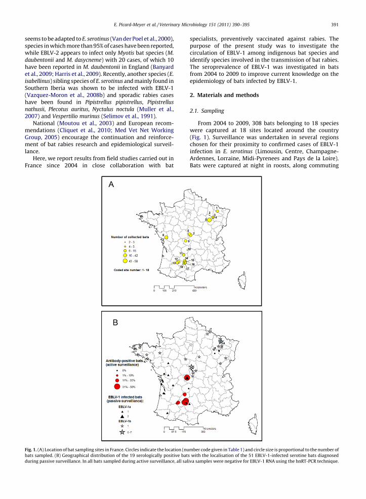
From 2004 to 2009, 308 bats belonging to 18 specieswere captured at 18 sites located around the country(Fig. 1). Surveillance was undertaken in several regionschosen for their proximity to confirmed cases of EBLV-1infection in E. serotinus (Limousin, Centre, Champagne-Ardennes, Lorraine, Midi-Pyrenees and Pays de la Loire).Bats were captured at night in roosts, along commuting
Fig. 1. (A) Location of bat sampling sites in France. Circles indicate the location (number code given in Table 1) and circle size is proportional to the number of
bats sampled. (B) Geographical distribution of the 19 serologically positive bats with the localisation of the 51 EBLV-1-infected serotine bats diagnosed
during passive surveillance. In all bats sampled during active surveillance, all saliva samples were negative for EBLV-1 RNA using the hnRT-PCR technique.
E. Picard-Meyer et al. / Veterinary Microbiology 151 (2011) 390–395 391
routes, at swarming sites or in parturition roosts when thebats left to forage. Bat specialists identified bats to thespecies level using morphological criteria (Dietz and VonHelversen, 2004; Schober and Grimmberger, 1991).Several parameters were recorded for each bat: sex,reproductive status, age, body weight, forearm length,behaviour and parasite load.
Oral swabs were taken to collect saliva from eachtrapped bat and stored in 1 mL of RNAlater (Ambion,France), a buffer solution designed for RNA preservation,for subsequent hemi-nested reverse-transcription PCR(hnRT-PCR) (n = 308 samples) and/or in a volume of0.3 mL of DMEM culture (Dulbecco’s minimum essentialmedium, Invitrogen, France) (n = 173) for the rabies tissueculture infection test (RTCIT). Blood samples were takenfrom 185 bats for a modified fluorescent antibody virusneutralisation test (mFAVNt) on EBLV-1. After sampling, allbats were released at the site of capture at night. Capture,handling and sampling were undertaken with authorisa-tion from the French Ministry of the Environment. Batspecies, study sites and the number of collected blood andsaliva samples are detailed in Table 1.
2.2. Laboratory methods
Oral swabs stored in 0.3 mL of culture medium (n = 173)were analysed using RTCIT on murine neuroblastoma cells(ATCC:CCL31) (Barrat et al., 1988) and by RT-PCR (Picard-
Meyer et al., 2004). The 135 oral swabs stored in RNAlaterwere tested only for the presence of EBLV-1 RNA by hnRT-PCR using universal primers (JW12-JW6) in the first roundof PCR and specific primers (JW12-JEBL1) for the secondround. RNA integrity was verified by amplifying 18S rRNAusing a commercial Competimer system (Ambion, France)in each RT-PCR.
To detect EBLV-1-specific neutralising antibodies inblood samples, a modified FAVN test was performed(Cliquet et al., 1998) with an EBLV-1 virus strain (ANSES,No. 121411) isolated in France (2000). Samples were testedin threefold dilutions on BHK-21 cells with a startingdilution of 1/27. Controls included uninfected BHK-21cells, OIE positive dog serum, negative dog serum andback-titration of the specific EBLV-1 virus. Levels of virus-neutralising antibodies are expressed in log D50. Thethreshold of antibody detection was calculated by usingthe Spearman–Karber formula and set at 1.67 log D50.
The 95% confidence intervals (95% CI) of seroprevalencedata were calculated using free open-source R software,version 2.8.1 (R Development Core Team, 2004).
3. Results
3.1. Presence of EBLV-1 antibodies (mFAVN test)
From 2004 to 2009, of the 185 bat blood samples, 4contained insufficient quantities to perform mFAVNt, 177
Table 1
Number of saliva (blood) samples collected, by bat species and by study site.
Region North-eastern France Central
France
North-western
France
South-western France
Species\site no. 1 2 3 4 5 6 7 9a 8 10 11a 12a 13a 14a 15 16 17a 18a
R. euryale 3
(0)
1
(0)
1
(0)
R. ferrumequinum 3 (3) 1 (0) 2 (1) 2 (2) 2 (2) 9 (2) 2 (2)
R. hipposideros 7 (0) 3 (0) 1 (1)
B. barbastellus 2 (0) 1 (1) 1 (1) 3 (0)
E. serotinus 11
(0)
33
(0)
12
(12)
2 (2) 7 (7) 1 (1) 1 (1) 5 (5)
M. alcathoe 2 (0)
M. bechsteinii 2 (0) 1(0) 3 (3) 1
(0)
M. blythii 3 (3) 2 (2)
M. daubentonii 1 (0) 5 (5) 2 (2) 1 (0) 2 (2) 7 (3) 8 (5) 1 (1)
M. emarginatus 8 (0) 18 (16) 1 (0)
M. myotis 1 (1) 8 (8) 1 (1) 17 (17) 4 (4) 9 (8)
M. myotis/blythii 2 (2) 7 (7) 1 (1) 1 (1)
M. mystacinus 1 (0) 0 (0) 2 (2)
M. nattereri 2 (0) 3(0) 3 (3) 2 (0)
M. schreibersii 14 (14) 3 (3) 1 (0)
P. auritus 1 (0) 1(0) 1 (1) 4 (4) 1 (0)
P. austriacus 15 (12) 1 (1)
P. kuhlii 1 (1)
P. pipistrellus 1 (0) 9 (8) 6 (1) 1 (0)
Total number
of saliva
samples
(blood)
15 (0) 2
(0)
13
(1)
5
(0)
39
(36)
11
(0)
33
(0)
12
(12)
31
(27)
3
(0)
11 (11) 36
(35)
15
(14)
13
(11)
13
(8)
10 (6) 42
(20)
4 (4)
The municipalities of study sites numbered 1–18 are as follows: Tincry (1), Montiers sur Saulx (2), Neufchateau-Rebeuville (3), Neufchateau-Landaville (4),
Chamarandes-Choignes (5), Saint Loup des Chaumes (6), Saint Amand Montrond (7), Fontenay le Comte (8), Gueret (9), Lafage (10), Rocamadour (11),
Gasques (12), Dunes (13), Loze (14), Feneyrols (15), Roussayrolles (16), Penne 1 and 2 (17), Saint Antonin Noble Val (18).a Sites positive for EBLV-1 antibodies. Values give the number of collected saliva samples; the number of collected blood samples for mFAVN test is given
in parentheses.
E. Picard-Meyer et al. / Veterinary Microbiology 151 (2011) 390–395392
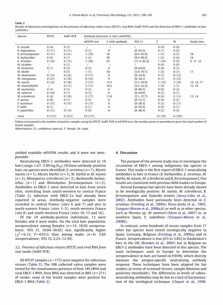
yielded readable mFAVNt results and 4 were not inter-pretable.
Neutralising EBLV-1 antibodies were detected in 19bats (range, 1.67–2.99 log D50). Of these antibody-positivebats, six species were identified: E. serotinus (n = 7), Myotis
myotis (n = 5), Myotis blythii (n = 1), M. blythii or M. myotis
(n = 2), Miniopterus schreibersii (n = 2), Barbastella barbas-
tellus (n = 1) and Rhinolophus ferrumequinum (n = 2).Antibodies to EBLV-1 were detected in bats from sevensites, stretching from south-western to central France(Table 2). Infection with EBLV-1a had already beenreported in areas. Antibody-negative samples wererecorded in central France (sites 6 and 7) and also innorth-eastern France (sites 1–5), north-western France(site 8) and south-western France (sites 10, 15 and 16).
Of the 19 antibody-positive individuals, 13 werefemales and 6 were males. On the total bats population,seroprevalence among females (n = 13; 18.6% seropreva-lence; 95% CI, 10.64–30.02) was significantly higher(x2 = 6.13; P = 0.013) than among males (n = 6; 5.6%seroprevalence; 95% CI, 2.23–12.30).
3.2. Presence of infectious viruses (RTCIT) and viral RNA from
oral swabs (hnRT-PCR)
All RTCIT samples (n = 173) were negative for infectiousviruses (Table 2). The 308 collected saliva samples weretested for the simultaneous presence of host 18S rRNA andviral EBLV-1 RNA. Host RNA was detected in 88% (n = 271)of swabs; none of the tested samples were positive forEBLV-1 RNA (Table 2).
4. Discussion
The purpose of the present study was to investigate thecirculation of EBLV-1 among indigenous bat species inFrance. This study is the first report of EBLV-1 neutralisingantibodies in bats in France (B. barbastellus, E. serotinus, M.
blythii, M. myotis, M. schreibersii and R. ferrumequinum). Ourresults are consistent with previous field studies in Europe.
Several European bat species have been already shownto be serologically positive: M. myotis, M. schreibersii, R.
ferrumequinum and Tadarida teniotis (Serra-Cobo et al.,2002). Antibodies have previously been detected in E.
serotinus (Freuling et al., 2009a; Perez-Jorda et al., 1995;Vazquez-Moron et al., 2008a,b) as well as in other species,such as Plecotus sp., M. nattereri (Klein et al., 2007) or, insouthern Spain, E. isabellinus (Vazquez-Moron et al.,2008b).
In contrast, some hundreds of serum samples from 17other bat species have tested serologically negative inSpain, Germany, Belgium (Freuling et al., 2009a) and inFrance. Seroprevalence is low [0.5 to 3.8%] in Daubenton’sbats in the UK (Brookes et al., 2005) but in Belgium noEBLV-2 antibodies have been detected in this species. Themain techniques used in Europe to determine theseroprevalence in bats are based on FAVNt, which directlymeasure the antigen-specific neutralising antibodyresponse. These techniques have been adapted for batstudies (in terms of screened viruses, sample dilutions andpositivity thresholds). The differences in levels of rabies-neutralising antibodies may be due to a lack of harmoniza-tion of the serological technique (Cliquet et al., 1998;
Table 2
Results of laboratory investigations on the presence of infectious rabies virus (RTCIT), viral RNA (hnRT-PCR) and the detection of EBLV-1 antibodies in bats
(mFAVNt).
Species RTCIT hnRT-PCR Antibody detection in bats (mFAVNt)
mFAVN test % with antibody 95% CI F M Study sites
R. euryale 0 (4) 0 (5) / / / 0 (0) 0 (0)
R. hipposideros 0 (11) 0 (11) 0 (1) 0 [0–95.5] 0 (1) 0 (0)
R. ferrumequinum 0 (11) 0 (21) 1 (10) 10 [0.5–45.9] 1 (5) 0 (5) 18
B. barbastellus 0 (6) 0 (7) 1 (2) 50 [9.5–90.5] 1 (2) 0 (0) 14
E. serotinus 0 (18) 0 (72) 7 (28) 25 [11.4–45.2] 7 (19) 0 (9) 9, 11, 12
M. alcathoe / 0 (1) / / / 0 (0) 0 (0)
M. bechsteini 0 (7) 0 (5) 0 (3) 0 [0–69.0] 0 (0) 0 (3)
M. blythii / 0 (5) 1 (5) 20 [1.0–70.1] 1 (2) 0 (3) 11
M. daubentonii 0 (15) 0 (22) 0 (17) 0 [0–22.9] 0 (5) 0 (12)
M. emarginatus 0 (27) 0 (10) 0 (16) 0 [0–24.1] 0 (1) 0 (15)
M. myotis 0 (22) 0 (36) 5 (37) 13.5 [5.1–29.6] 2 (19) 3 (18) 12, 14, 17
M. myotis/blythii / 0 (11) 2 (11) 18.2 [3.2–52.2] 1 (4) 1 (7) 12, 18
M. mystacinus 0 (3) 0 (3) 0 (2) 0 [0–80.2] 0 (0) 0 (2)
M. nattereri 0 (10) 0 (7) 0 (3) 0 [0–60.9] 0 (2) 0 (1)
M. schreibersii 0 (4) 0 (18) 2 (17) 11.8 [2.1–37.7] 0 (6) 2 (11) 13, 14
P. auritus 0 (8) 0 (7) 0 (5) 0 [0–53.7] 0 (0) 0 (5)
P. austriacus 0 (15) 0 (15) 0 (13) 0 [0–28.3] 0 (2) 0 (11)
P. kuhli 0 (1) / 0 (1) 0 [0–95.5] 0 (0) 0 (1)
P. pipistrellus 0 (11) 0 (15) 0 (6) 0 [0–48.3] 0 (2) 0 (4)
Total 0 (173) 0 (271) 19 (177) 13 (70) 6 (107)
Values correspond to the number of positive samples using the RTCIT, hnRT-PCR or mFAVN test; the number given in parentheses gives the total number of
tested samples.
Abbreviations: CI: confidence interval; F: female; M: male.
E. Picard-Meyer et al. / Veterinary Microbiology 151 (2011) 390–395 393
Freuling et al., 2009a) or to physiological stress in bats thatmay change the immune response with respect tolyssaviruses (Messenger et al., 2003). The level of detectedantibodies by mFAVNt could be the result of serologicalcross-reactivity (Wright et al., 2010) between Lyssavirus(EBLV-2, WCBV or an yet unknown serologically Lyssa-virus), even if the serological test has been adapted toEBLV-1. However, to date and despite the reinforcement ofbat rabies passive surveillance, no other rabies virus thanEBLV-1 was reported in France.
EBLV-1 antibodies in female bats are more frequentthan in males, with respectively 18.6% [95% CI, 10.64–30.02] and 5.6% [95% CI, 2.23–12.30] of seroprevalence.This difference in seroprevalence may arise from to thewell-known gregarious behaviour of female bats. Thepresence of antibodies in females suggests that virustransmission occurs within the breeding colonies duringsocial grooming, nursing or olfactory or lingual contactwith body fluids. Adult male serotines, known to inhabitmaternity colonies, may also play a role in virustransmission (Vos et al., 2007). Reproductive activity, alsorecently shown to be an important factor affecting rabiesseroprevalence, may also play a role in virus transmission(Turmelle et al., 2010). In this study, males from only twobat species (M. schreibersii and M. myotis) were shown to beserologically positive. However, this observation must beinterpreted with caution due to the limited sample size formFAVNt analysis. Further investigation is needed, parti-cularly in species that may contribute to the dispersal ofEBLV-1. M. myotis has a large activity space, covering aradius of about 25 km (Schober and Grimmberger, 1991)and the distance between their summer and winter roostsvaries between 20 and 253 km. In M. schreibersii, seasonalmigration distances are greater than 350 km (Schober andGrimmberger, 1991) between Spain and France (Serra-Cobo and Balcells, 1986; Serra-Cobo et al., 1998). Serotinebats are considered to be sedentary and non-migratory,although distances of 10–45 km have been reported incentral Europe (Havekost, 1960), given their strongphilopatry for hibernation and breeding roosts.
The effect of lyssavirus infection in bat populations hasstill not been resolved and the presence of rabies antibodiesin bat sera is difficult to interpret. It is currently unknownwhether antibody-positive animals have ever been infec-tious and have recovered from a rabies infection or whetherthe presence of antibodies is evidence of a peripheralinfection prevented by the immune response of the host(Constantine et al., 1968). Our study suggests that batspreviously exposed to EBLVs, have recovered from infectionand have developed a neutralising antibody response. Thishypothesis is supported by recapture data on EBLV-1 in M.
myotis species in Spain, indicating that there may be cyclesof infection and persistent immunity (Amengual et al.,2007). Detection of EBLV-1 antibodies in free-ranging batsprobably demonstrates that bats have been exposed torabies virus antigens, reflecting immunity rather than a viralincubation phase or ensuing illness (Shankar et al., 2004).The development of antibodies and subsequent immunity torabies may be attained via frequent exposure to small viralloads during social contact among bats, through biting,scratching or grooming.
The presence of antibodies in six different bat speciessuggests that viruses are also transmitted among bats, aspreviously reported in Europe and in the Americas(Constantine et al., 1968; Serra-Cobo et al., 2002; Shankaret al., 2004; Smith et al., 2006; Turmelle et al., 2010). Batsshare their roosts primarily with conspecifics, but also withother bat species (serotine bats are commonly observedwith P. pipistrellus, M. myotis, Nyctalus noctula and Vespertilo
murinus) offering many opportunities for interspecific virustransmission (Freuling et al., 2009a; Vos et al., 2007).
In the present study, all tested bats appeared to be‘‘healthy’’ and none exhibited any obvious clinical signs ofrabies. No viral RNA was detected in RT-PCRs performed onoral swabs during the study. These data suggest that thevirus was not excreted by the bats at the time of sampling, asdemonstrated in several experimental bat infections(Franka et al., 2008; Freuling et al., 2009b; Johnson et al.,2008) and other active surveys (Brookes et al., 2005; Harriset al., 2009; Kuzmin et al., 2008). In contrast to studies inSpain, we did not detect any viral RNA in oral swabs(Echevarria et al., 2001; Vazquez-Moron et al., 2008b). Thisdifference may be due to temporal fluctuations in viral RNA,to insufficient amounts of viral RNA for detection by RT-PCRor to the overall better apparent health status of all testedbats captured in flight.
The results of the present study suggest that serologicallypositive bats are concentrated in south-western France,where EBLV-1a-infected bats are most frequently encoun-tered, with the first case of EBLV-1a found in 2003. Incontrast, EBLV-1b infections and antibodies in bats do notappear to have the same geographical distribution as EBLV-1a. Further studies will be undertaken to determine thedetailed distribution of antibody-positive bats in France.
5. Conclusion
Continuing active and passive surveillance of bats willhelp increase knowledge of EBLV-1a and b distribution andepidemiology in France. Furthermore, owing to thenumerous serological techniques used in Europeanlaboratories working on rabies, there is an urgent needto standardise and harmonise the serological testing of batrabies in free-ranging bats.
Acknowledgements
We would like to thank the SFEPM (Societe Francaisepour l’Etude et la Protection des Mammiferes, Chiropteragroup), for their effective collaboration in passive andactive surveillance of bat rabies.
We gratefully acknowledge the proficient technicalsupport provided by the serology team (Anouck Labadieand Laetitia Tribout) for serological testing, the diagnosisteam (Alexandre Servat, Estelle Litaize, Josiane Ambert,and Valere Brogat) for rabies diagnosis, Melanie Biarnaisand Sebastien Kempff for PCR, the field unit (particularlyDr Franck Boue) and Dr Jacques Barrat.
We are also grateful to the Directorate General for Food(Direction Generale de l’Alimentation) of the FrenchMinistry of Agriculture and to the French Ministry of theEnvironment.
E. Picard-Meyer et al. / Veterinary Microbiology 151 (2011) 390–395394
References
Amengual, B., Bourhy, H., Lopez-Roig, M., Serra-Cobo, J., 2007. Temporaldynamics of European bat Lyssavirus type 1 and survival of Myotismyotis bats in natural colonies. PLoS ONE 2, e566.
Banyard, A.C., Johnson, N., Voller, K., Hicks, D., Nunez, A., Hartley, M.,Fooks, A.R., 2009. Repeated detection of European bat lyssavirus type2 in dead bats found at a single roost site in the UK. Arch. Virol. 154,1847–1850.
Barrat, J., Barrat, M., Picard, M., Aubert, M., 1988. Diagnostic de la rage surculture cellulaire. Comparaison des resultats de l’inoculation auneuroblastome murin et de l’inoculation a la souris. Comp. Immunol.,Microbiol. Infect. Dis. 11, 207–214.
Bourhy, H., Kissi, B., Tordo, N., 1993. Molecular diversity of the Lyssavirusgenus. Virology 194, 70–81.
Brookes, S.M., Aegerter, J.N., Smith, G.C., Healy, D.M., Jolliffe, T.A., Swift,S.M., Mackie, I.J., Pritchard, J.S., Racey, P.A., Moore, N.P., Fooks, A.R.,2005. European bat lyssavirus in Scottish bats. Emerg. Infect. Dis. 11,572–578.
Carstens, E.B., 2010. Ratification vote on taxonomic proposals to theInternational Committee on Taxonomy of Viruses (2009). Arch. Virol.155, 133–146.
Cliquet, F., Aubert, M., Sagne, L., 1998. Development of a fluorescentantibody virus neutralisation test (FAVN test) for the quantitationof rabies-neutralising antibody. J. Immunol. Methods 212, 79–87.
Cliquet, F., Freuling, C., Smreczak, M., Van der Poel, W., Horton, D., Fooks,A., Robardet, E., Picard-Meyer, E., Muller, T., 2010. Development ofharmonised schemes for monitoring and reporting of rabies in ani-mals in the European Union. EFSA Rep. 1–60.
Constantine, D.G., Tierkel, E.S., Kleckner, M.D., Hawkins, D.M., 1968.Rabies in New Mexico cavern bats. Public Health Rep. 83, 303–316.
Dietz, C., Von Helversen, O., 2004. Illustrated identification key to the batsof Europe. http://www.le-vespere.org/Ressources.php.
Echevarria, J.E., Avellon, A., Juste, J., Vera, M., Ibanez, C., 2001. Screen-ing of active lyssavirus infection in wild bat populations by viralRNA detection on oropharyngeal swabs. J. Clin. Microbiol. 39,3678–3683.
Franka, R., Johnson, N., Muller, T., Vos, A., Neubert, L., Freuling, C.,Rupprecht, C.E., Fooks, A.R., 2008. Susceptibility of North Americanbig brown bats (Eptesicus fuscus) to infection with European batlyssavirus type 1. J. Gen. Virol. 89, 1998–2010.
Freuling, C., Vos, A., Johnson, N., Fooks, A.R., Muller, T., 2009a. Bat rabies –a Gordian knot? Berl. Munch Tierarztl. Wochenschr. 122, 425–433.
Freuling, C., Vos, A., Johnson, N., Kaipf, I., Denzinger, A., Neubert, L.,Mansfield, K., Hicks, D., Nunez, A., Tordo, N., Rupprecht, C.E., Fooks,A.R., Muller, T., 2009b. Experimental infection of serotine bats (Epte-sicus serotinus) with European bat lyssavirus type 1a. J. Gen. Virol. 90,2493–2502.
Harris, S.L., Aegerter, J.N., Brookes, S.M., McElhinney, L.M., Jones, G., Smith,G.C., Fooks, A.R., 2009. Targeted surveillance for European bat lyssa-viruses in English bats (2003–06). J. Wildl. Dis. 45, 1030–1041.
Havekost, H., 1960. Die Beringung der Breitfluggelfledermaus (Eptesicusserotinus Schreber) im Oldenburger Land. Bonn. Zool. Beitr. 11,222–233.
Johnson, N., Vos, A., Neubert, L., Freuling, C., Mansfield, K.L., Kaipf, I.,Denzinger, A., Hicks, D., Nunez, A., Franka, R., Rupprecht, C.E., Muller,T., Fooks, A.R., 2008. Experimental study of European bat lyssavirustype-2 infection in Daubenton’s bats (Myotis daubentonii). J. Gen.Virol. 89, 2662–2672.
Klein, F., Audry, L., Fairon, J., Bourhy, H., 2007. First clue of circulation ofLyssaviruses in bat populations. In: Abstract Book of the SecondSymposium of the Belgian Wildlife Disease Society, Brussels, p. 45.
Kuzmin, I.V., Niezgoda, M., Franka, R., Agwanda, B., Markotter, W., Beag-ley, J.C., Urazova, O.Y., Breiman, R.F., Rupprecht, C.E., 2008. Possibleemergence of West Caucasian bat virus in Africa. Emerg. Infect. Dis.14, 1887–1889.
Med Vet Net Working Group, 2005. Passive and active surveillance of batLyssavirus infections. Rabies Bull. Europe 29, 5–6.
Messenger, S.L., Rupprecht, C.E., Smith, J.S., 2003. Bats, emerging virusinfections and the rabies paradigm. In: Kunz, T.F.M.E. (Ed.), Batecology. Univ. Chicago Press, Chicago, pp. 622–679.
Moutou, F., Dufour, B., Hattenberger, A.M., 2003. Rapport sur la rage deschiropteres en France metropolitaine. Rapport AFSSA 70 p.
Muller, T., Johnson, N., Freuling, C.M., Fooks, A.R., Selhorst, T., Vos, A.,2007. Epidemiology of bat rabies in Germany. Arch. Virol. 152,273–288.
Perez-Jorda, J.L., Ibanez, C., Munoz-Cervera, M., Tellez, A., 1995. Lyssavirusin Eptesicus serotinus (Chiroptera: Vespertilionidae). J. Wildl. Dis. 31,372–377.
Picard-Meyer, E., Bruyere, V., Barrat, J., Tissot, E., Barrat, M.J., Cliquet, F.,2004. Development of a hemi-nested RT-PCR method for the specificdetermination of European Bat Lyssavirus 1. Comparison with otherrabies diagnostic methods. Vaccine 22, 1921–1929.
R Development Core Team, 2004. R: A Language and Environment forStatistical Computing R. F. f. S. Computing, Vienna, Austria.
Sabeta, C.T., Markotter, W., Mohale, D.K., Shumba, W., Wandeler, A.I., Nel,L.H., 2007. Mokola virus in domestic mammals, South Africa. Emerg.Infect. Dis. 13, 1371–1373.
Schober, W., Grimmberger, E., 1991. Guide des chauves-souris d’Europe.Delachaux et Niestle, Lausanne, p. 225.
Selimov, M.A., Smekhov, A.M., Antonova, L.A., Shablovskaya, E.A., King,A.A., Kulikova, L.G., 1991. New strains of rabies-related viruses iso-lated from bats in the Ukraine. Acta Virol. 35, 226–231.
Serra-Cobo, J., Amengual, B., Abellan, C., Bourhy, H., 2002. European batlyssavirus infection in Spanish bat populations. Emerg. Infect. Dis. 8,413–420.
Serra-Cobo, J., Balcells, E., 1986. Mise a jour des resultats des campagnesde baguage de Miniopterus schreibersii dans le N.E. espagnol et le S.E.francais. In: IXeme Colloque Francophone de Mammalogie, ‘‘LesChiropteres’’, Rouen 19–20 Octobre. S.F.E.P.M., Paris, pp. 85–98.
Serra-Cobo, J., Sanz-Trullen, V., Martınez-Rica, J.P., 1998. Migratory move-ments of Miniopterus schreibersii in the north-east Spain. Acta Theriol.43, 271–283.
Shankar, V., Bowen, R.A., Davis, A.D., Rupprecht, C.E., O’Shea, T.J., 2004.Rabies in a captive colony of big brown bats (Eptesicus fuscus). J. Wildl.Dis. 40, 403–413.
Smith, G.C., Brookes, S.M., Harris, S.L., Aegerter, J.N., Jones, G., Fooks, A.R.,2006. EBLV-2 prevalence in the United Kingdom as determined bysurveillance testing. In: Dodet, B., Schudel, A., Pastoret, P.P., Lombard,M. (Eds.), First International Conference on Rabies in Europe, vol. 125.Karger Ed., Basel, pp. 265–271.
Turmelle, A.S., Allen, L.C., Jackson, F.R., Kunz, T.H., Rupprecht, C.E.,McCracken, G.F., 2010. Ecology of rabies virus exposure in coloniesof Brazilian free-tailed bats (Tadarida brasiliensis) at natural and man-made roosts in Texas. Vector Borne Zoon. Dis. 10, 165–175.
Van der Poel, W.H., Van der Heide, R., Van Amerongen, G., Van Keulen, L.J.,Wellenberg, G.J., Bourhy, H., Schaftenaar, W., Groen, J., Osterhaus,A.D., 2000. Characterisation of a recently isolated lyssavirus in fru-givorous zoo bats. Arch. Virol. 145, 1919–1931.
Vazquez-Moron, S., Juste, J., Ibanez, C., Aznar, C., Ruiz-Villamor, E., Eche-varria, J.E., 2008a. Asymptomatic rhabdovirus infection in meridonialserotine bats (Eptesicus isabellinus) from Spain. In: Dodet, B., Fooks,A.R., Muller, T., Tordo, N. (Eds.), Towards the Elimination of Rabies inEurasia, vol. 131. Karger Ed., Basel, pp. 311–316.
Vazquez-Moron, S., Juste, J., Ibanez, C., Ruiz-Villamor, E., Avellon, A., Vera,M., Echevarria, J.E., 2008b. Endemic circulation of European batlyssavirus type 1 in serotine bats, Spain. Emerg. Infect. Dis. 14,1263–1266.
Vos, A., Kaipf, I., Denzinger, A., Fooks, A.R., Johnson, N., Muller, T., 2007.European bat lyssaviruses – an ecological enigma. Acta Chiropt. 9,283–296.
World Health Organisation, 2008. Rabies. Fact Sheet No. 99.Wright, E., Hayman, D.T., Vaughan, A., Temperton, N.J., Wood, J.L., Cun-
ningham, A.A., Suu-Ire, R., Weiss, R.A., Fooks, A.R., 2010. Virus neu-tralising activity of African fruit bat (Eidolon helvum) sera againstemerging lyssaviruses. Virology 408, 183–189.
E. Picard-Meyer et al. / Veterinary Microbiology 151 (2011) 390–395 395