Embed Size (px)
Citation preview
Acupuncture Stimulation at Baihui Acupoint Reduced Cerebral Infarct and Increased
Dopamine Levels in Chronic Cerebral Hypoperfusion and Ischemia-Reperfusion
Injured Sprague-Dawley Rats
Chin-Min Chuang,* Ching-Liang Hsieh,†,‡ Tsai-Chung Li* and Jaung-Geng Lin*
*Graduate Institute of Chinese Medical Science†Graduate Institute of Integrated Medicine, College of Chinese Medicine
China Medical University, Taichung, Taiwan‡Department of Chinese Medicine, China Medical University Hospital, Taichung, Taiwan
Abstract: The Baihui acupoint has three Yang and five convergences; it is needled in order to activate spirit and resuscitate the brain in traditional Chinese medicine. Therefore, the purpose of the present study is to investigate the effect of acupuncture stimulation at the Baihui acupoint on cerebral infarct and dopamine levels. A chronic cerebral hypoperfusion animal model was established by permanent ligation of both common carotid arteries; a cerebral infarct animal model was established by blocking the blood flow of both common carotid arteries and the right middle cerebral artery for 90 min followed by reperfusion in Sprague-Dawly (SD) rats. The Baihui acupoint was stimulated for 20 min 3 days per week for 4 weeks. The cognitive and memory functions were evaluated by measuring the successful rates for rats to negotiate an 8-arm radial maze test; the test was performed after operation once a week for 4 weeks. Finally, the rats were sacrificed and their brains were removed; the dopamine levels in brain tissue were measured and the percentage of right to left hemisphere area was calculated. The results indicated that acupuncture stimulation (AS) did not increase the success rate of performing the 8-arm radial maze in chronic cerebral hypoperfusion and cerebral ischemia-reperfusion injured rat models. AS increased dopamine levels in the right cerebral cortex and hippocampus in the chronic cerebral hypoperfusion rats, and increased dopamine levels of the cerebral cortex in the cerebral ischemia-reperfusion injured rat’s models. The neurological deficit score was similar between control and AS groups 24 hours after reperfusion, whereas the AS group comprised of ischemia-reperfusion injured rats had a greater percentage of right to left hemisphere area than the control group.
Correspondence to: Dr. Jaung-Geng Lin, Graduate Institute of Chinese Medical Science, China Medical University, 91 Hsueh-Shih Road, Taichung, Taiwan. Tel: (+886) 4-2205-3366 (ext. 3311), Fax: (+886) 4-2203-5192, E-mail: [email protected]
The American Journal of Chinese Medicine, Vol. 35, No. 5, 779–791© 2007 World Scientific Publishing Company Institute for Advanced Research in Asian Science and Medicine
779
00526.indd 779 10/9/2007 1:36:44 PM
C.-M. CHUANG et al.780
In conclusion, AS at the Baihui acupoint for 4 weeks increased dopamine levels in the brain tissue of chronic cerebral hypoperfusion rats and of cerebral ischemia-reperfusion injured rats. The AS also reduced brain atrophy after cerebral infarct, suggesting that AS at the Baihui acupoint acts as neuroprotector. However, regular stimulation at the Baihui acupoint enhances cognition and memory functions need further study.
Keywords: Acupuncture; Baihui Acupoint; Chronic Cerebral Hypoperfusion; Ischemia- Reperfusion Injured; Dopamine; Eight-Arm Radial Maze Test.
Introduction
Cognitive and memory functions diminish rapidly after stroke; the incidence rate of post-stroke dementia or vascular dementia is 12.3% within one year after stroke (Hénon et al., 2001). The reduction of cerebral blood flow plays an important role in the initiation and progression of vascular and Alzheimer’s dementia (Pappas et al., 1996; Ohta et al., 1997). A research model of chronic cerebral hypoperfusion has been created by permanent ligation of both common carotid arteries in rats; after operation, the rats exhibited a learning and memory deficit in an 8-arm radial maze test (Ohta et al., 1997; Sopala and Danysz, 2001). Our previous studies have shown that a cerebral ischemia-reperfusion injured cerebral infarct model may be induced by blocking the cerebral blood flow of both common carotid arteries and the right middle cerebral artery for 90 min followed by reperfusion for 24 hours. The cerebral infarction and non-infarction areas can be differentiated by a 2% 2,3,5-triphenyl-tetrazolium chloride (TTC) solution stain (Hsieh et al., 2001; Lao et al., 2005; Hsieh et al., 2006). The 8-arm radial maze test may provide a sensitive method to evaluate cognitive function in cerebral ischemic rats, because the eight-arm radial maze does not involve food deprivation or immersion of the animals in water (Paganelli et al., 2004).
Dopamine has been reported to play an important role in modulation of neuronal plasticity, because dopamine is related to certain stages of learning and memory (Jay, 2003). The deterioration of sensorimotor and cognitive functions can be induced by middle cerebral artery occlusion (MCAo) in rats; it is believed that the deterioration in cognitive function results from damage to the striatal dopaminergic system (Borlongan et al., 1995b). Acupuncture stimulation (AS) has been shown to enhance the survival of dopamine neurons in a rat Parkinson’s disease model (Park et al., 2003). In addition, electroacupuncture (EA) has been reported to reduce neuronal death by suppressing caspase-9 activation in MCAo rats (Wang et al., 2002).
The Baihui (GV 20) acupoint has three Yang and five convergences, and acupuncture stimulation at Baihui activation of the spirit to resuscitate brain function in traditional Chinese medicine. Several studies have reported that EA pretreatment at Baihui can reduce neurological deficit and cerebral infarction volume in MCAo rats (Xiong et al., 2003; Wang et al., 2005). In addition, EA at Baihui has been shown to improve cognitive deficit in a pilocarpine-epileptic rat model (Dos Santos Jr. et al., 2005). Therefore, the purpose of the present study was to investigate the effect of AS at Baihui acupoint on cerebral infarct and
00526.indd 780 10/4/2007 9:49:35 AM
781
dopamine levels in chronic cerebral hypoperfusion and in ischemia-reperfusion injured Sprague-Dawley (SD) rat models. We blocked the blood flow of both common carotid arteries to establish chronic cerebral hypoperfusion, and blocked the cerebral blood flow of both common carotid arteries and the right middle cerebral artery for 90 min followed by reperfusion to induce an ischemia-reperfusion injured cerebral infarction in Sprague-Dawley rats. The cognitive and memory functions of the rats were evaluated by measuring how well rats could negotiate an 8-arm radial maze. The levels of dopamine in the brain tissue and cerebral infarction area were also measured.
Materials and Methods
Animal
Male SD rats, weighing 150–200 g, were purchased from the National Laboratory Animal Breeding and Research Center, National Science Council, Taiwan. The rats were housed in the animal center of the China Medical University. The humidity level was 55 ± 5%, and the rats were maintained on a 12 hour light-dark cycle at 22 ± 2°C. All experimental procedures were performed in accordance with the Care and Use of Laboratory Animals Committee approved by China Medical University.
Pre-Ischemic Training
The SD rat was placed into an 8-arm radial maze (Columbus instruments 02242-KP-02, USA) and allowed to move freely for 30 min a day for 3 days in order to be familiar with the environment prior to block the cerebral blood flow. Floodgates A, C, E and G of the 8-arm radial maze were opened and bait was placed at the end part of these arms; Floodgates B, D, F, and H were closed.
The Selection of the Experimental Animals
The 8-arm radial maze was used to select the experimental animals. Floodgates A, C, E, G, B, D, F and H were opened, and bait was placed at one of the B, D, F, and H arms. Then, rats were placed individually in the maze. If the rat succeeded in finding the bait within 3 min, the rat was considered to have passed the maze test and the test was stopped. The time it took for the rat to find the bait was recorded on a personal computer. Rats that successfully passed the 8-arm radial maze test in a 2-day period were selected for the experiments. The studies were divided into experiments A, B, and C as follows:
Experiment A: The Establishment of a Chronic Cerebral Hypoperfusion Animal Model: 8-Arm Radial Maze Test
Rats were anesthetized with an intraperitoneal injection (i.p.) of chloral hydrate (400 mg/kg). They were then placed in a supine position and the common carotid arteries were
ACUPUNCTURE REDUCED CEREBRAL INFARCT AND INCREASED DOPAMINE
00526.indd 781 10/4/2007 9:49:35 AM
C.-M. CHUANG et al.782
exposed through a midline incision in the neck. Both arteries were ligated with an 8-0 nylon line. The rat’s blood pressure and heart rate were monitored by a heart rate-blood pressure measuring apparatus (LE 5001 pressure meter, Panlab. S. L. L., Barcelona, Spain); rectal temperature was maintained at 37 ± 0.5°C with a heated pad throughout the experimental procedure.
The rats were randomly divided into 3 groups as follows: A) sham group (n = 3): the common carotid arteries were exposed, but the arteries were not ligated. After the operation, each rat was placed in the 8-arm radial maze test once a week for 4 continuous weeks; B) control group (n= 3): both common carotid arteries were ligated. After the operation, each rat was placed in the 8-arm radial maze test was once a week for 4 continuous weeks; C) acupuncture group (n = 3): the method was identical to in the control group, in addition, the manual twist acupuncture was needled at Baihui acupoint for 20 min, 3 days/week for 4 weeks that was according to clinical acupuncture treatment for 3 times per week in humans after ligation of both common arteries.
Experiment B: The Establishment of the Ischemia-Reperfusion Injured Animal Model: 8-Arm Radial Test
The rats were anesthetized with chloral hydrate (400 mg/kg) i.p. They were then were placed in a supine position and the common carotid arteries were exposed by a midline incision using an operative blade. Both common carotid arteries were wrapped by a plastic line loop (0.1 mm in diameter) and a PE-50 tube (0.2 mm in diameter), respectively. The head of each rat was then fixed in a stereotactic apparatus in a prone position. The scalp was incised from the midline of the binaural line to produce a wound (1.5 cm in length), and a bone window 3.5 mm in diameter was made after the temporal bone had been exposed. The olfactory tract and the right middle artery were thus clearly visible. A nylon line (8-0) was placed through a surgical needle on the right middle cerebral artery just at the upper margin of the olfactory tract. The markers of laser Doppler perfusion (DRT4, Moor Instruments Inc. Wilmington, USA) decreased from 900 to 200 when the blood flow of the common carotid arteries had been blocked by the plastic line; the monitor markers decreased from 200 to 50 when the blood flow of the right middle cerebral artery had been blocked by the nylon line. The blood flow of the common carotid arteries and the right middle cerebral artery was blocked for 90 min, followed by reperfusion. Blood pressure and heart rate were monitored by a heart rate-blood pressure measuring apparatus. Rectal temperature was maintained at 37 ± 0.5ºC with a heated pad throughout the experimental procedure.
The rats were randomly divided into 3 groups as follows: A) sham group (n = 3): the common carotid arteries and the right middle cerebral artery were exposed, but the blood flow was not blocked. Each rat performed the 8-arm radial maze test once a week for four weeks after operation; B) control group (n = 3): the blood flow of both common carotid arteries and the right middle cerebral artery was blocked for 90 min, followed by reperfusion. Each rat performed the 8-arm radial maze test once a week for 4 weeks after
00526.indd 782 10/4/2007 9:49:35 AM
783
ligation of both common arteries and right middle cerebral artery; C) acupuncture group (n = 3): the method was identical to that in the control group, except the Baihui acupoint was needed for 20 min, 3 days a week for 4 weeks after both common carotid arteries and right middle cerebral artery had been ligated.
The Measurement of Dopamine Levels
In experiments A and B, rats were anesthetized with chloral hydrate (400 mg/kg) i.p. Normal saline was then perfused intracardially after the rats had finished their fourth 8-arm radial maze test. Finally, rats were sacrificed; the rat brain was removed immediately. Phosphate buffer saline was added to the brain tissue and then it was homogenized by sonication. The homogenate was centrifuged at 12,000 rpm, 4ºC for 5 min. The suspension was stored in a refrigerator at −80ºC for enzyme linked immunoassay (ELISA) measurement of dopamine levels.
A rat dopamine kit (Labor Diagnostika Nor, German) was placed into different concentrations of protein suspension as a marker. The dopamine levels were determined by a colorimeteric method (optical density) at a wave-length of 450 nm, and an ELISA reader (Tecan, Sunrise Absorbance Reader, France).
Experiment C: The Establishment of Ischemia-Reperfusion Injured Cerebral Infarct Animal Model: Measurement of Cerebral Infarction Size
The establishment of cerebral infarct in ischemia-reperfusion injured SD rats was identical to that mentioned above.
The rats were randomly divided into 3 groups as follows: A) sham group (n = 3): the common carotid arteries and the right middle cerebral artery were exposed, but the blood flow was not blocked; B) control group (n = 4): the blood flow of both common carotid arteries and the right middle cerebral artery was blocked for 90 min followed by the reperfusion; C) acupuncture group (n = 4): the method was identical to that used in the control group, except that the Baihui acupoint was stimulated for 20 min 3 days/week for 4 weeks after liagtion of both common arteries.
Evaluation of Neurological Status
In experiment C, the neurological status was estimated by an evaluator who was blinded to the rat groups after reperfusion of 24 hours. The neurological status was determined according to the grading scale system of 0-3 (Bederson et al., 1986). Briefly, the rat tail was caught gently, and the rat was suspended in the air 1 m above the floor. The four grades of neurological deficit were defined as follows: Grade 0, no neurological deficit; Grade 1: left forelimb flexion; Grade 2, decreased resistance; and Grade 3, circles to the left.
ACUPUNCTURE REDUCED CEREBRAL INFARCT AND INCREASED DOPAMINE
00526.indd 783 10/4/2007 9:49:35 AM
C.-M. CHUANG et al.784
Measurement of Cerebral Infarction Size
In experiment C, the rat was anesthetized with chloral hydrate (400 mg/kg) i.p. 4 weeks after reperfusion. The rat was sacrificed with transcardiac perfusion of 0.9% NaCl and 4% paraformaldehyde and the brain was removed. The brain of each rat was placed in plastic model and sectioned in the coronals plane into pieces 2 mm thick. The samples were then placed in 2% TTC solution in a 37ºC room for 15 min and were fixed by 10% formalin solution. The brain areas of the first six pieces from the frontal lobe were measured using an image-analysis system (Image-Pro Lito Version 3.0, Media Cybernetics, USA). The ratio of right to left hemisphere area was calculated, and the data were represented as a percentage (%).
Statistical Analysis
The data are represented as mean ± SD. Groups were compared by one-way analysis of variance (ANOVA) followed by Scheffe’s test. p < 0.05 was considered statistically significant.
Results
Effect of AS on 8-Arm Radial Maze Test in Chronic Cerebral Hypoperfusion SD Rats
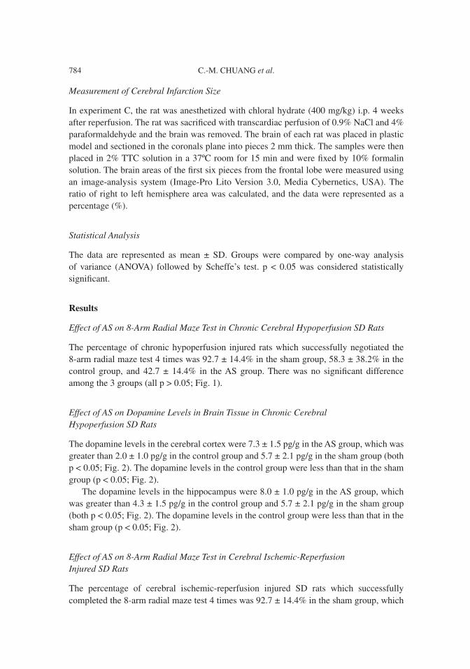
The percentage of chronic hypoperfusion injured rats which successfully negotiated the 8-arm radial maze test 4 times was 92.7 ± 14.4% in the sham group, 58.3 ± 38.2% in the control group, and 42.7 ± 14.4% in the AS group. There was no significant difference among the 3 groups (all p > 0.05; Fig. 1).
Effect of AS on Dopamine Levels in Brain Tissue in Chronic Cerebral Hypoperfusion SD Rats
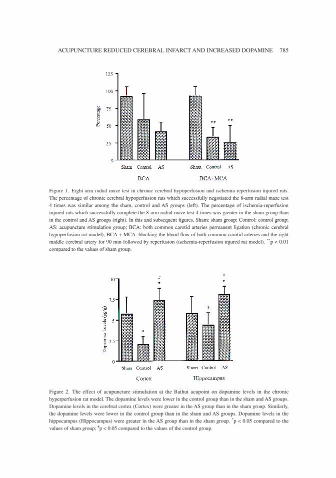
The dopamine levels in the cerebral cortex were 7.3 ± 1.5 pg/g in the AS group, which was greater than 2.0 ± 1.0 pg/g in the control group and 5.7 ± 2.1 pg/g in the sham group (both p < 0.05; Fig. 2). The dopamine levels in the control group were less than that in the sham group (p < 0.05; Fig. 2).
The dopamine levels in the hippocampus were 8.0 ± 1.0 pg/g in the AS group, which was greater than 4.3 ± 1.5 pg/g in the control group and 5.7 ± 2.1 pg/g in the sham group (both p < 0.05; Fig. 2). The dopamine levels in the control group were less than that in the sham group (p < 0.05; Fig. 2).
Effect of AS on 8-Arm Radial Maze Test in Cerebral Ischemic-Reperfusion Injured SD Rats
The percentage of cerebral ischemic-reperfusion injured SD rats which successfully completed the 8-arm radial maze test 4 times was 92.7 ± 14.4% in the sham group, which
00526.indd 784 10/4/2007 9:49:35 AM
785
Figure 1. Eight-arm radial maze test in chronic cerebral hypoperfusion and ischemia-reperfusion injured rats. The percentage of chronic cerebral hypoperfusion rats which successfully negotiated the 8-arm radial maze test 4 times was similar among the sham, control and AS groups (left). The percentage of ischemia-reperfusion injured rats which successfully complete the 8-arm radial maze test 4 times was greater in the sham group than in the control and AS groups (right). In this and subsequent figures, Sham: sham group; Control: control group; AS: acupuncture stimulation group; BCA: both common carotid arteries permanent ligation (chronic cerebral hypoperfusion rat model); BCA + MCA: blocking the blood flow of both common carotid arteries and the right middle cerebral artery for 90 min followed by reperfusion (ischemia-reperfusion injured rat model). **p < 0.01 compared to the values of sham group.
Figure 2. The effect of acupuncture stimulation at the Baihui acupoint on dopamine levels in the chronic hyperperfusion rat model. The dopamine levels were lower in the control group than in the sham and AS groups. Dopamine levels in the cerebral cortex (Cortex) were greater in the AS group than in the sham group. Similarly, the dopamine levels were lower in the control group than in the sham and AS groups. Dopamine levels in the hippocampus (Hippocampus) were greater in the AS group than in the sham group. *p < 0.05 compared to the values of sham group; #p < 0.05 compared to the values of the control group.
ACUPUNCTURE REDUCED CEREBRAL INFARCT AND INCREASED DOPAMINE
00526.indd 785 10/4/2007 9:49:36 AM
C.-M. CHUANG et al.786
was greater than 33.3 ± 14.4% in the control group and 25.0 ± 25% in the AS group (both p < 0.01; Fig. 1). The percentage was similar in the control group and AS group (p > 0.05; Fig. 1).
Effect of AS on Dopamine Levels in Brain Tissues of Cerebral Ischemic-Reperfusion Injured SD Rats
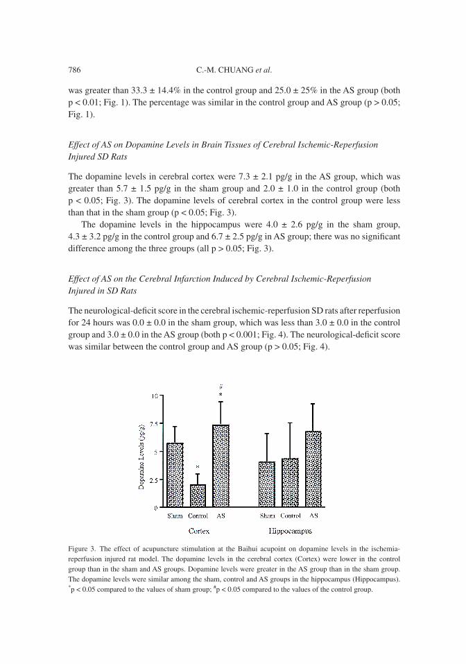
The dopamine levels in cerebral cortex were 7.3 ± 2.1 pg/g in the AS group, which was greater than 5.7 ± 1.5 pg/g in the sham group and 2.0 ± 1.0 in the control group (both p < 0.05; Fig. 3). The dopamine levels of cerebral cortex in the control group were less than that in the sham group (p < 0.05; Fig. 3).
The dopamine levels in the hippocampus were 4.0 ± 2.6 pg/g in the sham group, 4.3 ± 3.2 pg/g in the control group and 6.7 ± 2.5 pg/g in AS group; there was no significant difference among the three groups (all p > 0.05; Fig. 3).
Effect of AS on the Cerebral Infarction Induced by Cerebral Ischemic-Reperfusion Injured in SD Rats
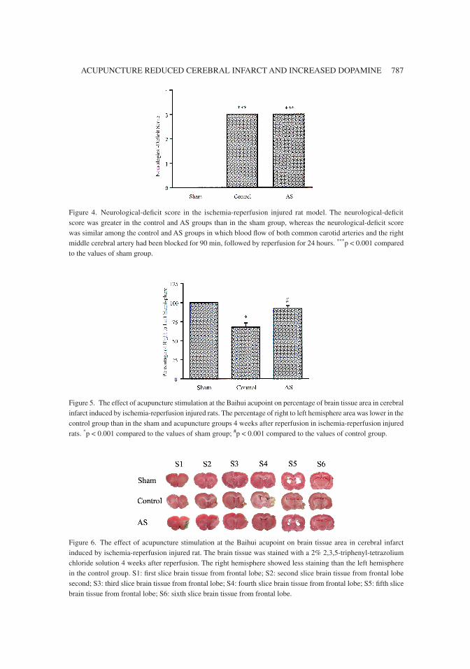
The neurological-deficit score in the cerebral ischemic-reperfusion SD rats after reperfusion for 24 hours was 0.0 ± 0.0 in the sham group, which was less than 3.0 ± 0.0 in the control group and 3.0 ± 0.0 in the AS group (both p < 0.001; Fig. 4). The neurological-deficit score was similar between the control group and AS group (p > 0.05; Fig. 4).
Figure 3. The effect of acupuncture stimulation at the Baihui acupoint on dopamine levels in the ischemia-reperfusion injured rat model. The dopamine levels in the cerebral cortex (Cortex) were lower in the control group than in the sham and AS groups. Dopamine levels were greater in the AS group than in the sham group. The dopamine levels were similar among the sham, control and AS groups in the hippocampus (Hippocampus). *p < 0.05 compared to the values of sham group; #p < 0.05 compared to the values of the control group.
00526.indd 786 10/4/2007 9:49:36 AM
787
Figure 6. The effect of acupuncture stimulation at the Baihui acupoint on brain tissue area in cerebral infarct induced by ischemia-reperfusion injured rat. The brain tissue was stained with a 2% 2,3,5-triphenyl-tetrazolium chloride solution 4 weeks after reperfusion. The right hemisphere showed less staining than the left hemisphere in the control group. S1: first slice brain tissue from frontal lobe; S2: second slice brain tissue from frontal lobe second; S3: third slice brain tissue from frontal lobe; S4: fourth slice brain tissue from frontal lobe; S5: fifth slice brain tissue from frontal lobe; S6: sixth slice brain tissue from frontal lobe.
Figure 4. Neurological-deficit score in the ischemia-reperfusion injured rat model. The neurological-deficit score was greater in the control and AS groups than in the sham group, whereas the neurological-deficit score was similar among the control and AS groups in which blood flow of both common carotid arteries and the right middle cerebral artery had been blocked for 90 min, followed by reperfusion for 24 hours. ***p < 0.001 compared to the values of sham group.
Figure 5. The effect of acupuncture stimulation at the Baihui acupoint on percentage of brain tissue area in cerebral infarct induced by ischemia-reperfusion injured rats. The percentage of right to left hemisphere area was lower in the control group than in the sham and acupuncture groups 4 weeks after reperfusion in ischemia-reperfusion injured rats. *p < 0.001 compared to the values of sham group; #p < 0.001 compared to the values of control group.
ACUPUNCTURE REDUCED CEREBRAL INFARCT AND INCREASED DOPAMINE
00526.indd 787 10/4/2007 9:49:36 AM
C.-M. CHUANG et al.788
The percentage of right to left hemisphere area was 68.0 ± 5.6% in the control group, which was less than 100.1 ± 0.4% in the sham group and 92.6 ± 3.6% in the AS group (both p < 0.001; Figs. 5 and 6). The percentage of right to left hemisphere area was similar between the sham group and AS group (p > 0.05; Figs. 5 and 6).
Discussion
The 8-arm radial maze test is a sensitive test for evaluating cognition and memory functions (Sopala and Danysz, 2001; Paganelli et al., 2004). The present results indicate that the percentage of rates which successfully negotiated the 8-arm radial maze test 4 times was similar among the sham, the chronic cerebral hypoperfusion with AS, and chronic cerebral hypoperfusion without AS groups. The percentage of rates which successfully completed the 8-arm radial maze test 4 times was greater in the sham group than in the cerebral ischemia-reperfusion injured group with or without AS. The results indicate that AS at Baihui acupoint for 4 weeks does not improve the cognitive and memory deficit of rats with ischemia-reperfusion injury. Our results also indicate that cognitive and memory deficits develop more rapidly in cerebral ischemia-reperfusion injured SD rats than in chronic cerebral hypoperfusion SD rats. Results from previous studies showed that cerebral infarct can be induced by blocking the blood flow of both common carotid arteries and the right middle cerebral artery for 90 min followed by reperfusion for 24 hours (Hsieh et al., 2001; Lao et al., 2005; Hsieh et al., 2006). In addition, the results of the present study were similar to those that demonstrated that dementia develops within three months after stroke in humans (Hénon et al., 2001). Our results also supported the result reported by Sopala and Danysz (2001) which indicated that cognitive deficits were not found within 16 months in SD rats with chronic cerebral hypoperfusion induced by permanent ligation of both common carotid arteries.
Our results indicated that AS at the Baihui acupoint for 4 weeks increased the levels of dopamine in the cerebral cortex and hippocampus in chronic cerebral hypoperfusion rats, and also increased the levels of dopamine in the right cerebral cortex in the cerebral ischemia-reperfusion injured rats. Dopamine plays a critical role in the regulation of synaptic plasticity in different brain areas including prefrontal cortex, hippocampus and striatum, and in the regulation of memory process (Jay, 2003). The deterioration of sensorimotor and cognitive function has been shown to be closely related to the dopamine receptor system in MCAo rats with striatal damage (Borlongan et al., 1995a, b). In addition, both D-amphetamine and Levodopa have been reported to enhance learning in humans (Breitenstein et al., 2006). Furthermore, stroke produces neurological deficits by damaging dopamine nerve terminals (Weinberger, 2002). Acupuncture has been shown to stimulate the expression of tyrosine kinase receptor B (trkB), which is known to reduce neuronal death of the nigostriatal dopaminergic system in a 6-hydroxydopamine- induced Parkinson’s disease model (Park et al., 2003). In our study, AS at the Baihui acupoint for 4 weeks increased dopamine levels in the cerebral cortex and hippocampus, but did not lead to an increase in the percentage of rates which could successfully negotiate the 8-arm radial maze test. One possible explanation is that dopamine involves in memory
00526.indd 788 10/4/2007 9:49:36 AM
789
performance possibly through the regulation of acetylcholine (Umegaki et al., 2001). Alzheimer’s disease is characterized by the progressive deterioration of memory, behavior and cognition, and lesions involving acetylcholine have been reported in humans (Coyle et al., 1983).
Our results also indicate that AS at the Baihui acupoit for 4 weeks leads to a higher percentage of right to left hemisphere area, suggesting that AS stimulates neuroprotective molecules that reduce the degree of cerebral atrophy after cerebral infarct. Our results showed that the neurological deficit scores were similar between the AS and the control groups after reperfusion for 24 hours. These results are consistent with those reported by Xiong et al., (2003) and Wang et al., (2005) which showed that EA pretreatment at Baihui acupoint reduces cerebral infarction volume in MCAo rats. It has been reported that acupuncture suppresses apoptosis in the cerebral ischemic rats (Jang et al., 2003), and that EA increases the activity of superoxide dismutase (SOD) and glutathione peroxidase (GPx), thereby reducing the extent of lipid peroxidation (Siu et al., 2004a) and the amount of malondialdehyde (Siu et al., 2004b).
In conclusion, AS at the Baihui acupoint for 4 weeks increased dopamine levels in brain tissues in chronic cerebral hypoperfusion rats and in cerebral ischemia-reperfusion injured rats. AS also reduces brain atrophy after cerebral infarct, suggesting that AS acts as a neuroprotector. AS at the Baihui acupoint 4 weeks does not enhance cognition and memory deficits.
Acknowledgments
This study was supported by grant NSC95-2320-B-039-029 from the National Science Council, Taiwan.
References
Bederson, J.B., L.H. Pittis, M. Tsuji, M.C. Nishimura, R.L. Davis and H. Bartkowski. Rat middle cerebral artery occlusion: evaluation of the model and development of a neurologic examination. Stroke 17: 472–476, 1986.
Borlongan, C.V., D.W. Cahill and P.R. Sanberg. Locomotor and passive avoidance deficits following occlusion of the middle cerebral artery. Physiol. Behav. 58: 909–917, 1995a.
Borlongan, C.V., R. Martinez, R.D. Shytle, T.B. Freeman, D.W. Cahill and P.R. Sanberg. Striatal dopamine-mediated motor behavior is altered following occlusion of the middle cerebral artery. Pharmacol. Biochem. Behav. 52: 225–229, 1995b.
Breitenstein, C., A. Flöel, C. Korsukewitz, S. Wailke, S. Bushuven and S. Knecht. A shift of paradigm: from noradrenergic to dopaminergic modulation of learning? J. Neurol. Sci. 248: 42–47, 2006.
Coyle, J.T., D.L. Price and M.R. DeLong. Alzheimer’s disease: a disorder of cortical cholinergic innervation. Science 219: 1184–1190, 1983.
Dos Santos, J.G., Jr., A. Tabosa, F.H. do Monte, M.M. Blanco, A. de Oliveira Freire and L.E. Mello. Electroacupuncture prevents cognitive deficits in pilocarpine-epileptic rats. Neurosci. Lett. 384: 234–238, 2005.
ACUPUNCTURE REDUCED CEREBRAL INFARCT AND INCREASED DOPAMINE
00526.indd 789 10/4/2007 9:49:37 AM
C.-M. CHUANG et al.790
Hénon, H., I. Durieu, F. Lebert, F. Pasquier and D. Leys. Poststroke dementia incidence and relationship to prestroke cognitive decline. Neurology 57: 1216–1222, 2001.
Hsieh, C.L, E.T. Liao, S.Y. Chiang, C.J. Lao, N.Y. Tang, C.T. Hsieh and J.G. Lin. Effect of Rhizoma Corydalis on focal cerebral infarct in ischemia-reperfusion injured rats. Acta Pharmacol. Sin. 22: 1143–1148, 2001.
Hsieh, C.L., C.Y. Cheng, T.H. Tsai, I.H. Lin, C.H. Liu, S.Y. Chiang, J.G. Lin, C.J. Lao and N.Y. Tang. Paeonol reduced cerebral infarction involving the superoxide anion and microglia activation in ischemia-reperfusion injured rats. J. Ethnopharmacol. 106: 208–215, 2006.
Jang, M.H., M.C. Shin, T.H. Lee, B.V. Lim, M.S. Shin, B.I. Min, H. Kim, S. Cho, E.H. Kim and C.J. Kim. Acupuncture suppresses ischemia-induced increase in c-Fos expression and apoptosis in the hippocampal CA1 region in gerbils. Neurosci. Lett. 347: 5–8, 2003.
Jay, T.M. Dopamine: a potential substrate for synaptic plasticity and memory mechanisms. Prog. Neurobiol. 69: 375–390, 2003.
Lao, C.J., J.G. Lin, J.S. Kuo, P.D. Chao, C.Y. Cheng, N.Y. Tang and C.L. Hsieh. Microglia, apoptosis and interleukin-1β expression in the effect of Sophora Japonica L. on cerebral infarct induced by ischemia-reperfusion in rats. Am. J. Chin. Med. 33: 425–438, 2005.
Ohta, H., H. Nishikawa, H. Kimura, H. Anayama and M. Miyamoto. Chronic cerebral hypoperfusion by permanent internal carotid ligation produces learning impairment without brain damage in rats. Neuroscience 79: 1039–1050, 1997.
Paganelli, R.A., A. Benetolli, K.C.M. Lima, L.A. Cestari-Junior, L.A.F. Filho and H. Milani. A novel version of the 8-arm radial maze: effects of cerebral ischemia on learning and memory. J. Neurosci. Methods 132: 9–18, 2004.
Pappas, B.A., J.C. de la Torre, C.M. Davidson, M.T. Keyes and T. Fortin. Chronic reduction of cerebral blood flow in the adult rat: late-emerging CA1 cell loss and memory dysfunction. Brain Res. 708: 50–58, 1996.
Park, H.J., S. Lim, W.S. Joo, C.S. Yin, H.S. Lee, H.J. Lee, J.C. Seo, K. Leem, Y.S. Son, Y.J. Kim, C.J. Kim, Y.S. Kim and J.H. Chung. Acupuncture prevents 6-hydroxydopamine-induced neuronal death in the nigrostriatal dopaminergic system in the rat Parkinson’s disease model. Exp. Neurol. 180: 92–97, 2003.
Siu, F.K.W., S.C.L. Lo and M.C.P. Leung. Effectiveness of multiple pre-ischemia electro-acupuncture on attenuating lipid peroxidation induced by cerebral ischemia in adult rats. Life Sci. 75: 1323–1332, 2004a.
Siu, F.K.W., S.C.L. Lo and M.C.P. Leung. Electroacupuncture reduces the extent of lipid peroxidation by increasing superoxide dismutase and glutathione peroxidase activities in ischemic-reperfused rat brains. Neurosci. Lett. 354: 158–162, 2004b.
Sopala, M. and W. Danysz. Chronic cerebral hypoperfusion in the rat enhances age-related deficits in spatial memory. J. Neural Transm. 108: 1445–1456, 2001.
Umegaki, H., J. Munoz, R.C. Meyer, E.L. Spangler, J. Yoshimura, H. Ikari, A. Iguchi and D.K. Ingram. Involvement of dopamine D2 receptors in complex maze learning and acetylcholine release in ventral hippocampus of rats. Neuroscience 103: 27–33, 2001.
Wang, S.J., N. Omori, F. Jin, G. Li, W.R. Zhang, Y. Hamakawa, K. Sato, I. Nagano, M. Shoji and K. Abe. Potentiation of Akt and suppression of caspase-9 activations by electroacupuncture after transient middle cerebral artery occlusion in rats. Neurosci. Lett. 331: 115–118, 2002.
Wang, Q., L. Xiong, S. Chen, Y. Liu and X. Zhu. Rapid tolerance to focal cerebral ischemia in rats is induced by preconditioning with electroacupuncture: window of protection and the role of adenosine. Neurosci. Lett. 381: 158–162, 2005.
00526.indd 790 10/4/2007 9:49:37 AM

791
Weinberger, J. The role of dopamine in cerebral ischemic damage: a review of studies with Gerald Cohen. Parkinsonism Relat. Disord. 8: 413–416, 2002.
Xiong, L., Z. Lu, L. Hou, H. Zheng, Z. Zhu, Q. Wang and S. Chen. Pretreatment with repeated electroacupuncture attenuates transient focal cerebral ischemic injury in rats. Chin. Med. J. 116: 108–111, 2003.
ACUPUNCTURE REDUCED CEREBRAL INFARCT AND INCREASED DOPAMINE
00526.indd 791 10/4/2007 9:49:37 AM