Embed Size (px)
Citation preview
Acute anterior ST segment elevation myocardial infarction
complicated with transient intraventricular conduction disturbance
https://ekgvcg.wordpress.com/
Raimundo Barbosa-Barros M.D1; Andrés Ricardo Pérez-Riera M.D.Ph.D.2; Luiz Carlos de Abreu P.h.D.3
1. Chief of the Coronary Center of the Hospital de Messejana Dr. Carlos Alberto Studart Gomes. Fortaleza
–CE- Brazil
2. Post-Graduates Advisor at Design of Studies and Scientific Writing Laboratory in the ABC Faculty of
Medicine - ABC Foundation - Santo André – São Paulo – Brazil
3. Visiting Scientist at Program in Molecular and Integrative Physiological Sciences (MIPS), Department of
Environmental Health | Harvard T.H. Chan School of Public Health.
Case report
A 58-year-old Caucasian man was admitted to our emergency department due to prolonged oppressive
retrosternal chest pain for 3 hours associated with shortness of breath and cold diaphoresis.
He had several risk factors: hypertension, low-density lipoprotein cholesterol elevated levels, central obesity
(waist circumference = 112 cm and body mass index (BMI) =34), diabetic intolerance, and addiction to
tobacco.
On physical examination, the jugular veins were noted 3 cm above the clavicles with the patient in a 45°
semirecumbent position.
Absence of hepatojugular reflux (the height of the neck veins increases only 2 cm with moderate pressure
applied over the middle of the abdomen for 30 seconds).
His blood pressure was 95/80 mm Hg (pulse pressure/systolic pressure ratio > 0.25) and alternating discrete
irregular heart rate 107-111 bpm.
The point of maximal impulse was located in the midclavicular line at the left fifth intercostal space and it is
covered by the tip of one index finger.
A third protodiastolic gallop or ventricular gallop (S3) was heard without murmurs with gallop cadence.
Absence of rales in both pulmonary bases.
The liver edge was palpable smooth, uniform, non-tender and non painful at 1 cm of costal border.
Absence of lower extremity edema.
The electrocardiograms are shown in the next slides.
Question
What are the diagnoses of both electrocardiograms?
Homem caucasiano de 58 anos, admitido em nosso departamento de emergência por dor retroesternal típica
opressiva prolongada que havia iniciado há 3 horas associada a falta de ar e sudorese fria.
Vários fatores de risco para doença arterial coronariana estavam presentes: hipertensão arterial, níveis LDL-C
elevado, obesidade centrípeta (circunferência da cintura = 112 cm e índice de massa corporal (IMC) = 34),
intolerância a glicose e tabagista.
Ao exame físico, jugulares a 3 cm acima da clavícula com o paciente em uma posição reclinada a 45°.
Ausência de refluxo hepatojugular.
Pressão arterial 95/80 mm de Hg (relação da pressão do pulso / pressão sistólica > 0,25) ritmo cardíaco
irregular com discreta variação da FC 107-111 bpm.
Choque da ponta foi localizado na linha médio clavicular no quinto espaço intercostal esquerdo e é coberto
com um dedo indicador.
Presença de terceira bulha (S3) protossistólica com cadencia de galope. Ausência de sopros.
Ausência de estertores em ambas as bases pulmonares.
Fígado liso, uniforme, não doloroso e palpável a 1 cm da borda costal direita.
Ausência de edema de membros inferiores.
Os eletrocardiogramas são mostrados nos próximos slides.
Pergunta
Quais os diagnósticos de ambos os eletrocardiogramas?
Português reporte de caso
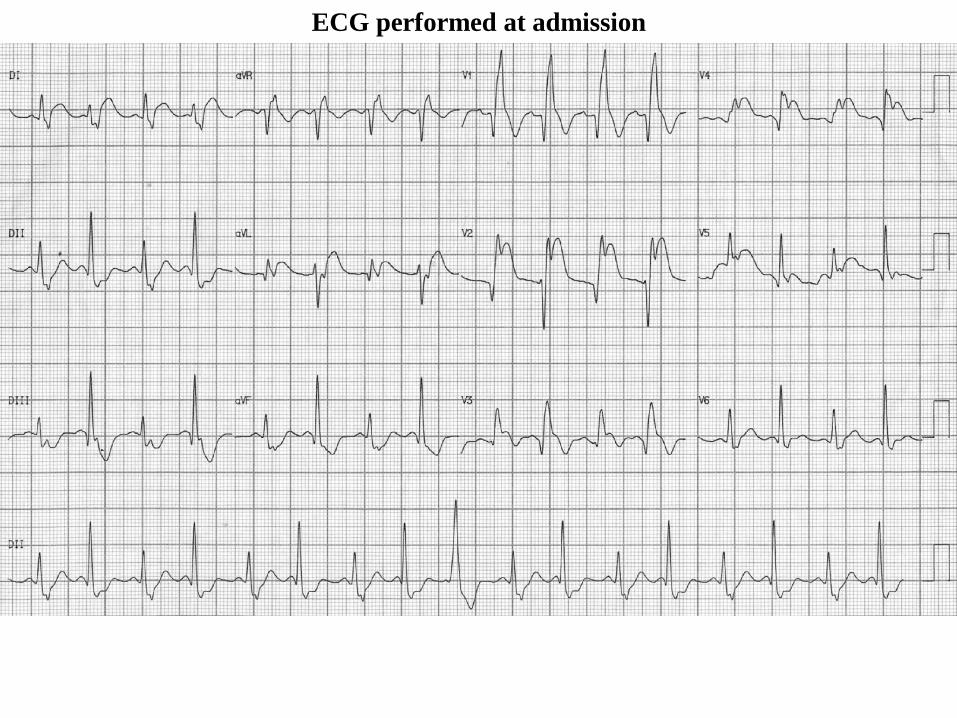
ECG performed at admission
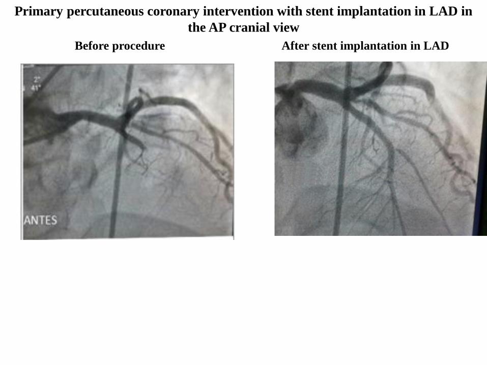
Before procedure After stent implantation in LAD
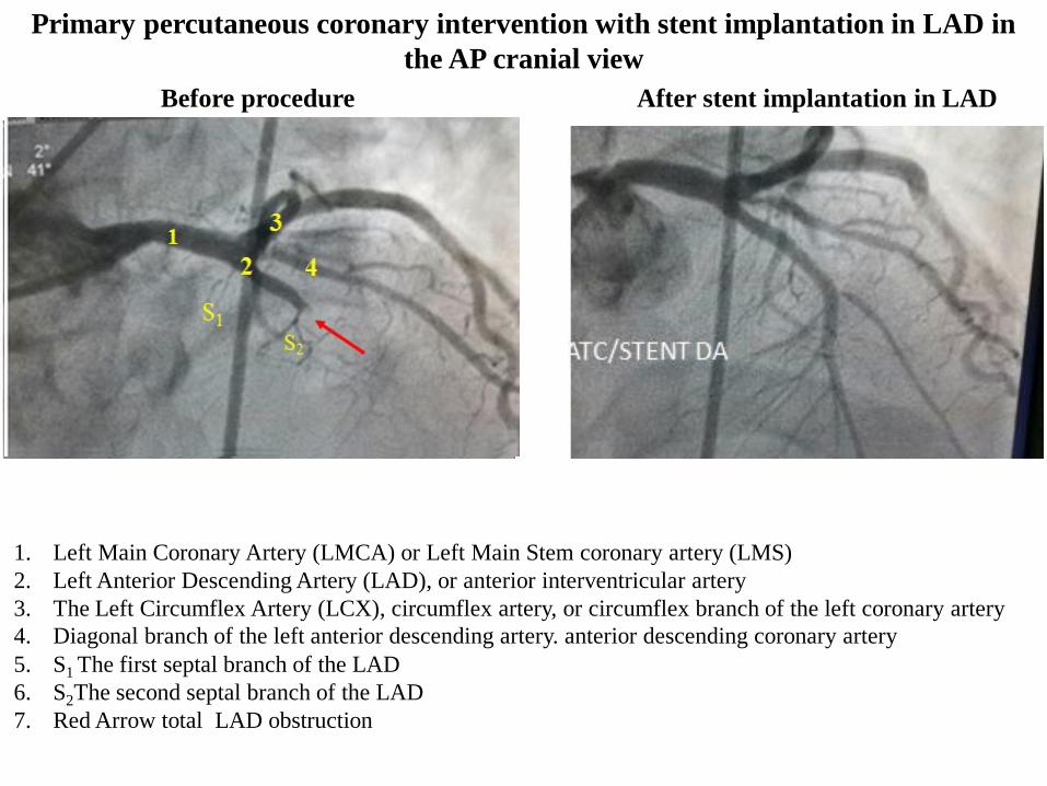
Primary percutaneous coronary intervention with stent implantation in LAD in
the AP cranial view
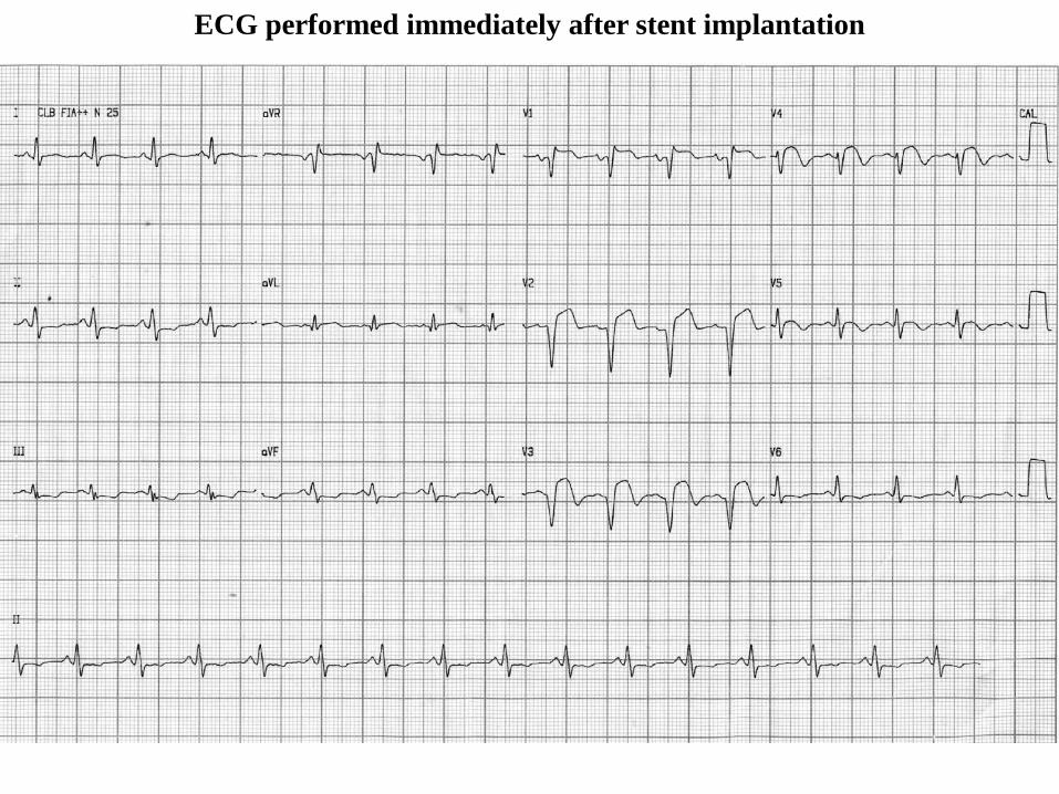
ECG performed immediately after stent implantation
The ECG shows an acute anteroseptal MI with rate alternate left posterior hemiblock pattern. This resolves
after PCI of LAD and shows resolving anteroseptal MI. Of interest is the RBBB and 2:1 block in the
Posterior fascicle which is associated with a decrease in the anterior q waves, probably because the initial
forces are oriented left and superior
Melvin M Sheinman
Department of Cardiac Electrophysiology, University of California San Francisco, San Francisco, California,
USA. [email protected]
Professor of Medicine
Colleagues opinions
Dear friends, Professor Andrés Ricardo, Raimundo, and Luiz Carlos,
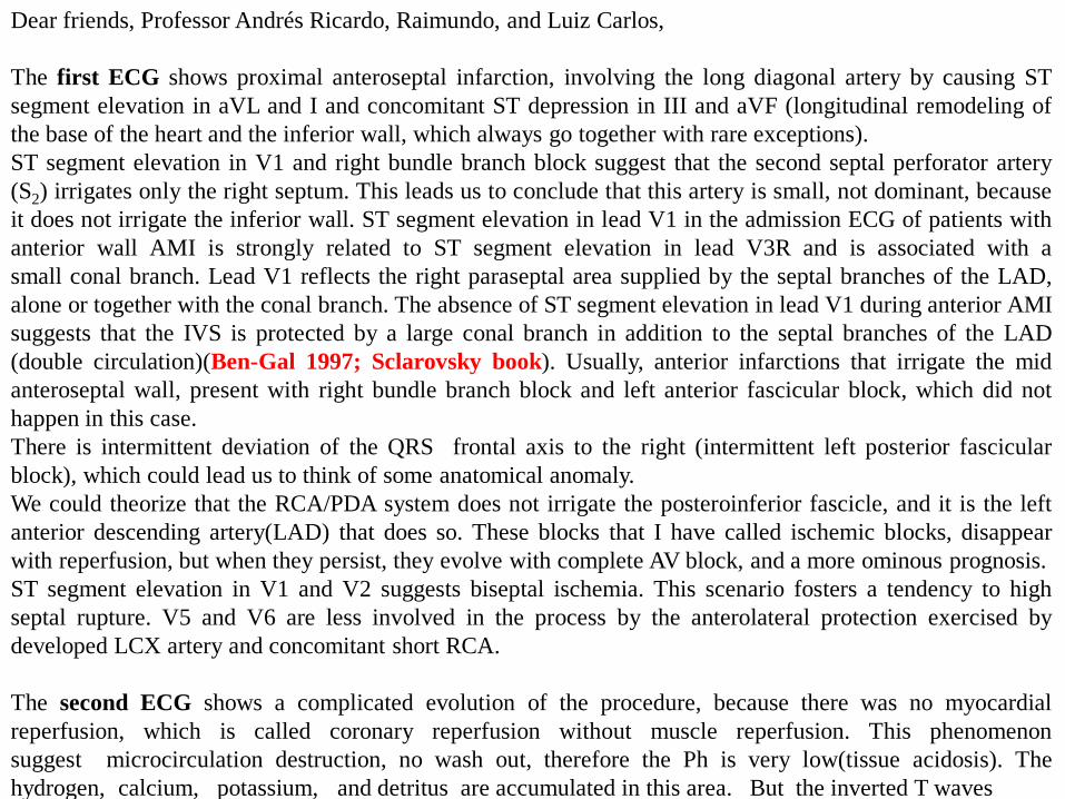
The first ECG shows proximal anteroseptal infarction, involving the long diagonal artery by causing ST
segment elevation in aVL and I and concomitant ST depression in III and aVF (longitudinal remodeling of
the base of the heart and the inferior wall, which always go together with rare exceptions).
ST segment elevation in V1 and right bundle branch block suggest that the second septal perforator artery
(S2) irrigates only the right septum. This leads us to conclude that this artery is small, not dominant, because
it does not irrigate the inferior wall. ST segment elevation in lead V1 in the admission ECG of patients with
anterior wall AMI is strongly related to ST segment elevation in lead V3R and is associated with a
small conal branch. Lead V1 reflects the right paraseptal area supplied by the septal branches of the LAD,
alone or together with the conal branch. The absence of ST segment elevation in lead V1 during anterior AMI
suggests that the IVS is protected by a large conal branch in addition to the septal branches of the LAD
(double circulation)(Ben-Gal 1997; Sclarovsky book). Usually, anterior infarctions that irrigate the mid
anteroseptal wall, present with right bundle branch block and left anterior fascicular block, which did not
happen in this case.
There is intermittent deviation of the QRS frontal axis to the right (intermittent left posterior fascicular
block), which could lead us to think of some anatomical anomaly.
We could theorize that the RCA/PDA system does not irrigate the posteroinferior fascicle, and it is the left
anterior descending artery(LAD) that does so. These blocks that I have called ischemic blocks, disappear
with reperfusion, but when they persist, they evolve with complete AV block, and a more ominous prognosis.
ST segment elevation in V1 and V2 suggests biseptal ischemia. This scenario fosters a tendency to high
septal rupture. V5 and V6 are less involved in the process by the anterolateral protection exercised by
developed LCX artery and concomitant short RCA.
The second ECG shows a complicated evolution of the procedure, because there was no myocardial
reperfusion, which is called coronary reperfusion without muscle reperfusion. This phenomenon
suggest microcirculation destruction, no wash out, therefore the Ph is very low(tissue acidosis). The
hydrogen, calcium, potassium, and detritus are accumulated in this area. But the inverted T waves
in V5-V6 the inferior lateral wall are reperfused, and V4 is the ischemic border area The upper area
of the lateral wall is not reperfused.
There is inferior anteroseptal aneurysmatic dilatation. This is very important and should be taken into
account, because after 48 hours, intracavitary thrombi frequently appear, with a high risk of embolism(Sagie
1991: Sclarovsky book).
The cases of stroke that appear during the evolution of infarctions, are due to this cause. The hope in this case
is that from V5, V6, lateral hypertrophic remodeling develops, helping to maintain LV ejection fraction.
Warm regards and congratulations to this fantastic Brazilian trio.
1. Ben-Gal T, Sclarovsky S, Herz I, Strasberg B, Zlotikamien B, Sulkes J, Birnbaum Y, Wagner GS, Sagie
A. Importance of the conal branch of the right coronary artery in patients with acute anterior wall
myocardial infarction: electrocardiographic and angiographic correlation. J Am Coll Cardiol.
1997;29(3):506-1.
2. Sagie A, Strasberg B, Imbar S, Rechavia E, Sclarovsky S. Value of the electrocardiogram for prediction
of left ventricular mural thrombus in anterior wall acute myocardial infarction. Am J Cardiol.
1991;68(9):957-9.
3. Sclarovsky S. Electrocardiography of Acute Myocardial Ischemia and Infarction 1st Edition: Chapter 4, 5
and 7 ISBN-13: 978-1853173806; ISBN-10: 1853173800.
ISBN-10: 1853173800
Samuel Sclarovsky MD Israel
- Professor Emeritus - Tel Aviv University
- Director of Telemedicine, Procardia Medical Center
Spanish
Queridos amigos Profesor, Andrés Ricardo , Raimundo y Luiz Carlos
El primer ECG muestra um infarto antero-septal proximal que involucra la artéria diagonal larga por
ocasionar elevación del segmento ST en aVL y I y concomitante depresión del ST en III y aVF(remodelado
longitudinal de la base cardíaca y de la pared inferior las cuales siempre van juntas con raras excepciones).
La elevación del segmento ST en V1 y el bloqueo de rama derecha sugere que la segunda perforante septal
irriga apenas el septo derecho. Esto tambíén hace pensar que esta artéria es pequeña no dominante porque no
irriga la pared inferior. Habitualmente, los infartos anteriores que irrigan la pared media antero-septal , se
presentan con bloqueo rama derecha y hemibloqueo antero-superior izquierdo, lo que no ha ocurrido en este
caso Hay desviación intermitente del eje frontal hacia la derecha, (hemibloqueo posterior intermitente), lo
que podria hacer pensar en alguna anomalia anatómica. Se podria especular que el Sistema de la
coronária derecha/ desendente posterior no irriga al fascículo póstero-inferior y si la descendente anterior,
Estos bloqueos que yo he denominado bloqueos isquémicos desaparecen com la reperfusión, más cuando
persisten desarrollan um bloqueo completo de pronóstico mas sombrio.
La elevación del segmento ST en V1 y V2 sugiere una isquemia biseptal. Este escenário propicia una
tendencia a ruptura septal alta
V5 y V6, estan menos involucradas en el proceso por la protección antero-latera ejercida por una artéria
circunfleja izquierda desarrollada y concomitante coronária derecha corta
El segundo ECG muestra una evolución complicada del procedimiento porque no ocurrió reperfusión
miocárdica, lo que se denomina reperfusion coronaria sin reperfusion muscular.
Existe una dilatación aneurismática anteroseptal inferior. Esto es muy importante de tener en cuenta porque a
las 48 horas frecuentemente aparecen trombos intracavitários, con alto riesgo de embolia Los casos de CVA
, que aparecen durante la evolucion del infartos , se deben a esta causa. La esperanza de este caso es que a
partir de V5,V6 comiencen a desarrollar una remodelacion hipertrófica lateral, que ayude a mantener el la
fracción de eyección
Un fraternal abrazo y felitaciones a este fantástico trio Brasileño
Samuel Sclarovsky
Português: Achamos que é taquicardia sinusal, BRD + BDPI intermitente no primeiro ECG com
desaparecimento do distúrbio de condução intraventricular após o implante do stent em DA.--------------------------------------------------------------------------------We think that the first ECG has sinus tachycardia, RBBB and intermittent LPFB
In the second ECG intraventricular conduction defect disappear after stent implantation.
Drs José Grindler, Acácio F. Cardoso Alfredo J. Fonseca
Serviço: Eletrocardiologia HC Faculdade de Medicina -Universidade de São Paulo-USP
Service: Electrocardiology HC Faculty of Medicine - University of São Paulo-USP
Diretor de Serviço
This is extensive anterior STEMI due to proximal LAD occlusion before the first diagonal branch with ST
elevation in I, aVL V2-V4 and reciprocal ST depression in the inferior leads.
The initial ECG shows RBBB that is unclear whether it is new or old.
There is electrical alternans, most probably secondary to extensive ischemia.
You give hints that there is no tamponade.
After stenting QRS width decreases, there are Q waves in aVL, V1-V3
Dr. Yochai Birnbaum, MD., FAHA, FACC
The Section of Cardiology, The Department of Medicine, Baylor College of Medicine, Houston, TX, USA.

Final comments by Andrés Ricardo Pérez-Riera, Raimundo
Barbosa-Barros & Luiz Carlos de Abreu
Before procedure After stent implantation in LAD
Primary percutaneous coronary intervention with stent implantation in LAD in
the AP cranial view
1
1. Left Main Coronary Artery (LMCA) or Left Main Stem coronary artery (LMS)
2. Left Anterior Descending Artery (LAD), or anterior interventricular artery
3. The Left Circumflex Artery (LCX), circumflex artery, or circumflex branch of the left coronary artery
4. Diagonal branch of the left anterior descending artery. anterior descending coronary artery
5. S1 The first septal branch of the LAD
6. S2The second septal branch of the LAD
7. Red Arrow total LAD obstruction
2
3
4
S2
S1
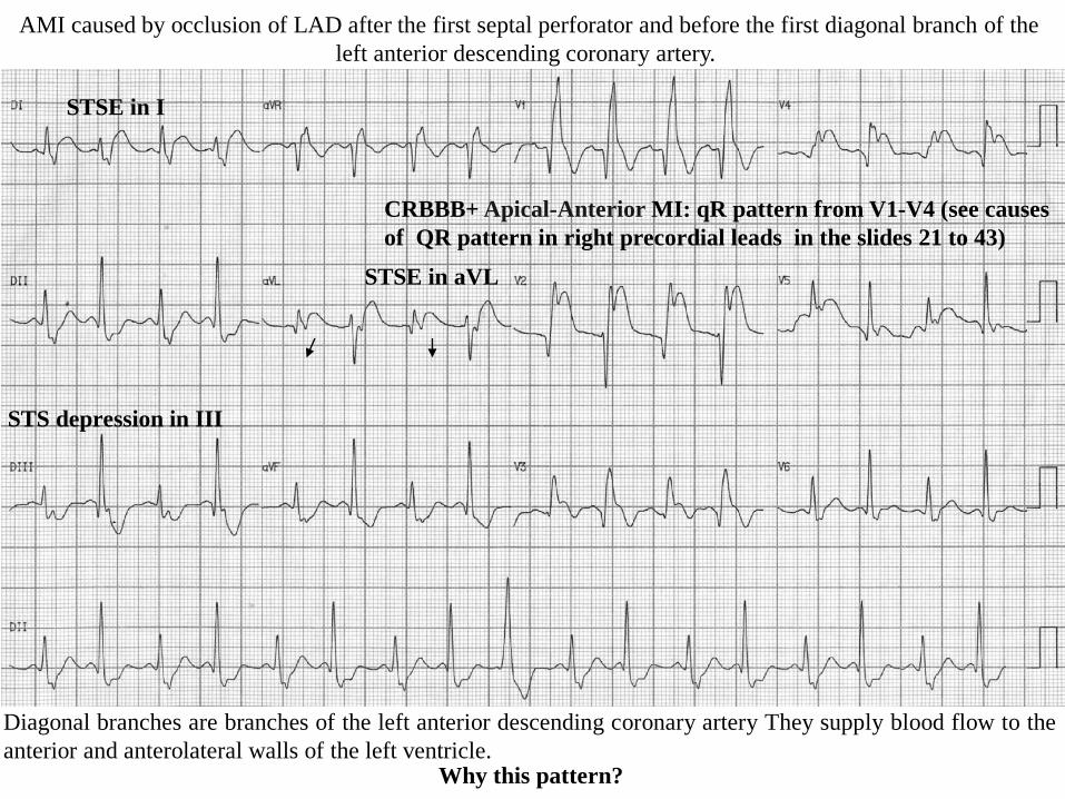
Diagonal branches are branches of the left anterior descending coronary artery They supply blood flow to the
anterior and anterolateral walls of the left ventricle.
CRBBB+ Apical-Anterior MI: qR pattern from V1-V4 (see causes
of QR pattern in right precordial leads in the slides 21 to 43)
STSE in I
STS depression in III
STSE in aVL
AMI caused by occlusion of LAD after the first septal perforator and before the first diagonal branch of the
left anterior descending coronary artery.
Why this pattern?
X
Y
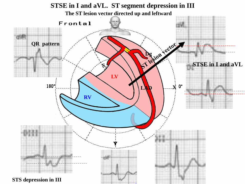
The ST lesion vector directed up and leftward
S1
Dg
LAD
RV
LV
STSE in I and aVL. ST segment depression in III
STS depression in III
QR pattern
STSE in I and aVL
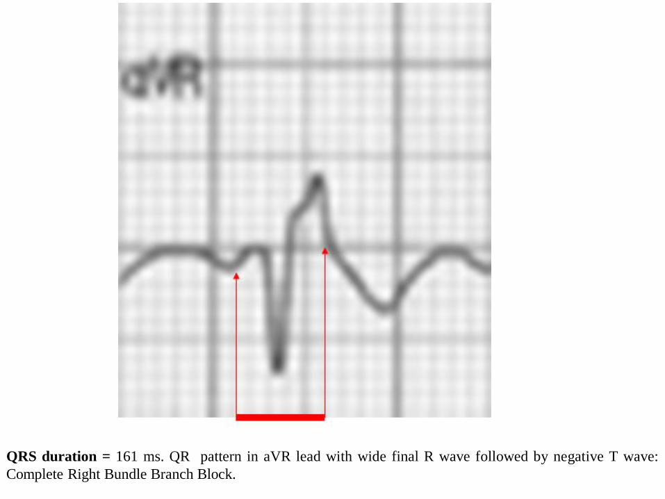
QRS duration = 161 ms. QR pattern in aVR lead with wide final R wave followed by negative T wave:
Complete Right Bundle Branch Block.
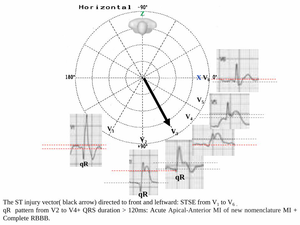
The ST injury vector( black arrow) directed to front and leftward: STSE from V1 to V6 .
qR pattern from V2 to V4+ QRS duration > 120ms: Acute Apical-Anterior MI of new nomenclature MI +
Complete RBBB.
X V6
V1
V4
V5
V2
V3
Z
qR
qR
qR
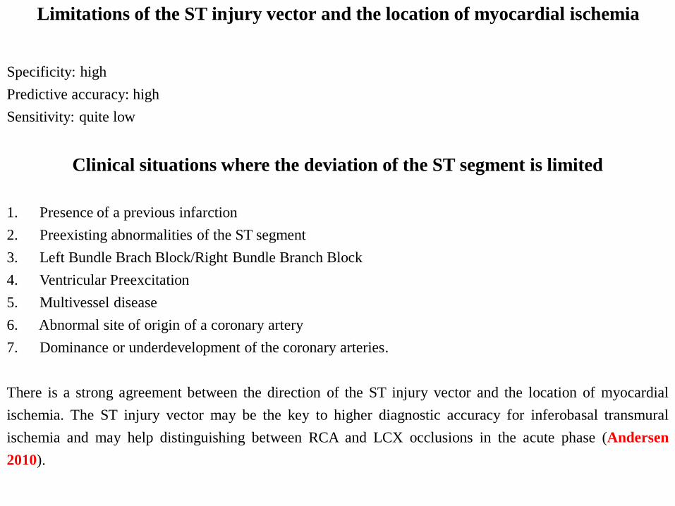
Limitations of the ST injury vector and the location of myocardial ischemia
Specificity: high
Predictive accuracy: high
Sensitivity: quite low
Clinical situations where the deviation of the ST segment is limited
1. Presence of a previous infarction
2. Preexisting abnormalities of the ST segment
3. Left Bundle Brach Block/Right Bundle Branch Block
4. Ventricular Preexcitation
5. Multivessel disease
6. Abnormal site of origin of a coronary artery
7. Dominance or underdevelopment of the coronary arteries.
There is a strong agreement between the direction of the ST injury vector and the location of myocardial
ischemia. The ST injury vector may be the key to higher diagnostic accuracy for inferobasal transmural
ischemia and may help distinguishing between RCA and LCX occlusions in the acute phase (Andersen
2010).
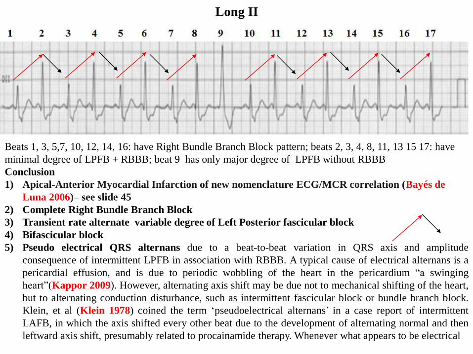
Long II
Beats 1, 3, 5,7, 10, 12, 14, 16: have Right Bundle Branch Block pattern; beats 2, 3, 4, 8, 11, 13 15 17: have
minimal degree of LPFB + RBBB; beat 9 has only major degree of LPFB without RBBB
Conclusion
1) Apical-Anterior Myocardial Infarction of new nomenclature ECG/MCR correlation (Bayés de
Luna 2006)– see slide 45
2) Complete Right Bundle Branch Block
3) Transient rate alternate variable degree of Left Posterior fascicular block
4) Bifascicular block
5) Pseudo electrical QRS alternans due to a beat-to-beat variation in QRS axis and amplitude
consequence of intermittent LPFB in association with RBBB. A typical cause of electrical alternans is a
pericardial effusion, and is due to periodic wobbling of the heart in the pericardium “a swinging
heart”(Kappor 2009). However, alternating axis shift may be due not to mechanical shifting of the heart,
but to alternating conduction disturbance, such as intermittent fascicular block or bundle branch block.
Klein, et al (Klein 1978) coined the term ‘pseudoelectrical alternans’ in a case report of intermittent
LAFB, in which the axis shifted every other beat due to the development of alternating normal and then
leftward axis shift, presumably related to procainamide therapy. Whenever what appears to be electrical
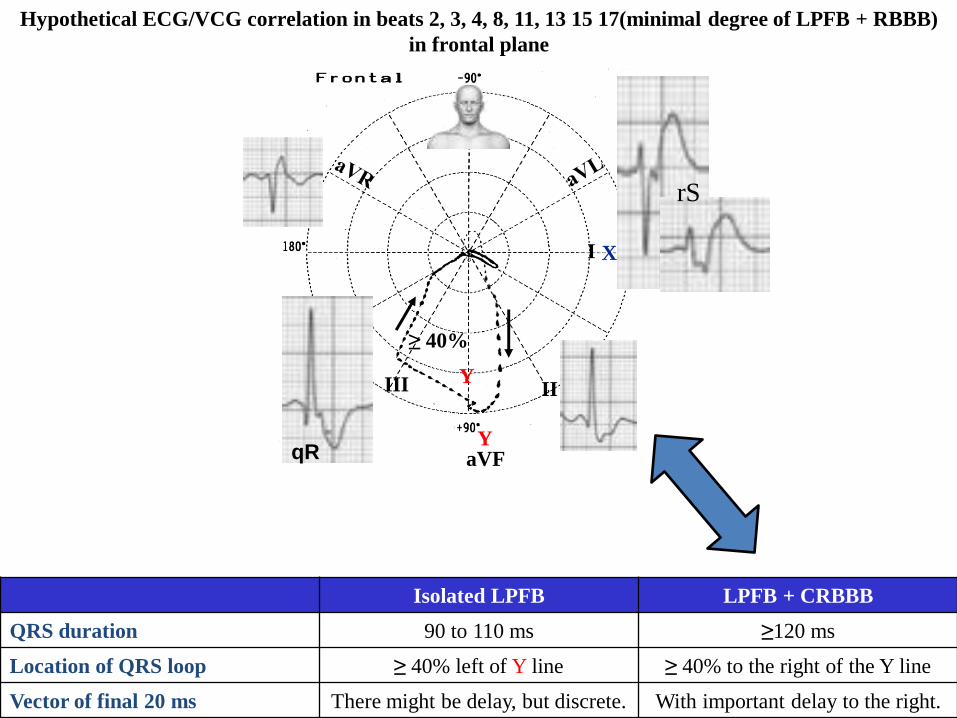
Hypothetical ECG/VCG correlation in beats 2, 3, 4, 8, 11, 13 15 17(minimal degree of LPFB + RBBB)
in frontal plane
≥ 40%
X
Y
Isolated LPFB LPFB + CRBBB
QRS duration 90 to 110 ms ≥120 ms
Location of QRS loop ≥ 40% left of Y line ≥ 40% to the right of the Y line
Vector of final 20 ms There might be delay, but discrete. With important delay to the right.
IIIII
aVF
I
Y
rS
qR
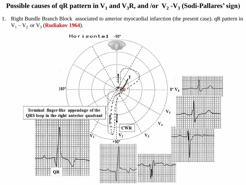
1. Right Bundle Branch Block associated to anterior myocardial infarction (the present case). qR pattern in
V1 – V2 or V3 (Rudiakov 1964).
Possible causes of qR pattern in V1 and V3R, and /or V2 -V3 (Sodi-Pallares’ sign)
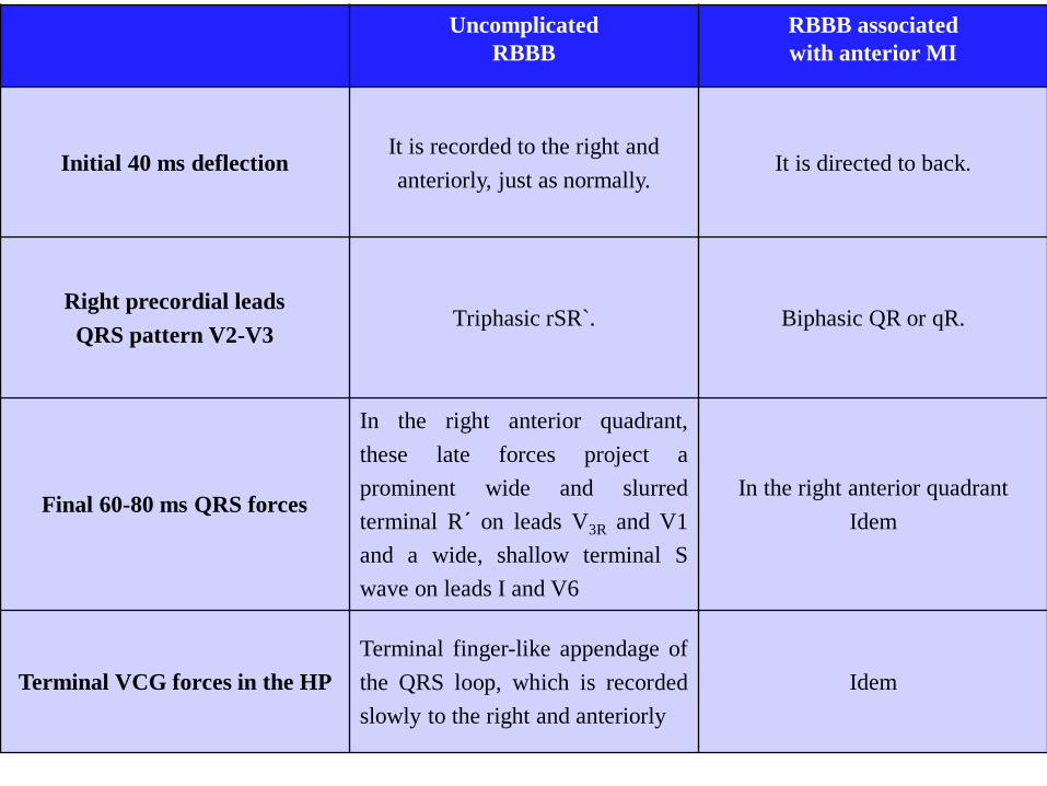
Uncomplicated
RBBB
RBBB associated
with anterior MI
Initial 40 ms deflection It is recorded to the right and
anteriorly, just as normally.It is directed to back.
Right precordial leads
QRS pattern V2-V3Triphasic rSR`. Biphasic QR or qR.
Final 60-80 ms QRS forces
In the right anterior quadrant,
these late forces project a
prominent wide and slurred
terminal R´ on leads V3R and V1
and a wide, shallow terminal S
wave on leads I and V6
In the right anterior quadrant
Idem
Terminal VCG forces in the HP
Terminal finger-like appendage of
the QRS loop, which is recorded
slowly to the right and anteriorly
Idem
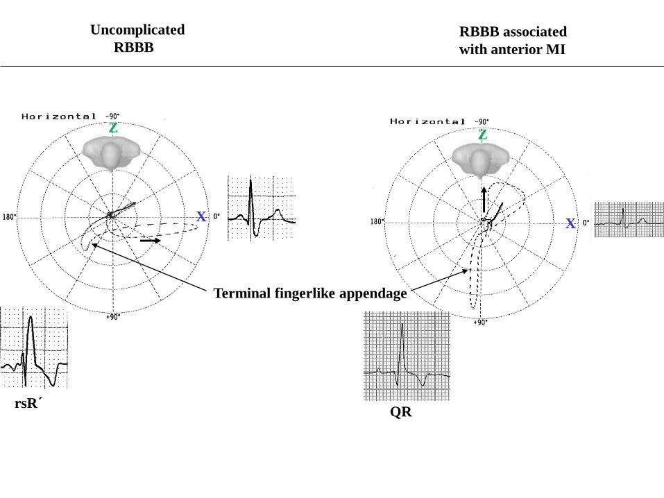
Uncomplicated
RBBB RBBB associated
with anterior MI
rsR´
Terminal fingerlike appendage
QR
X
Z
X
Z
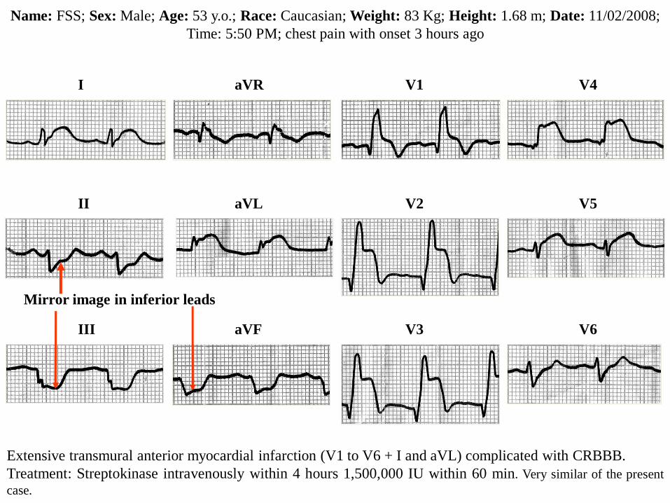
Name: FSS; Sex: Male; Age: 53 y.o.; Race: Caucasian; Weight: 83 Kg; Height: 1.68 m; Date: 11/02/2008;
Time: 5:50 PM; chest pain with onset 3 hours ago
I
II
III
aVR
aVL
aVF
V1
V2
V3
V4
V5
V6
Extensive transmural anterior myocardial infarction (V1 to V6 + I and aVL) complicated with CRBBB.
Treatment: Streptokinase intravenously within 4 hours 1,500,000 IU within 60 min. Very similar of the present
case.
Mirror image in inferior leads
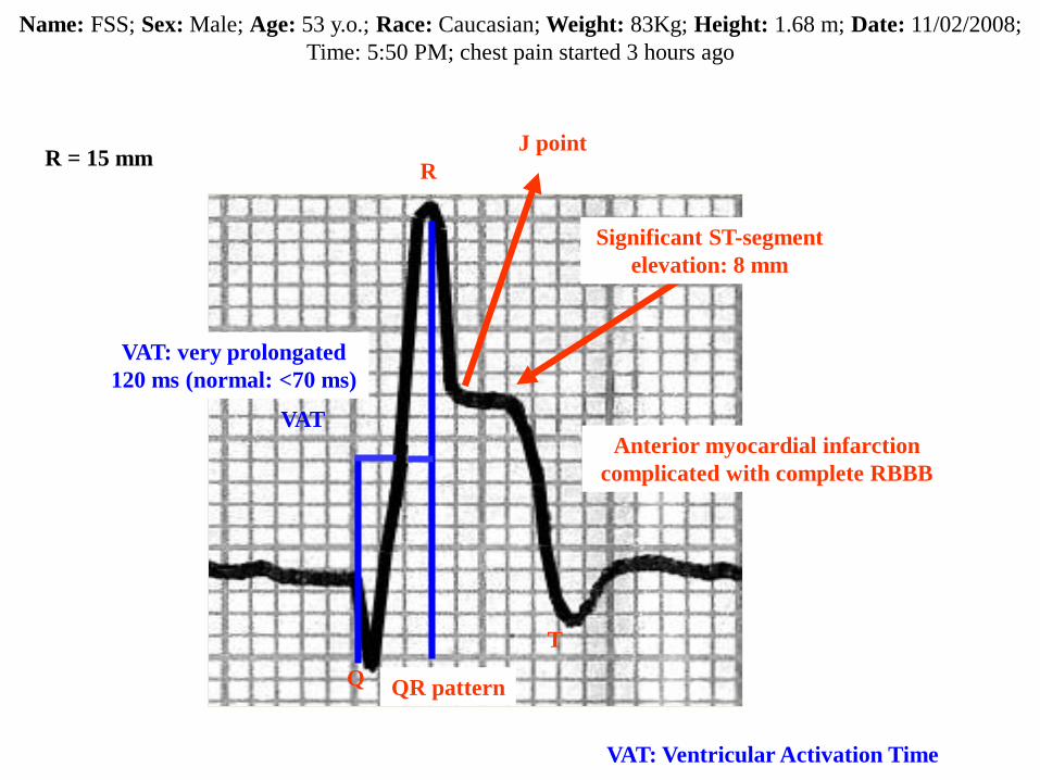
V2
Q
R
T
Significant ST-segment
elevation: 8 mm
J point
VAT
VAT: Ventricular Activation Time
VAT: very prolongated
120 ms (normal: <70 ms)
QR pattern
Anterior myocardial infarction
complicated with complete RBBB
R = 15 mm
Name: FSS; Sex: Male; Age: 53 y.o.; Race: Caucasian; Weight: 83Kg; Height: 1.68 m; Date: 11/02/2008;
Time: 5:50 PM; chest pain started 3 hours ago
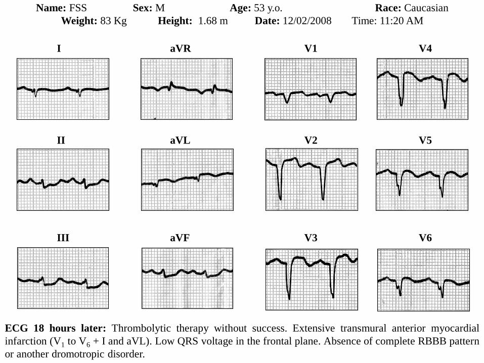
Name: FSS Sex: M Age: 53 y.o. Race: Caucasian
Weight: 83 Kg Height: 1.68 m Date: 12/02/2008 Time: 11:20 AM
I
II
III
aVR
aVL
aVF
V1
V2
V3
V4
V5
V6
ECG 18 hours later: Thrombolytic therapy without success. Extensive transmural anterior myocardial
infarction (V1 to V6 + I and aVL). Low QRS voltage in the frontal plane. Absence of complete RBBB pattern
or another dromotropic disorder.
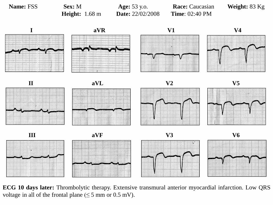
Name: FSS Sex: M Age: 53 y.o. Race: Caucasian Weight: 83 Kg
Height: 1.68 m Date: 22/02/2008 Time: 02:40 PM
I
II
III
aVR
aVL
aVF
V1
V2
V3
V4
V5
V6
ECG 10 days later: Thrombolytic therapy. Extensive transmural anterior myocardial infarction. Low QRS
voltage in all of the frontal plane (≤ 5 mm or 0.5 mV).
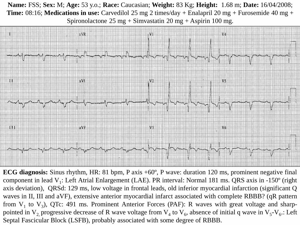
Name: FSS; Sex: M; Age: 53 y.o.; Race: Caucasian; Weight: 83 Kg; Height: 1.68 m; Date: 16/04/2008;
Time: 08:16; Medications in use: Carvedilol 25 mg 2 times/day + Enalapril 20 mg + Furosemide 40 mg +
Spironolactone 25 mg + Simvastatin 20 mg + Aspirin 100 mg.
ECG diagnosis: Sinus rhythm, HR: 81 bpm, P axis +60º, P wave: duration 120 ms, prominent negative final
component in lead V1: Left Atrial Enlargement (LAE). PR interval: Normal 181 ms. QRS axis in -150º (right
axis deviation), QRSd: 129 ms, low voltage in frontal leads, old inferior myocardial infarction (significant Q
waves in II, III and aVF), extensive anterior myocardial infarct associated with complete RBBB? (qR pattern
from V1 to V3), QTc: 491 ms. Prominent Anterior Forces (PAF): R waves with great voltage and sharp-
pointed in V2, progressive decrease of R wave voltage from V4 to V6, absence of initial q wave in V5-V6.: Left
Septal Fascicular Block (LSFB), probably associated with some degree of RBBB.
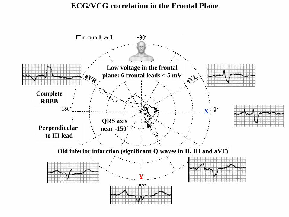
ECG/VCG correlation in the Frontal Plane
DI
DIIDIII aVF
X
Y
T
P
Old inferior infarction (significant Q waves in II, III and aVF)
Q
Complete
RBBB
QRS axis
near -150ºPerpendicular
to III lead
Low voltage in the frontal
plane: 6 frontal leads < 5 mV
LAE
X
Y
V6
V1
V4
V5
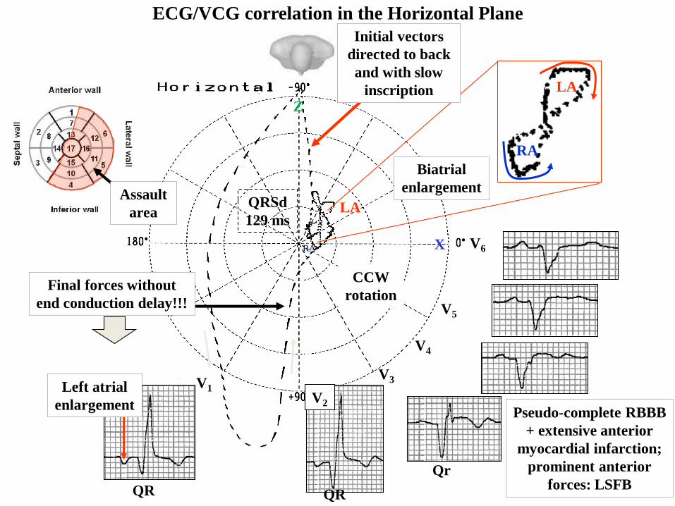
ECG/VCG correlation in the Horizontal Plane
V3
CCW
rotation
T
V2
Left atrial
enlargement
RA
LA
RA
LA
Biatrial
enlargement
QR QR
Qr
QRSd
129 ms
Pseudo-complete RBBB
+ extensive anterior
myocardial infarction;
prominent anterior
forces: LSFB
PSEUDO CRBBB
Final forces without
end conduction delay!!!
Initial vectors
directed to back
and with slow
inscription
Assault
area
X
Z
V2
QR
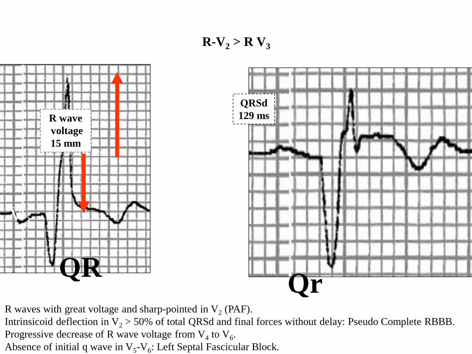
R-V2 > R V3
V3
R waves with great voltage and sharp-pointed in V2 (PAF).
Intrinsicoid deflection in V2 > 50% of total QRSd and final forces without delay: Pseudo Complete RBBB.
Progressive decrease of R wave voltage from V4 to V6.
Absence of initial q wave in V5-V6: Left Septal Fascicular Block.
R wave
voltage
15 mm
Qr
QRSd
129 ms
Y
aVF
Z
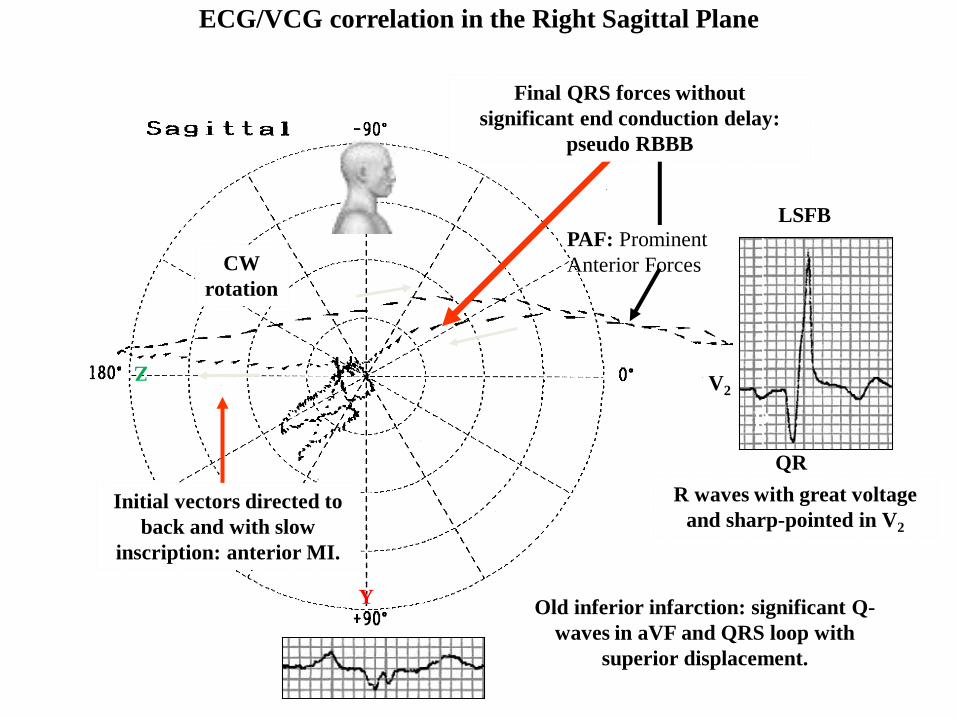
ECG/VCG correlation in the Right Sagittal Plane
V2
CW
rotation
T
P
R
Old inferior infarction: significant Q-
waves in aVF and QRS loop with
superior displacement.
QR
P
R waves with great voltage
and sharp-pointed in V2
PAF
LSFB
PAF: Prominent
Anterior Forces
Initial vectors directed to
back and with slow
inscription: anterior MI.
Final QRS forces without
significant end conduction delay:
pseudo RBBB
LAE
Y
Z
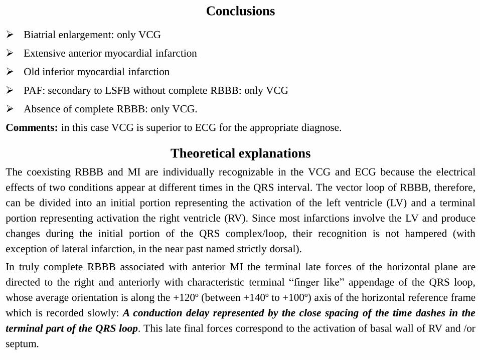
Conclusions
The coexisting RBBB and MI are individually recognizable in the VCG and ECG because the electrical
effects of two conditions appear at different times in the QRS interval. The vector loop of RBBB, therefore,
can be divided into an initial portion representing the activation of the left ventricle (LV) and a terminal
portion representing activation the right ventricle (RV). Since most infarctions involve the LV and produce
changes during the initial portion of the QRS complex/loop, their recognition is not hampered (with
exception of lateral infarction, in the near past named strictly dorsal).
In truly complete RBBB associated with anterior MI the terminal late forces of the horizontal plane are
directed to the right and anteriorly with characteristic terminal “finger like” appendage of the QRS loop,
whose average orientation is along the +120º (between +140º to +100º) axis of the horizontal reference frame
which is recorded slowly: A conduction delay represented by the close spacing of the time dashes in the
terminal part of the QRS loop. This late final forces correspond to the activation of basal wall of RV and /or
septum.
Theoretical explanations
Biatrial enlargement: only VCG
Extensive anterior myocardial infarction
Old inferior myocardial infarction
PAF: secondary to LSFB without complete RBBB: only VCG
Absence of complete RBBB: only VCG.
Comments: in this case VCG is superior to ECG for the appropriate diagnose.
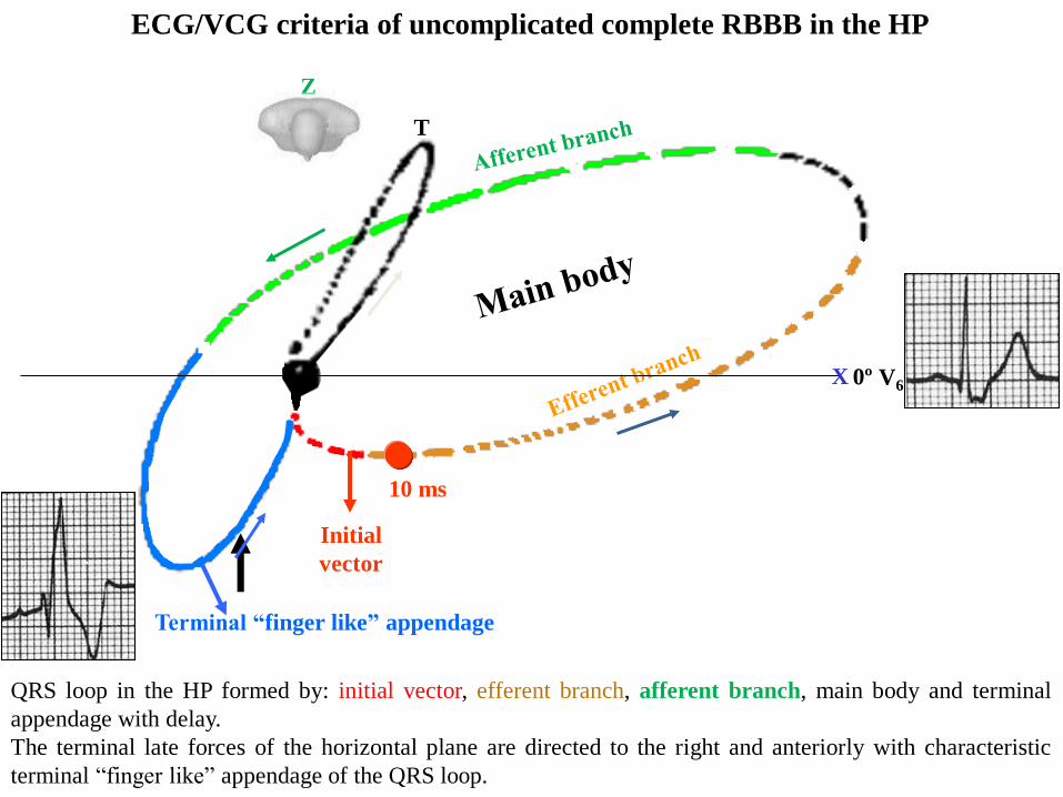
ECG/VCG criteria of uncomplicated complete RBBB in the HP
QRS loop in the HP formed by: initial vector, efferent branch, afferent branch, main body and terminal
appendage with delay.
The terminal late forces of the horizontal plane are directed to the right and anteriorly with characteristic
terminal “finger like” appendage of the QRS loop.
Initial
vector
10 ms
Terminal “finger like” appendage
T
X
Z
V60º
rSR’
V1
qRS
I
II
III
IIIIV
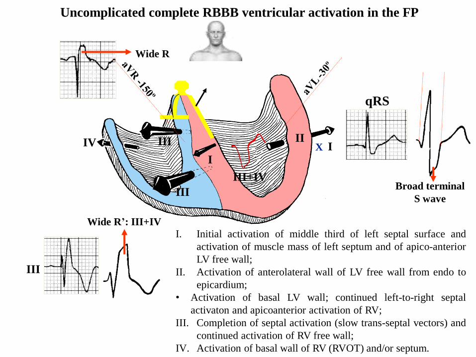
I. Initial activation of middle third of left septal surface and
activation of muscle mass of left septum and of apico-anterior
LV free wall;
II. Activation of anterolateral wall of LV free wall from endo to
epicardium;
• Activation of basal LV wall; continued left-to-right septal
activaton and apicoanterior activation of RV;
III. Completion of septal activation (slow trans-septal vectors) and
continued activation of RV free wall;
IV. Activation of basal wall of RV (RVOT) and/or septum.
aVR
QR
Broad terminal
S wave
Wide R’: III+IV
Wide R
120 ms
I
II
III+IV
II
A-V NLB
III+IV
Uncomplicated complete RBBB ventricular activation in the FP
XIV I
III
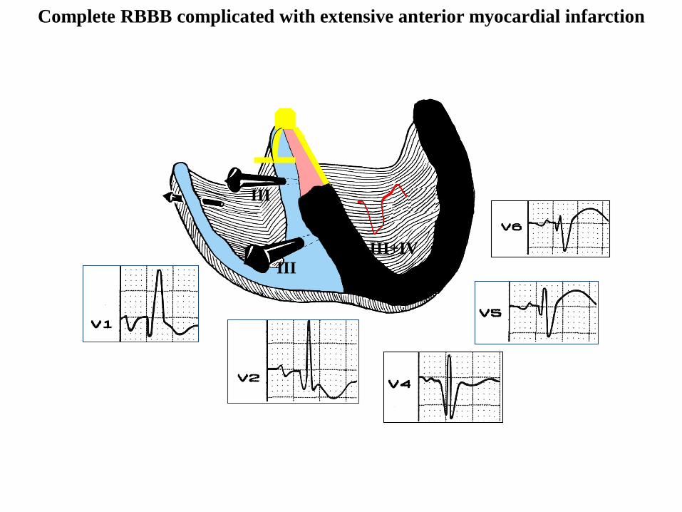
Complete RBBB complicated with extensive anterior myocardial infarction
I
II
III
IIIIV
QR
III+IV
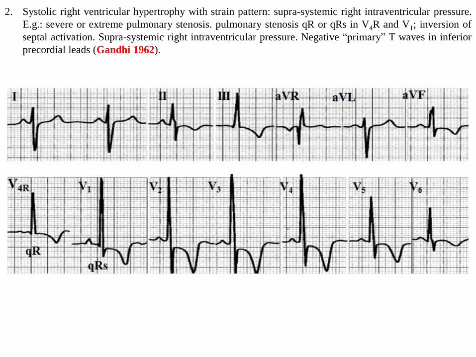
2. Systolic right ventricular hypertrophy with strain pattern: supra-systemic right intraventricular pressure.
E.g.: severe or extreme pulmonary stenosis. pulmonary stenosis qR or qRs in V4R and V1; inversion of
septal activation. Supra-systemic right intraventricular pressure. Negative “primary” T waves in inferior
precordial leads (Gandhi 1962).
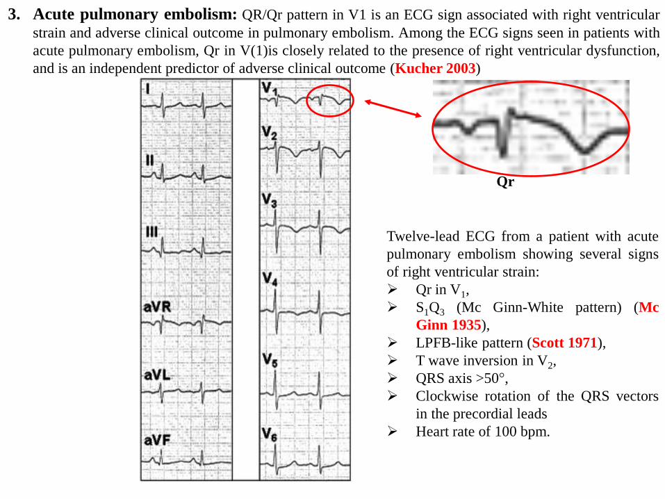
3. Acute pulmonary embolism: QR/Qr pattern in V1 is an ECG sign associated with right ventricular
strain and adverse clinical outcome in pulmonary embolism. Among the ECG signs seen in patients with
acute pulmonary embolism, Qr in V(1)is closely related to the presence of right ventricular dysfunction,
and is an independent predictor of adverse clinical outcome (Kucher 2003)
Twelve-lead ECG from a patient with acute
pulmonary embolism showing several signs
of right ventricular strain:
Qr in V1,
S1Q3 (Mc Ginn-White pattern) (Mc
Ginn 1935),
LPFB-like pattern (Scott 1971),
T wave inversion in V2,
QRS axis >50°,
Clockwise rotation of the QRS vectors
in the precordial leads
Heart rate of 100 bpm.
Qr
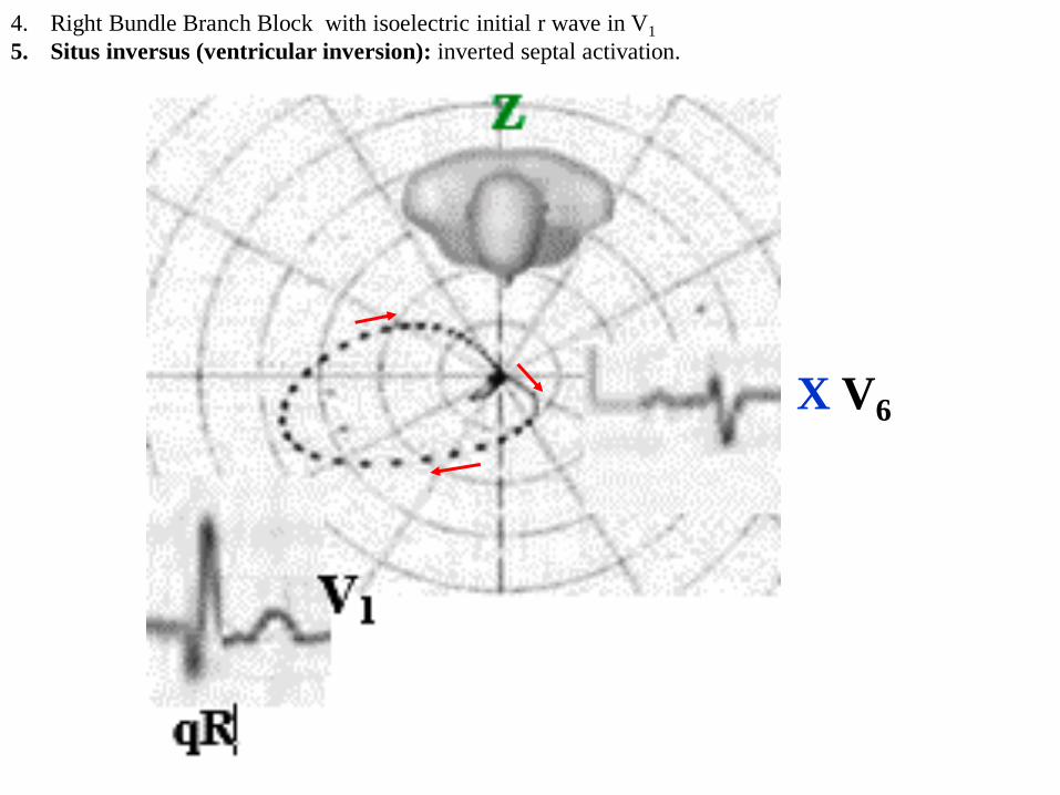
4. Right Bundle Branch Block with isoelectric initial r wave in V1
5. Situs inversus (ventricular inversion): inverted septal activation.
X V6
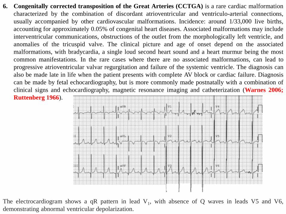
The electrocardiogram shows a qR pattern in lead V1, with absence of Q waves in leads V5 and V6,
demonstrating abnormal ventricular depolarization.
6. Congenitally corrected transposition of the Great Arteries (CCTGA) is a rare cardiac malformation
characterized by the combination of discordant atrioventricular and ventriculo-arterial connections,
usually accompanied by other cardiovascular malformations. Incidence: around 1/33,000 live births,
accounting for approximately 0.05% of congenital heart diseases. Associated malformations may include
interventricular communications, obstructions of the outlet from the morphologically left ventricle, and
anomalies of the tricuspid valve. The clinical picture and age of onset depend on the associated
malformations, with bradycardia, a single loud second heart sound and a heart murmur being the most
common manifestations. In the rare cases where there are no associated malformations, can lead to
progressive atrioventricular valvar regurgitation and failure of the systemic ventricle. The diagnosis can
also be made late in life when the patient presents with complete AV block or cardiac failure. Diagnosis
can be made by fetal echocardiography, but is more commonly made postnatally with a combination of
clinical signs and echocardiography, magnetic resonance imaging and catheterization (Warnes 2006;
Ruttenberg 1966).
7. Endomyocardiofibrosis (Tobias 1992).
8. Pectus excavatum
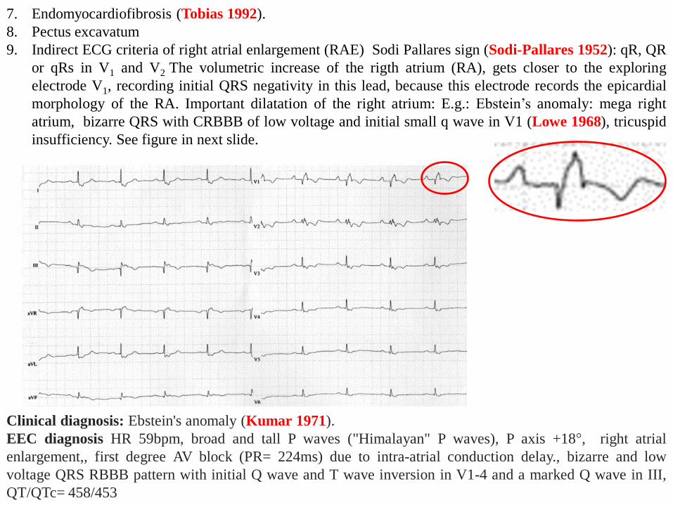
9. Indirect ECG criteria of right atrial enlargement (RAE) Sodi Pallares sign (Sodi-Pallares 1952): qR, QR
or qRs in V1 and V2 The volumetric increase of the rigth atrium (RA), gets closer to the exploring
electrode V1, recording initial QRS negativity in this lead, because this electrode records the epicardial
morphology of the RA. Important dilatation of the right atrium: E.g.: Ebstein’s anomaly: mega right
atrium, bizarre QRS with CRBBB of low voltage and initial small q wave in V1 (Lowe 1968), tricuspid
insufficiency. See figure in next slide.
Clinical diagnosis: Ebstein's anomaly (Kumar 1971).
EEC diagnosis HR 59bpm, broad and tall P waves ("Himalayan" P waves), P axis +18°, right atrial
enlargement,, first degree AV block (PR= 224ms) due to intra-atrial conduction delay., bizarre and low
voltage QRS RBBB pattern with initial Q wave and T wave inversion in V1-4 and a marked Q wave in III,
QT/QTc= 458/453
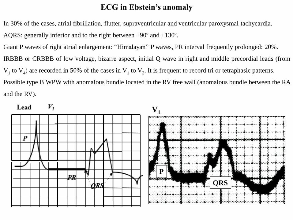
ECG in Ebstein’s anomaly
In 30% of the cases, atrial fibrillation, flutter, supraventricular and ventricular paroxysmal tachycardia.
AQRS: generally inferior and to the right between +90º and +130º.
Giant P waves of right atrial enlargement: “Himalayan” P waves, PR interval frequently prolonged: 20%.
IRBBB or CRBBB of low voltage, bizarre aspect, initial Q wave in right and middle precordial leads (from
V1 to V4) are recorded in 50% of the cases in V1 to V3. It is frequent to record tri or tetraphasic patterns.
Possible type B WPW with anomalous bundle located in the RV free wall (anomalous bundle between the RA
and the RV).
V1
P
QRS
RAE
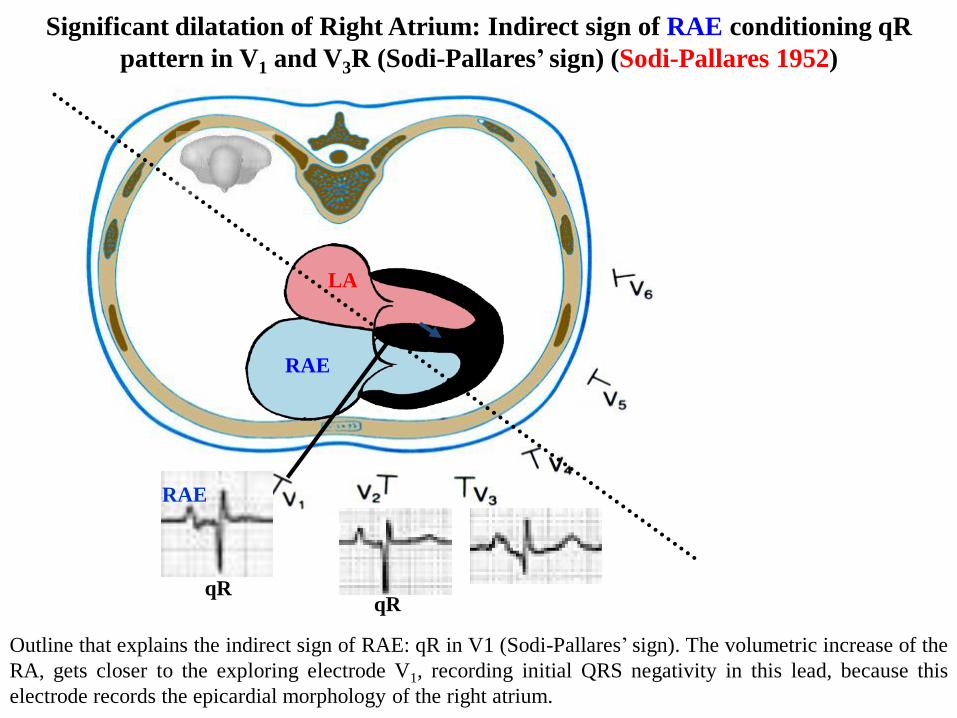
Significant dilatation of Right Atrium: Indirect sign of RAE conditioning qR
pattern in V1 and V3R (Sodi-Pallares’ sign) (Sodi-Pallares 1952)
Outline that explains the indirect sign of RAE: qR in V1 (Sodi-Pallares’ sign). The volumetric increase of the
RA, gets closer to the exploring electrode V1, recording initial QRS negativity in this lead, because this
electrode records the epicardial morphology of the right atrium.
LA
qR
RAE
qR
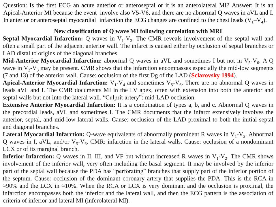
Question: Is the first ECG an acute anterior or anteroseptal or it is an anterolateral MI? Answer: It is an
Apical-Anterior MI because the event involve also V5-V6, and there are no abnormal Q waves in aVL and I.
In anterior or anteroseptal myocardial infarction the ECG changes are confined to the chest leads (V1–V4).
New classification of Q wave MI following correlation with MRI
Septal Myocardial Infarction: Q waves in V1-V2. The CMR reveals involvement of the septal wall and
often a small part of the adjacent anterior wall. The infarct is caused either by occlusion of septal branches or
LAD distal to origins of the diagonal branches.
Mid-Anterior Myocardial Infarction: abnormal Q waves in aVL and sometimes I but not in V5-V6. A Q
wave in V2-V3 may be present. CMR shows that the infarction encompasses especially the mid-low segments
(7 and 13) of the anterior wall. Cause: occlusion of the first Dg of the LAD (Sclarovsky 1994).
Apical-Anterior Myocardial Infarction: V1-V4 and sometimes V5-V6. There are no abnormal Q waves in
leads aVL and I. The CMR documents MI in the LV apex, often with extension into both the anterior and
septal walls but not into the lateral wall. “Culprit artery”: mid-LAD occlusion.
Extensive Anterior Myocardial Infarction: It is a combination of types a, b, and c. Abnormal Q waves in
the precordial leads, aVL and sometimes I. The CMR documents that the infarct extensively involves the
anterior, septal, and mid-low lateral walls. Cause: occlusion of the LAD proximal to both the initial septal
and diagonal branches.
Lateral Myocardial Infarction: Q-wave equivalents of abnormally prominent R waves in V1-V2. Abnormal
Q waves in I, aVL, and/or V5-V6. CMR: infarction in the lateral walls. Cause: occlusion of a nondominant
LCX or of its marginal branch.
Inferior Infarction: Q waves in II, III, and VF but without increased R waves in V1-V2. The CMR shows
involvement of the inferior wall, very often including the basal segment. It may be involved by the inferior
part of the septal wall because the PDA has “perforating” branches that supply part of the inferior portion of
the septum. Cause: occlusion of the dominant coronary artery that supplies the PDA. This is the RCA in
≈90% and the LCX in ≈10%. When the RCA or LCX is very dominant and the occlusion is proximal, the
infarction encompasses both the inferior and the lateral wall, and then the ECG pattern is the association of
criteria of inferior and lateral MI (inferolateral MI).
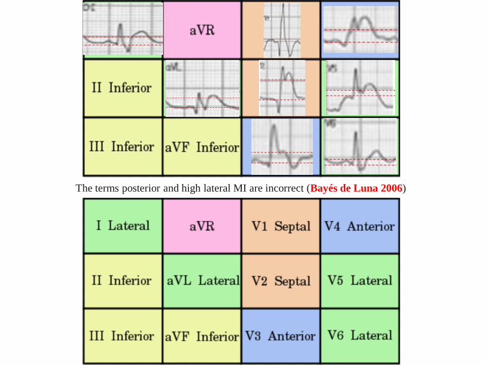
The terms posterior and high lateral MI are incorrect (Bayés de Luna 2006)
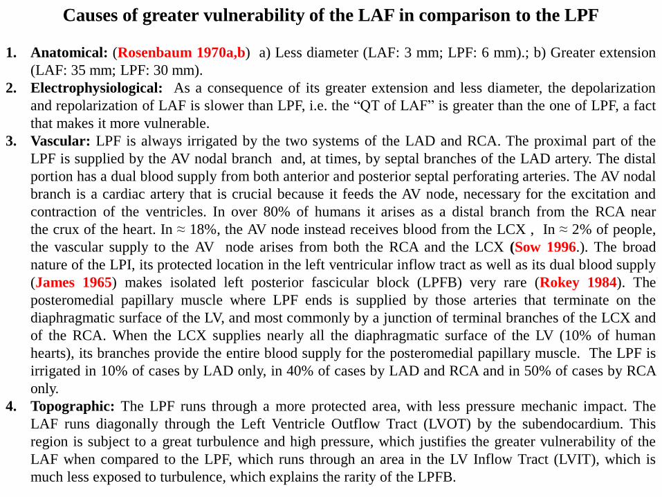
Causes of greater vulnerability of the LAF in comparison to the LPF
1. Anatomical: (Rosenbaum 1970a,b) a) Less diameter (LAF: 3 mm; LPF: 6 mm).; b) Greater extension
(LAF: 35 mm; LPF: 30 mm).
2. Electrophysiological: As a consequence of its greater extension and less diameter, the depolarization
and repolarization of LAF is slower than LPF, i.e. the “QT of LAF” is greater than the one of LPF, a fact
that makes it more vulnerable.
3. Vascular: LPF is always irrigated by the two systems of the LAD and RCA. The proximal part of the
LPF is supplied by the AV nodal branch and, at times, by septal branches of the LAD artery. The distal
portion has a dual blood supply from both anterior and posterior septal perforating arteries. The AV nodal
branch is a cardiac artery that is crucial because it feeds the AV node, necessary for the excitation and
contraction of the ventricles. In over 80% of humans it arises as a distal branch from the RCA near
the crux of the heart. In ≈ 18%, the AV node instead receives blood from the LCX , In ≈ 2% of people,
the vascular supply to the AV node arises from both the RCA and the LCX (Sow 1996.). The broad
nature of the LPI, its protected location in the left ventricular inflow tract as well as its dual blood supply
(James 1965) makes isolated left posterior fascicular block (LPFB) very rare (Rokey 1984). The
posteromedial papillary muscle where LPF ends is supplied by those arteries that terminate on the
diaphragmatic surface of the LV, and most commonly by a junction of terminal branches of the LCX and
of the RCA. When the LCX supplies nearly all the diaphragmatic surface of the LV (10% of human
hearts), its branches provide the entire blood supply for the posteromedial papillary muscle. The LPF is
irrigated in 10% of cases by LAD only, in 40% of cases by LAD and RCA and in 50% of cases by RCA
only.
4. Topographic: The LPF runs through a more protected area, with less pressure mechanic impact. The
LAF runs diagonally through the Left Ventricle Outflow Tract (LVOT) by the subendocardium. This
region is subject to a great turbulence and high pressure, which justifies the greater vulnerability of the
LAF when compared to the LPF, which runs through an area in the LV Inflow Tract (LVIT), which is
much less exposed to turbulence, which explains the rarity of the LPFB.
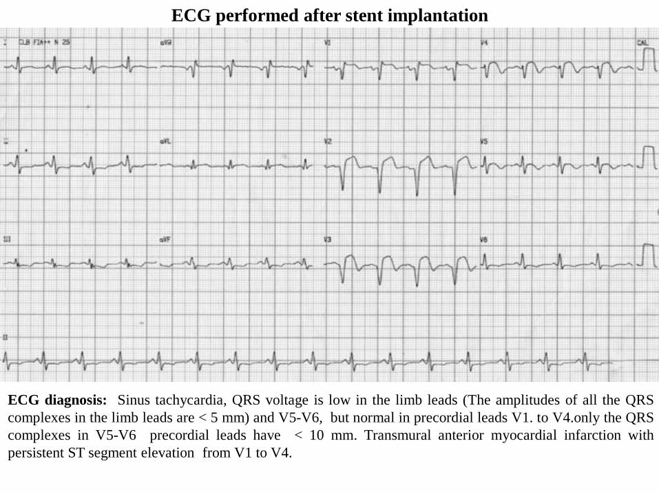
ECG performed after stent implantation
ECG diagnosis: Sinus tachycardia, QRS voltage is low in the limb leads (The amplitudes of all the QRS
complexes in the limb leads are < 5 mm) and V5-V6, but normal in precordial leads V1. to V4.only the QRS
complexes in V5-V6 precordial leads have < 10 mm. Transmural anterior myocardial infarction with
persistent ST segment elevation from V1 to V4.
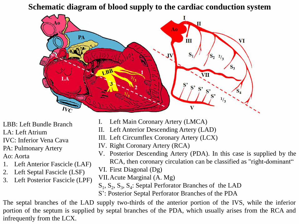
Schematic diagram of blood supply to the cardiac conduction system
I. Left Main Coronary Artery (LMCA)
II. Left Anterior Descending Artery (LAD)
III. Left Circumflex Coronary Artery (LCX)
IV. Right Coronary Artery (RCA)
V. Posterior Descending Artery (PDA). In this case is supplied by the
RCA, then coronary circulation can be classified as "right-dominant“
VI. First Diagonal (Dg)
VII.Acute Marginal (A. Mg)
S1, S2, S3, S4: Septal Perforator Branches of the LAD
S’: Posterior Septal Perforator Branches of the PDA
LBB: Left Bundle Branch
LA: Left Atrium
IVC: Inferior Vena Cava
PA: Pulmonary Artery
Ao: Aorta
1. Left Anterior Fascicle (LAF)
2. Left Septal Fascicle (LSF)
3. Left Posterior Fascicle (LPF)
The septal branches of the LAD supply two-thirds of the anterior portion of the IVS, while the inferior
portion of the septum is supplied by septal branches of the PDA, which usually arises from the RCA and
infrequently from the LCX.
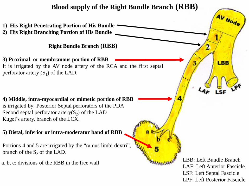
1) His Right Penetrating Portion of His Bundle
2) His Right Branching Portion of His Bundle
Right Bundle Branch (RBB)
3) Proximal or membranous portion of RBB
It is irrigated by the AV node artery of the RCA and the first septal
perforator artery (S1) of the LAD.
4) Middle, intra-myocardial or mimetic portion of RBB
is irrigated by: Posterior Septal perforators of the PDA
Second septal perforator artery(S2) of the LAD
Kugel’s artery, branch of the LCX.
5) Distal, inferior or intra-moderator band of RBB
Portions 4 and 5 are irrigated by the “ramus limbi dextri”,
branch of the S2 of the LAD.
Blood supply of the Right Bundle Branch (RBB)
LBB: Left Bundle Branch
LAF: Left Anterior Fascicle
LSF: Left Septal Fascicle
LPF: Left Posterior Fascicle
a, b, c: divisions of the RBB in the free wall
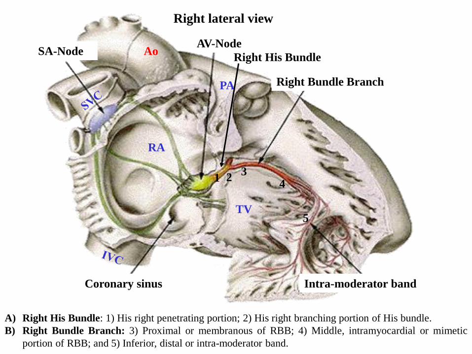
A) Right His Bundle: 1) His right penetrating portion; 2) His right branching portion of His bundle.
B) Right Bundle Branch: 3) Proximal or membranous of RBB; 4) Middle, intramyocardial or mimetic
portion of RBB; and 5) Inferior, distal or intra-moderator band.
Right Bundle Branch
Intra-moderator bandCoronary sinus
RA
TV
PA
Ao
Right lateral view
AV-NodeSA-Node
1 23
4
5
Right His Bundle
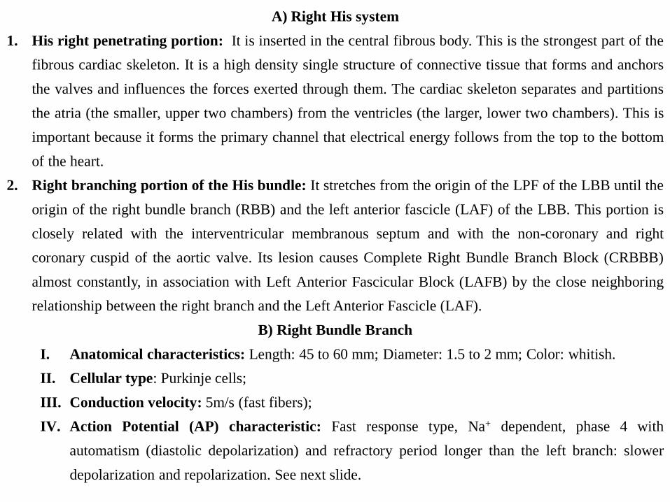
A) Right His system
1. His right penetrating portion: It is inserted in the central fibrous body. This is the strongest part of the
fibrous cardiac skeleton. It is a high density single structure of connective tissue that forms and anchors
the valves and influences the forces exerted through them. The cardiac skeleton separates and partitions
the atria (the smaller, upper two chambers) from the ventricles (the larger, lower two chambers). This is
important because it forms the primary channel that electrical energy follows from the top to the bottom
of the heart.
2. Right branching portion of the His bundle: It stretches from the origin of the LPF of the LBB until the
origin of the right bundle branch (RBB) and the left anterior fascicle (LAF) of the LBB. This portion is
closely related with the interventricular membranous septum and with the non-coronary and right
coronary cuspid of the aortic valve. Its lesion causes Complete Right Bundle Branch Block (CRBBB)
almost constantly, in association with Left Anterior Fascicular Block (LAFB) by the close neighboring
relationship between the right branch and the Left Anterior Fascicle (LAF).
B) Right Bundle Branch
I. Anatomical characteristics: Length: 45 to 60 mm; Diameter: 1.5 to 2 mm; Color: whitish.
II. Cellular type: Purkinje cells;
III. Conduction velocity: 5m/s (fast fibers);
IV. Action Potential (AP) characteristic: Fast response type, Na+ dependent, phase 4 with
automatism (diastolic depolarization) and refractory period longer than the left branch: slower
depolarization and repolarization. See next slide.
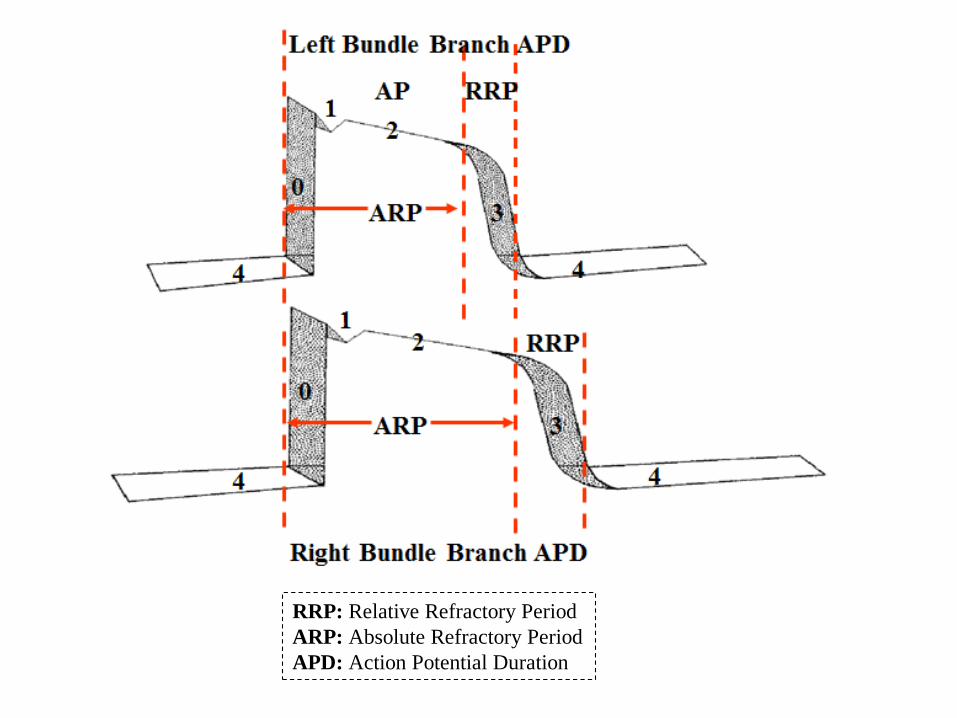
RRP: Relative Refractory Period
ARP: Absolute Refractory Period
APD: Action Potential Duration
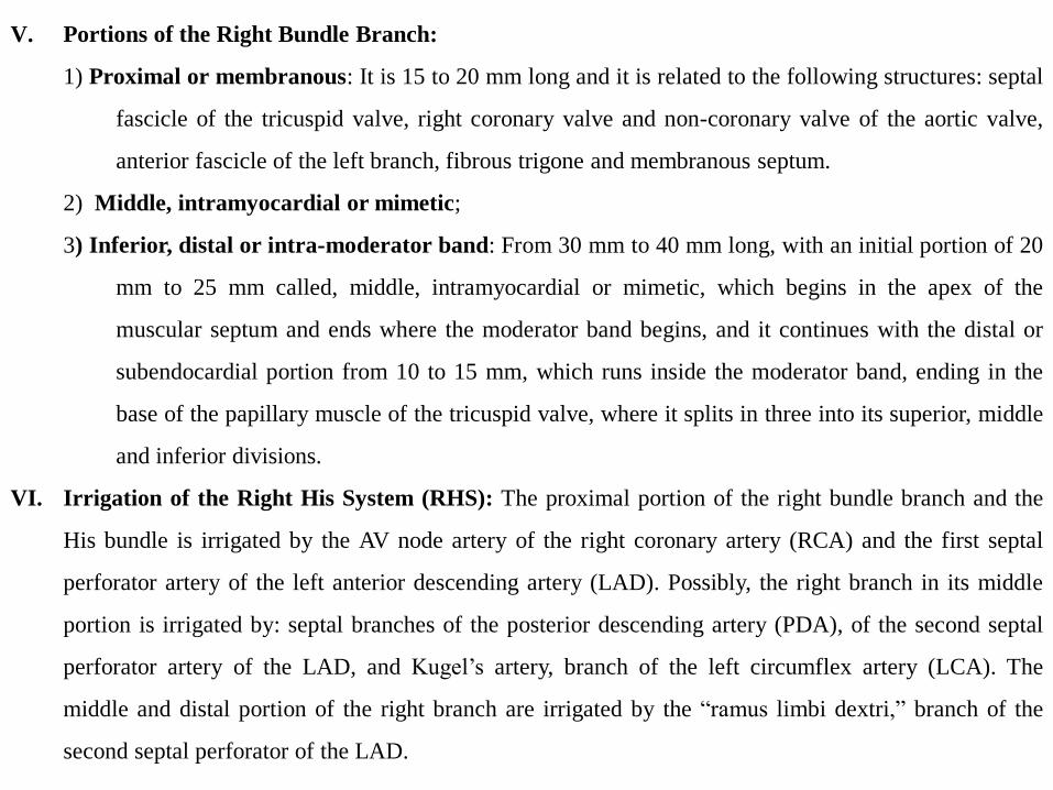
V. Portions of the Right Bundle Branch:
1) Proximal or membranous: It is 15 to 20 mm long and it is related to the following structures: septal
fascicle of the tricuspid valve, right coronary valve and non-coronary valve of the aortic valve,
anterior fascicle of the left branch, fibrous trigone and membranous septum.
2) Middle, intramyocardial or mimetic;
3) Inferior, distal or intra-moderator band: From 30 mm to 40 mm long, with an initial portion of 20
mm to 25 mm called, middle, intramyocardial or mimetic, which begins in the apex of the
muscular septum and ends where the moderator band begins, and it continues with the distal or
subendocardial portion from 10 to 15 mm, which runs inside the moderator band, ending in the
base of the papillary muscle of the tricuspid valve, where it splits in three into its superior, middle
and inferior divisions.
VI. Irrigation of the Right His System (RHS): The proximal portion of the right bundle branch and the
His bundle is irrigated by the AV node artery of the right coronary artery (RCA) and the first septal
perforator artery of the left anterior descending artery (LAD). Possibly, the right branch in its middle
portion is irrigated by: septal branches of the posterior descending artery (PDA), of the second septal
perforator artery of the LAD, and Kugel’s artery, branch of the left circumflex artery (LCA). The
middle and distal portion of the right branch are irrigated by the “ramus limbi dextri,” branch of the
second septal perforator of the LAD.
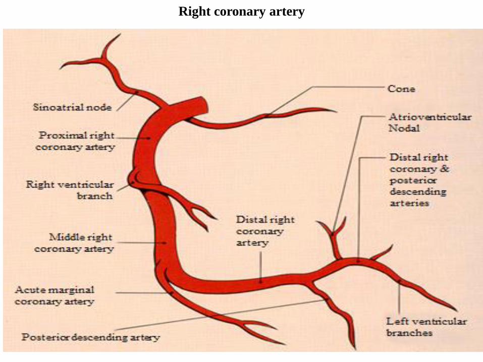
Right coronary artery
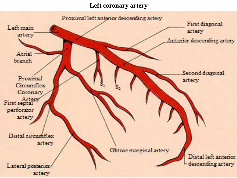
Left coronary artery
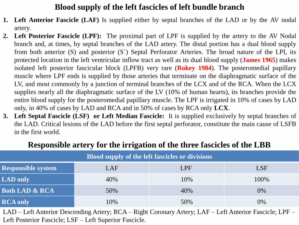
1. Left Anterior Fascicle (LAF) Is supplied either by septal branches of the LAD or by the AV nodal
artery.
2. Left Posterior Fascicle (LPF): The proximal part of LPF is supplied by the artery to the AV Nodal
branch and, at times, by septal branches of the LAD artery. The distal portion has a dual blood supply
from both anterior (S) and posterior (S`) Septal Perforator Arteries. The broad nature of the LPI, its
protected location in the left ventricular inflow tract as well as its dual blood supply (James 1965) makes
isolated left posterior fascicular block (LPFB) very rare (Rokey 1984). The posteromedial papillary
muscle where LPF ends is supplied by those arteries that terminate on the diaphragmatic surface of the
LV, and most commonly by a junction of terminal branches of the LCX and of the RCA. When the LCX
supplies nearly all the diaphragmatic surface of the LV (10% of human hearts), its branches provide the
entire blood supply for the posteromedial papillary muscle. The LPF is irrigated in 10% of cases by LAD
only, in 40% of cases by LAD and RCA and in 50% of cases by RCA only LCX.
3. Left Septal Fascicle (LSF) or Left Median Fascicle: It is supplied exclusively by septal branches of
the LAD. Critical lesions of the LAD before the first septal perforator, constitute the main cause of LSFB
in the first world.
Blood supply of the left fascicles of left bundle branch
Blood supply of the left fascicles or divisions
Responsible system LAF LPF LSF
LAD only 40% 10% 100%
Both LAD & RCA 50% 40% 0%
RCA only 10% 50% 0%
Responsible artery for the irrigation of the three fascicles of the LBB
LAD – Left Anterior Descending Artery; RCA – Right Coronary Artery; LAF – Left Anterior Fascicle; LPF –
Left Posterior Fascicle; LSF – Left Superior Fascicle.
References
1. Andersen MP, Terkelsen CJ, Sørensen JT, et al. The ST injury vector: electrocardiogram-based estimation
of location and extent of myocardial ischemia. J Electrocardiol. 2010;43(2):121-31.
2. Basualdo CA, Haraphongse M, Rossall RE. Intraventricular blocks in acute myocardial infarction. Chest.
1975; 67(1):75-8.
3. Bayés de Luna A, Wagner G, Birnbaum Y, et al. A new terminology for left ventricular walls and location
of myocardial infarcts that present Q wave based on the standard of cardiac magnetic resonance imaging:
a statement for healthcare professionals from a committee appointed by the International Society for
Holter and Noninvasive Electrocardiography. Circulation. 2006;114(16):1755-60.
4. Botvinick EH, Perez-Gonzalez JF, Dunn R, Ports T, Chatterjee K, Parmley W. Late prognostic value of
scintigraphic parameters of acute myocardial infarction size in complicated myocardial infarction
without heart failure. Am J Cardiol. 1983;51(7):1045-51.
5. Elizari MV, Acunzo RS, Ferreiro M. Hemiblocks revisited. Circulation. 2007;115(9):1154-63.
6. Gandhi MJ, Dattey KK, Kulkarni TP, Hansoti RC. Genesis of qR pattern in right precordial leads in right
ventricular overload. J Assoc Physicians India. 1962;10:217-23.
7. James TN. Anatomy of the coronary arteries in health and disease. Circulation. 1965;32(6):1020-33.
8. Kapoor J, McConnell M. A swinging heart. N Engl J Med. 2009;361(18):e 37.
9. Klein H, Di Segni E, Kaplinsky E. Procainamide-induced left anterior hemiblock of the 2:1 type
(pseudoelectrical alternans). Chest. 1978;74(2):230–3.
10. Kucher N, Walpoth N, Wustmann K, Noveanu M, Gertsch M. QR in V1--an ECG sign associated with
right ventricular strain and adverse clinical outcome in pulmonary embolism. Eur Heart J.
2003;24(12):1113-9.
11. Kumar AE, Fyler DC, Miettinen OS, Nadas AS. Ebstein's anomaly. Clinical profile and natural history.
Am J Cardiol. 1971;28(1):84-95.
12. Lowe KG, Emslie-Smith D, Robertson PG, et al. Scalar, Vector, and Intracardiac Electrocardiograms in
Ebstein's Anomaly. Br Heart J. 1968;30(5):617-29.
13. Marriott HJ, Hogan P. Hemiblock in acute myocardial infarction. Chest. 1970;58(4):342-4.
14. McGinn S, White PD. Acute cor pulmonale resulting from pulmonary embolism: its clinical recognition.
JAMA 1935;114:1473.
15. Rathore SS, Gersh BJ, Berger PB, et al. Acute myocardial infarction complicated by heart block in the
elderly: prevalence and outcomes. Am Heart J. 2001;141(1):47-54.
16. Rokey R, Chahine RA. Isolated left posterior fascicular block associated with acquired ventricular septal
defect.Clin Cardiol. 1984;7(6):364-9.
17. Rosenbaum, MB. The Hemiblocks: Diagnostic criteria and clinical significance. Mod. Concepts
Cardiovasc. Dis.1970;39(12):141-6.
18. Rosenbaum, MB, Elizari MV, Lazzari JO. The Hemiblocks. New Concepts of intraventricular
Conduction Based on Human Anatomical Physiological, and Clinical Studies. Oldsmar, Florida: Tampa
Tracings, 1970.
19. Rudiakov LaI. On The Diagnostic Significance Of The qR Type QRS Complex In Right
Electrocardiographic Leads. Kardiologiia. 1964;18:72-3.
20. Ruttenberg HD, Elliott LP, Anderson RC, Adams P Jr, Tuna N. Congenital corrected transposition of the
great vessels. Correlation of electrocardiograms and vector cardiograms with associated cardiac
malformations and hemodynamic states. Am J Cardiol. 1966; 17(3):339-54.
21. Sclarovsky S, Birnbaum Y, Solodky A, Zafrir N, Wurzel M, Rechavia E. Isolated mid-anterior
myocardial infarction: a special electrocardiographic sub-type of acute myocardial infarction consisting
of ST-elevation in non-consecutive leads and two different morphologic types of ST-depression. Int J
Cardiol. 1994; 46: 37–47.
22. Scott RC. The S1-Q3 (Mc Ginn-White) pattern in acute cor pulmonale: A form of transient left posterior
hemiblock? Am Heart J. 1971;82(1):135-7.
23. Sodi Pallares D, Bisteni A, Hermann GR. Some views on the significance of qR and QR type complexes
in right precordial leads in the absence of myocardial infarction. Am Heart J. 1952;43(5):716-34.
24. Sow ML, Ndoye JM, Lo EA. The artery of the atrioventricular node: an anatomic study based on 38
injection-dissections. Surg Radiol Anat 1996;18(3):183–7.
25. Tobias NM, Moffa PJ, Pastore CA, et al. The electrocardiogram in endomyocardial fibrosis. Arq Bras
Cardiol. 1992;59(4):249-53.
26. Warnes CA. Transposition of the great arteries. Circulation 2006;114(24):2699-709.

43rd International Congress on Electrocardiology Palma, Balearic Island, Spain - June, 4-6
2016
The registration process for the ICE2016 is actually running. Hurry up and register now, don´t miss this
opportunity to join this World class event.
Registration Deadline is just a few days away, by May 10th the process will be over.
The full set of activities programmed for this event, the scientific committee, speakers and guidelines are
available at the official ICE2016 website. Don´t hesitate to check it all ! Where? The ICE 2016 will take
place in Palma de Mallorca, Balearic Islands, Spain.
Mallorca is well known for its beaches and coves, but is also a perfect destination to enjoy countryside, golf,
culture, water sports, and entertainment... Registration process is very simple, and there are special fees for
students, SEC Members or visitors.For further info: AGA TRAVEL Paseo mallorca, 11 07011, Palma de
Mallorca, Spain +34 971 222 292 [email protected]