Embed Size (px)
Citation preview
In India, quality surveillance for acute encephalitis syn-drome (AES), including laboratory testing, is necessary for understanding the epidemiology and etiology of AES, plan-ning interventions, and developing policy. We reviewed AES surveillance data for January 2011–June 2012 from Kushi-nagar District, Uttar Pradesh, India. Data were cleaned, in-cidence was determined, and demographic characteristics of cases and data quality were analyzed. A total of 812 AES case records were identified, of which 23% had illogical en-tries. AES incidence was highest among boys <6 years of age, and cases peaked during monsoon season. Records for laboratory results (available for Japanese encephalitis but not AES) and vaccination history were largely incom-plete, so inferences about the epidemiology and etiology of AES could not be made. The low-quality AES/Japanese encephalitis surveillance data in this area provide little evi-dence to support development of prevention and control measures, estimate the effect of interventions, and avoid the waste of public health resources.
Acute encephalitis syndrome (AES) is a clinical condi-tion caused by infection with Japanese encephalitis
virus (JEV) or other infectious and noninfectious causes. A confirmed etiology is generally not required for the clinical management of AES. Thus, surveillance for JEV infection in India has focused on identifying AES cases rather than JE cases; this approach is more feasible given
the limitations of public health resources (1). However, identification of the etiologic agent is necessary for plan-ning relevant interventions. The standard for determining the etiology of AES is examination of cerebrospinal fluid (CSF) during the acute phase of illness; pathogen-specific IgM capture ELISA or nucleic acid amplification tech-niques are used to detect pathogens in the CSF. Serologic tests for pathogen-specific antibodies and virus detection in serum are also recommended. However, examination of CSF is preferred because serologic test results may indi-cate the presence of antibodies in the serum, but the AES may have a cause different than the agent producing the detected antibodies (1–3).
A good quality surveillance system with laboratory support is essential for understanding the causes of AES and responding appropriately. Accordingly, the National Vector Borne Diseases Control Programme in New Del-hi, India, has developed guidelines for AES surveillance that promote the need for a strong surveillance system as a critical component for any control activities. In these guidelines, the goals outlined for AES surveillance are to 1) assess and characterize the burden of JE, 2) detect early warning signals for an outbreak, 3) assess the ef-fect of vaccination, and 4) guide future strategies (1). The National Vector Borne Diseases Control Programme has also implemented several measures to strengthen local health systems, including building on the capacity of the health workforce to provide better clinical management, extending referral diagnostic facilities by upgrading the existing Baba Raghav Das (BRD) Medical College facili-ties and setting up a National Institute of Virology field unit; and establishing a dedicated surveillance unit in the Department of Preventive and Social Medicine at BRD Medical College to provide improved surveillance and outbreak responses (4).
Acute Encephalitis Syndrome Surveillance, Kushinagar District, Uttar Pradesh, India, 2011–2012
Manish Kakkar, Elizabeth T. Rogawski, Syed Shahid Abbas, Sanjay Chaturvedi, Tapan N. Dhole, Shaikh Shah Hossain, and Sampath K. Krishnan
Emerging Infectious Diseases • www.cdc.gov/eid • Vol. 19, No. 9, September 2013 Emerging Infectious Diseases • www.cdc.gov/eid • Vol. 19, No. 9, September 2013 1361
Author affiliations: Public Health Foundation of India, New Delhi, India (M. Kakkar, E.T. Rogawski, S.S. Abbas); University College of Medical Sciences, New Delhi (S. Chaturvedi); Sanjay Gandhi Postgraduate Institute of Medical Sciences, Lucknow, India (T.N. Dhole); Centers for Disease Control and Prevention, New Delhi (S.S. Hossain); and Office of the World Health Organization Repre-sentative to India, New Delhi (S.K. Krishnan)
DOI: http://dx.doi.org/10.3201/eid1909.121855
PERSPECTIVE
From the 1970s until around 2010, JEV infection was considered to be the leading cause of AES in the traditional JE belt of India, which includes Kushinagar District in the state of Uttar Pradesh (5–11). However, because of a large number of JE cases of unknown etiology, AES patterns alone have not suggested a clear picture of the epidemiol-ogy of the disease. In recent years, despite of the introduc-tion of a JE vaccine, an increased number of AES cases have been reported in India, including Uttar Pradesh, and the disease has spread to new districts, urban areas, and villages without pigs, which are not usually associated with JE transmission (12,13). Thus, the assertion that JEV is the leading cause of AES has been questioned, and other infec-tious agents, such as enteroviruses, have been reported as a cause of AES in Uttar Pradesh and other parts of India (14–19). A substantial contributor to the ambiguity about the etiology of AES could be the fact that surveillance data for AES have not been analyzed to assess reasons for the increased cases and other reported causes. We examined the completeness and quality of AES surveillance data from Kushinagar District, an area where JEV is highly en-demic. Herein, we discuss the ability to make inferences about AES epidemiology and etiology from these data and the implications of our findings for policy planning and program implementation.
Methods
Source of Surveillance DataThe sentinel site for JE surveillance in Kushinagar
District is the district hospital. Using a standard format, the hospital reports all AES/JE cases that meet the standard World Health Organization case definition (3) to the district malaria officer; this officer then forwards compiled data to the state program officer for transmission to National Vec-tor Borne Disease Control Programme (http://nvbdcp.gov.in/Doc/AES%20guidelines.pdf). BRD Medical College in Gorakhpur, Uttar Pradesh, is a nearby regional field labo-ratory of the Indian Council of Medical Research, which receives clinical samples from patients admitted to the dis-trict hospital in Kushinagar and completes most laboratory testing for JEV infections. In addition, BRD Medical Col-lege serves as a tertiary care center for patients who directly seek medical care or who are referred from nearby districts, including Kushinagar. A line list with laboratory results of these patients is reported by BRD Medical College to the district malaria officer in Kushinagar for submission to state program officers.
In July 2012, we obtained the AES line lists data for January 2011–June 2012 that were submitted by BRD Medical College to Kushinagar District Headquarters in Padrauna. The list was originally to be used to identify blocks in Kushinagar District with high, medium, and
low numbers of AES cases so that sites could be select-ed for a larger study of the drivers of JEV transmission. Cases recorded in the line lists represented case-patients from Kushinagar District who were 1) admitted directly to BRD Medical College, 2) referred to BRD Medical College by the district hospital in Padrauna, and 3) admit-ted to the district hospital in Padrauna but had serum sam-ples referred to BRD Medical College because no diagnos-tic kits were available and laboratory testing was limited at the district hospital. An individual AES patient tracking system does not exist in the state of Uttar Pradesh, so pa-tients treated in the private sector or at the district hospital in Gorakhpur were not included in the surveillance lists.
Preparation of Surveillance Database for AnalysisIndividual identifiers for the case-patients were re-
moved from the line lists to ensure confidentiality. Data were cleaned (i.e., extraneous data were removed) in mul-tiple steps, resulting in 4 changes being made to the AES line lists. First, the spelling of residential localities (block, village, or police station) were matched and standardized to result in a list of 32 residential areas. Residential areas were then categorized and combined by block (14 blocks in Kushinagar). Modifications were validated by cross-checking with village population lists provided by Sav-era, a local nongovernmental organization. Second, all age values were standardized to a uniform decimal sys-tem (e.g., 1 year and 6 months was changed to 1.5 years, and 8 months was changed to 0.67 year). Third, the dates of symptom onset, hospital admission, sample collection, and outcome (i.e., discharged, left against medical advice, or death) were standardized to 1 format. In the original list, dates were variously coded by using different nota-tions (e.g., 4 March 2011 was mentioned within the same row as both 04/03/11 and 03/04/11). Assuming that the case data were entered in chronological order, we stan-dardized dates by using, as a cue, the dates of the preced-ing and following AES cases. Fourth, case-patients who died, were absent, or left against medical advice were considered to have been discharged.
Data AnalysisCleaned data were imported into SAS version 9.2.2
(SAS Institute, Inc., Cary, NC, USA) for analysis. AES cases were plotted by week and month by using the re-corded dates of symptom onset. We tabulated demographic characteristics of AES case-patients for 2011 and 2012 and stratified case-patients by block, vaccination status, laboratory test result for JEV infection, and clinical out-come. Incidence for 2011 was calculated by using popula-tion denominators from the 2011 Census of India (20). We calculated incidence overall, by sex, and for children 0–6 years of age by using the age stratification available in the
1362 Emerging Infectious Diseases • www.cdc.gov/eid • Vol. 19, No. 9, September 2013
Acute Encephalitis Syndrome Surveillance, India
district-level census data. We estimated the incidence for each block by using block-specific population denomina-tors projected with the decadal growth rate from the 2001 Census of India (20). Crude incidence rate ratios were esti-mated with Wald-based CIs.
We also determined the median number of days be-tween key points in AES disease progression and diagnosis: time between onset of symptoms and hospital admission, onset of symptoms and serum sample collection, hospital admission and serum sample collection, and hospital ad-mission and discharge or death. Using previously described methods (21), we evaluated the quality of the surveillance data by assessing the amount of data cleaning required, the proportion of missing or incomplete values in line list fields, and inconsistencies in dates recorded for key points in AES disease progression and diagnosis.
Results
AES EpidemiologyIn 2011, a total of 721 AES cases from Kushinagar
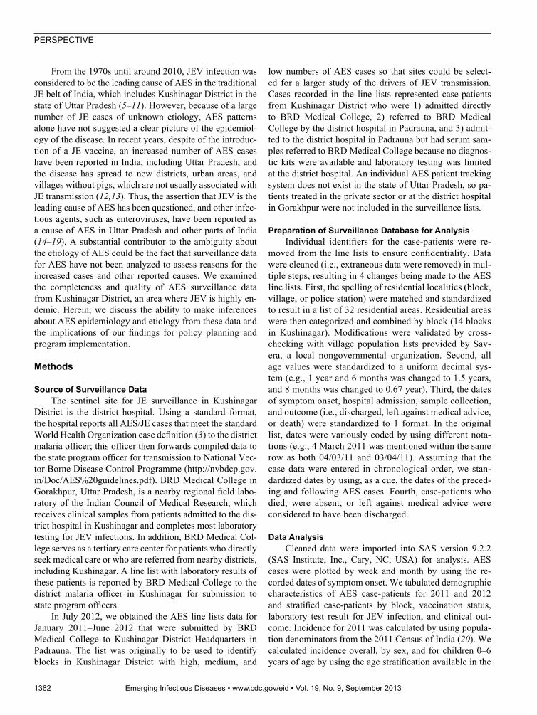
District were identified through BRD Medical College; in 2012 (January–June), 91 cases were identified. Using the cleaned line lists, we determined the weekly number of AES cases reported during January 2011–June 2012 (Fig-ure 1). Cases peaked during August–October 2011; >150 cases were identified in each of these 3 months. This sea-sonal trend corresponds with an expected increase in cases during the monsoon season, when transmission of both wa-terborne and vector-borne diseases increases (vector den-sity is at its maximum).
In 2011 and 2012, most case-patients were male (57.4% and 59.3%, respectively) (Table 1). In 2011, al-most half of the AES cases were in children <5 years of age (44.7%); the distribution of cases by age group was not substantially different in 2012. The case-fatality rate was 18.0% in 2011 and 19.8% in 2012.
Using 2011 population data (20), we estimated that there were 20.2 AES cases/100,000 population in Kushi-nagar District in 2011 (Table 2). The incidence was higher among male residents than female residents (incidence rate ratio 1.29, 95% CI 1.11–1.49), and it was highest among children 0–6 years of age. The crude incidence rate ratio, comparing case-patients 0–6 years of age with those >6 years of age, was 7.97 (95% CI 6.87–9.25). Boys 0–6 years of age were at highest risk for AES. The incidence among 0- to 6-year-old boys was almost 50% greater than that among girls of the same age.
Emerging Infectious Diseases • www.cdc.gov/eid • Vol. 19, No. 9, September 2013 1363
Figure 1. Weekly number of acute encephalitis syndrome cases, by month, in Kushinagar District, Uttar Pradesh State, India, 2011–2012. Numbers are based on data obtained from Baba Raghav Das Medical College, Gorakhpur, Uttar Pradesh, India.
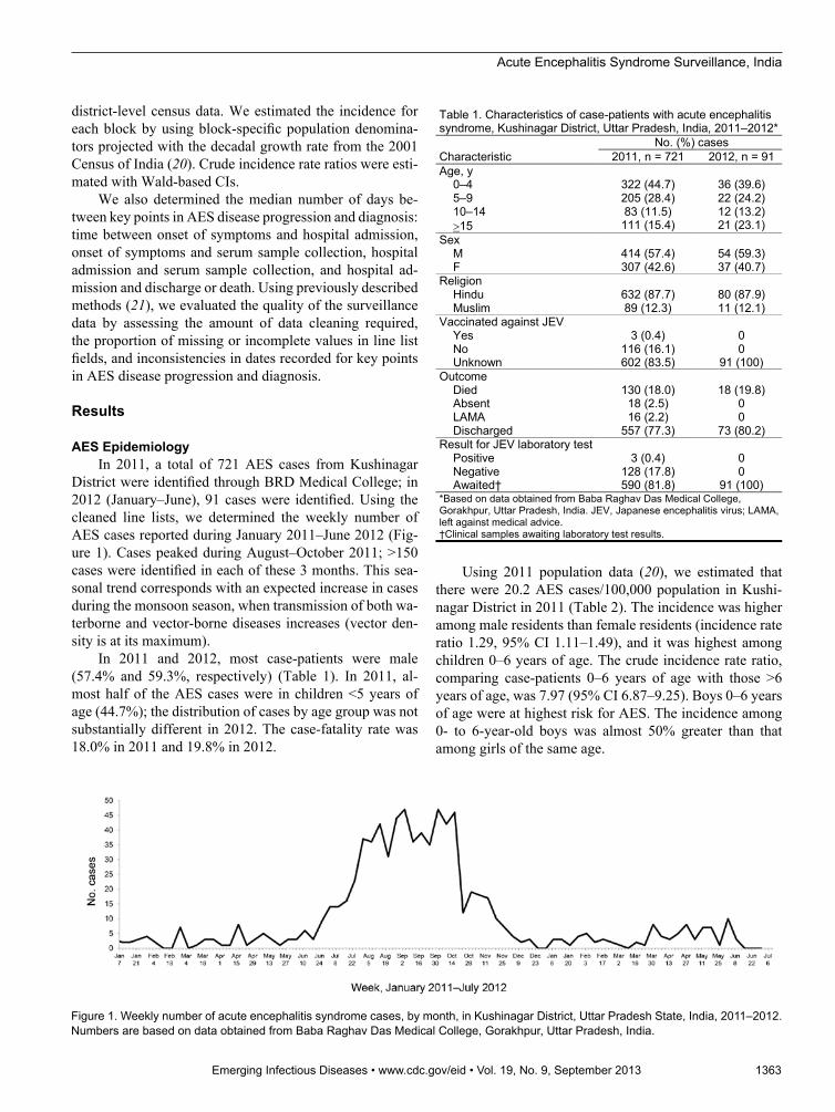
Table 1. Characteristics of case-patients with acute encephalitis syndrome, Kushinagar District, Uttar Pradesh, India, 2011–2012*
Characteristic No. (%) cases
2011, n = 721 2012, n = 91 Age, y 0–4 322 (44.7) 36 (39.6) 5–9 205 (28.4) 22 (24.2) 10–14 83 (11.5) 12 (13.2) 15 111 (15.4) 21 (23.1) Sex M 414 (57.4) 54 (59.3) F 307 (42.6) 37 (40.7) Religion Hindu 632 (87.7) 80 (87.9) Muslim 89 (12.3) 11 (12.1) Vaccinated against JEV Yes 3 (0.4) 0 No 116 (16.1) 0 Unknown 602 (83.5) 91 (100) Outcome Died 130 (18.0) 18 (19.8) Absent 18 (2.5) 0 LAMA 16 (2.2) 0 Discharged 557 (77.3) 73 (80.2) Result for JEV laboratory test Positive 3 (0.4) 0 Negative 128 (17.8) 0 Awaited† 590 (81.8) 91 (100) *Based on data obtained from Baba Raghav Das Medical College, Gorakhpur, Uttar Pradesh, India. JEV, Japanese encephalitis virus; LAMA, left against medical advice. †Clinical samples awaiting laboratory test results.
PERSPECTIVE
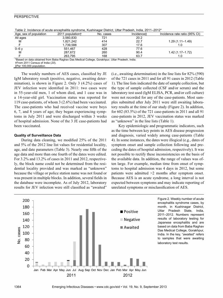
The weekly numbers of AES cases, classified by JE IgM laboratory result (positive, negative, awaiting deter-mination), is shown in Figure 2. Only 3 (4.2%) cases of JEV infection were identified in 2011: two cases were in 55-year-old men, 1 of whom died, and 1 case was in a 14-year-old girl. Vaccination status was reported for 119 case-patients, of whom 3 (2.6%) had been vaccinated. The case-patients who had received vaccine were boys 6, 7, and 8 years of age; they began experiencing symp-toms in July 2011 and were discharged within 3 weeks of hospital admission. None of the 3 JE case-patients had been vaccinated.
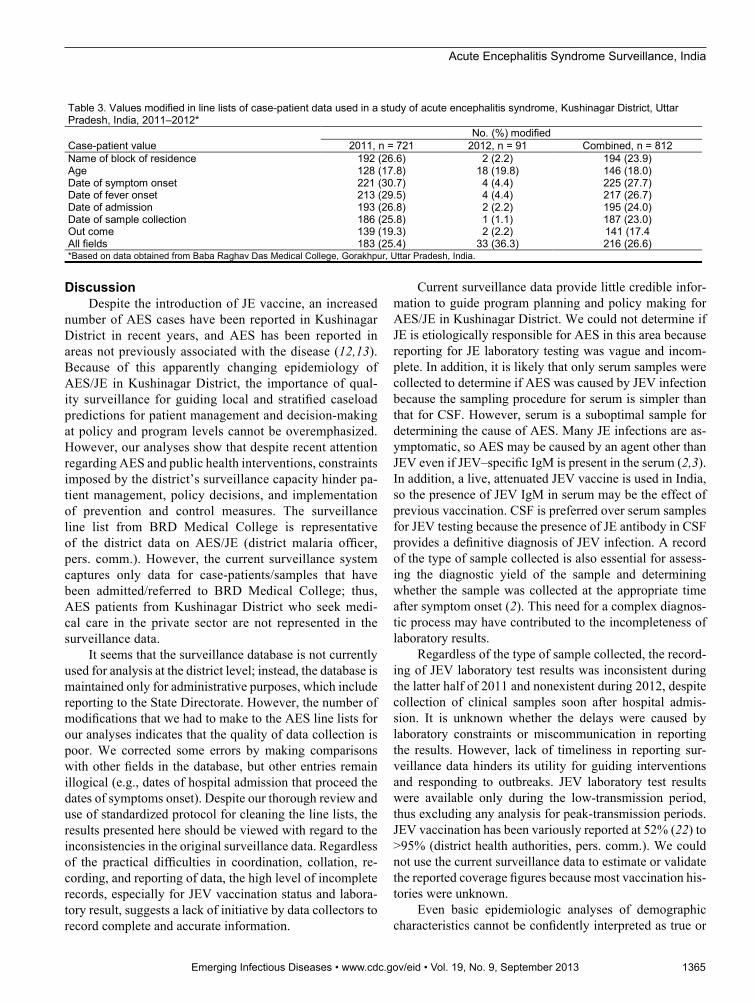
Quality of Surveillance Data During data cleaning, we modified 25% of the 2011
and 5% of the 2012 line list values for residential locality, age, and date parameters (Table 3). Nearly one fifth of the age data and more than one fourth of the dates were edited. For 3.2% and 13.2% of cases in 2011 and 2012, respective-ly, the block name could not be determined from the resi-dential locality provided and was marked as “unknown” because the village or police station name was not found or was present in multiple blocks. In addition, several fields in the database were incomplete. As of July 2012, laboratory results for JEV infection were still classified as “awaited”
(i.e., awaiting determination) in the line lists for 82% (590) of the 721 cases in 2011 and for all 91 cases in 2012 (Table 1). The line lists indicated the date of sample collection, but the type of sample collected (CSF and/or serum) and the laboratory test used (IgM ELISA, PCR, and/or cell culture) were not recorded for any of the case-patients. Most sam-ples submitted after July 2011 were still awaiting labora-tory results at the time of our study (Figure 2). In addition, for 602 (83.5%) of the 721 case-patients in 2011 and all 91 case-patients in 2012, JEV vaccination status was marked as “unknown” in the line lists (Table 1).
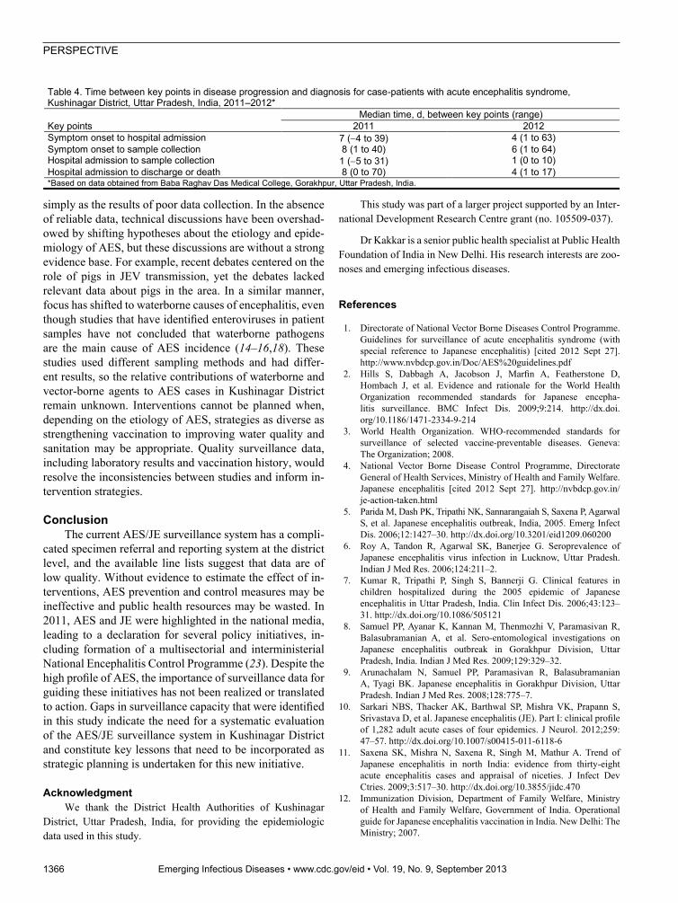
Key epidemiologic and programmatic indicators, such as the time between key points in AES disease progression and diagnosis, varied widely among case-patients (Table 4). In some instances, the dates were illogical (e.g., dates of symptom onset and sample collection following and pre-ceding the dates of hospital admission, respectively). It was not possible to rectify these inconsistencies on the basis of the available data. In addition, the range of values was of-ten large. For example, median time from onset of symp-toms to hospital admission was 4 days in 2012, but some patients were admitted >2 months after symptom onset. Because AES is an acute syndrome, a long interval is not expected between symptoms and may indicate reporting of unrelated symptoms or misclassification of AES.
1364 Emerging Infectious Diseases • www.cdc.gov/eid • Vol. 19, No. 9, September 2013
Table 2. Incidence of acute encephalitis syndrome, Kushinagar District, Uttar Pradesh, India, 2011–2012* Age, sex of population 2011 population† No. cases Incidence‡ Incidence rate ratio (95% CI) All ages 3,560,830 721 20.2 M 1,821,242 414 22.7 1.29 (1.11–1.49) F 1,739,588 307 17.6 1.0 0–6 y 551,467 428 77.6 M 287,672 260 90.4 1.42 (1.17–1.72) F 263,795 168 63.7 1.0 *Based on data obtained from Baba Raghav Das Medical College, Gorakhpur, Uttar Pradesh, India. †From 2011 Census of India (20). ‡Per 100,000 population.
Figure 2. Weekly number of acute encephalitis syndrome cases, by month, in Kushinagar District, Uttar Pradesh State, India, 2011–2012. Numbers represent results of laboratory testing for Japanese encephalitis and are based on data from Baba Raghav Das Medical College, Gorakhpur, India. In the key, “awaited” refers to samples that were awaiting laboratory test results.
Acute Encephalitis Syndrome Surveillance, India
DiscussionDespite the introduction of JE vaccine, an increased
number of AES cases have been reported in Kushinagar District in recent years, and AES has been reported in areas not previously associated with the disease (12,13). Because of this apparently changing epidemiology of AES/JE in Kushinagar District, the importance of qual-ity surveillance for guiding local and stratified caseload predictions for patient management and decision-making at policy and program levels cannot be overemphasized. However, our analyses show that despite recent attention regarding AES and public health interventions, constraints imposed by the district’s surveillance capacity hinder pa-tient management, policy decisions, and implementation of prevention and control measures. The surveillance line list from BRD Medical College is representative of the district data on AES/JE (district malaria officer, pers. comm.). However, the current surveillance system captures only data for case-patients/samples that have been admitted/referred to BRD Medical College; thus, AES patients from Kushinagar District who seek medi-cal care in the private sector are not represented in the surveillance data.
It seems that the surveillance database is not currently used for analysis at the district level; instead, the database is maintained only for administrative purposes, which include reporting to the State Directorate. However, the number of modifications that we had to make to the AES line lists for our analyses indicates that the quality of data collection is poor. We corrected some errors by making comparisons with other fields in the database, but other entries remain illogical (e.g., dates of hospital admission that proceed the dates of symptoms onset). Despite our thorough review and use of standardized protocol for cleaning the line lists, the results presented here should be viewed with regard to the inconsistencies in the original surveillance data. Regardless of the practical difficulties in coordination, collation, re-cording, and reporting of data, the high level of incomplete records, especially for JEV vaccination status and labora-tory result, suggests a lack of initiative by data collectors to record complete and accurate information.
Current surveillance data provide little credible infor-mation to guide program planning and policy making for AES/JE in Kushinagar District. We could not determine if JE is etiologically responsible for AES in this area because reporting for JE laboratory testing was vague and incom-plete. In addition, it is likely that only serum samples were collected to determine if AES was caused by JEV infection because the sampling procedure for serum is simpler than that for CSF. However, serum is a suboptimal sample for determining the cause of AES. Many JE infections are as-ymptomatic, so AES may be caused by an agent other than JEV even if JEV–specific IgM is present in the serum (2,3). In addition, a live, attenuated JEV vaccine is used in India, so the presence of JEV IgM in serum may be the effect of previous vaccination. CSF is preferred over serum samples for JEV testing because the presence of JE antibody in CSF provides a definitive diagnosis of JEV infection. A record of the type of sample collected is also essential for assess-ing the diagnostic yield of the sample and determining whether the sample was collected at the appropriate time after symptom onset (2). This need for a complex diagnos-tic process may have contributed to the incompleteness of laboratory results.
Regardless of the type of sample collected, the record-ing of JEV laboratory test results was inconsistent during the latter half of 2011 and nonexistent during 2012, despite collection of clinical samples soon after hospital admis-sion. It is unknown whether the delays were caused by laboratory constraints or miscommunication in reporting the results. However, lack of timeliness in reporting sur-veillance data hinders its utility for guiding interventions and responding to outbreaks. JEV laboratory test results were available only during the low-transmission period, thus excluding any analysis for peak-transmission periods. JEV vaccination has been variously reported at 52% (22) to >95% (district health authorities, pers. comm.). We could not use the current surveillance data to estimate or validate the reported coverage figures because most vaccination his-tories were unknown.
Even basic epidemiologic analyses of demographic characteristics cannot be confidently interpreted as true or
Emerging Infectious Diseases • www.cdc.gov/eid • Vol. 19, No. 9, September 2013 1365
Table 3. Values modified in line lists of case-patient data used in a study of acute encephalitis syndrome, Kushinagar District, Uttar Pradesh, India, 2011–2012*
Case-patient value No. (%) modified
2011, n = 721 2012, n = 91 Combined, n = 812 Name of block of residence 192 (26.6) 2 (2.2) 194 (23.9) Age 128 (17.8) 18 (19.8) 146 (18.0) Date of symptom onset 221 (30.7) 4 (4.4) 225 (27.7) Date of fever onset 213 (29.5) 4 (4.4) 217 (26.7) Date of admission 193 (26.8) 2 (2.2) 195 (24.0) Date of sample collection 186 (25.8) 1 (1.1) 187 (23.0) Out come 139 (19.3) 2 (2.2) 141 (17.4 All fields 183 (25.4) 33 (36.3) 216 (26.6) *Based on data obtained from Baba Raghav Das Medical College, Gorakhpur, Uttar Pradesh, India.
PERSPECTIVE
simply as the results of poor data collection. In the absence of reliable data, technical discussions have been overshad-owed by shifting hypotheses about the etiology and epide-miology of AES, but these discussions are without a strong evidence base. For example, recent debates centered on the role of pigs in JEV transmission, yet the debates lacked relevant data about pigs in the area. In a similar manner, focus has shifted to waterborne causes of encephalitis, even though studies that have identified enteroviruses in patient samples have not concluded that waterborne pathogens are the main cause of AES incidence (14–16,18). These studies used different sampling methods and had differ-ent results, so the relative contributions of waterborne and vector-borne agents to AES cases in Kushinagar District remain unknown. Interventions cannot be planned when, depending on the etiology of AES, strategies as diverse as strengthening vaccination to improving water quality and sanitation may be appropriate. Quality surveillance data, including laboratory results and vaccination history, would resolve the inconsistencies between studies and inform in-tervention strategies.
ConclusionThe current AES/JE surveillance system has a compli-
cated specimen referral and reporting system at the district level, and the available line lists suggest that data are of low quality. Without evidence to estimate the effect of in-terventions, AES prevention and control measures may be ineffective and public health resources may be wasted. In 2011, AES and JE were highlighted in the national media, leading to a declaration for several policy initiatives, in-cluding formation of a multisectorial and interministerial National Encephalitis Control Programme (23). Despite the high profile of AES, the importance of surveillance data for guiding these initiatives has not been realized or translated to action. Gaps in surveillance capacity that were identified in this study indicate the need for a systematic evaluation of the AES/JE surveillance system in Kushinagar District and constitute key lessons that need to be incorporated as strategic planning is undertaken for this new initiative.
AcknowledgmentWe thank the District Health Authorities of Kushinagar
District, Uttar Pradesh, India, for providing the epidemiologic data used in this study.
This study was part of a larger project supported by an Inter-national Development Research Centre grant (no. 105509-037).
Dr Kakkar is a senior public health specialist at Public Health Foundation of India in New Delhi. His research interests are zoo-noses and emerging infectious diseases.
References
1. Directorate of National Vector Borne Diseases Control Programme. Guidelines for surveillance of acute encephalitis syndrome (with special reference to Japanese encephalitis) [cited 2012 Sept 27]. http://www.nvbdcp.gov.in/Doc/AES%20guidelines.pdf
2. Hills S, Dabbagh A, Jacobson J, Marfin A, Featherstone D, Hombach J, et al. Evidence and rationale for the World Health Organization recommended standards for Japanese encepha-litis surveillance. BMC Infect Dis. 2009;9:214. http://dx.doi.org/10.1186/1471-2334-9-214
3. World Health Organization. WHO-recommended standards for surveillance of selected vaccine-preventable diseases. Geneva: The Organization; 2008.
4. National Vector Borne Disease Control Programme, Directorate General of Health Services, Ministry of Health and Family Welfare. Japanese encephalitis [cited 2012 Sept 27]. http://nvbdcp.gov.in/ je-action-taken.html
5. Parida M, Dash PK, Tripathi NK, Sannarangaiah S, Saxena P, Agarwal S, et al. Japanese encephalitis outbreak, India, 2005. Emerg Infect Dis. 2006;12:1427–30. http://dx.doi.org/10.3201/eid1209.060200
6. Roy A, Tandon R, Agarwal SK, Banerjee G. Seroprevalence of Japanese encephalitis virus infection in Lucknow, Uttar Pradesh. Indian J Med Res. 2006;124:211–2.
7. Kumar R, Tripathi P, Singh S, Bannerji G. Clinical features in children hospitalized during the 2005 epidemic of Japanese encephalitis in Uttar Pradesh, India. Clin Infect Dis. 2006;43:123–31. http://dx.doi.org/10.1086/505121
8. Samuel PP, Ayanar K, Kannan M, Thenmozhi V, Paramasivan R, Balasubramanian A, et al. Sero-entomological investigations on Japanese encephalitis outbreak in Gorakhpur Division, Uttar Pradesh, India. Indian J Med Res. 2009;129:329–32.
9. Arunachalam N, Samuel PP, Paramasivan R, Balasubramanian A, Tyagi BK. Japanese encephalitis in Gorakhpur Division, Uttar Pradesh. Indian J Med Res. 2008;128:775–7.
10. Sarkari NBS, Thacker AK, Barthwal SP, Mishra VK, Prapann S, Srivastava D, et al. Japanese encephalitis (JE). Part I: clinical profile of 1,282 adult acute cases of four epidemics. J Neurol. 2012;259: 47–57. http://dx.doi.org/10.1007/s00415-011-6118-6
11. Saxena SK, Mishra N, Saxena R, Singh M, Mathur A. Trend of Japanese encephalitis in north India: evidence from thirty-eight acute encephalitis cases and appraisal of niceties. J Infect Dev Ctries. 2009;3:517–30. http://dx.doi.org/10.3855/jidc.470
12. Immunization Division, Department of Family Welfare, Ministry of Health and Family Welfare, Government of India. Operational guide for Japanese encephalitis vaccination in India. New Delhi: The Ministry; 2007.
1366 Emerging Infectious Diseases • www.cdc.gov/eid • Vol. 19, No. 9, September 2013
Table 4. Time between key points in disease progression and diagnosis for case-patients with acute encephalitis syndrome, Kushinagar District, Uttar Pradesh, India, 2011–2012*
Key points Median time, d, between key points (range)
2011 2012 Symptom onset to hospital admission 7 (4 to 39) 4 (1 to 63) Symptom onset to sample collection 8 (1 to 40) 6 (1 to 64) Hospital admission to sample collection 1 (5 to 31) 1 (0 to 10) Hospital admission to discharge or death 8 (0 to 70) 4 (1 to 17) *Based on data obtained from Baba Raghav Das Medical College, Gorakhpur, Uttar Pradesh, India.
Acute Encephalitis Syndrome Surveillance, India
13. Directorate of National Vector Borne Diseases Control Programme—Delhi. Details of AES/JE cases and deaths from 2008–2013 [cited 2013 Jan 20]. http://nvbdcp.gov.in/Doc/je-aes-cd-till1Apr13.pdf
14. Sapkal GN, Bondre VP, Fulmali PV, Patil P, Gopalkrishna V, Dadhania V, et al. Enteroviruses in patients with acute encepha-litis, Uttar Pradesh, India. Emerg Infect Dis. 2009;15:295–8. http://dx.doi.org/10.3201/eid1502.080865
15. Lewthwaite P, Perera D, Ooi MH, Last A, Kumar R, Desai A, et al. Enterovirus 75 encephalitis in children, southern India. Emerg Infect Dis. 2010;16:1780–2. http://dx.doi.org/10.3201/eid1611.100672
16. Karmarkar SA, Aneja S, Khare S, Saini A, Seth A, Chauhan BKY. A study of acute febrile encephalopathy with special reference to viral etiology. Indian J Pediatr. 2008;75:801–5. http://dx.doi.org/10.1007/s12098-008-0150-2
17. Bhatt GC, Bondre VP, Sapkal GN, Sharma T, Kumar S, Gore MM, et al. Changing clinico-laboratory profile of encephalitis patients in the eastern Uttar Pradesh region of India. Trop Doct. 2012;42: 106–8. http://dx.doi.org/10.1258/td.2011.110391
18. Beig FK, Malik A, Rizvi M, Acharya D, Khare S. Etiology and clinico-epidemiological profile of acute viral encephalitis in children of western Uttar Pradesh, India. Int J Infect Dis. 2010;14:e141–6. http://dx.doi.org/10.1016/j.ijid.2009.03.035
19. Planning Commission of India. Report of the Working Group on Disease Burden for the 12th Five Year Plan [cited 2013 Nov 3]. http://planningcommission.nic.in/aboutus/committee/wrkgrp12/health/WG_3_1communicable.pdf
20. Office of the Registrar General and Census Commissioner (India). India population and housing census 2011. New Delhi (India): The Office; 2011.
21. German RR, Lee LM, Horan JM, Milstein RL, Pertowski CA, Waller MN. Updated guidelines for evaluating public health surveillance systems: recommendations from the Guidelines Working Group. MMWR Recomm Rep. 2001;50(RR-13):1–35.
22. UNICEF. Japanese encephalitis coverage evaluation survey report 2008 [cited 2012 Oct 8]. http://www.unicef.org/india/Japanese_ Encephalitis_CES_2008_report.pdf
23. Press Information Bureau, Government of India. Implementation of intervention/activities for prevention and control of Japanese encephalitis (JE)/acute encephalitis syndrome (AES). 2012 [cited 2012 Nov 3]. http://pib.nic.in/newsite/erelease.aspx?relid=88512
Address for correspondence: Manish Kakkar, Public Health Foundation of India, ISID Campus, 4 Institutional Area, Vasant Kunj, New Delhi 110070, India; email: [email protected]
Emerging Infectious Diseases • www.cdc.gov/eid • Vol. 19, No. 9, September 2013 1367