Embed Size (px)
Citation preview
clinical practice
T h e n e w e ng l a nd j o u r na l o f m e dic i n e
This Journal feature begins with a case vignette highlighting a common clinical problem. Evidence supporting various strategies is then presented, followed by a review of formal guidelines,
when they exist. The article ends with the authors’ clinical recommendations.
n engl j med 353;26 www.nejm.org december 29, 20052788
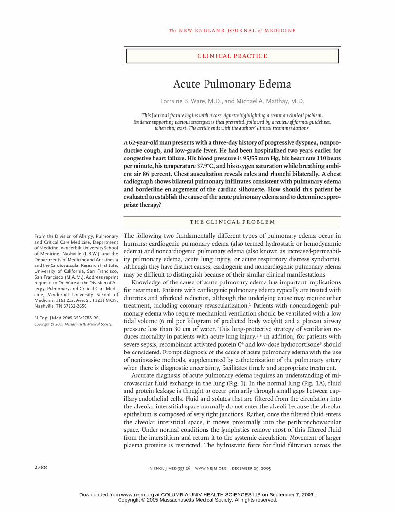
Acute Pulmonary EdemaLorraine B. Ware, M.D., and Michael A. Matthay, M.D.
From the Division of Allergy, Pulmonary and Critical Care Medicine, Department of Medicine, Vanderbilt University School of Medicine, Nashville (L.B.W.); and the Departments of Medicine and Anesthesia and the Cardiovascular Research Institute, University of California, San Francisco, San Francisco (M.A.M.). Address reprint requests to Dr. Ware at the Division of Al-lergy, Pulmonary and Critical Care Medi-cine, Vanderbilt University School of Medicine, 1161 21st Ave. S., T1218 MCN, Nashville, TN 37232-2650.
N Engl J Med 2005;353:2788-96.Copyright © 2005 Massachusetts Medical Society.
A 62-year-old man presents with a three-day history of progressive dyspnea, nonpro-ductive cough, and low-grade fever. He had been hospitalized two years earlier for congestive heart failure. His blood pressure is 95/55 mm Hg, his heart rate 110 beats per minute, his temperature 37.9°C, and his oxygen saturation while breathing ambi-ent air 86 percent. Chest auscultation reveals rales and rhonchi bilaterally. A chest radiograph shows bilateral pulmonary infiltrates consistent with pulmonary edema and borderline enlargement of the cardiac silhouette. How should this patient be evaluated to establish the cause of the acute pulmonary edema and to determine appro-priate therapy?
the clinical problem
The following two fundamentally different types of pulmonary edema occur in humans: cardiogenic pulmonary edema (also termed hydrostatic or hemodynamic edema) and noncardiogenic pulmonary edema (also known as increased-permeabil-ity pulmonary edema, acute lung injury, or acute respiratory distress syndrome). Although they have distinct causes, cardiogenic and noncardiogenic pulmonary edema may be difficult to distinguish because of their similar clinical manifestations.
Knowledge of the cause of acute pulmonary edema has important implications for treatment. Patients with cardiogenic pulmonary edema typically are treated with diuretics and afterload reduction, although the underlying cause may require other treatment, including coronary revascularization.1 Patients with noncardiogenic pul-monary edema who require mechanical ventilation should be ventilated with a low tidal volume (6 ml per kilogram of predicted body weight) and a plateau airway pressure less than 30 cm of water. This lung-protective strategy of ventilation re-duces mortality in patients with acute lung injury.2,3 In addition, for patients with severe sepsis, recombinant activated protein C4 and low-dose hydrocortisone5 should be considered. Prompt diagnosis of the cause of acute pulmonary edema with the use of noninvasive methods, supplemented by catheterization of the pulmonary artery when there is diagnostic uncertainty, facilitates timely and appropriate treatment.
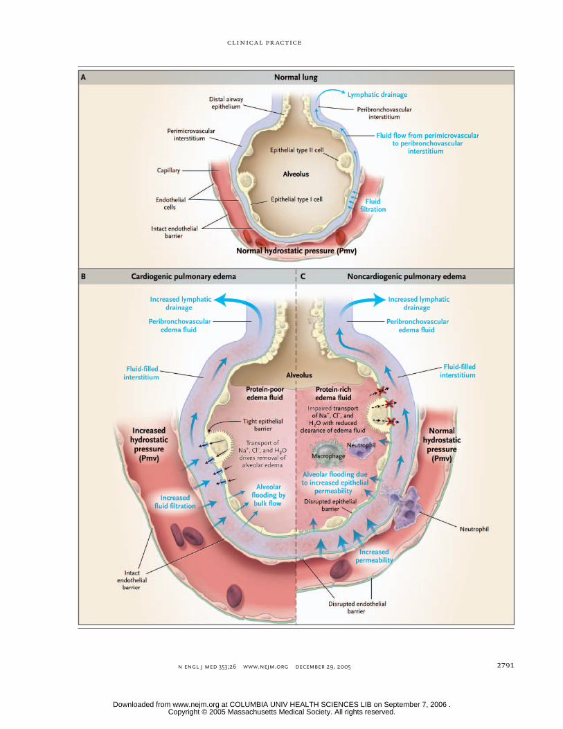
Accurate diagnosis of acute pulmonary edema requires an understanding of mi-crovascular fluid exchange in the lung (Fig. 1). In the normal lung (Fig. 1A), fluid and protein leakage is thought to occur primarily through small gaps between cap-illary endothelial cells. Fluid and solutes that are filtered from the circulation into the alveolar interstitial space normally do not enter the alveoli because the alveolar epithelium is composed of very tight junctions. Rather, once the filtered fluid enters the alveolar interstitial space, it moves proximally into the peribronchovascular space. Under normal conditions the lymphatics remove most of this filtered fluid from the interstitium and return it to the systemic circulation. Movement of larger plasma proteins is restricted. The hydrostatic force for fluid filtration across the
Copyright © 2005 Massachusetts Medical Society. All rights reserved. Downloaded from www.nejm.org at COLUMBIA UNIV HEALTH SCIENCES LIB on September 7, 2006 .
clinical pr actice
n engl j med 353;26 www.nejm.org december 29, 2005 2789
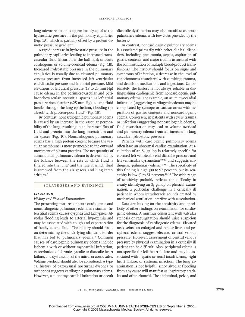
lung microcirculation is approximately equal to the hydrostatic pressure in the pulmonary capillaries (Fig. 1A), which is partially offset by a protein os-motic pressure gradient.
A rapid increase in hydrostatic pressure in the pulmonary capillaries leading to increased trans-vascular f luid filtration is the hallmark of acute cardiogenic or volume-overload edema (Fig. 1B). Increased hydrostatic pressure in the pulmonary capillaries is usually due to elevated pulmonary venous pressure from increased left ventricular end-diastolic pressure and left atrial pressure. Mild elevations of left atrial pressure (18 to 25 mm Hg) cause edema in the perimicrovascular and peri-bronchovascular interstitial spaces.7 As left atrial pressure rises further (>25 mm Hg), edema fluid breaks through the lung epithelium, flooding the alveoli with protein-poor fluid7 (Fig. 1B).
By contrast, noncardiogenic pulmonary edema is caused by an increase in the vascular permea-bility of the lung, resulting in an increased flux of fluid and protein into the lung interstitium and air spaces (Fig. 1C). Noncardiogenic pulmonary edema has a high protein content because the vas-cular membrane is more permeable to the outward movement of plasma proteins. The net quantity of accumulated pulmonary edema is determined by the balance between the rate at which fluid is filtered into the lung7 and the rate at which fluid is removed from the air spaces and lung inter-stitium.6
strategies and evidence
evaluation
History and Physical ExaminationThe presenting features of acute cardiogenic and noncardiogenic pulmonary edema are similar. In-terstitial edema causes dyspnea and tachypnea. Al-veolar flooding leads to arterial hypoxemia and may be associated with cough and expectoration of frothy edema fluid. The history should focus on determining the underlying clinical disorder that has led to pulmonary edema.8 Common causes of cardiogenic pulmonary edema include ischemia with or without myocardial infarction, exacerbation of chronic systolic or diastolic heart failure, and dysfunction of the mitral or aortic valve. Volume overload should also be considered. A typi-cal history of paroxysmal nocturnal dyspnea or orthopnea suggests cardiogenic pulmonary edema. However, a silent myocardial infarction or occult
diastolic dysfunction may also manifest as acute pulmonary edema, with few clues provided by the history.9
In contrast, noncardiogenic pulmonary edema is associated primarily with other clinical disor-ders, including pneumonia, sepsis, aspiration of gastric contents, and major trauma associated with the administration of multiple blood-product trans-fusions.3 The history should focus on signs and symptoms of infection, a decrease in the level of consciousness associated with vomiting, trauma, and details of medications and ingestions. Unfor-tunately, the history is not always reliable in dis-tinguishing cardiogenic from noncardiogenic pul-monary edema. For example, an acute myocardial infarction (suggesting cardiogenic edema) may be complicated by syncope or cardiac arrest with as-piration of gastric contents and noncardiogenic edema. Conversely, in patients with severe trauma or infection (suggesting noncardiogenic edema), fluid resuscitation may lead to volume overload and pulmonary edema from an increase in lung vascular hydrostatic pressure.
Patients with cardiogenic pulmonary edema often have an abnormal cardiac examination. Aus-cultation of an S3 gallop is relatively specific for elevated left ventricular end-diastolic pressure and left ventricular dysfunction10-13 and suggests car-diogenic pulmonary edema.10-12 The specificity of this finding is high (90 to 97 percent), but its sen-sitivity is low (9 to 51 percent).10-13 The wide range of sensitivity probably reflects the difficulty in clearly identifying an S3 gallop on physical exami-nation, a particular challenge in a critically ill patient in whom intrathoracic sounds created by mechanical ventilation interfere with auscultation.
Data are lacking on the sensitivity and speci-ficity of other findings on examination for cardio-genic edema. A murmur consistent with valvular stenosis or regurgitation should raise suspicion for the diagnosis of cardiogenic edema. Elevated neck veins, an enlarged and tender liver, and pe-ripheral edema suggest elevated central venous pressure. However, assessment of central venous pressure by physical examination in a critically ill patient can be difficult. Also, peripheral edema is not specific for left heart failure and may be as-sociated with hepatic or renal insufficiency, right heart failure, or systemic infection. The lung ex-amination is not helpful, since alveolar flooding from any cause will manifest as inspiratory crack-les and often rhonchi. The abdominal, pelvic, and
Copyright © 2005 Massachusetts Medical Society. All rights reserved. Downloaded from www.nejm.org at COLUMBIA UNIV HEALTH SCIENCES LIB on September 7, 2006 .

T h e n e w e ng l a nd j o u r na l o f m e dic i n e
n engl j med 353;26 www.nejm.org december 29, 20052790
rectal examinations are important. An intraab-dominal crisis such as perforation of a viscus can cause acute lung injury with noncardiogenic edema, and patients who are mechanically ven-tilated may be unable to provide a history of ab-dominal symptoms. Patients with noncardiogenic edema often have warm extremities, even in the absence of sepsis, whereas patients with cardio-genic edema and poor cardiac output usually have cool extremities.
Laboratory TestingElectrocardiographic findings may suggest myo-cardial ischemia or infarction. Elevated troponin levels may indicate damage to myocytes. However, elevated troponin levels can occur in patients with severe sepsis in the absence of evidence for an acute coronary syndrome.14-17 In a patient who is obtunded and has pulmonary edema of an un-known cause, measurement of electrolytes, the se-rum osmolarity, and a toxicology screen may lead to the diagnosis of an unsuspected ingestion. El-evated levels of serum amylase and lipase suggest acute pancreatitis.
Plasma levels of brain natriuretic peptide (BNP) are often used in the evaluation of pulmonary edema. BNP is secreted predominantly by the car-diac ventricles in response to wall stretch or in-creased intracardiac pressures. In patients with congestive heart failure, plasma BNP levels corre-late with left ventricular end-diastolic pressure18-21 and pulmonary-artery occlusion pressure.22 Ac-cording to a consensus panel, a BNP level below 100 pg per milliliter indicates that heart failure is unlikely (negative predictive value, >90 percent), whereas a BNP level greater than 500 pg per mil-liliter indicates that heart failure is likely (posi-tive predictive value, >90 percent).23 However, BNP levels between 100 and 500 pg per milliliter pro-vide inadequate diagnostic discrimination.
BNP levels must be interpreted with caution in critically ill patients, since the predictive value of BNP levels is uncertain in this group. Some reports indicate that BNP levels can be elevated in criti-cally ill patients even in the absence of heart fail-ure.24,25 Levels between 100 and 500 pg per milli-liter are common in these patients.26 In one report, all eight patients with sepsis with normal left ventricular function had BNP levels above 500 pg per milliliter.27 Thus, measuring BNP is most use-ful in critically ill patients if the level is below 100 pg per milliliter. BNP levels are also higher in
patients with renal failure independent of heart failure, and a cutoff of below 200 pg per milliliter has been suggested to exclude heart failure when the estimated glomerular filtration rate is below 60 ml per minute.23 BNP can also be secreted by
Figure 1 (facing page). Physiology of Microvascular Fluid Exchange in the Lung.
In the normal lung (Panel A), fluid moves continuously outward from the vascular to the interstitial space according to the net difference between hydrostatic and protein osmotic pressures, as well as to the per-meability of the capillary membrane. The following Starling equation for filtration of fluid across a semi-permeable membrane describes the factors that deter-mine the amount of fluid leaving the vascular space: Q = K[(Pmv−Ppmv) − (πmv−πpmv)], where Q is the net transvascular flow of fluid, K is the membrane permea-bility, Pmv is the hydrostatic pressure in the microves-sels, Ppmv is the hydrostatic pressure in the perimicro-vascular interstitium, πmv is the plasma protein osmotic pressure in the circulation, and πpmv is the protein osmotic pressure in the perimicrovascular interstitium. When hydrostatic pressure increases in the microcirculation, the rate of transvascular fluid fil-tration rises (Panel B). When lung interstitial pressure exceeds pleural pressure, fluid moves across the vis-ceral pleura, creating pleural effusions. Since the per-meability of the capillary endothelium remains normal, the filtered edema fluid leaving the circulation has a low protein content. The removal of edema fluid from the air spaces of the lung depends on active transport of sodium and chloride across the alveolar epithelial barrier. The primary sites of sodium and chloride reab-sorption are the epithelial ion channels located on the apical membrane of alveolar epithelial type I and II cells and distal airway epithelia. Sodium is actively extruded into the interstitial space by means of the Na+/K+–ATPase located on the basolateral membrane of type II cells. Water follows passively, probably through aquaporins, which are water channels that are found predominantly on alveolar epithelial type I cells.6 Noncardiogenic pulmonary edema (Panel C) occurs when the permeability of the microvascular membrane increases because of direct or indirect lung injury (including the acute respiratory distress syndrome), resulting in a marked increase in the amount of fluid and protein leaving the vascular space. Noncardiogen-ic pulmonary edema has a high protein content because the more permeable microvascular membrane has a reduced capacity to restrict the outward move-ment of larger molecules such as plasma proteins. The degree of alveolar flooding depends on the extent of interstitial edema, the presence or absence of injury to the alveolar epithelium, and the capacity of the alveo-lar epithelium to actively remove alveolar edema fluid. In edema due to acute lung injury, alveolar epithelial injury commonly causes a decrease in the capacity for the removal of alveolar fluid, delaying the resolution of pulmonary edema.6
Copyright © 2005 Massachusetts Medical Society. All rights reserved. Downloaded from www.nejm.org at COLUMBIA UNIV HEALTH SCIENCES LIB on September 7, 2006 .
clinical pr actice
n engl j med 353;26 www.nejm.org december 29, 2005 2791
Copyright © 2005 Massachusetts Medical Society. All rights reserved. Downloaded from www.nejm.org at COLUMBIA UNIV HEALTH SCIENCES LIB on September 7, 2006 .
T h e n e w e ng l a nd j o u r na l o f m e dic i n e
n engl j med 353;26 www.nejm.org december 29, 20052792
the right ventricle, and moderate elevations have been reported in patients with acute pulmonary embolism, cor pulmonale, and pulmonary hyper-tension.23
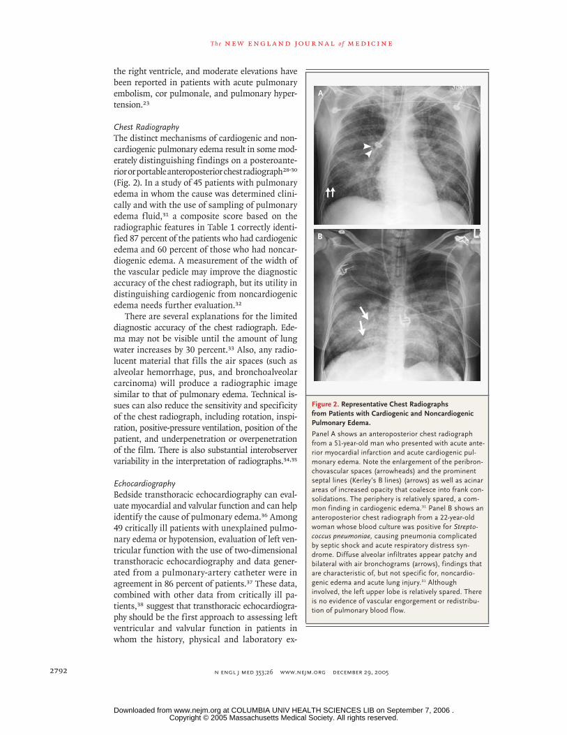
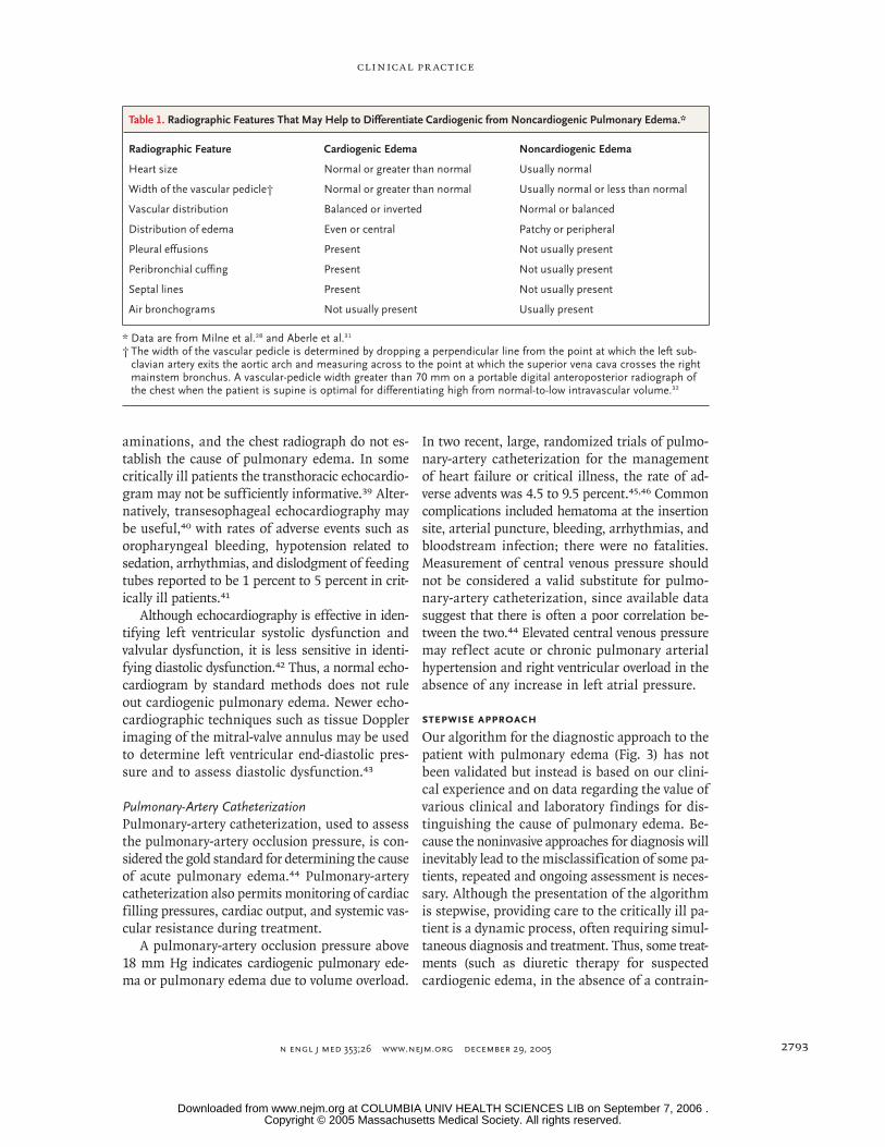
Chest RadiographyThe distinct mechanisms of cardiogenic and non-cardiogenic pulmonary edema result in some mod-erately distinguishing findings on a posteroante-rior or portable anteroposterior chest radiograph28-30 (Fig. 2). In a study of 45 patients with pulmonary edema in whom the cause was determined clini-cally and with the use of sampling of pulmonary edema fluid,31 a composite score based on the radiographic features in Table 1 correctly identi-fied 87 percent of the patients who had cardiogenic edema and 60 percent of those who had noncar-diogenic edema. A measurement of the width of the vascular pedicle may improve the diagnostic accuracy of the chest radiograph, but its utility in distinguishing cardiogenic from noncardiogenic edema needs further evaluation.32
There are several explanations for the limited diagnostic accuracy of the chest radiograph. Ede-ma may not be visible until the amount of lung water increases by 30 percent.33 Also, any radio-lucent material that fills the air spaces (such as alveolar hemorrhage, pus, and bronchoalveolar carcinoma) will produce a radiographic image similar to that of pulmonary edema. Technical is-sues can also reduce the sensitivity and specificity of the chest radiograph, including rotation, inspi-ration, positive-pressure ventilation, position of the patient, and underpenetration or overpenetration of the film. There is also substantial interobserver variability in the interpretation of radiographs.34,35
EchocardiographyBedside transthoracic echocardiography can eval-uate myocardial and valvular function and can help identify the cause of pulmonary edema.36 Among 49 critically ill patients with unexplained pulmo-nary edema or hypotension, evaluation of left ven-tricular function with the use of two-dimensional transthoracic echocardiography and data gener-ated from a pulmonary-artery catheter were in agreement in 86 percent of patients.37 These data, combined with other data from critically ill pa-tients,38 suggest that transthoracic echocardiogra-phy should be the first approach to assessing left ventricular and valvular function in patients in whom the history, physical and laboratory ex-
A
B
Figure 2. Representative Chest Radiographs from Patients with Cardiogenic and Noncardiogenic Pulmonary Edema.
Panel A shows an anteroposterior chest radiograph from a 51-year-old man who presented with acute ante-rior myocardial infarction and acute cardiogenic pul-monary edema. Note the enlargement of the peribron-chovascular spaces (arrowheads) and the prominent septal lines (Kerley’s B lines) (arrows) as well as acinar areas of increased opacity that coalesce into frank con-solidations. The periphery is relatively spared, a com-mon finding in cardiogenic edema.31 Panel B shows an anteroposterior chest radiograph from a 22-year-old woman whose blood culture was positive for Strepto-coccus pneumoniae, causing pneumonia complicated by septic shock and acute respiratory distress syn-drome. Diffuse alveolar infiltrates appear patchy and bilateral with air bronchograms (arrows), findings that are characteristic of, but not specific for, noncardio-genic edema and acute lung injury.31 Although involved, the left upper lobe is relatively spared. There is no evidence of vascular engorgement or redistribu-tion of pulmonary blood flow.
Copyright © 2005 Massachusetts Medical Society. All rights reserved. Downloaded from www.nejm.org at COLUMBIA UNIV HEALTH SCIENCES LIB on September 7, 2006 .
clinical pr actice
n engl j med 353;26 www.nejm.org december 29, 2005 2793
aminations, and the chest radiograph do not es-tablish the cause of pulmonary edema. In some critically ill patients the transthoracic echocardio-gram may not be sufficiently informative.39 Alter-natively, transesophageal echocardiography may be useful,40 with rates of adverse events such as oropharyngeal bleeding, hypotension related to sedation, arrhythmias, and dislodgment of feeding tubes reported to be 1 percent to 5 percent in crit-ically ill patients.41
Although echocardiography is effective in iden-tifying left ventricular systolic dysfunction and valvular dysfunction, it is less sensitive in identi-fying diastolic dysfunction.42 Thus, a normal echo-cardiogram by standard methods does not rule out cardiogenic pulmonary edema. Newer echo-cardiographic techniques such as tissue Doppler imaging of the mitral-valve annulus may be used to determine left ventricular end-diastolic pres-sure and to assess diastolic dysfunction.43
Pulmonary-Artery CatheterizationPulmonary-artery catheterization, used to assess the pulmonary-artery occlusion pressure, is con-sidered the gold standard for determining the cause of acute pulmonary edema.44 Pulmonary-artery catheterization also permits monitoring of cardiac filling pressures, cardiac output, and systemic vas-cular resistance during treatment.
A pulmonary-artery occlusion pressure above 18 mm Hg indicates cardiogenic pulmonary ede-ma or pulmonary edema due to volume overload.
In two recent, large, randomized trials of pulmo-nary-artery catheterization for the management of heart failure or critical illness, the rate of ad-verse advents was 4.5 to 9.5 percent.45,46 Common complications included hematoma at the insertion site, arterial puncture, bleeding, arrhythmias, and bloodstream infection; there were no fatalities. Measurement of central venous pressure should not be considered a valid substitute for pulmo-nary-artery catheterization, since available data suggest that there is often a poor correlation be-tween the two.44 Elevated central venous pressure may reflect acute or chronic pulmonary arterial hypertension and right ventricular overload in the absence of any increase in left atrial pressure.
stepwise approach
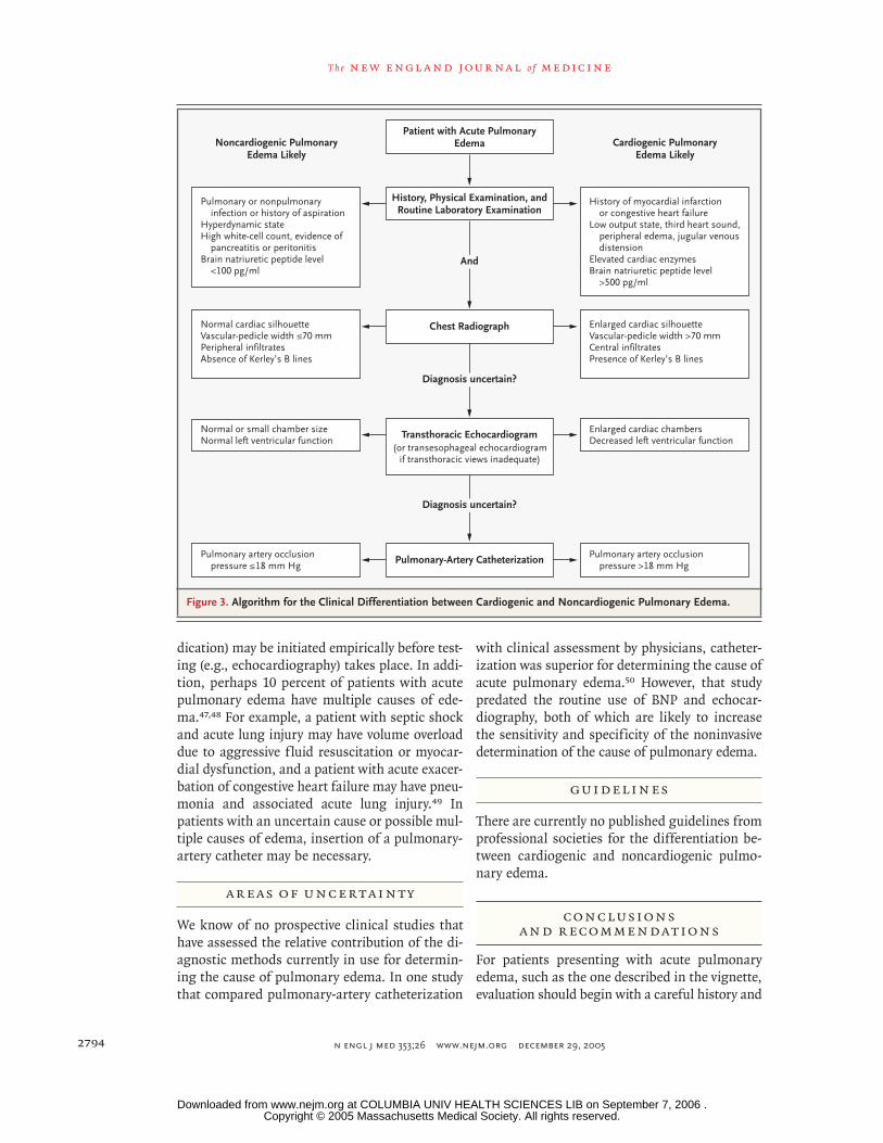
Our algorithm for the diagnostic approach to the patient with pulmonary edema (Fig. 3) has not been validated but instead is based on our clini-cal experience and on data regarding the value of various clinical and laboratory findings for dis-tinguishing the cause of pulmonary edema. Be-cause the noninvasive approaches for diagnosis will inevitably lead to the misclassification of some pa-tients, repeated and ongoing assessment is neces-sary. Although the presentation of the algorithm is stepwise, providing care to the critically ill pa-tient is a dynamic process, often requiring simul-taneous diagnosis and treatment. Thus, some treat-ments (such as diuretic therapy for suspected cardiogenic edema, in the absence of a contrain-
Table 1. Radiographic Features That May Help to Differentiate Cardiogenic from Noncardiogenic Pulmonary Edema.*
Radiographic Feature Cardiogenic Edema Noncardiogenic Edema
Heart size Normal or greater than normal Usually normal
Width of the vascular pedicle† Normal or greater than normal Usually normal or less than normal
Vascular distribution Balanced or inverted Normal or balanced
Distribution of edema Even or central Patchy or peripheral
Pleural effusions Present Not usually present
Peribronchial cuffing Present Not usually present
Septal lines Present Not usually present
Air bronchograms Not usually present Usually present
* Data are from Milne et al.28 and Aberle et al.31
† The width of the vascular pedicle is determined by dropping a perpendicular line from the point at which the left sub-clavian artery exits the aortic arch and measuring across to the point at which the superior vena cava crosses the right mainstem bronchus. A vascular-pedicle width greater than 70 mm on a portable digital anteroposterior radiograph of the chest when the patient is supine is optimal for differentiating high from normal-to-low intravascular volume.32
Copyright © 2005 Massachusetts Medical Society. All rights reserved. Downloaded from www.nejm.org at COLUMBIA UNIV HEALTH SCIENCES LIB on September 7, 2006 .
T h e n e w e ng l a nd j o u r na l o f m e dic i n e
n engl j med 353;26 www.nejm.org december 29, 20052794
dication) may be initiated empirically before test-ing (e.g., echocardiography) takes place. In addi-tion, perhaps 10 percent of patients with acute pulmonary edema have multiple causes of ede-ma.47,48 For example, a patient with septic shock and acute lung injury may have volume overload due to aggressive fluid resuscitation or myocar-dial dysfunction, and a patient with acute exacer-bation of congestive heart failure may have pneu-monia and associated acute lung injury.49 In patients with an uncertain cause or possible mul-tiple causes of edema, insertion of a pulmonary-artery catheter may be necessary.
areas of uncertainty
We know of no prospective clinical studies that have assessed the relative contribution of the di-agnostic methods currently in use for determin-ing the cause of pulmonary edema. In one study that compared pulmonary-artery catheterization
with clinical assessment by physicians, catheter-ization was superior for determining the cause of acute pulmonary edema.50 However, that study predated the routine use of BNP and echocar-diography, both of which are likely to increase the sensitivity and specificity of the noninvasive determination of the cause of pulmonary edema.
guidelines
There are currently no published guidelines from professional societies for the differentiation be-tween cardiogenic and noncardiogenic pulmo-nary edema.
conclusions
and recommendations
For patients presenting with acute pulmonary edema, such as the one described in the vignette, evaluation should begin with a careful history and
History, Physical Examination, andRoutine Laboratory Examination
Patient with Acute PulmonaryEdemaNoncardiogenic Pulmonary
Edema LikelyCardiogenic Pulmonary
Edema Likely
Pulmonary or nonpulmonaryinfection or history of aspiration
Hyperdynamic stateHigh white-cell count, evidence of
pancreatitis or peritonitisBrain natriuretic peptide level <100 pg/ml
History of myocardial infarction or congestive heart failure
Low output state, third heart sound,peripheral edema, jugular venousdistension
Elevated cardiac enzymesBrain natriuretic peptide level >500 pg/ml
Chest Radiograph
And
Diagnosis uncertain?
Diagnosis uncertain?
Normal cardiac silhouetteVascular-pedicle width ≤70 mmPeripheral infiltratesAbsence of Kerley’s B lines
Transthoracic EchocardiogramNormal or small chamber sizeNormal left ventricular function
(or transesophageal echocardiogramif transthoracic views inadequate)
Enlarged cardiac chambersDecreased left ventricular function
Pulmonary-Artery CatheterizationPulmonary artery occlusion pressure ≤18 mm Hg
Pulmonary artery occlusion pressure >18 mm Hg
Enlarged cardiac silhouetteVascular-pedicle width >70 mmCentral infiltratesPresence of Kerley’s B lines
Figure 3. Algorithm for the Clinical Differentiation between Cardiogenic and Noncardiogenic Pulmonary Edema.
Copyright © 2005 Massachusetts Medical Society. All rights reserved. Downloaded from www.nejm.org at COLUMBIA UNIV HEALTH SCIENCES LIB on September 7, 2006 .
clinical pr actice
n engl j med 353;26 www.nejm.org december 29, 2005 2795
physical examination. Special attention should be paid to signs and symptoms of acute or chronic cardiac disease, as well as evidence for a primary pulmonary process such as pneumonia or a non-pulmonary source of infection such as peritoni-tis. An electrocardiogram should be obtained to rule out ischemic changes, although such chang-es alone would not establish that the pulmonary edema was cardiogenic. Measurement of plasma BNP is warranted and is most useful if the value is below 100 pg per milliliter, a level at which congestive heart failure is unlikely. The chest ra-diograph should be reviewed with attention to features suggesting cardiogenic edema (e.g., in-creased heart size and central distribution of ede-ma) as opposed to noncardiogenic edema. If the diagnosis remains uncertain, a transthoracic echo-cardiogram can evaluate left ventricular systolic function and aortic- and mitral-valve function.
With the use of the stepwise approach in the
diagnostic algorithm, the majority of patients with acute pulmonary edema will be diagnosed non-invasively, and treatment can be provided while the diagnostic steps are taken. For example, if in-fection is suspected, antibiotic therapy should be initiated after obtaining appropriate cultures. Sim-ilarly, if the patient requires mechanical ventila-tion, and there is uncertainty about the cause of the pulmonary edema, then a lung-protective strat-egy of ventilation with a low tidal volume is rec-ommended. In some patients, particularly those in whom shock complicates the pulmonary edema, insertion of a pulmonary-artery catheter is need-ed to identify the cause of the pulmonary edema and target therapy appropriately.
Supported by grants from the National Heart, Lung, and Blood Institute (NHLBI HL51856 and HL74005, to Dr. Matthay; and NHLBI 70521 and 081332, to Dr. Ware).
No potential conflict of interest relevant to this article was reported.
references
Jessup M, Brozena S. Heart failure. N Engl J Med 2003;348:2007-18.
The Acute Respiratory Distress Syn-drome Network. Ventilation with lower tidal volumes as compared with tradition-al tidal volumes for acute lung injury and the acute respiratory distress syndrome. N Engl J Med 2000;342:1301-8.
Ware LB, Matthay MA. The acute re-spiratory distress syndrome. N Engl J Med 2000;342:1334-49.
Bernard GR, Vincent J-L, Laterre P-F, et al. Efficacy and safety of recombinant human activated protein C for severe sep-sis. N Engl J Med 2001;344:699-709.
Annane D, Sebille V, Charpentier C, et al. Effect of treatment with low doses of hydrocortisone and fludrocortisone on mortality in patients with septic shock. JAMA 2002;288:862-71.
Matthay MA, Folkesson HG, Clerici C. Lung epithelial f luid transport and the resolution of pulmonary edema. Physiol Rev 2002;82:569-600.
Staub NC. Pulmonary edema. Physiol Rev 1974;54:678-811.
Sibbald WJ, Cunningham DR, Chin DN. Non-cardiac or cardiac pulmonary edema? A practical approach to clinical differentiation in critically ill patients. Chest 1983;84:452-61.
Graham SP, Vetrovec GW. Comparison of angiographic findings and demographic variables in patients with coronary artery disease presenting with acute pulmonary edema versus those presenting with chest pain. Am J Cardiol 1991;68:1614-8.
Shah PM, Gramiak R, Kramer DH, Yu PN. Determinants of atrial (S4) and ven-tricular (S3) gallop sounds in primary
1.
2.
3.
4.
5.
6.
7.
8.
9.
10.
myocardial disease. N Engl J Med 1968;278:753-8.
Patel R, Bushnell DL, Sobotka PA. Im-plications of an audible third heart sound in evaluating cardiac function. West J Med 1993;158:606-9.
Rihal CS, Davis KB, Kennedy JW, Gersh BJ. The utility of clinical, electro-cardiographic, and roentgenographic vari-ables in the prediction of left ventricular function. Am J Cardiol 1995;75:220-3.
Marcus GM, Gerber IL, McKeown BH, et al. Association between phonocardio-graphic third and fourth heart sounds and objective measures of left ventricular func-tion. JAMA 2005;293:2238-44.
Ammann P, Fehr T, Minder EI, Gunter C, Bertel O. Elevation of troponin I in sep-sis and septic shock. Intensive Care Med 2001;27:965-9.
ver Elst KM, Spapen HD, Nguyen DN, Garbar C, Huyghens LP, Gorus FK. Car-diac troponins I and T are biological markers of left ventricular dysfunction in septic shock. Clin Chem 2000;46:650-7.
Arlati S, Brenna S, Prencipe L, et al. Myocardial necrosis in ICU patients with acute non-cardiac disease: a prospective study. Intensive Care Med 2000;26:31-7.
Spies C, Haude V, Fitzner R, et al. Se-rum cardiac troponin T as a prognostic marker in early sepsis. Chest 1998;113:1055-63.
Maisel AS, Krishnaswamy P, Nowak RM, et al. Rapid measurement of B-type natriuretic peptide in the emergency di-agnosis of heart failure. N Engl J Med 2002;347:161-7.
Maisel AS, Koon J, Krishnaswamy P, et al. Utility of B-natriuretic peptide as a
11.
12.
13.
14.
15.
16.
17.
18.
19.
rapid, point-of-care test for screening pa-tients undergoing echocardiography to de-termine left ventricular dysfunction. Am Heart J 2001;141:367-74.
Omland T, Aakvaag A, Bonarjee VV, et al. Plasma brain natriuretic peptide as an indicator of left ventricular systolic function and long-term survival after acute myo-cardial infarction: comparison with plasma atrial natriuretic peptide and N-terminal proatrial natriuretic peptide. Circulation 1996;93:1963-9.
Krishnaswamy P, Lubien E, Clopton P, et al. Utility of B-natriuretic peptide levels in identifying patients with left ventricu-lar systolic or diastolic dysfunction. Am J Med 2001;111:274-9.
Kazanegra R, Cheng V, Garcia A, et al. A rapid test for B-type natriuretic peptide correlates with falling wedge pressures in patients treated for decompensated heart failure: a pilot study. J Card Fail 2001;7:21-9.
Silver MA, Maisel A, Yancy CW, et al. BNP Consensus Panel 2004: a clinical ap-proach for the diagnostic, prognostic, screening, treatment monitoring, and therapeutic roles of natriuretic peptides in cardiovascular diseases. Congest Heart Fail 2004;10:Suppl 3:1-30.
Tung RH, Garcia C, Morss AM, et al. Utility of B-type natriuretic peptide for the evaluation of intensive care unit shock. Crit Care Med 2004;32:1643-7.
Maeder M, Ammann P, Rickli H, Di-ethelm M. Elevation of B-type natriuretic peptide levels in acute respiratory distress syndrome. Swiss Med Wkly 2003;133:515-8.
Jefic D, Lee JW, Jefic D, Savoy-Moore
20.
21.
22.
23.
24.
25.
26.
Copyright © 2005 Massachusetts Medical Society. All rights reserved. Downloaded from www.nejm.org at COLUMBIA UNIV HEALTH SCIENCES LIB on September 7, 2006 .
n engl j med 353;26 www.nejm.org december 29, 20052796
clinical pr actice
RT, Rosman HS. Utility of B-type natri-uretic peptide and N-terminal pro B-type natriuretic peptide in evaluation of respi-ratory failure in critically ill patients. Chest 2005;128:288-95.
Maeder M, Ammann P, Kiowski W, Rickli H. B-type natriuretic peptide in pa-tients with sepsis and preserved left ven-tricular ejection fraction. Eur J Heart Fail (in press).
Milne EN, Pistolesi M, Miniati M, Gi-untini C. The radiologic distinction of cardiogenic and noncardiogenic edema. AJR Am J Roentgenol 1985;144:879-94.
Miniati M, Pistolesi M, Paoletti P, et al. Objective radiographic criteria to dif-ferentiate cardiac, renal, and injury lung edema. Invest Radiol 1988;23:433-40.
Pistolesi M, Miniati M, Milne EN, Gi-untini C. The chest roentgenogram in pulmonary edema. Clin Chest Med 1985;6:315-44.
Aberle DR, Wiener-Kronish JP, Webb WR, Matthay MA. Hydrostatic versus in-creased permeability pulmonary edema: diagnosis based on radiographic criteria in critically ill patients. Radiology 1988;168:73-9.
Ely EW, Haponik EF. Using the chest radiograph to determine intravascular vol-ume status: the role of vascular pedicle width. Chest 2002;121:942-50.
Pistolesi M, Guintini C. Assessment of extravascular lung water. Radiol Clin North Am 1978;16:551-74.
Rubenfeld GD, Caldwell E, Granton J, Hudson LD, Matthay MA. Interobserver variability in applying a radiographic def-inition for ARDS. Chest 1999;116:1347-53.
Meade MO, Cook RJ, Guyatt GH, et al. Interobserver variation in interpreting chest radiographs for the diagnosis of
27.
28.
29.
30.
31.
32.
33.
34.
35.
acute respiratory distress syndrome. Am J Respir Crit Care Med 2000;161:85-90.
Duane PG, Colice GL. Impact of non-invasive studies to distinguish volume overload from ARDS in acutely ill patients with pulmonary edema: analysis of the medical literature from 1966 to 1998. Chest 2000;118:1709-17.
Kaul S, Stratienko AA, Pollock SG, Marieb MA, Keller MW, Sabia PJ. Value of two-dimensional echocardiography for de-termining the basis of hemodynamic com-promise in critically ill patients: a prospec-tive study. J Am Soc Echocardiogr 1994;7:598-606.
Cheitlin MD, Armstrong WF, Auri-gemma GP, et al. ACC/AHA/ASE 2003 guideline update for the clinical applica-tion of echocardiography: a report of the American College of Cardiology/Ameri-can Heart Association Task Force on Prac-tice Guidelines. (Accessed December 5, 2005, at http://www.acc.org/clinical/guidelines/echo/index.pdf.)
Cook CH, Praba AC, Beery PR, Martin LC. Transthoracic echocardiography is not cost-effective in critically ill surgical pa-tients. J Trauma 2002;52:280-4.
Poelaert JI, Trouerbach J, De Buyzere M, Everaert J, Colardyn FA. Evaluation of transesophageal echocardiography as a diagnostic and therapeutic aid in a criti-cal care setting. Chest 1995;107:774-9.
Huttemann E, Schelenz C, Kara F, Chatzinikolaou K, Reinhart K. The use and safety of transoesophageal echocardiog-raphy in the general ICU — a minireview. Acta Anaesthesiol Scand 2004;48:827-36.
Aurigemma GP, Gaasch WH. Diastol-ic heart failure. N Engl J Med 2004;351:1097-105.
Nagueh SF, Kopelen HA, Zoghbi WA. Feasibility and accuracy of Doppler echo-
36.
37.
38.
39.
40.
41.
42.
43.
cardiographic estimation of pulmonary artery occlusive pressure in the intensive care unit. Am J Cardiol 1995;75:1256-62.
Swan HJ, Ganz W, Forrester J, Marcus H, Diamond G, Chonette D. Catheteriza-tion of the heart in man with use of a flow-directed balloon-tipped catheter. N Engl J Med 1970;283:447-51.
Binanay C, Califf RM, Hasselblad V, et al. Evaluation study of congestive heart failure and pulmonary artery catheteriza-tion effectiveness: the ESCAPE trial. JAMA 2005;294:1625-33.
Harvey S, Harrison DA, Singer M, et al. Assessment of the clinical effective-ness of pulmonary artery catheters in management of patients in intensive care (PAC-Man): a randomised controlled trial. Lancet 2005;366:472-7.
Neff M, Rubenfeld G, Caldwell E, et al. Exclusion of patients with elevated pulmonary capillary wedge pressure from acute respiratory distress syndrome. Am J Respir Crit Care Med 1999;159:A716. ab-stract.
Ratnoff WD, Matthay MA, Wong MY, et al. Sulfidopeptide-leukotriene peptidas-es in pulmonary edema fluid from patients with the adult respiratory distress syn-drome. J Clin Immunol 1988;8:250-8.
Zimmerman GA, Morris AH, Cengiz M. Cardiovascular alterations in the adult respiratory distress syndrome. Am J Med 1982;73:25-34.
Fein AM, Goldberg SK, Walkenstein MD, Dershaw B, Braitman L, Lippmann ML. Is pulmonary artery catheterization necessary for the diagnosis of pulmonary edema? Am Rev Respir Dis 1984;129:1006-9.Copyright © 2005 Massachusetts Medical Society.
44.
45.
46.
47.
48.
49.
50.
JOURNAL INDEX
The index to volume 353 of the Journal will be available on February 23, 2006. At that time, it can be downloaded free in PDF format from www.nejm.org.
Copyright © 2005 Massachusetts Medical Society. All rights reserved. Downloaded from www.nejm.org at COLUMBIA UNIV HEALTH SCIENCES LIB on September 7, 2006 .