Embed Size (px)
Citation preview
Acute respiratory distress syndrome and acute lung injury
Acute respiratory distress syndrome
• In 1967, Ashbaugh et al coined the term adult respiratory distress syndrome.
• Other names–▫ Acute respiratory distress syndrome▫ Adult hyaline membrane disease▫ Shock lung▫ Stiff lung syndrome▫ Non-cardiogenic pulmonary edema▫ Ventilator-associated lung injury ▫ Traumatic wet lung▫ Transfusion wet lung▫ Post perfusion lung (Transplant lung: reperfusion
pulmonary edema)
Acute respiratory distress syndrome
• It is a state of acute, diffuse alveolar damagecharacterised by increased capillary permeability,pulmonary edema and refractory hypoxemia.
• It is characterized by▫ acute onset of severe hypoxemia▫ signs of respiratory distress (dyspnoea, tachypnoea)▫ diffuse bilateral infiltrates in the CXR▫ no evidence of left atrial hypertension (PAWP<18mm
Hg).
Incidence
• ARDS accounts for 10-15% of all ICU admissions
• ARDS mortality in trauma patients is 10-15% and in medical ICU patients is 60%.
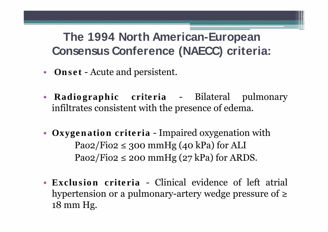
The 1994 North American-European Consensus Conference (NAECC) criteria:
• Onset - Acute and persistent.
• Radiographic criteria - Bilateral pulmonaryinfiltrates consistent with the presence of edema.
• Oxygenation criteria - Impaired oxygenation withPao2/Fio2 ≤ 300 mmHg (40 kPa) for ALIPao2/Fio2 ≤ 200 mmHg (27 kPa) for ARDS.
• Exclusion criteria - Clinical evidence of left atrialhypertension or a pulmonary-artery wedge pressure of ≥18 mm Hg.
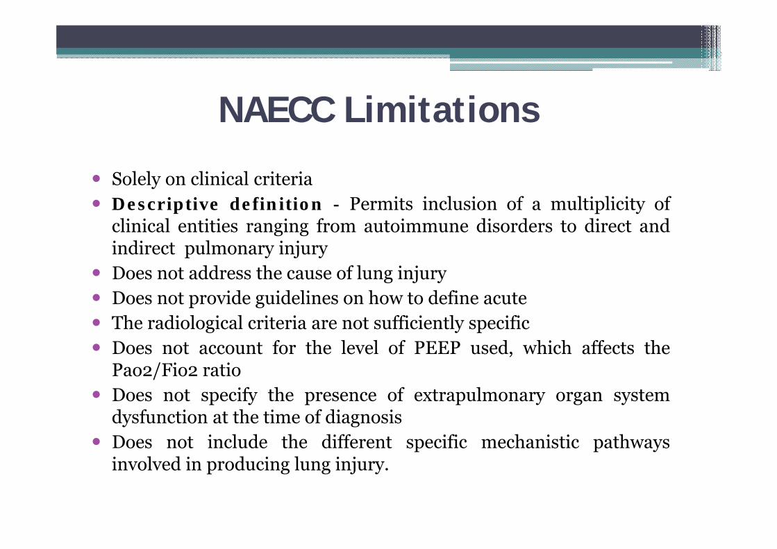
NAECC Limitations
Solely on clinical criteria Descriptive definition - Permits inclusion of a multiplicity of
clinical entities ranging from autoimmune disorders to direct andindirect pulmonary injury
Does not address the cause of lung injury Does not provide guidelines on how to define acute The radiological criteria are not sufficiently specific Does not account for the level of PEEP used, which affects the
Pao2/Fio2 ratio Does not specify the presence of extrapulmonary organ system
dysfunction at the time of diagnosis Does not include the different specific mechanistic pathways
involved in producing lung injury.
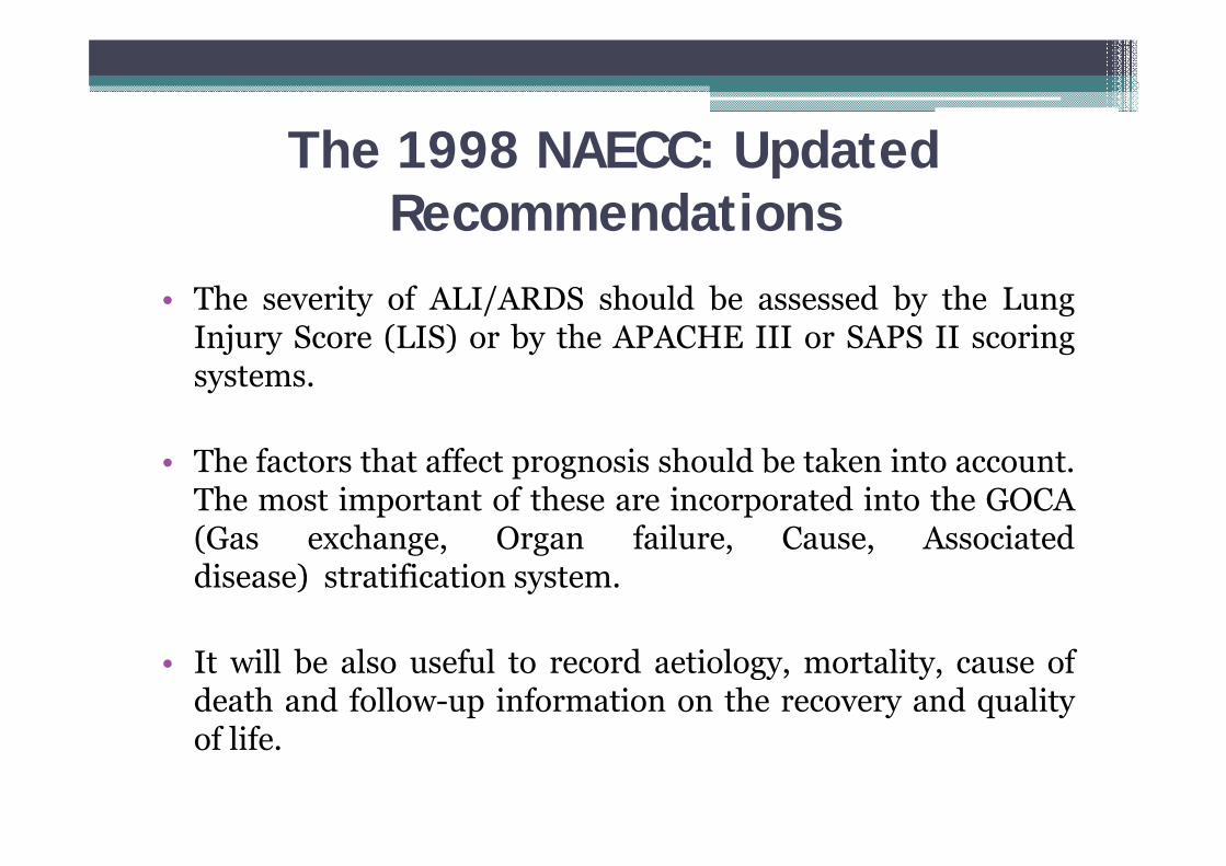
The 1998 NAECC: UpdatedRecommendations
• The severity of ALI/ARDS should be assessed by the LungInjury Score (LIS) or by the APACHE III or SAPS II scoringsystems.
• The factors that affect prognosis should be taken into account.The most important of these are incorporated into the GOCA(Gas exchange, Organ failure, Cause, Associateddisease) stratification system.
• It will be also useful to record aetiology, mortality, cause ofdeath and follow-up information on the recovery and qualityof life.
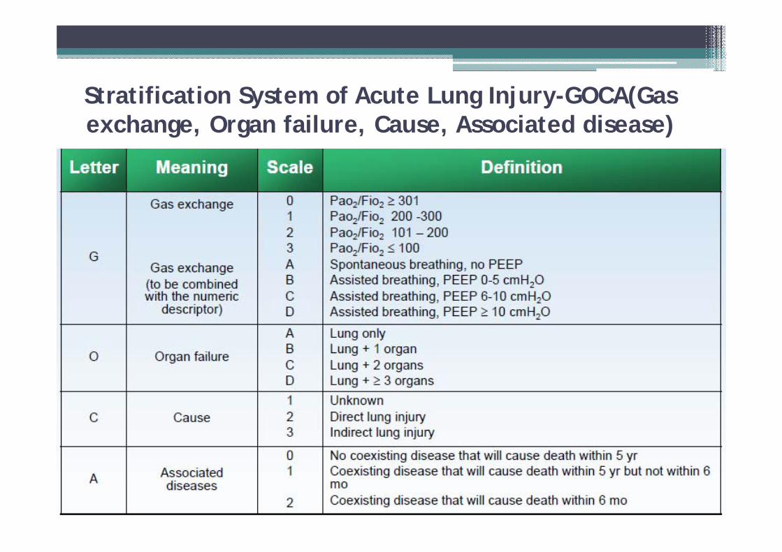
Stratification System of Acute Lung Injury-GOCA(Gas exchange, Organ failure, Cause, Associated disease)
Lung injury score
Four components:
1. Chest radiograph2. PEEP score3. Hypoxemia score4. Respiratory system compliance score
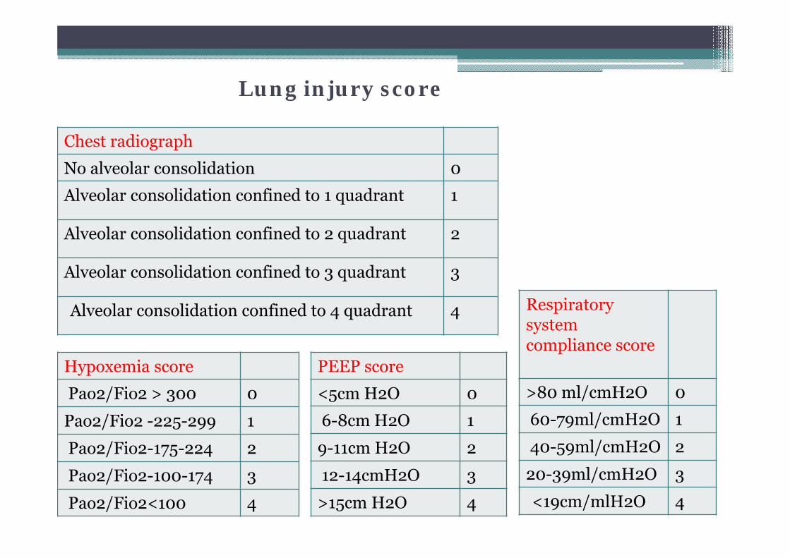
Chest radiograph
No alveolar consolidation 0
Alveolar consolidation confined to 1 quadrant 1
Alveolar consolidation confined to 2 quadrant 2
Alveolar consolidation confined to 3 quadrant 3
Alveolar consolidation confined to 4 quadrant 4
PEEP score
<5cm H2O 0
6-8cm H2O 1
9-11cm H2O 2
12-14cmH2O 3
>15cm H2O 4
Hypoxemia score
Pao2/Fio2 > 300 0
Pao2/Fio2 -225-299 1
Pao2/Fio2-175-224 2
Pao2/Fio2-100-174 3
Pao2/Fio2<100 4
Respiratory system compliance score
>80 ml/cmH2O 0
60-79ml/cmH2O 1
40-59ml/cmH2O 2
20-39ml/cmH2O 3
<19cm/mlH2O 4
Lung injury score
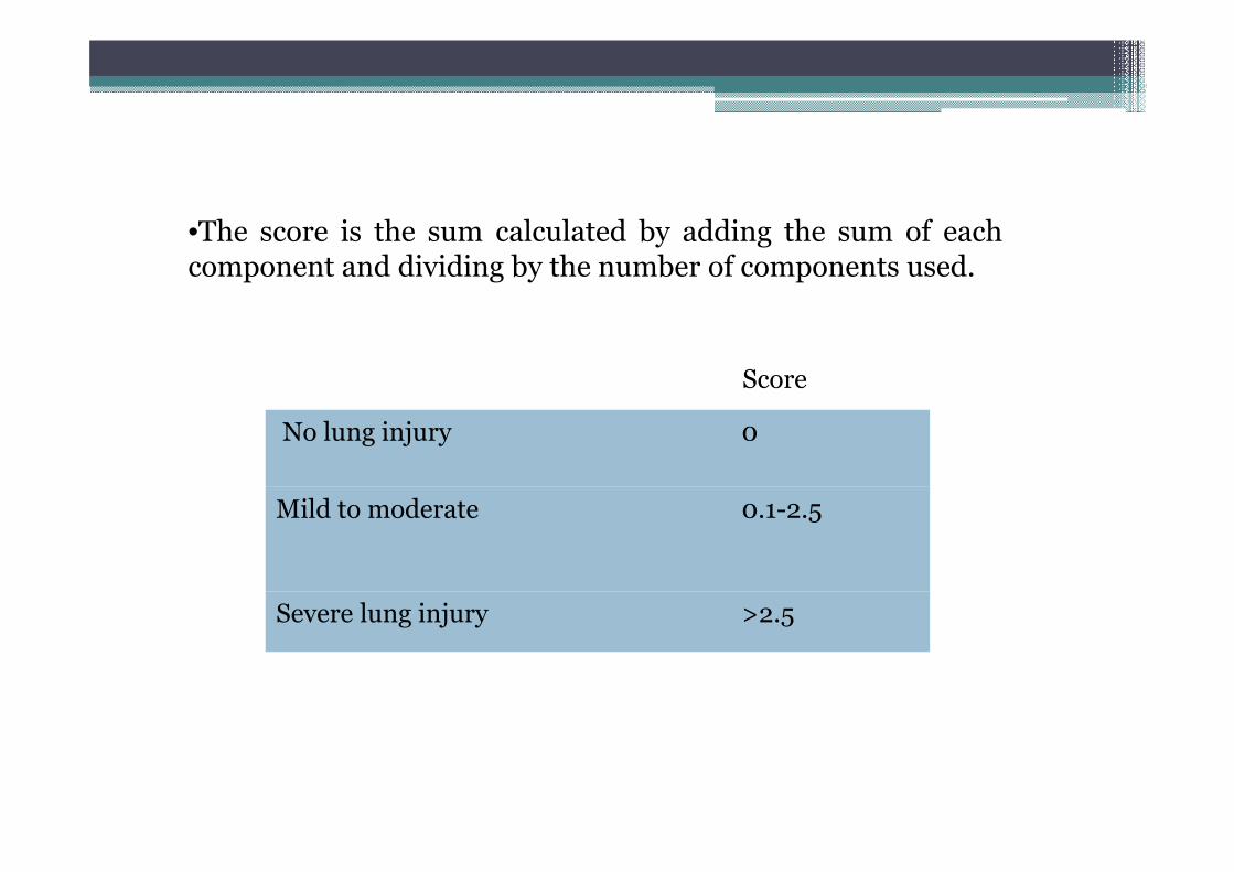
•The score is the sum calculated by adding the sum of eachcomponent and dividing by the number of components used.
No lung injury 0
Mild to moderate 0.1-2.5
Severe lung injury >2.5
Score
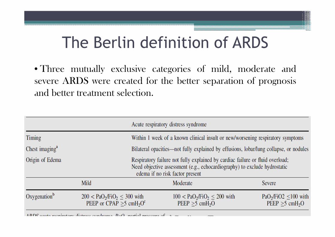
The Berlin definition of ARDS• Three mutually exclusive categories of mild, moderate andsevere ARDS were created for the better separation of prognosisand better treatment selection.
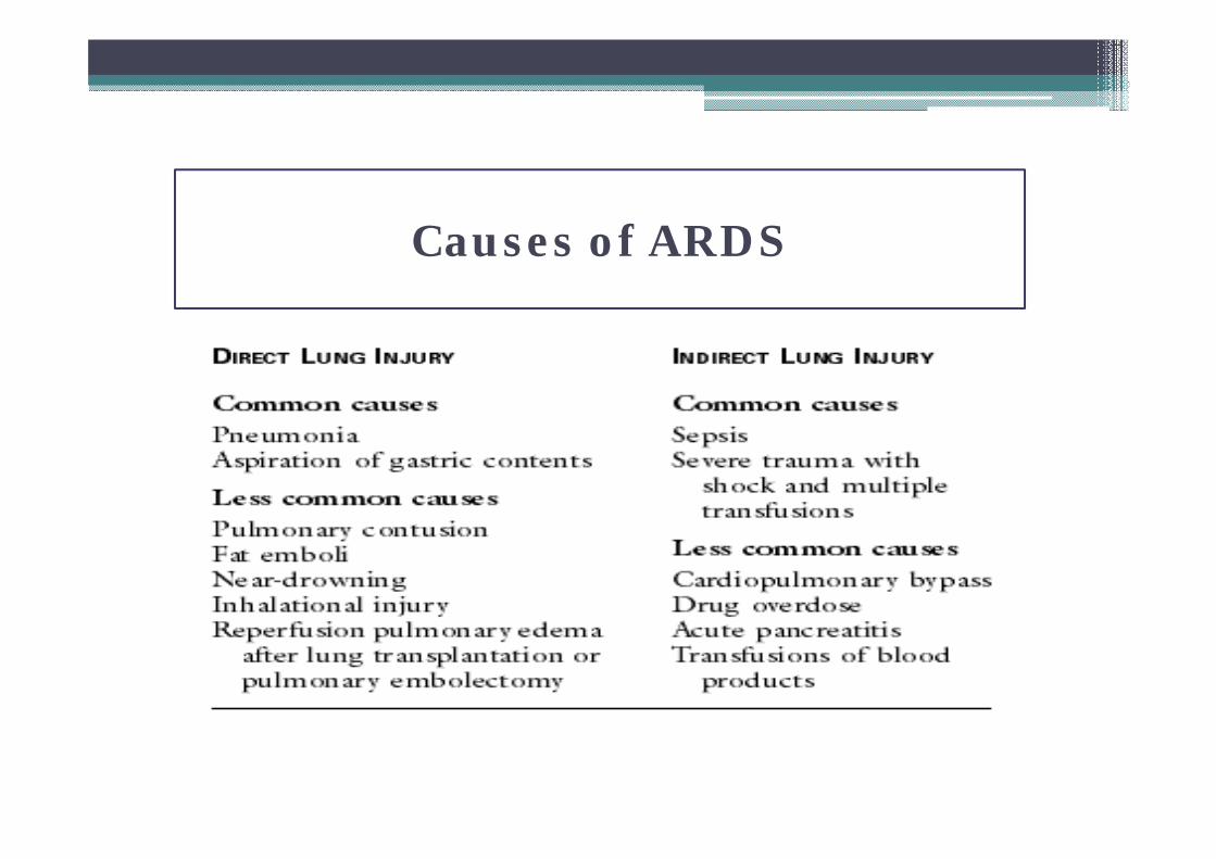
Causes of ARDS and ALICauses of ARDS
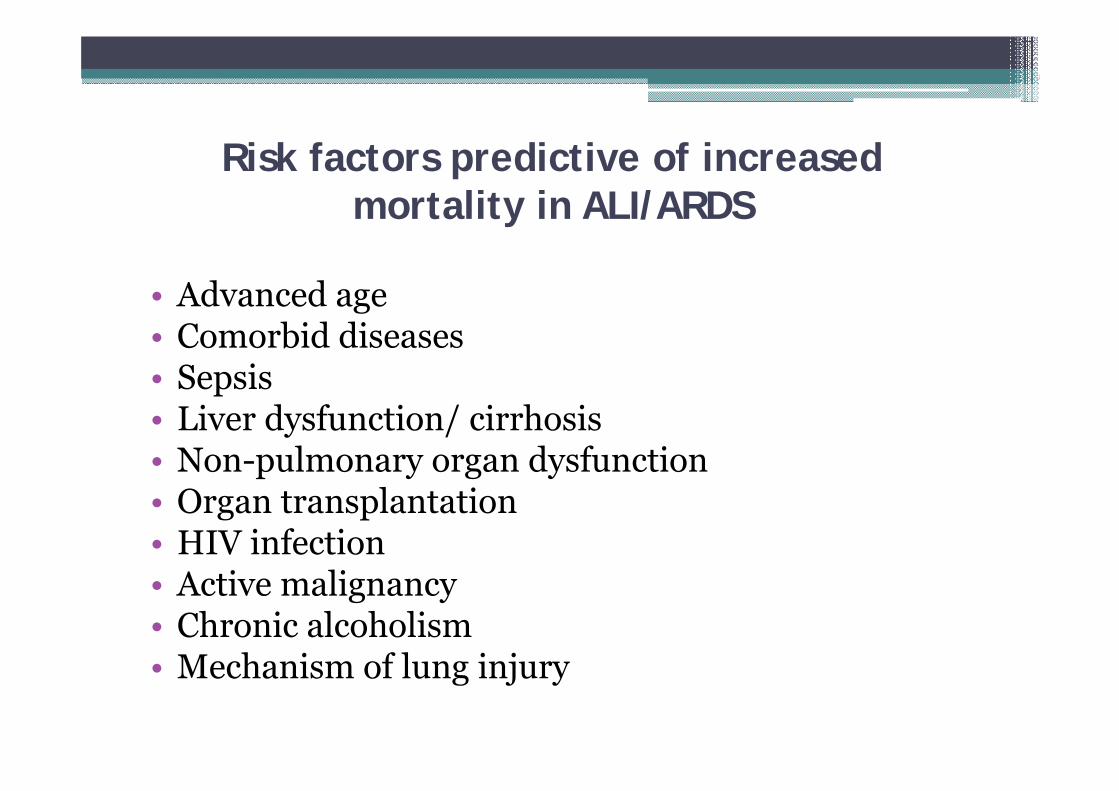
Risk factors predictive of increased mortality in ALI/ARDS
• Advanced age• Comorbid diseases• Sepsis• Liver dysfunction/ cirrhosis• Non-pulmonary organ dysfunction• Organ transplantation• HIV infection• Active malignancy• Chronic alcoholism• Mechanism of lung injury
Pathophysiology
3 phases –
1. Exudative/ Inflammatory
2. Proliferative
3. Fibrotic
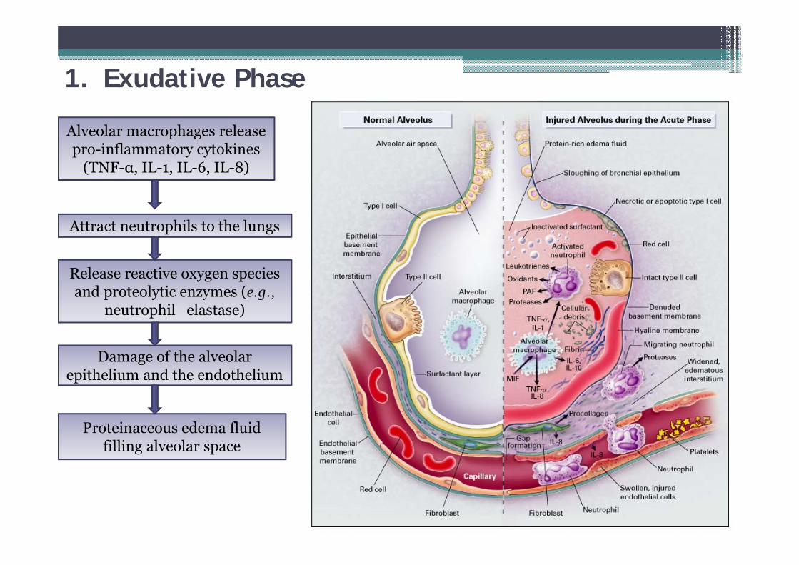
1. Exudative Phase
Damage of the alveolar epithelium and the endothelium
Alveolar macrophages release pro-inflammatory cytokines
(TNF-α, IL-1, IL-6, IL-8)
Proteinaceous edema fluid filling alveolar space
Release reactive oxygen species and proteolytic enzymes (e.g.,
neutrophil elastase)
Attract neutrophils to the lungs
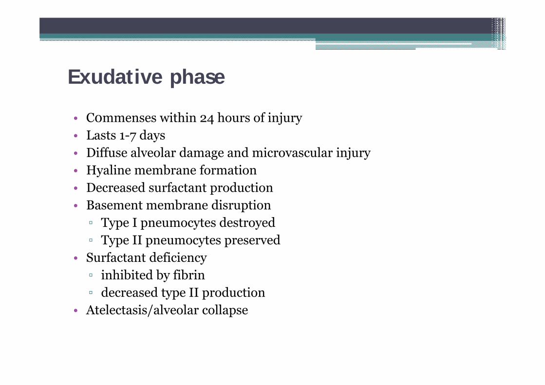
Exudative phase
• C0mmenses within 24 hours of injury• Lasts 1-7 days• Diffuse alveolar damage and microvascular injury• Hyaline membrane formation• Decreased surfactant production• Basement membrane disruption▫ Type I pneumocytes destroyed▫ Type II pneumocytes preserved
• Surfactant deficiency▫ inhibited by fibrin▫ decreased type II production
• Atelectasis/alveolar collapse
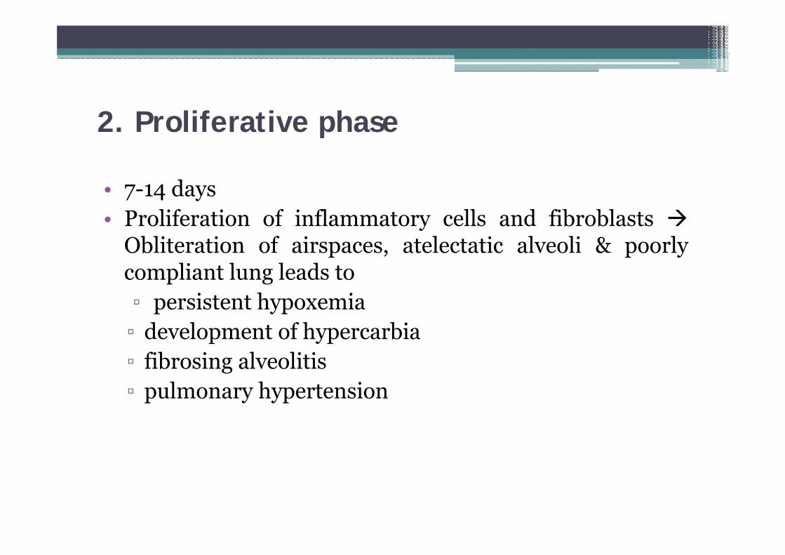
2. Proliferative phase
• 7-14 days• Proliferation of inflammatory cells and fibroblasts
Obliteration of airspaces, atelectatic alveoli & poorlycompliant lung leads to▫ persistent hypoxemia▫ development of hypercarbia▫ fibrosing alveolitis▫ pulmonary hypertension
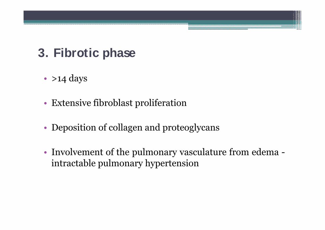
3. Fibrotic phase
• >14 days
• Extensive fibroblast proliferation
• Deposition of collagen and proteoglycans
• Involvement of the pulmonary vasculature from edema -intractable pulmonary hypertension
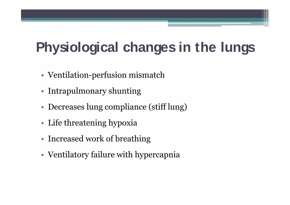
Physiological changes in the lungs
• Ventilation-perfusion mismatch
• Intrapulmonary shunting
• Decreases lung compliance (stiff lung)
• Life threatening hypoxia
• Increased work of breathing
• Ventilatory failure with hypercapnia
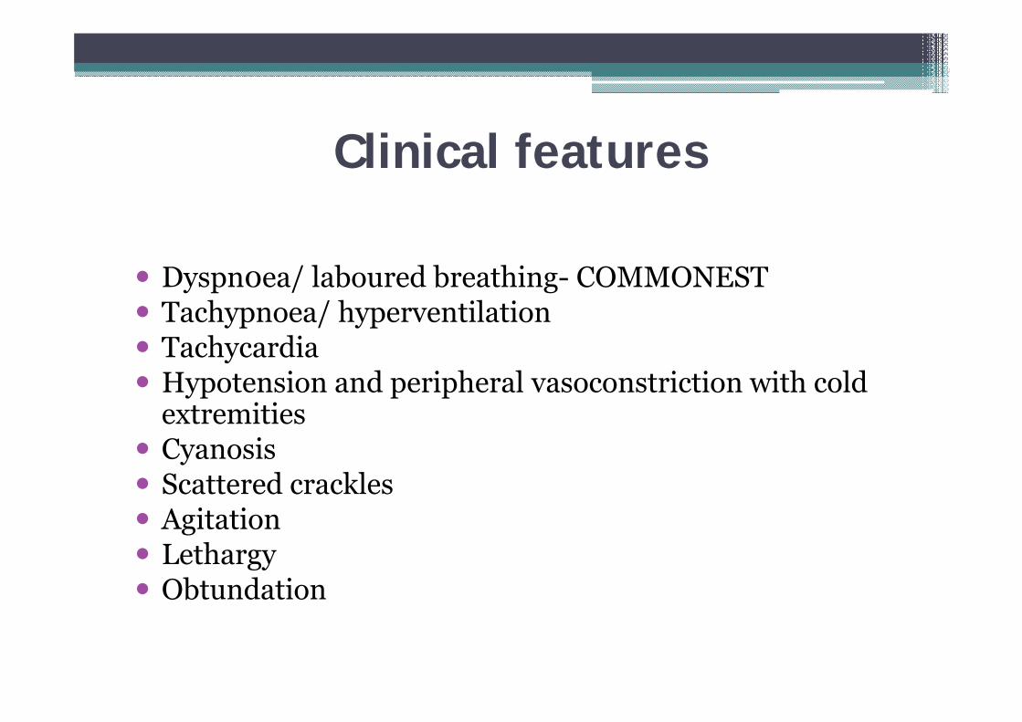
Clinical features
Dyspn0ea/ laboured breathing- COMMONEST Tachypnoea/ hyperventilation Tachycardia Hypotension and peripheral vasoconstriction with cold
extremities Cyanosis Scattered crackles Agitation Lethargy Obtundation
Management
3 cornerstones:
1. Recognizing patients with acute lung injury and ruling out other causes of acute hypoxemia
2. Identifying and treating the underlying disease3. Mechanical ventilation to secure oxygenation
and ventilation whilst protecting the lung from ventilation-induced lung injury
Approach to clinical diagnosis1. Chest x rayEarly stages- interstitial edemaEstablished cases- full blown pulmonary edema
Not reliable :“normal” for a period of time (hours -days) following the precipitating event [e.g. sepsis]
Progression to diffuse, bilateral alveolar infiltrates within 4 - 24 hours after the first abnormal radiographic signs appear
Diagnostic confusion: cardiac failure, pneumonia, pulmonary embolism
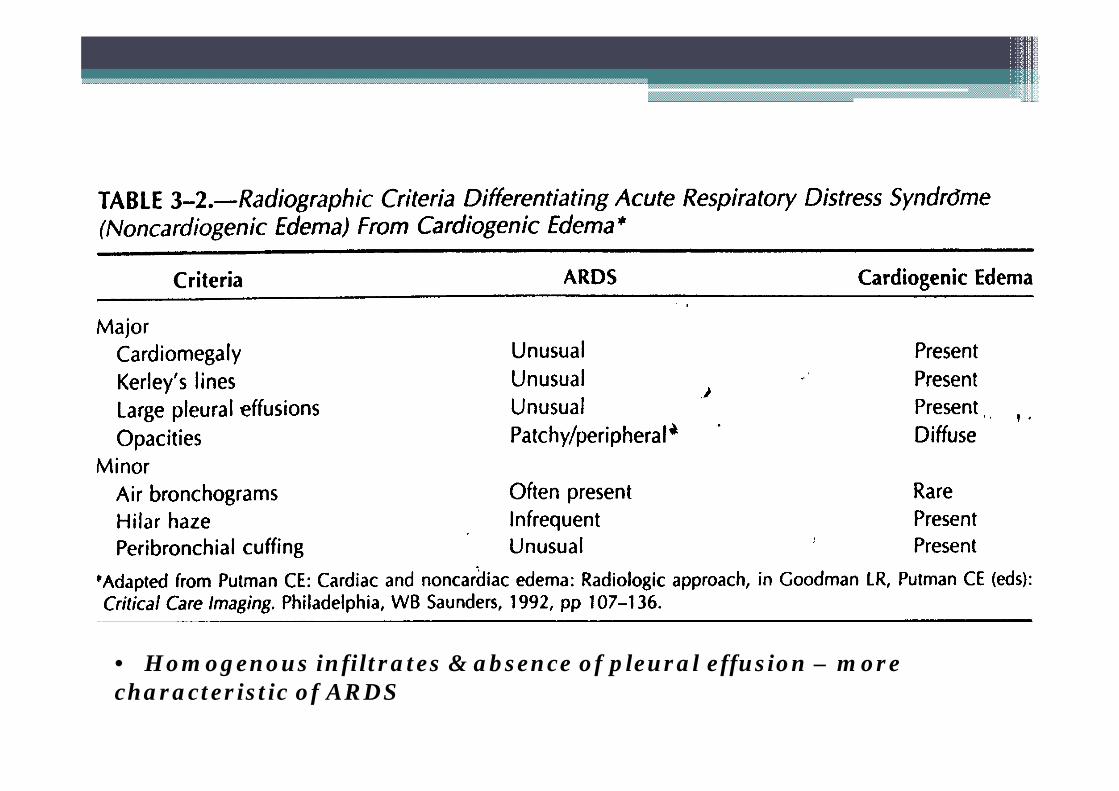
• Homogenous infiltrates & absence of pleural effusion – more characteristic of ARDS
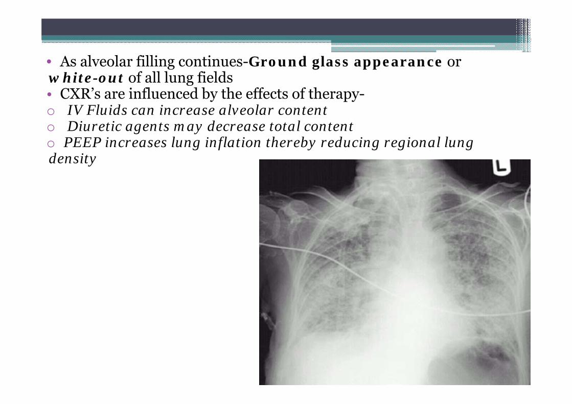
• As alveolar filling continues-Ground glass appearance orwhite-out of all lung fields• CXR’s are influenced by the effects of therapy-o IV Fluids can increase alveolar content o Diuretic agents may decrease total content o PEEP increases lung inflation thereby reducing regional lung density
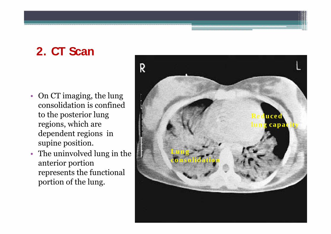
2. CT Scan
• On CT imaging, the lung consolidation is confined to the posterior lung regions, which are dependent regions in supine position.
• The uninvolved lung in the anterior portion represents the functional portion of the lung.
Reduced lung capacity
Lung consolidation
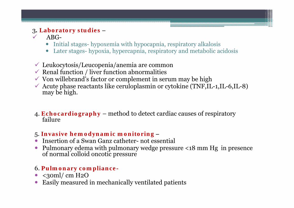
3. Laboratory studies – ABG-
Initial stages- hypoxemia with hypocapnia, respiratory alkalosis Later stages- hypoxia, hypercapnia, respiratory and metabolic acidosis
Leukocytosis/Leucopenia/anemia are common Renal function / liver function abnormalities Von willebrand’s factor or complement in serum may be high Acute phase reactants like ceruloplasmin or cytokine (TNF,IL-1,IL-6,IL-8)
may be high.
4. Echocardiography – method to detect cardiac causes of respiratory failure
5. Invasive hemodynamic monitoring – Insertion of a Swan Ganz catheter- not essential Pulmonary edema with pulmonary wedge pressure <18 mm Hg in presence
of normal colloid oncotic pressure
6. Pulmonary compliance- <30ml/ cm H2O Easily measured in mechanically ventilated patients
7. Full bacteriological screen – Blood , urine and tracheal aspirate cultures Culture of other body secretions and discharges Culture of bronchoalveolar lavage fluid.
8. Bronchoalveolar Lavage – Very reliable & underutilized In normal subjects, less than 5% of cells are neutrophils. In patients
with ARDS, upto 80% cells are neutrophils. Lavage fluid is rich in protein in patients of ARDS as the
inflammatory exudates are rich in protein.Hydrostatic edema: Lavage protein/ Plasma protein < 0.5ARDS : Lavage protein/ Plasma protein > 0.7
Presence of a marker of pulmonary fibrosis called procollagen peptide III (PCPIII) correlates with mortality.
Ventilatory management in ARDS
• In the early stages of ARDS, the hypoxia may be
corrected by 40 to 60% inspired oxygen with CPAP.
• If the patient is well oxygenated on ≤ 60 % inspired
oxygen and apparently stable, then close ward
monitoring , continuous oximetry and regular blood gas
estimation is required.
Indications of ventilatory support
Inadequate Oxygenation(PaO2 < 60mm Hg on FiO2 >= 0.6)
Rising or elevated PaCO2(>= 45 mmHg)
Clinical signs of incipient respiratory failure
Conventional mechanical ventilation
• Tidal volumes are 12-15ml/kg
• In ARDS, the functional portion of the lungs is greatly reduced
• High inflation volumes results in overdistension and rupture of the distal airspaces.
• Ventilator associated lung injury:
High inflation pressure - Barotrauma
Over distension - Volutrauma
Repetitive opening & closing of alveoli - Atelectrauma
SIRS & cytokines release - Biotrauma.
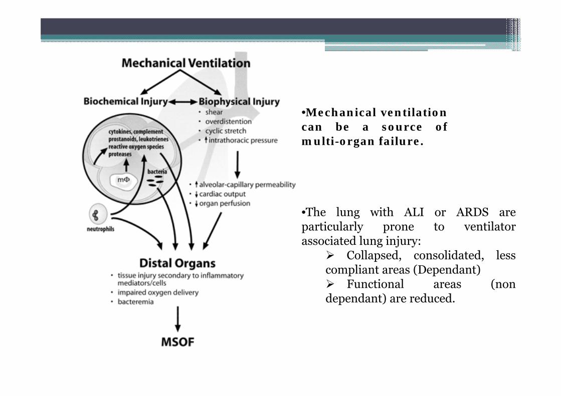
•Mechanical ventilationcan be a source ofmulti-organ failure.
•The lung with ALI or ARDS areparticularly prone to ventilatorassociated lung injury:
Collapsed, consolidated, lesscompliant areas (Dependant) Functional areas (nondependant) are reduced.
Lung protective ventilation
• Tidal volumes – 6ml/kg
• Limits risk of volutrauma and biotrauma
• Uses positive end expiratory pressure to limit the risk of
atelectrauma.
• Currently, the only therapy that has been proven to be
effective at reducing mortality in ALI/ARDS in a large,
randomized, multi-center, controlled trial is a protective
ventilatory strategy.
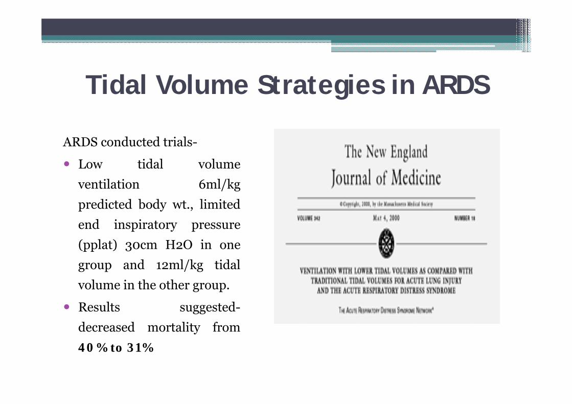
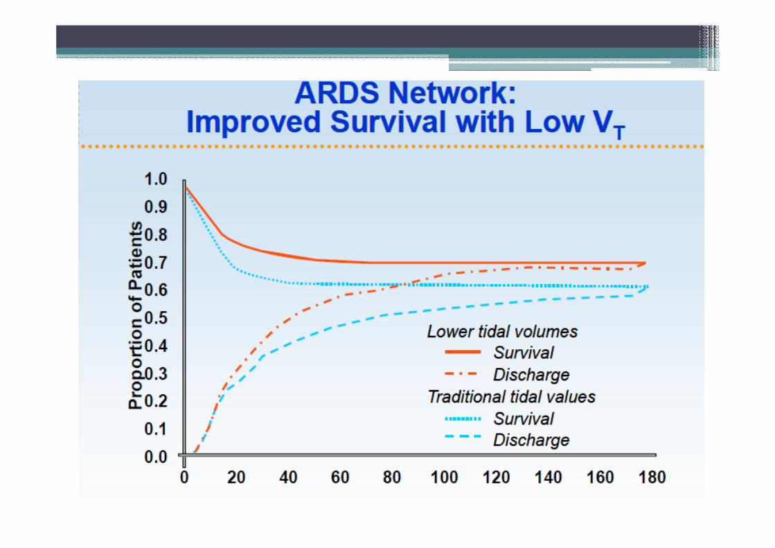
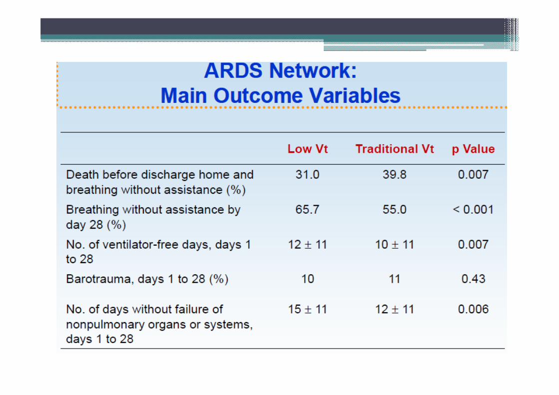
Tidal Volume Strategies in ARDS
ARDS conducted trials-
Low tidal volumeventilation 6ml/kgpredicted body wt., limitedend inspiratory pressure(pplat) 30cm H2O in onegroup and 12ml/kg tidalvolume in the other group.
Results suggested-decreased mortality from40% to 31%
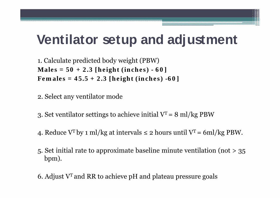
Ventilator setup and adjustment1. Calculate predicted body weight (PBW)Males = 50 + 2.3 [height (inches) - 60]Females = 45.5 + 2.3 [height (inches) -60]
2. Select any ventilator mode
3. Set ventilator settings to achieve initial VT = 8 ml/kg PBW
4. Reduce VT by 1 ml/kg at intervals ≤ 2 hours until VT = 6ml/kg PBW.
5. Set initial rate to approximate baseline minute ventilation (not > 35bpm).
6. Adjust VT and RR to achieve pH and plateau pressure goals
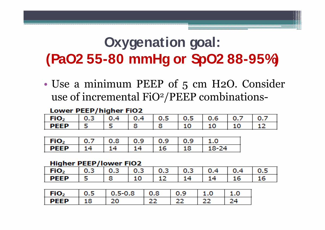
Oxygenation goal: (PaO2 55-80 mmHg or SpO2 88-95%)
• Use a minimum PEEP of 5 cm H2O. Consideruse of incremental FiO2/PEEP combinations-
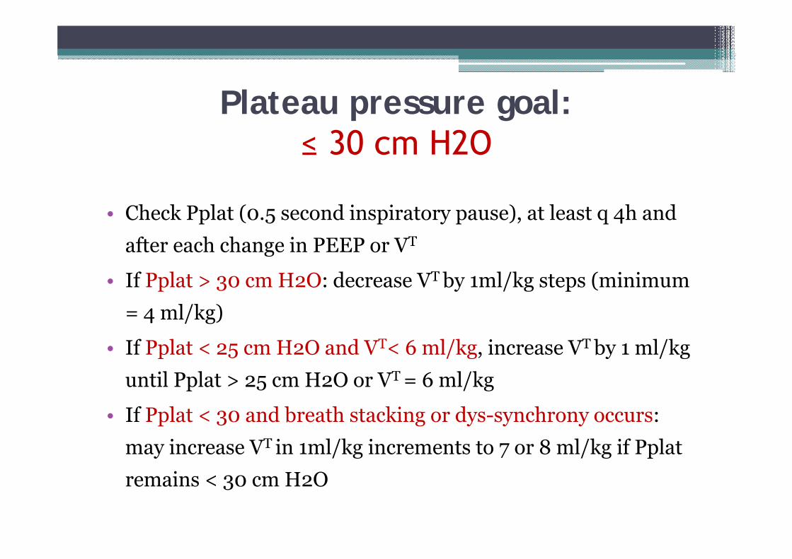
Plateau pressure goal: ≤ 30 cm H2O
• Check Pplat (0.5 second inspiratory pause), at least q 4h and after each change in PEEP or VT
• If Pplat > 30 cm H2O: decrease VT by 1ml/kg steps (minimum = 4 ml/kg)
• If Pplat < 25 cm H2O and VT< 6 ml/kg, increase VT by 1 ml/kg until Pplat > 25 cm H2O or VT = 6 ml/kg
• If Pplat < 30 and breath stacking or dys-synchrony occurs: may increase VT in 1ml/kg increments to 7 or 8 ml/kg if Pplat remains < 30 cm H2O
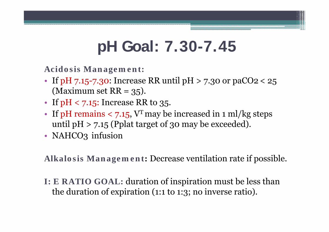
pH Goal: 7.30-7.45Acidosis Management:• If pH 7.15-7.30: Increase RR until pH > 7.30 or paCO2 < 25
(Maximum set RR = 35).• If pH < 7.15: Increase RR to 35. • If pH remains < 7.15, VT may be increased in 1 ml/kg steps
until pH > 7.15 (Pplat target of 30 may be exceeded). • NAHCO3 infusion
Alkalosis Management: Decrease ventilation rate if possible.
I: E RATIO GOAL: duration of inspiration must be less than the duration of expiration (1:1 to 1:3; no inverse ratio).
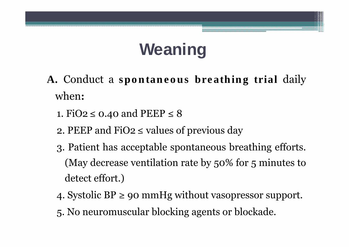
Weaning
A. Conduct a spontaneous breathing trial dailywhen:1. FiO2≤ 0.40 and PEEP ≤ 8
2. PEEP and FiO2≤ values of previous day
3. Patient has acceptable spontaneous breathing efforts.(May decrease ventilation rate by 50% for 5 minutes todetect effort.)
4. Systolic BP ≥ 90 mmHg without vasopressor support.
5. No neuromuscular blocking agents or blockade.
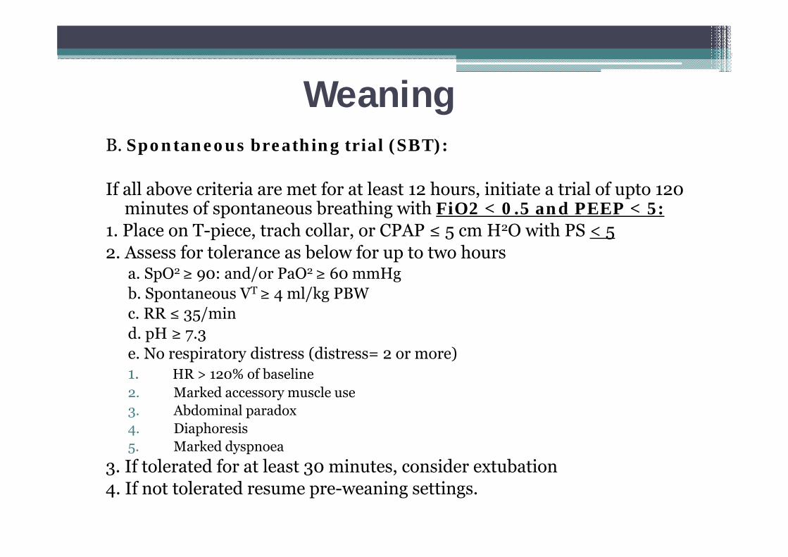
WeaningB. Spontaneous breathing trial (SBT):
If all above criteria are met for at least 12 hours, initiate a trial of upto 120 minutes of spontaneous breathing with FiO2 < 0.5 and PEEP < 5:
1. Place on T-piece, trach collar, or CPAP ≤ 5 cm H2O with PS < 5 2. Assess for tolerance as below for up to two hours
a. SpO2 ≥ 90: and/or PaO2 ≥ 60 mmHg b. Spontaneous VT ≥ 4 ml/kg PBW c. RR ≤ 35/min d. pH ≥ 7.3 e. No respiratory distress (distress= 2 or more) 1. HR > 120% of baseline 2. Marked accessory muscle use 3. Abdominal paradox 4. Diaphoresis 5. Marked dyspnoea
3. If tolerated for at least 30 minutes, consider extubation4. If not tolerated resume pre-weaning settings.
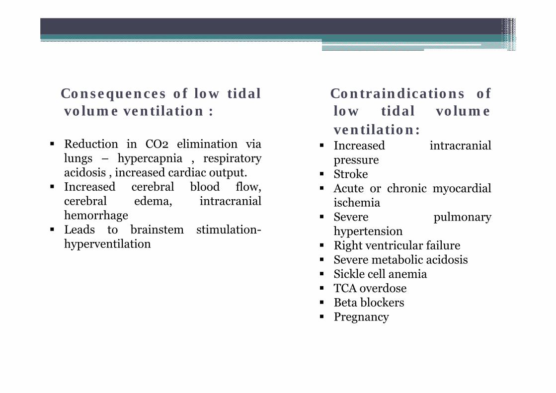
Consequences of low tidalvolume ventilation :
Reduction in CO2 elimination vialungs – hypercapnia , respiratoryacidosis , increased cardiac output.
Increased cerebral blood flow,cerebral edema, intracranialhemorrhage
Leads to brainstem stimulation-hyperventilation
Contraindications oflow tidal volumeventilation:
Increased intracranialpressure
Stroke Acute or chronic myocardial
ischemia Severe pulmonary
hypertension Right ventricular failure Severe metabolic acidosis Sickle cell anemia TCA overdose Beta blockers Pregnancy
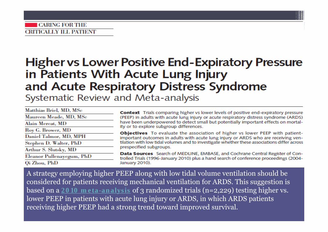
A strategy employing higher PEEP along with low tidal volume ventilation should be considered for patients receiving mechanical ventilation for ARDS. This suggestion is based on a 2010 meta-analysis of 3 randomized trials (n=2,229) testing higher vs. lower PEEP in patients with acute lung injury or ARDS, in which ARDS patients receiving higher PEEP had a strong trend toward improved survival.
Role of PEEP in ARDS
• PEEP avoids repetitive opening and collapse of atelectatic lung
units – could protect against VILI
• Improves arterial oxygenation
• Lung is kept open by using PEEP to avoid end expiratory
collapse
▫ Preserves inspiratory recruitment
▫ Re-establish end expiratory lung volume
▫ Prevent surfactant loss in airway
▫ Decreases intrapulmonary shunt
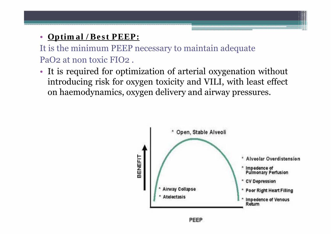
• Optimal /Best PEEP:It is the minimum PEEP necessary to maintain adequatePaO2 at non toxic FIO2 .• It is required for optimization of arterial oxygenation without
introducing risk for oxygen toxicity and VILI, with least effecton haemodynamics, oxygen delivery and airway pressures.
Adverse effects of PEEP
1. Decreased cardiac output
2. Increased pulmonary edema formation
3. Increased dead space & increased resistance of the bronchial
circulation
4. Increased lung volume and stretch during inspiration
lung injury or barotrauma.
• These adverse effects are more pronounced in patients with
direct lung injury
Recruitment maneuvers
Application of continuous positive airway pressure aimed at recruiting or
opening totally or partially collapsed alveoli.
▫ The most well known method of recruitment maneuver is sustained
application of CPAP of 30- 50 cm H2O for 30 seconds
▫ Periodic recruitment with a series of traditional sigh breaths
▫ Intermittently raising PEEP over several breaths
▫ Extended sigh maneuver with step wise increase in PEEP while Vt
is decreased
▫ Intermittent application of pressure controlled ventilation with
incremental high PEEP
Other approaches to ventilatory management
1. Pressure control mode – PCV limits maximal peak airway
pressure , end inspiratory pressure.
2. Inverse ratio ventilation with PCV – inspiratory time >
expiratory time , some case reports – patients with refractory
hypoxemia responded to PCV IRV
Due to intrinsic PEEP , alveolar recruitment
Can compromise cardiac output – risk of non pulmonary organ
dysfunction
Used as salvage mode
3. Modes that allow spontaneous breathing during positivepressure ventilation –
BiPAP, APRV- airway pressure cycles between higher and lowerlevels of PEEP
▫ Decreases- intrathoracic pressure
▫ Improved ventilation perfusion mismatch
▫ Improved cardiac output
▫ Decreases shunt fraction , dead space
▫ Less patient ventilator asynchrony
4. High frequency oscillatory ventilation (HFOV)
Delivers small tidal volumes (1-2 ml/kg) Rapid oscillations (300 cycles/min) Ventilation is achieved by oscillation of air around set airway pressure Constant airway pressures maintain open lung, optimize lung
recruitment Lower Vt – less alveolar distension High mean pressures- higher end expiratory lung volumes and
recruitment- improvement in oxygenation Disadvantages: A special ventilator is required Aerosolized bronchodilators are ineffective Cardiac output is decreased due to high mean airway pressures
Adjuncts to protective lung ventilation
• Permissive hypercapnia
• Fluid and hemodynamic management
• Inhaled nitric oxide
• Prone position ventilation
• Tracheal gas insufflation
• Steroids
• Nutritional support
• Other drug therapies
1.Permissive hypercapnia
• Low tidal volume ventilation results in hypercapnia.
• Clinicians have allowed hypercapnia to persist in patients of
ARDS as long as there is no evidence of harm.
• Clinical trials have shown that arterial pCO2 level of 60-70
mmHg and arterial pH of 7.2-7.25 are safe for most of the
patients.
2. Fluid management
• NIH/ARDS Network Fluid Management Trial (The FACTT Study)– FACTT (Fluid and Catheter Therapy Trial; 2005-6) - fluid restriction results in improved outcome (may reduce pulmonary edema due to increased pulmonary vascular permeability).
• FACTT – fluid management protocol based on –1. Mean arterial pressure >60mmHg2. Urine output >0.5ml/kg3. Effectiveness of circulation CI >2.5l/min/m24. Intravascular pressure –CVP <4mmHg
• Result – improved lung function, improved central nervous system function, less sedation, mechanical ventilation , less complications
• Avoiding a positive fluid balance in ARDS patients reduces the time on mechanical ventilation and also mortality.
3. Haemodynamic management
• ARDSNet FACTT compared hemodynamic management guided -CVP/PAC SUPPORT – use of PAC increases mortality
PAC allows measurement of central venous and pulmonary arterialpressure, pulmonary artery occlusion pressure , mixed venous bloodgases, cardiac output.
no added benefit, increasing complications CVP better indicator of measurement than PAC
Inotropes Dobutamine is preferred- because vasodilators increase
intrapulmonary shunt Dopamine – constricts pulmonary veins , rise in pulmonary capillary
hydrostatic pressure
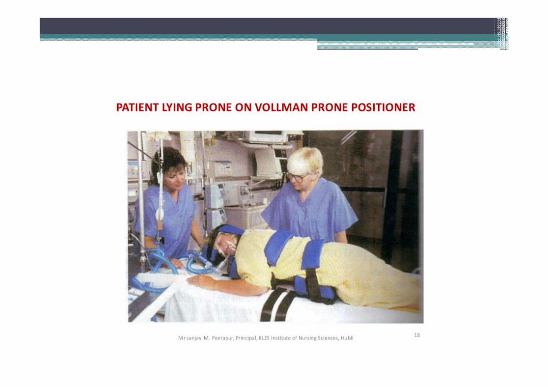
4. Prone ventilation
• Routine use of prone positioning in all patients with ALI / ARDS cannot be currently recommended due to a lack of clinical data support.
• Indications:– as an adjunctive therapy to improve oxygenation in established
ALI and ARDS – considered in patients who require PEEP >12 cmH2O and a FiO2
>0.60• should better be used early within 36 hours of the onset of ARDS• optimum duration unknown
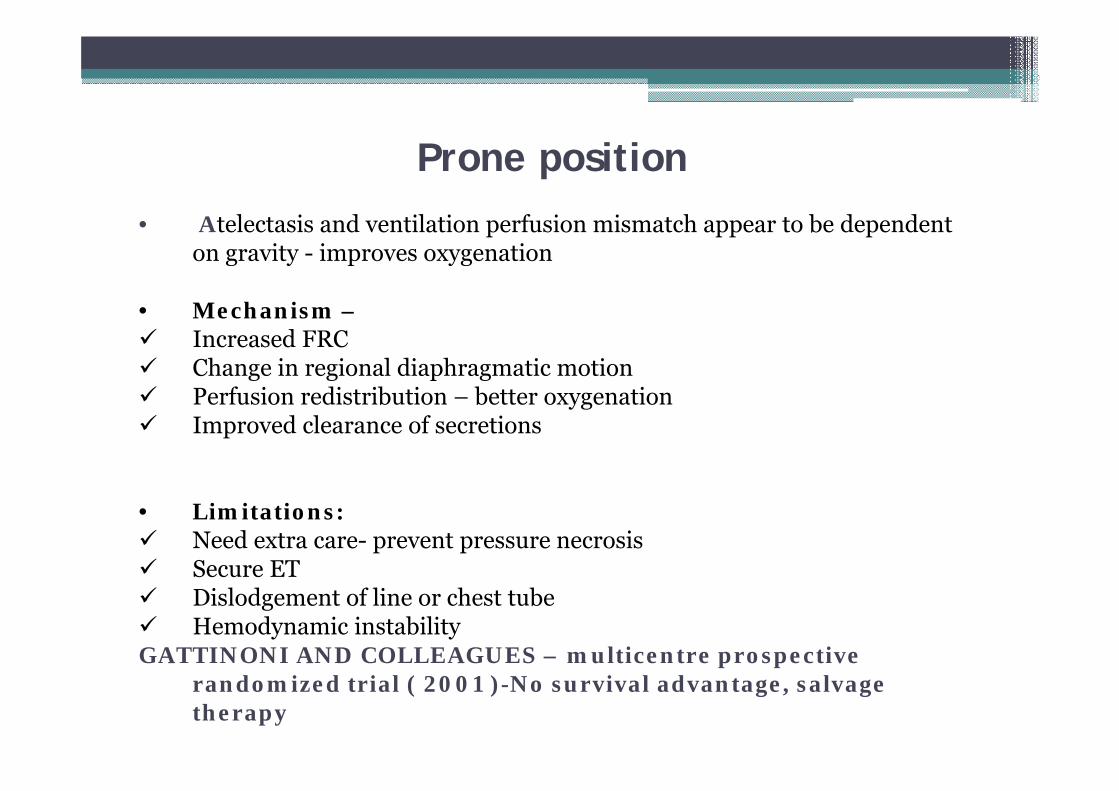
Prone position• Atelectasis and ventilation perfusion mismatch appear to be dependent
on gravity - improves oxygenation
• Mechanism – Increased FRC Change in regional diaphragmatic motion Perfusion redistribution – better oxygenation Improved clearance of secretions
• Limitations: Need extra care- prevent pressure necrosis Secure ET Dislodgement of line or chest tube Hemodynamic instabilityGATTINONI AND COLLEAGUES – multicentre prospective
randomized trial ( 2001 )-No survival advantage, salvage therapy
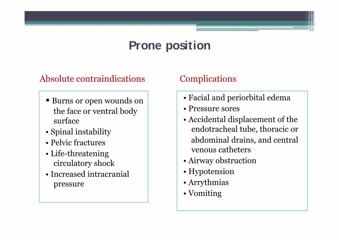
Prone position
• Burns or open wounds on the face or ventral body surface
• Spinal instability• Pelvic fractures• Life-threatening
circulatory shock• Increased intracranial
pressure
• Facial and periorbital edema• Pressure sores• Accidental displacement of the
endotracheal tube, thoracic orabdominal drains, and central venous catheters
• Airway obstruction• Hypotension• Arrythmias• Vomiting
Absolute contraindications Complications
5. Inhaled nitric oxide
• Selectively dilates pulmonary capillaries• Decreases pulmonary capillary pressure• Selectively vasodilates ventilated areas of lung• Inhibit neutrophils activation• Used as a salvage therapy to improve oxygenation• Adverse effects- methemoglobinemia, renal dysfunction.
6. Tracheal gas insufflation
Delivering fresh gas through modified ET just above carina
Removes CO2 rich gas- from trachea, small airways Reduces anatomic dead space
Risks- Tracheal erosion Oxygen toxicity due to increased Fio2 Hemodynamic compromise Barotraumas
Salvage therapy
7. Steroids
• Steroid therapy is currently recommended only for early
severe and unresolving ARDS:
• Early severe ARDS (PaO2/FiO2<200 mmHg,
PEEP=10 cm H2O):
Methylprednisolone IV loading dose of 1 mg/kg over 30
min, followed by 1 mg/kg/day for 14 days, then taper
over next 14 days and then discontinue.
Steroids
Unresolving ARDS
• In cases where ARDS has not started to resolve after 7 days, high
dose steroid therapy should be started ( but it should not begin after
14 days of onset)
Methylprednisolone IV loading dose of 2 mg/kg over 30 min,
followed by 2 mg/kg/day for 14 days, then 1 mg/kg/day for next 7
days. Gradually taper the dose and discontinue therapy at 2 weeks
after extubation.
5 days after the patient is able to ingest oral medications, oral
prednisone or prednisolone can be started as a single daily dose.
8. Nutrition
• Goals-▫ Provision of adequate nutrition▫ Prevention and treatment of deficiencies
Enteral nutrition superior because Prevents bacterial translocation Lowers levels of TNF, arterial glucagon & epinephrine Lesser febrile responses Lower incidence of infection Less costly
- However parenteral maybe needed in some patients
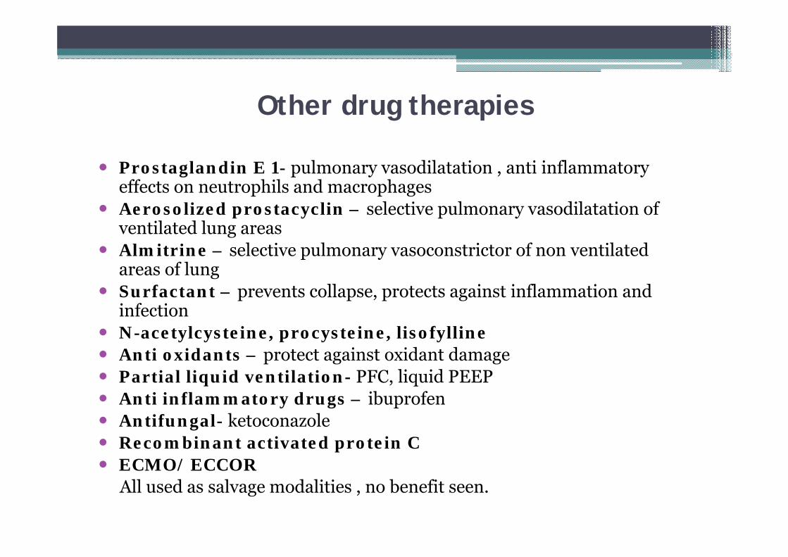
Other drug therapies
Prostaglandin E 1- pulmonary vasodilatation , anti inflammatory effects on neutrophils and macrophages
Aerosolized prostacyclin – selective pulmonary vasodilatation of ventilated lung areas
Almitrine – selective pulmonary vasoconstrictor of non ventilated areas of lung
Surfactant – prevents collapse, protects against inflammation and infection
N-acetylcysteine, procysteine, lisofylline Anti oxidants – protect against oxidant damage Partial liquid ventilation- PFC, liquid PEEP Anti inflammatory drugs – ibuprofen Antifungal- ketoconazole Recombinant activated protein C ECMO/ ECCOR
All used as salvage modalities , no benefit seen.
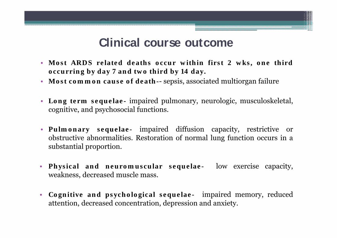
Clinical course outcome• Most ARDS related deaths occur within first 2 wks, one third
occurring by day 7 and two third by 14 day.• Most common cause of death-- sepsis, associated multiorgan failure
• Long term sequelae- impaired pulmonary, neurologic, musculoskeletal,cognitive, and psychosocial functions.
• Pulmonary sequelae- impaired diffusion capacity, restrictive orobstructive abnormalities. Restoration of normal lung function occurs in asubstantial proportion.
• Physical and neuromuscular sequelae- low exercise capacity,weakness, decreased muscle mass.
• Cognitive and psychological sequelae- impaired memory, reducedattention, decreased concentration, depression and anxiety.
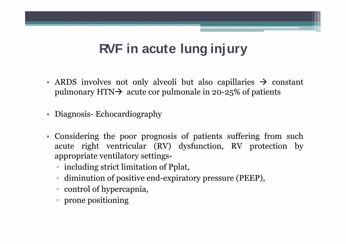
RVF in acute lung injury
• ARDS involves not only alveoli but also capillaries constantpulmonary HTN acute cor pulmonale in 20-25% of patients
• Diagnosis- Echocardiography
• Considering the poor prognosis of patients suffering from suchacute right ventricular (RV) dysfunction, RV protection byappropriate ventilatory settings-▫ including strict limitation of Pplat,▫ diminution of positive end-expiratory pressure (PEEP),▫ control of hypercapnia,▫ prone positioning
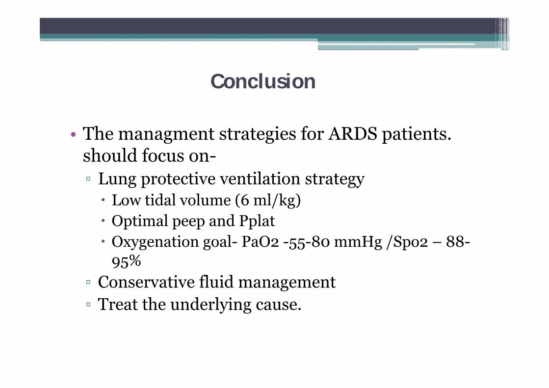
Conclusion
• The managment strategies for ARDS patients. should focus on-▫ Lung protective ventilation strategy Low tidal volume (6 ml/kg) Optimal peep and Pplat Oxygenation goal- PaO2 -55-80 mmHg /Spo2 – 88-
95%▫ Conservative fluid management▫ Treat the underlying cause.

Thank you