Embed Size (px)
Citation preview
Acute rheumatic heart disease
Dr.I.P.Sukumar
Commonest form of acquired heart disease in children especially in the developing countries.Contributes to the larger extent of the heart disease in adults.The long term outcome of the acute rheumatic process depends on the state of the patient during the initial attack. Those who do not have carditis nearly always remain free of chronic valvar heart disease.
Epidemiology
Prevalence in (1937)western countries 4-5/1000 children dropped to 2.9/100000 in the late 1960s.
In India, the prevalence was 6-11/1000 (padmavathi,1978).
In south india a study done by Dr.Koshy et el in 1981 showed the prevalence of rheumatic fever and heart disease was 4.9/1000 in a survey in school children.
Streptococcal throat infection peaks in winter months in western countriesbut there was o seasonal variation in tropical countries.
The incidence of rheumatic fever following the documented streptococcal upper respiratory infection varies from 0.3% during sporadic cases to 3% during the epidemics.
Attack rate is higher in individuals who have had a previous rheumatic episode than in the general population.There are rebound and recurrences in rheumatic fever. Rebound usually occurs within two weeks of stopping the treatment. Recurrence occur around two months or more after discontinuationn of treatment and always follow a new streptococcal infection.
The recurrences frequently show the appearance of c/f present in the first attack ie.,mimetic or true to type.There is lower incidence of carditis during the recurrences in patients initially free of it. Rheumatogenic strains – M type of group A beta hemolytic streptococci -3,5,6,12,18,19,24.
Etiology
Rheumatic fever follows 2-4weeks after a Group A streptococcal infection of the throat and not of the skin.
The infection can be identified in about 25% by bacteriological and in over 90% by serological methods.
Individuals with vigorous antibody response develop rheumatic fever with greater frequency.
Prerequisites for development of ARF – group A streptococcus infection especially of URI, streptococcal antibody response and relative persistence of the organism at the site of infection.
Following is the picture of structure of Streptococcus…
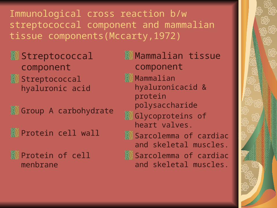
Immunological cross reaction b/w streptococcal component and mammalian tissue components(Mccarty,1972)
Streptococcal componentStreptococcal hyaluronic acid
Group A carbohydrate
Protein cell wall
Protein of cell menbrane
Mammalian tissue componentMammalian hyaluronicacid & protein polysaccharide
Glycoproteins of heart valves.
Sarcolemma of cardiac and skeletal muscles.
Sarcolemma of cardiac and skeletal muscles.
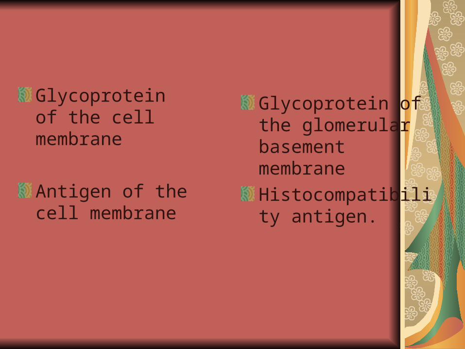
Glycoprotein of the cell membrane
Antigen of the cell membrane
Glycoprotein of the glomerular basement membrane
Histocompatibility antigen.
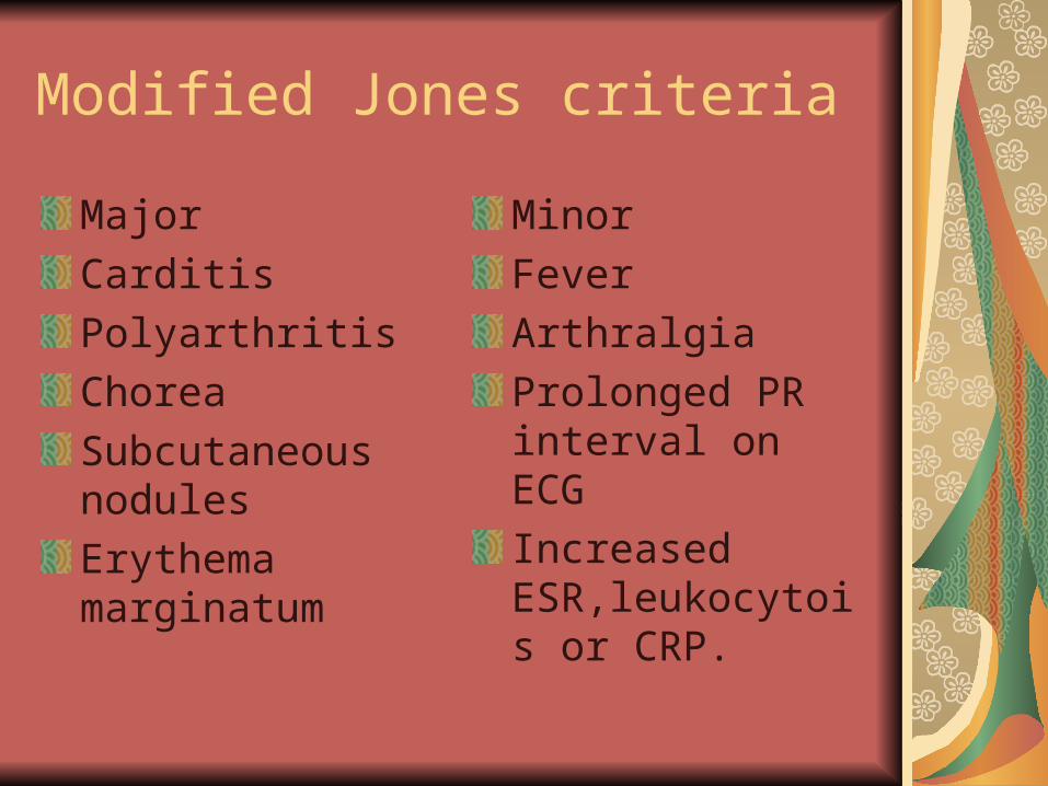
Modified Jones criteria
Major
Carditis
Polyarthritis
Chorea
Subcutaneous nodules
Erythema marginatum
Minor
Fever
Arthralgia
Prolonged PR interval on ECG
Increased ESR,leukocytois or CRP.
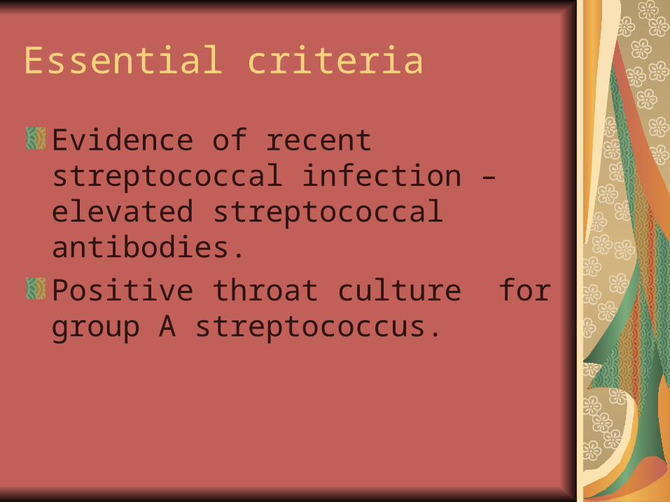
Essential criteria
Evidence of recent streptococcal infection – elevated streptococcal antibodies.
Positive throat culture for group A streptococcus.
Diagnosis
2 major or 1 major and two minor criteria indicate high probability of acute rheumatic process.
These criteria are oly guidelines not meant to substitute clinician ‘s judgement .
Acute Rheumatic heart disease
Carditis is the most serious manifestation of the acute rheumatic process since it causes significant morbidity and mortality.It is the common manifestation occuring in 1/3rd -1/2 of the patients with ARF.It is a pancarditis.Endocarditis – valvar malfunctionMyocarditis – CCFPericarditis – Fibrinoid pericarditis or pericardial effusion.
Cardiac tamponade was unusual.Cardiac involvement tend to appear early in the attack of acute process and rarely delayed for up to 3 weeks.At times, the carditis may be initially sub clinical and may become overt during a recurrent episode.A history of rheumatic fever without carditis does not confer immunity from future rheumatic heart disease.
On examination
Apical murmur- pan systolic murmur of the mitral regurgitation is heard. It is soft and are transmitted to the axilla. The intensity and duration of the murmur can wane and may disappear towards recovery.The development of the low pitched apical mid diastolic murmur (carey coombs) murmur which is attributed to mitral valvulitis helps to confirm the organic significance of MR murmur.It tend to disappear during recovery.
AR- a early decrescendo murmur .It tends to be more persistent and indicates permanent valvar injury.But development of severe AR following a single episode of ARF is unusual.Murmurs due to valvar stenosis are seldom seen in initial attack of active carditis.
The murmurs in acute rheumatic carditis typically change from day to day depending on the alteration in the myocardial and valvar function with or without treatment.
S1- may be diminished in about half the children with MR in the presence of carditis. It is attributed to lengthened conducted time which allows for early closure of mitral valve and diminishes the valvar component of the first heart sound.
In a patient with earlier RHD or past h/o ARF documentation of a change in the established murmur or appearance of new murmur is significant.
CCF – occurs with varying frequency with cardiomegaly, persistent elevation of the sleeping pulse rate and a gallop rhythm.
They donot occur without organic murmur.
The sudden onset of CCF with cardiomegaly may the first manifestation in acute rheumatic valvar disease
Cardiac failure alone should not be accepted as sole evidence of active carditis without supporting evidence.
Pericarditis – always associated with valvar involvement.
Commonly manifests as friction rub heard over the pericardium, especially over the sternum, with a to and fro character in systole and diastole.
It lasts from a few days to weeks.
Pericardial effusion is not uncommon.
In the presence of CCF with myocarditis , pericardial effusion may be unnoticed.
ECG – abnormal ST segment and T wave changes in the left praecardium
ECHO – helps to confirm the diagnosis.
During active phase of carditis, chest radiograph may show rapid changes in the heart size, significant pulmonary congestion or even frank pulmonary edema.It doesnot carry grave prognosis for immediate outcome or longterm valve dysfunction.ECG – show disproportionate sinus tachycardia, a prolonged P-R interval or a prolonged QT interval. Conduction defects due to AV or fascicular blocks may develop transiently but are rarely permanent.
Prolonged PR interval – occurs in 40% of patients with ARF.
In isolation, of much help.
It does not correlate with active carditis, prognosis or residual heart disease.
Natural history
TOMPKIN et el 1972 –reported that about 2/3rd of patients with mitral regurgitation or AR became free of murmurs within 5-10 years.Mitral stenosis did not develop in any patient who was on regular penicillin prophylaxis.New valvular lesions do not develop usually in the absence of recurrences, even in the patient who had carditis as an initial attack but old valvar lesions may evolve further.