Embed Size (px)
Citation preview
J. exp. Biol. 115, 55-68 (1985) 55Printed in Great Britain © The Company ofBiobgists Limited 1985
ADAPTABILITY OF ULTRASTRUCTURE IN THEMAMMALIAN MUSCLE
BY BRENDA R. EISENBERG
Department of Physiology, Rush Medical College, 1750 West Harrison St, Chicago,IL 60612, U.SA.
SUMMARY
All the skeletal muscle fibres taken from an adult mammal do not look alike.The structural differences are a result of adaptations which allow gradationsin mechanical output to be achieved. The anatomy is described and theamounts of the subcellular components are measured by stereologicaltechniques from electron micrographs. A population of normal, adult fibres isclassified by the Z-line width, by the amounts of the mitochondria, T-systemand terminal cisternae (TC), and by the isoforms of contractile proteinspresent. Classification of fibres by some of these ultrastructural componentsgives clusters named fast-twitch and slow-twitch types, but classification byother components gives a continuum of overlapping properties. Trans-formation from the fast- to the slow-twitch type or vice versa follows a specificalteration in the use of the fibre. The mechanical demand on the fibre ismodified by changing the frequency of stimulation in the nerve with animplanted electrode. The time course of the changes in subcellularcomposition in the fibre during adaptation is followed for many weeks.Changes in the membrane systems begin within hours and are complete indays. Changes in the contractile proteins and metabolic systems begin in daysand are complete in weeks. During these transitional phases of adaptation thefibres have an unusual complement of components never seen in a normaladult fibre. Extreme alterations, such as myofibril disassembly or8upranormal amounts of mitochondria also result during some adaptivetransitions. The aberrant appearance in the transitional fibres may be a resultof doing the required mechanical work with a less than optimal set of proteins.At the end of the fibre type transformation, the fibre ultrastructure isindistinguishable from normal.
Modern cell biology is concerned with the ways by which the cell is able to producespecific proteins by the regulation of gene expression. This is often studied indeveloping tissues where a predetermined sequence of changes occurs involving anorderly progression from the embryonic to the adult forms of the protein. However,there are also naturally occurring changes which take place in adulthood and these givethe animal the evolutionary advantage of surviving transient changes in the adultenvironment. Cardiac and skeletal muscles show an example of adult adaptation at the
Key words: Fibre-type, myofibrils, adaptation, sarcoplasmic reticulum.
B. R. ElSENBERG
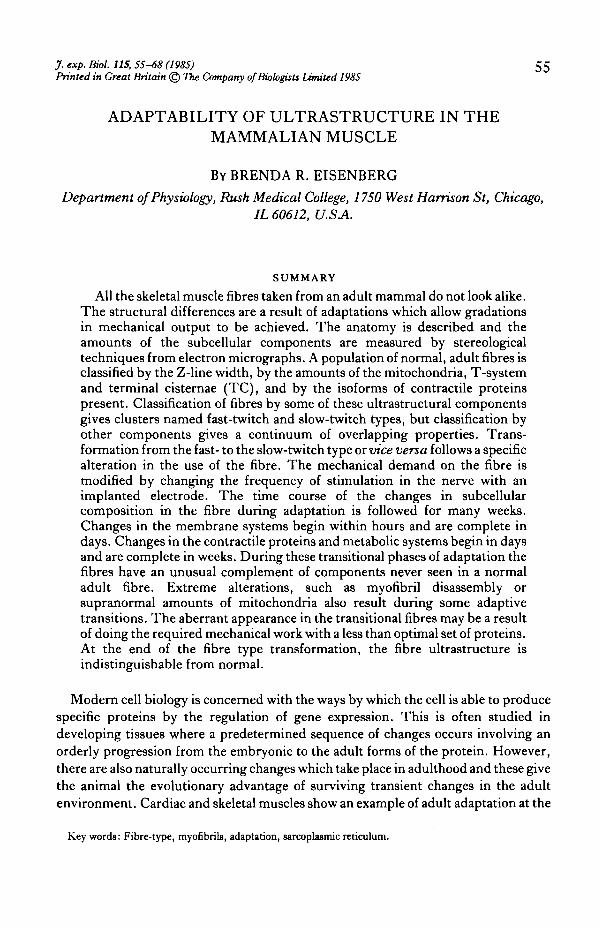
Figs 1, 2
Adaptability of muscle ultrastructure 57
molecular level. A sudden drastic change in the physiological requirement of the kindof mechanical output of the cell is met at first by using the existing contractile proteinsin a non-optimal, perhaps energetically inefficient way. Within a period of a few daysthe cell adjusts the gene expression so that a different and better suited protein isproduced instead. This protein exchange can sometimes be accomplished without acatastrophic interruption of function which would end in destruction of the animal.The heart beats and pumps blood throughout the protein exchange.
Changes which require alteration of the contractile protein composition are not acommonly occurring natural event in skeletal muscle. We are normally moreconcerned with the changes that involve merely an alteration in the amounts of thesubcellular components rather than a change in kind. For example, strength trainingor immobilization regulates fibre hypertrophy and atrophy, and endurance trainingregulates metabolic content (Salmons & Henriksson, 1981). There are thus complexinteractions possible between several regulatory systems which determine the finalappearance and performance of any one muscle fibre.
In the normal adult mammal, the activity patterns in the motor nerve are restrictedto a limited repertoire and therefore there is not an infinite variety of performancedemanded from the muscle fibres. As the activity pattern largely determines thecomposition of the fibre it follows that the fibre composition is necessarily also limitedto restricted types (Burke & Edgerton, 1975). Thus the normal skeletal muscle fibrescan be classified by their composition into types. In some cases the structure withinthe fibre can differ in the extent of the organelles and in other cases in the kind ofmolecule used to build the organelles.
The first problem in the study of the anatomy within mammalian skeletal musclewas to find an objective definition of the fibre types rather than the use of qualitativedescriptions of the subcellular organelles and the arbitrary assignment to a type.Electron microscopy is one of the few methods which views more than one system at atime. I combined stereological techniques (Weibel, 1979) to estimate the amount ofthe organelles from many fibres and used a multivariate discriminant analysis toallocate them into types. A series of papers gave structural data for these systems innormal muscle (Eisenberg, Kuda & Peter, 1974; Eisenberg & Kuda, 1975, 1976;Eisenberg, 1983). I found that in many cellular components there was a broad,continuous distribution in the quantitative amount of the organelle present ratherthan a very narrow, limited amount. Classification could not be made from some of thecomponents as claimed previously but it was possible to classify fibres into types bycombinations of several systems and by assays which depended on the discretemolecular isoforms found in the contractile proteins. These many systems work
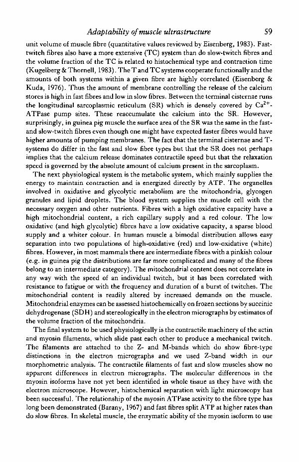
Fig. 1. Electron micrograph of longitudinal section of normal fast-twitch skeletal muscle from theguinea pig white vastua lateralis muscle, showing narrow Z-bands, few mitochondria and extensivesarcoplasmic reticulum (SR) and T-system. (From Eisenberg & Kuda, 1975.) Scale bar, 1 /an.
Fig. 2. Longitudinal section from normal slow-twitch soleus muscle of guinea pig showing wider Z-bands, more mitochondria (nut) and sparser SR »nd T-systema than fast-twitch muscle. (FromEisenberg, Kuda & Peter, 1974.) Scale bar, 1 /an.
58 B. R. ElSENBERG
together to produce ranges in the properties of the mechanical twitch, the time course,output of force and resistance to fatigue.
I will illustrate the major ultrastructural differences by micrographs of a fast-twitchand a slow-twitch guinea pig muscle selected to represent the typical properties(Figs 1,2). The quantitative distribution of each organelle within a muscle fibre isrelated to its own physiological function. The functional role of the membranesystems of muscle fibres is to regulate the speed and duration of the contraction andperhaps to control the peak twitch tension. The T-system carries inward the electricalsignal which activates the inner fibrils; then the terminal cisternae release calcium toinitiate contraction. Fast-twitch fibres have a T-system that is about twice as extensiveas that of slow-twitch fibres of the same species. The membrane area of the T-systemhas the most functional significance because the action potential travels over thissurface, which can be estimated by surface density and length of T-luminal axis per
8 -
E _
MATP SDHX light dark
• dark dark
O dark light
Width of the Z-line (nm)
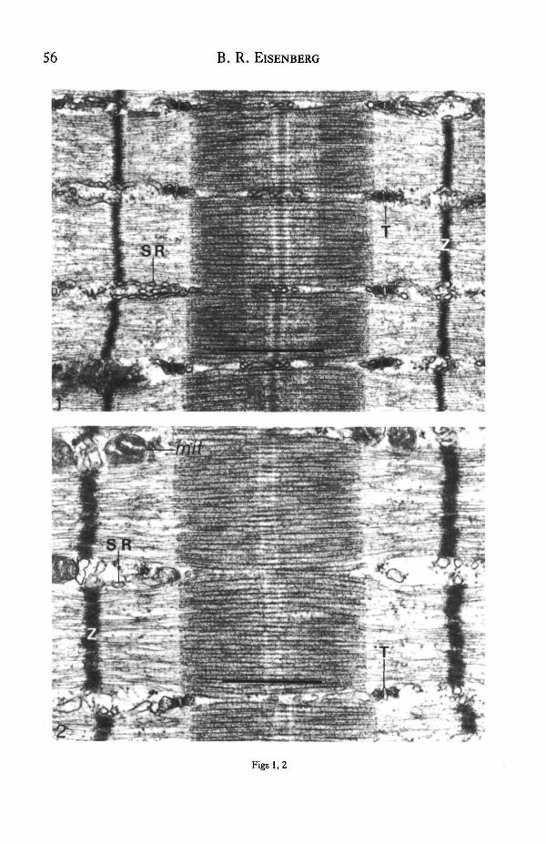
Fig. 3. Scattergram of Z-line width vs mitochondrial volume measured from electron micrographs ofmedial gastrocnemius guinea pig muscle fibres that had been frozen, thawed and then fixed. Serialcryostat sections were used to determine histochemical stains of myofibrillar ATP (MATP) andsuccinic dehydrogenase (SDH) of each fibre to give the conventional fibre types: x , slow-twitch,oxidative; • , fast-twitch, oxidative, glycolytic; and O, fast-twitch, glyeolytic. The three types formone large cluster that can be separated into three subclusters only by reference to histochemical profileof the fibre. (From Eisenberg & Kuda, 1977, reprinted fromJ. Histochem. Cytochem. Copyright 1977by The Histochemical Society, Inc.)
Adaptability of muscle ultrastructure 59unit volume of muscle fibre (quantitative values reviewed by Eisenberg, 1983). Fast-twitch fibres also have a more extensive (TC) system than do slow-twitch fibres andthe volume fraction of the TC is related to histochemical type and contraction time(Kugelberg & Thornell, 1983). The T and TC systems cooperate functionally and theamounts of both systems within a given fibre are highly correlated (Eisenberg &Kuda, 1976). Thus the amount of membrane controlling the release of the calciumstores is high in fast fibres and low in slow fibres. Between the terminal cisternae runsthe longitudinal sarcoplasmic reticulum (SR) which is densely covered by Ca -ATPase pump sites. These reaccumulate the calcium into the SR. However,surprisingly, in guinea pig muscle the surface area of the SR was the same in the fast-and slow-twitch fibres even though one might have expected faster fibres would havehigher amounts of pumping membranes. The fact that the terminal cisternae and T-systems do differ in the fast and slow fibre types but that the SR does not perhapsimplies that the calcium release dominates contractile speed but that the relaxationspeed is governed by the absolute amount of calcium present in the sarcoplasm.
The next physiological system is the metabolic system, which mainly supplies theenergy to maintain contraction and is energized directly by ATP. The organellesinvolved in oxidative and glycolytic metabolism are the mitochondria, glycogengranules and lipid droplets. The blood system supplies the muscle cell with thenecessary oxygen and other nutrients. Fibres with a high oxidative capacity have ahigh mitochondrial content, a rich capillary supply and a red colour. The lowoxidative (and high glycolytic) fibres have a low oxidative capacity, a sparse bloodsupply and a whiter colour. In human muscle a bimodal distribution allows easyseparation into two populations of high-oxidative (red) and low-oxidative (white)fibres. However, in most mammals there are intermediate fibres with a pinkish colour(e.g. in guinea pig the distributions are far more complicated and many of the fibresbelong to an intermediate category). The mitochondrial content does not correlate inany way with the speed of an individual twitch, but it has been correlated withresistance to fatigue or with the frequency and duration of a burst of twitches. Themitochondrial content is readily altered by increased demands on the muscle.Mitochondrial enzymes can be assessed histochemically on frozen sections by succinicdehydrogenase (SDH) and stereologically in the electron micrographs by estimates ofthe volume fraction of the mitochondria.
The final system to be used physiologically is the contractile machinery of the actinand myosin filaments, which slide past each other to produce a mechanical twitch.The filaments are attached to the Z- and M-bands which do show fibre-typedistinctions in the electron micrographs and we used Z-band width in ourmorphometric analysis. The contractile filaments of fast and slow muscles show noapparent differences in electron micrographs. The molecular differences in themyosin isoforms have not yet been identified in whole tissue as they have with theelectron microscope. However, histochemical separation with light microscopy hasbeen successful. The relationship of the myosin ATPase activity to the fibre type haslong been demonstrated (Barany, 1967) and fast fibres split ATP at higher rates thando slow fibres. In skeletal muscle, the enzymatic ability of the myosin isoform to use
B. R. ElSENBERG
Figs 4, S
Adaptability of muscle ultrastructure 61
ATP is differentially affected after incubation in solutions of altered pH. This pHsensitivity is a useful tool for selective inhibition of only one isoform while the other isspared (Dubowitz & Brooke, 1973). Acid preincubation allows histochemicalseparation of the two fast subtypes of skeletal muscle fibres and alkaline preincubationallows the slow type to be separated.
Fibres in serial frozen sections were analysed by ATPase, SDH and quantitativeelectron microscopy of all elements within many fibres. The distribution of fibreproperties assessed in several ways at once is shown in Fig. 3. Clusters of fibres intotypes can be formed but clearly there is the ability to show a continuum of overlappingproperties. We found from this histochemical study (Eisenberg & Kuda, 1977) andthe earlier morphometric studies (see review by Eisenberg, 1983) that the width of theZ-band was correlated with the ATPase fibre-typing and the set of contractile proteinisoforms. With these anatomical methods well defined, we can proceed to look at theway in which a muscle adapts to altered physiological demands.
The great diversity in the anatomical composition within mammalian skeletalmuscle led to the rejection of the concept of a rigid, immutable, fibre type in favour ofthe concept of plasticity. Several books and reviews cover this extensive area ofresearch (Salmons & Henriksson, 1981; Pette, 1980; Jolesz & Sreter, 1981). Musclefibres of the fast-twitch type are normally activated by brief bursts of high-frequencyimpulse activity. Using the technique of chronic electrical stimulation of the motornerve, this 'fast' pattern may be overlaid with continuous low-frequency activity at10 Hz. Under these conditions fast-twitch fibres gradually acquire the physiologicaland biochemical characteristics of fibres of the slow-twitch type. The mechanisms bywhich this fibre plasticity are accomplished can be followed ultrastructurally and thetime course of fibre transformation studied (Salmons, Gale & Sreter, 1978; Eisenberg& Salmons, 1981). The implantable stimulator could then be switched off, leaving themuscle under the influence of its normal, phasic pattern of activity. The transformedslow fibres then recover their original fast morphologial characteristics under theseconditions (Eisenberg, Brown & Salmons, 1984). In both fast-to-slow and slow-to-fast directions the membrane systems regulating the cell calcium begin to change first,followed by the contractile proteins. The metabolic system is quadrupled abovenormal during the fast-to-slow transformation at the time when the fibre gives slowtwitches with fast isomyosin composition. We concluded that this inappropriate use ofthe fast crossbridge wastes energy.
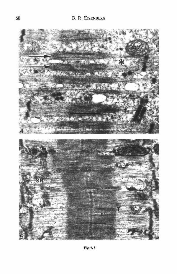
A few hours after initiation of nerve stimulation at 10 Hz the fast tibialis anterior(TA) muscle began to adapt with longitudinal SR swelling in many fibres. By 5—12days there was a generalized reduction in the amount of T-system and terminal
Fig. 4. Rabbit tibialis anterior (TA) muscle stimulated at 10 Hz for 9 days. Narrow myofibrils(arrows) separated by abundant amorphous material (asterisk), few mitochondria (mit) and scant SRand T-systems. (From Eisenberg & Salmons, 1981.) Scale bar, 1 jxra.
Fig. 5. Electron micrograph of longitudinal section of a rabbit tibialis anterior (TA) muscle fibreafter 6 weeks' indirect stimulation and 2 weeks' recovery. T-system (T) and SR returning towards fastfibre levels. Myofibrils are well organized. (From Eisenberg, Brown & Salmons, 1984.) Scale bar,\fim.
m B. R. ElSENBERG
Figs 6, 7
Adaptability of muscle ultrastructure 61
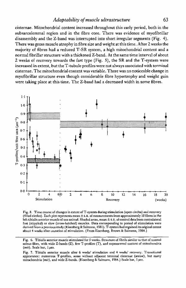
cisternae. Mitochondrial content increased throughout this early period, both in thesubsarcolemmal region and in the fibre core. There was evidence of myofibrillardisassembly and the Z-band was interrupted into short irregular segments (Fig. 4).There was gross muscle atrophy in fibre size and weight at this time. After 2 weeks themajority of fibres had a reduced T-SR system, a high mitochondrial content and anormal fibrillar structure with a thickened Z-band. At the same time interval of about2 weeks of recovery towards the fast type (Fig. 5), the SR and the T-system wereincreased in extent, but the T-tubule profiles were not always associated with terminalcisternae. The mitochondrial content was variable. There was no noticeable change inmyofibrillar structure even though considerable fibre hypertrophy and weight gainwere taking place at this time. The Z-band had a decreased width in some fibres.
0 2 4 6|0
Stimulation10 12
Recovery
14 16 18 20
(weeks)
Fig. 8. Time course of changes in extent of T-system during stimulation (open circles) and recovery(filled circles). Each plot represents mean ± S.E. of measurements from approximately 10 fibres in theleft tibialis anterior muscle of one animal. Shaded areas, mean ± S.E. of control data from contralateralfast (stippled) or slow (cross-hatched) muscles. Data corresponding to period of stimulation werederived from a previous study (Eisenberg & Salmons, 1981). T-system had regained its original extentabout 4 weeks after cessation of stimulation. (From Eisenberg, Brown & Salmons, 1984.)
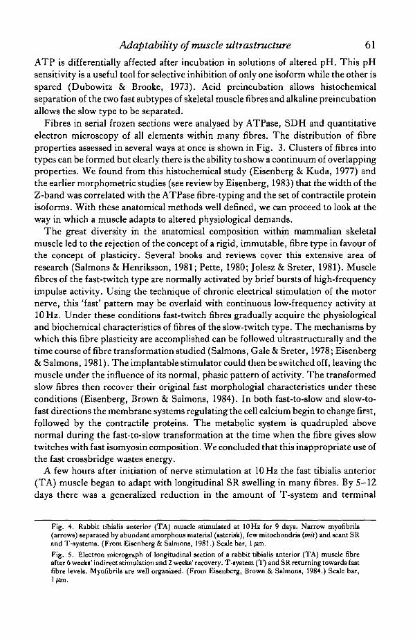
Fig. 6. Tibialis anterior muscle stimulated for 5 weeks. Structure of fibrils similar to that of normalsoleus fibre, with wide Z-bands (Z), few T-profiles (T), and supranormal content of mitochondria(mit). Scale bar, 1/tm.
Fig. 7. Tibialis anterior muscle after 6 weeks' stimulation and 4 weeks' recovery. Transitionalappearance: numerous T-profiles, some without adjacent terminal cisternae (arrow), but manymitochondria (mit), and wide Z-bands. (Eisenberg & Salmons, 1984.) Scale bar, 1 fim.
64 B. R. ElSENBERG
Fast muscle fibres which had been stimulated for more than 3 weeks were difficultto distinguish from normal slow soleus fibres except that the mitochondrial contentwas greater (Fig. 6). When chronic stimulation was ended the fibres regained theappearance of fast-twitch muscles of normal fibre size. During the stage of 3-6 weeksof recovery many of the fibres showed a mixture of features of both fast and slowfibres. It was possible to find a wide Z-band and high mitochondrial content incombination with an extensive T-SR system (Fig. 7). After 6 weeks of recovery fibreshad regained an appearance typical of the fast-twitch muscle fibre type.
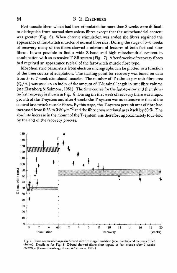
Morphometric parameters from electron micrographs can be plotted as a functionof the time course of adaptation. The starting point for recovery was based on datafrom 5- to 7-week stimulated muscles. The number of T-tubules per unit fibre area(Qt/Af) was used an an index of the amount of T-luminal length in unit fibre volume(see Eisenberg & Salmons, 1981). The time course for the fast-to-slow and then slow-to-fast recovery is shown in Fig. 8. During the first week of recovery there was a rapidgrowth of the T-system and after 4 weeks the T-system was as extensive as that of thecontrol fast-twitch muscle fibres. By this stage, the T-system per unit area of fibre hadincreased from 0-33 to 0-80 jzm~2 and the fibre cross sectional area itself by 60 %. Theabsolute increase in the mount of the T-system was therefore approximately four-foldby the end of the recovery process.
2 4 6J0Stimulation
8 10 12
Recovery
14 16 18 20
(weeks)
Fig, 9. Time course of changes in Z-band width during stimulation (open circles) and recovery (filledcircles). Details as for Fig. 8. Z-band showed dimensions typical of fast muscle after 7 weeks'recovery. (From Eisenberg, Brown & Salmons, 1984.)
Adaptability of muscle ultrastructure 65
In both fast-to-slow (Heilmann & Pette, 1979) and slow-to-fast transformations themembrane systems were the most rapidly changing organelles. Physiologicalrecordings of isometric twitch contractions during the transformation processesshowed that times to peak tension and hah0 relaxation could partly be explained bychanges in the isoform of myosin. Initially, however, alterations in the time course ofactivation could also have arisen from transient calcium changes in the sarcoplasm.The transient calcium distribution within a muscle depends in a complex way on theregulatory proteins and the membrane systems that have been calculated for a givenset of parameters by computer simulations (Gillis, Thomason, Lefevre & Kretsinger,1982). Changes in the kinetics of calcium release, binding and reaccumulation couldhave resulted not only from the observed addition of membrane area, but also from achange in the density of pump sites in the SR.
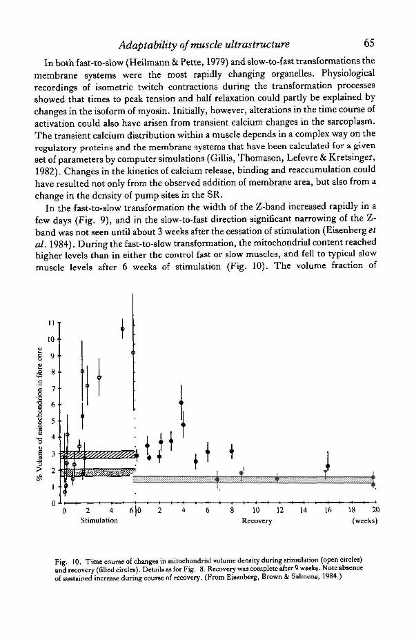
In the fast-to-slow transformation the width of the Z-band increased rapidly in afew days (Fig. 9), and in the slow-to-fast direction significant narrowing of the Z-band was not seen until about 3 weeks after the cessation of stimulation (Eisenbergefal. 1984). During the fast-to-slow transformation, the mitochondrial content reachedhigher levels than in either the control fast or slow muscles, and fell to typical slowmuscle levels after 6 weeks of stimulation (Fig. 10). The volume fraction of
11
10
i .1 »c 7o1 6o
I 4
I 3"3> 2t£
1
0
t j ( ' . ( •+•§ iW:¥:::¥:-:¥:W:*:ra^
0 2 4 6 |0 2 4 6Stimulation
10 12 14 16 18 20
Recovery (weeks)
Fig. 10. Time course of changes in mitochondrial volume density during stimulation (open circles)and recovery (filled circles). Details as for Fig. 8. Recovery was complete after 9 weeks. Note absenceof sustained increase during course of recovery. (From Eisenberg, Brown & Salmons, 1984.)
66 B. R. ElSENBERG
mitochondria did not make a comparable overshoot during recovery even aftercompensation for fibre hypertrophy was considered. There was a rapid reduction inmitochondrial content at 0-3 weeks of the slow-to-fast recovery.
The economical performance of sustained work requires the slow isoform ofmyosin, with its lower rate of crossbridge cycling (Goldspink, 1975, 1983; Alpert,Mulieri & Litten, 1983). During the fast-to-slow transformation the speed ofcontraction begins to alter before the conversion of myosin isoforms is complete(Salmons & Vrbovd, 1969; Pette, Muller, Leisner & Vrbovsi, 1976; Brown, Salmons& Whalen, 1983). During this period we observed a supranormal mitochondrialcontent within the fibre when the utili2ation of ATP was high because the fast bridgeswere cycling inefficiently (Eisenberg & Salmons, 1981). After about 6 weeks ofchronic stimulation, when the isomyosin was 50 % converted (Brown et al. 1983) themitochondrial content declined to a new level, not much higher than that of controlslow muscle. During the slow-to-fast transformation, the energy demands have beenremoved, so that the energetically less efficient faster myosin is reinstated underconditions in which overall ATP consumption was very much reduced and themetabolic stimulus removed.
The net balance between protein synthesis and protein degradation determinesfibre size. In fast-to-slow transformation the net protein balance is negative and thefibre atrophies, and in slow-to-fast transformation it is positive and the fibrehypertrophies. Fibre atrophy occurred over the period in which the fibres had theirhighest mitochondrial content and were using fast cycling contractile proteins tomaintain forces. These fibres must have a rapid rate of oxygen consumption and theoxygen partial pressure would be greatly reduced as it diffuses to the innermostmyofibrils. The low level of oxygen at the centre of the fibre might eventually limitfunction and in fact the mitochondrial content is lowest in the centre of fibres(Hoppeler et al. 1981). It is possible that oxygen deprivation is involved in myofibrilsurvival.
During fibre transformation of the contractile proteins, many other cellularprocesses are affected. The membrane systems and metabolic systems are altered inquantity but not in kind. The contractile machinery is being altered in kind, with theisomyosin form being particularly significant. This transformation must involvesuppression of genes of 'old' proteins, perhaps it also involves an increase in the rate ofdegradation of the 'old' proteins. Transformation also requires switching on genes tomake 'new' proteins. All these processes must be triggered and controlled by themechanical function of the fibre. The mechanism of fibre-type transformation is as yetunknown. And there may be more than one way the fibre can transform. The task isnow to apply biologically relevant challenges to a muscle and to monitor biochemicaland structural changes relevant to the eventual physiological adaptation. A majorproblem in skeletal muscle research is that the population of fibres is mixed andtherefore the staging of the transformation varies from one fibre to the next within thesame muscle. This makes control of the experimental manipulation and interpretationof data quite complex. In skeletal muscle it takes over 8 weeks to convert all the myosinto the 'new' kind. The skeletal muscle is not an ideal system for the study of the
Adaptability of muscle ultrastructure 67
fundamental mechanisms involved in adaptation. The nerve is able to deliver a verylarge variety of signals by packaging bursts of activity into different frequencies anddurations. Therefore it is not obvious what kind of complex mechanical activity is themain signal for transformation.
This research was supported by the American Heart Association. The technicalassistance of Ms Darlene Bruner is gratefully acknowledged.
R E F E R E N C E S
ALPBRT, N. R., MULJEJU, L. A. StLrrrEN, R. Z. (1983). Isoenzyme contribution to economy of contraction andrelaxation in normal and hypertrophied hearts. In Cardiac Adaptation to Hemodynamic Overload, Trainingand Stress, (eds R. Jacob, R. Gulch & G. Kissling), pp. 147-157. Darmstadt: Steinkopff Verlag.
BARANY, M. (1967). ATPase activity of myosin correlated with speed of muscle shortening. J. gen. Physiol. 50,197-216.
BROWN, W. E., SALMONS, S. SCWHALEN, R.G. (1983). The sequential replacement of myosin subunit isoformsduring muscle type transformation induced by long term electrical stimulation. J. biol. Chem. 258,14686-14692.
BURKE, R. E. & EDOERTON, V. R. (1975). Motor unit properties and selective involvement in movement. Ex.Sports Sci. Rev. 3, 31-81.
DUBOWTTZ, V., & BROOKE, M. H. Muscle Biopsy; a Modern Approach. London: Saunders.ElSENBEJtG, B. R. (1983). Quantitative ultrastructure of mammalian skeletal muscle. InHandbook of Physiology:
Section 10, Skeletal Muscle, (eds L. D. Peachey & R. H. Adrian), Chapter 3, pp. 73-112. Baltimore, MD:Williams & Wilkins.
EISENBERO, B. R., BROWN, J. M. C. & SALMONS, S. (1984). Restoration of fast muscle characteristics followingcessation of chronic stimulation: the ultrastructure of slow-to-fast transformation. Cell Tissue Res. 238,221-230.
ElSENBERG, B. R. &K.UDA, A. M. (1975). Stereolo^ical analysis of mammalian skeletal muscle. II . White vastusmuscle of the adult guinea pig. J. Ultrastruct. Ris. 51, 176-187.
ElSENBERG, B. R. & KUDA, A. M. (1976). Discrimination between fiber populations in mammalian skeletalmuscle by using ultrastructural parameters. J. Ultrastruct. Res. 54, 76-88.
ElSENBERG, B. R. & KUDA, A. M. (1977). Retrieval of cryoetat sections for comparison of histochemistry andquantitative electron microscopy in a muscle fiber. J. Histochem. Cytochem. 25, 1169-1177.
EISENBERO, B. R., KUDA, A. M. & PETER, J. B. (1974). Stereological analysis of mammalian skeletal muscle. I.Soleus muscle of the adult guinea pig. J. Cell Biol. 60, 732-754.
EISENBEJIG, B. R. & SALMONS, S. (1981). The reorganization of subcellukr structure in muscle undergoing fast-to-slow type transformation: a stereological study. Cell Tissue Res. 220, 449—471.
GILUS, J. M., THOMASON, D., LEFEVRE, J. JCKRETSINGER, R. H. (1982). Parvalbumins and muscle relaxation:a computer simulation study. J Muse. Res. CellMotil. 3, 377-398.
GOLDSPINK, G. (1975). Biochemical energetics for fast and slow muscles. In Comparative Physiology:Functional Aspects of Structural Materials, (eds L. Bolis, S. H. P. Maddrell & K. Schmidt-Nielsen).Amsterdam: Elsevier.
GOLDSPINK, G. (1983). Alteration of myofibril size and structure during growth, exercise, and changes inenvironmental temperature. In Handbook of Physiology: Section 10, Skeletal Muscle, (eds L. D. Peachey &R. H.Adrian), pp. 539-555. Baltimore, MD: Williams & Wilkins.
HEJLMANN, C. & Pette, D. (1979). Molecular transformations in sarcoplasmic reticulum of fast-twitch muscle byelectro-stimulation. Eur.J. Biochem. 93, 437-446.
HOPPELER, H., MATHIEU, O., KRAUER, R., CLAASSEN, H., ARMSTRONG, R. B. & WEIBEL, E. R. (1981). Design
of the mammalian respiratory system. VI. Distribution of mitochondria and capillaries in various muscles.Respir. Physiol. 44, 87-111.
JOLESZ, F. &SRETER, F. A. (1981). Development, innervation, and activity-pattern induced changes in skeletalmuscle. A, Rev. Physiol. 43, 531-552.
KUGELBERG, E. & THORNELL, L.-E. (1983). Contraction time, hintochemical type, and terminal cisternaevolume of rat motor units. Muscle Nerve 6, 149—153.
PETTE, D. (1980). Plasticity of Muscle. Berlin: Walter de Gruyter.PETTE, D., MULLER, W., LEISNES, E. & VRBOVA, G. (1976). Time dependent effects on contractile properties,
fibre population, myosin light chains and enzymes of energy metabolism in intermittently and continuouslystimulated fast twitch muscles of the rabbit. Pflugers Arch, ges. Physiol. 364, 103-112.
68 B. R. ElSENBERGSALMONS, S., G A L E . D . R. &SRBTER, F. A. (1978). Ultrastructural aspects of the transformation of muscle fibre
type by long term stimulation: changes in Z discs and mitochondria. J. Anat. 127, 17-31.SALMONS, S. & HENRIKSSON, J. (1981). The adaptive response of skeletal muscle to increased use. Muscle Nerve
4, 94-105.SALMONS, S. & VRBOVA, G. (1969). The influence of activity on some characteristics of mammalian fast and slow
muscles. J . Pkysioi, Land. 201, S3S-S49.WEIBEL, E. R. (1979). Stereobgical Methods. I. Practical Methods for Biological Morphometry. New York:
Academic Press.

![Practice For May: Cell Ultrastructure [114 marks]blogs.4j.lane.edu/.../2018/02/Cell-Ultrastructure-Test-1.pdfPractice For May: Cell Ultrastructure [114 marks]1. Which structure found](https://img.pdfslide.net/doc/110x75/5eda4db5b3745412b5711d9c/practice-for-may-cell-ultrastructure-114-marksblogs4jlaneedu201802cell-ultrastructure-test-1pdf.jpg)