Embed Size (px)
Citation preview
Endocrine, vol. 4, no. 3, 239-247, june 1996 0969-711X/96/4:239-247/$7.25 �9 1996 by Humana Press Inc. All rights of any nature whatsoever reserved.
Adenosine 5'-Triphosphate (ATP) Receptors Induce Intracellular Calcium Changes in Mouse Leydig Cells
Ella Martha POrez-Armendariz, 1,2 Angel Nadal, 1 Esther Fuentes, 1 and David C. Spray 1
I Department of Neuroscience, Albert Einstein College of Medicine, Bronx, NY; 2Departamento de Fisiologfa, Bioffsica y Neurociencias, Centro de In vestigaci6n y Estudios A vanzados, del I.P.N., MExico
Cytoplasmic calcium ([Ca2+]i) changes evoked by aden- osine 5'-triphosphate (ATP) were recorded in cultured individual Leydig cells within 10-18 h after cell disper- sion. [Ca2+] i was monitored using Fura-2AM loaded cells with a digital ratio imaging system. Five micro- molars ATP induced biphasic [Ca2+] i responses in most cells (94%, n -- 100), characterized by a fast increase from a basal level (126 _+ 5 nM SF, n = 60 cells) to a peak (5-7 times above basal levels) within seconds, followed by a slow decrease toward a plateau level (2-3 times above basal) within 5 rain. The peak phase of the tea2+] i response increased with ATP concentrations (1-100 btM A-I-P) in a dose-dependent manner with an IC~0 of 5.9 + 1.2 btM, and it desensitized in a reversible manner with repeated application of 5 BM ATP at < 5-min intervals. The [Ca2+] i peak response was dependent on Ca 2+ release from an intracellular pool, whereas the plateau phase was dependent on extracellular [Ca2+]. ATP did not appear to induce formation of nonspecific membrane pores, since stimulation for 10 min with ATP (10-100 btM) in the presence of extracel- lular Lucifer yellow (LY) (5 mg/mL) did not result in dye loading of the cells. [Ca2+] i transients were elicited by other adenosine nucleotides with an order of potencies (ATP > Adenosine diphosphate [ADP] > Adenosine > Adenosine monophosphate [AMP]) that was compat- ible with the expression of P2 receptors. [Ca2+] i responses were suppressed by the purinergic P2 recep- tor antagonist, suramin. These results provide func- tional evidence for the expression of purinergic P2 receptors in Leydig cells.
Key Words: Testis; purinergic receptors; secretion; gap junctions.
Received October 17, 1995; Revised January 23 and February 14, 1996; Accepted February 23, 1996.
Author to whom all correspondence and reprint requests should be addressed: Dr. E. M. Pdrez-Armendariz, c/o M. Romano, Dept. Fisiologia, Biofisica y Neurociencias, CTNVESTAV, I.P.N., Ap. Postal 14-740, M6xico, D. F., 07000.
Introduction
Extracellular adenosine 5'-triphosphate (ATP) is believed to regulate physiological processes, including cell contraction or relaxation, aggregation, cell growth, and secretion in numerous different cell systems, including neu- rons, smooth muscle (arterial and visceral), cardiac myo- cytes, endothelial, hematopoietic, and secretory cells. In these systems, ATP produces various effects, such as acti- vation of ionic channels or induction of membrane pores, and/or activation of G-proteins, stimulation of phospho- lipid metabolism, and mobilization of intracellular Ca 2+ (reviewed in: Gordon, 1986; Burnstock 1990; Coh-nan, 1990; EI-Moatassim et al., 1992). These changes appear to be mediated by activation of a diversity ofpurinergic recep- tor types. Purinergic receptors were originally classified as types Pl and P2, where P1 receptors were mainly sensitive to adenosine and P2 receptors were most sensitive to ATP (Burnstock, 1978, 1990). More recently, P2-purinorecep- tors have been subdivided into at least four subtypes, P2x, P2y, P2z, P2t, according to actions underlying cell responses and the rank order of potency or new synthetic agonists (Gordon 1986; Cusak and Hournai, 1990).
The expression of the P2z, receptor subtype has recently been suggested to be associated with the expression of the gap junction protein connexin43 (Cx43), possibly through an ATP-induced activation of conducting Cx43 hemi- channels (Beyer and Steinberg, 1991). In the testis, the interstitial or Leydig cells that surround the spermatic cords directly communicate through large gap junctions, which express abundant Cx43 in situ and in vitro (Risley et al., 1992; P6rez-Armendariz et al., 1994, 1995, 1996; Varanda and Campos de Carvahlo 1994). For this reason, we have explored the possibility that Leydig cells might express purinergic P2z-type receptor activity.
Moreover, ATP released from purinergic nerves or coreleased with other neurotransmitters at both sympathetic adrenergic and cholinergic synapses (Burnstock, 1972, 1978) has been proposed as the primary source of ATP for different cell types which express P2 receptor activity. In
239
240 Expression of ATP Receptors in Leydig Cells/Perez-Armendariz et al. Endocr ine
the testis, there is no consensus about autonomic innervation of Leydig cells (Hodson, 1970). Nevertheless, ultrastruc- tural evidence of both direct and indirect autonomic innerva- tion of these cells has now been provided in several species, including bird (Baumgarten and Holstein, 1968), reptile (Unsicker, 1973), telost (Gresik, 1973), and most recently mammals (Prince, 1992). Thus, anatomical studies on testis raise the possibility that ATP might be released or coreleased at sites ofinnervation, and has also led us to examine whether P2 purinergic receptors are expressed in Leydig cells.
In this article, we provide functional evidence for the expression of P2 receptors in Leydig cells. Our demonstra- tion that [Ca2+]i is quite sensitive to ATP and its metabolites suggests that in Leydig cells ATP may be physiologically important in regulating Ca 2+ homeostasis. The possible relevance of P2 receptors for Leydig cell function is dis- cussed. Part of these data have been presented in a prelimi- nary form (Nadal et al., 1993).
R e s u l t s
A TP induces [Ca2+]i Transients in Leydig Cells
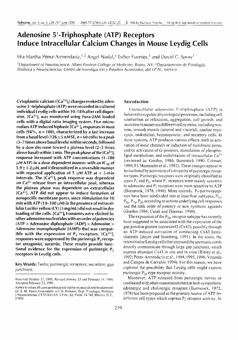
In most Leydig cells maintained under resting condi- tions, basal [Ca2+]i values were stably recorded for at least 30 min. Application of low extracellular ATP concentra- tions (1-100 laM) in the presence of [ mM MgCI 2 in the external media induced transient increases in [Ca2+]i in most of the cells (94 of 100). Figure 1 shows traces repre- sentative of those recorded in most of the cells in response to 5 gM ATP, where four phases are resolved. In the first phase, [Ca2+]i increased from a basal level (126 + 5 nM, SE, n = 60 cells) to a peak (610 + 98 nM, SE, n = 10 cells) within 2-5 s. In the second phase, Ca 2+ levels decreased rapidly also within seconds. In the third phase, Ca 2+ levels de- creased more slowly, tending to reach a plateau level (418 + 39 nM, n = 10 cells) within 5 rain. The fourth phase was triggered by ATP removal, where Ca 2+ levels dropped and reached their resting values within 2-3 rain.
Evidence that [Ca2+]i Transients Induced by A TP in Leydig Cells are Receptor-Mediated
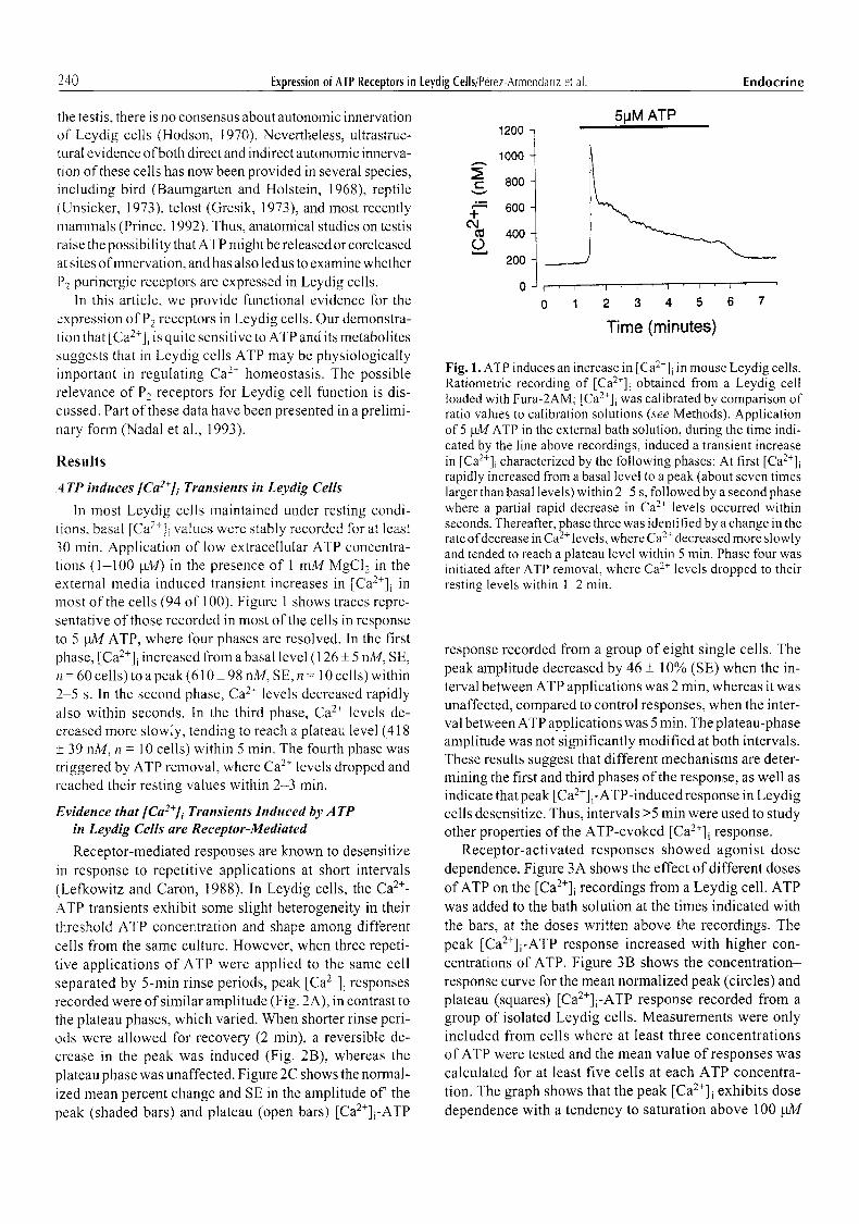
Receptor-mediated responses are known to desensitize in response to repetitive applications at short intervals (Lefkowitz and Caron, 1988). In Leydig cells, the Ca 2+- ATP transients exhibit some slight heterogeneity in their threshold ATP concentration and shape among different ceils from the same culture. However, when three repeti- tive applications of ATP were applied to the same cell separated by 5-rain rinse periods, peak [Ca2+]i responses recorded were of similar amplitude (Fig. 2A), in contrast to the plateau phases, which varied. When shorter rinse peri- ods were allowed for recovery (2 rain), a reversible de- crease in the peak was induced (Fig. 2B), whereas the plateau phase was unaffected. Figure 2C shows the normal- ized mean percent change and SE in the amplitude of' the peak (shaded bars) and plateau (open bars) [Ca2+]i-ATP
5pM ATP 1200
t- v
~
.-I-
0
1000
800
600
400
200
0 F ' I
0 1
I J I t I
2 3 4 5 6 7
Time (minutes)
Fig. 1. ATP induces an increase in [Ca2+]i in mouse Leydig cells. Ratiometric recording of [Ca2+]i obtained from a Leydig cell loaded with Fura-2AM; [Ca2+]i was calibrated by comparison of ratio values to calibration solutions (see Methods). Application of 5 pM ATP in the external bath solution, during the time indi- cated by the line above recordings, induced a transient increase in [Ca2+]i characterized by the following phases: At first [Ca2+]i rapidly increased from a basal level to a peak (about seven times larger than basal levels) within 2 3 s, followed by a second phase where a partial rapid decrease in Ca 2+ levels occurred within seconds. Thereafter, phase three was identified by a change in the rate of decrease in Ca 2+ levels, where Ca 2+ decreased more slowly and tended to reach a plateau level within 5 min. Phase four was initiated after ATP removal, where Ca 2+ levels dropped to their resting levels within 1-2 min.
response recorded from a group of eight single cells. The peak amplitude decreased by 46 + 10% (SE) when the in- terval between ATP applications was 2 min, whereas it was unaffected, compared to control responses, when the inter- val between ATP applications was 5 min. The plateau-phase amplitude was not significantly modified at both intervals. These results suggest that different mechanisms are deter- mining the first and third phases of the response, as well as indicate that peak [Ca 2+] i-ATP-induced response in Leydig cells desensitize. Thus, intervals >5 min were used to study other properties of the ATP-evoked [Ca2+]i response.
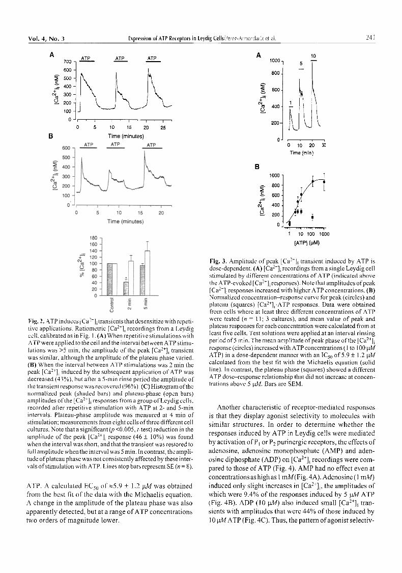
Receptor-activated responses showed agonist dose dependence. Figure 3A shows the effect of different doses of ATP on the [Ca2+]i recordings from a Leydig cell. ATP was added to the bath solution at the times indicated with the bars, at the doses written above the recordings. The peak [Ca2+]i-ATP response increased with higher con- centrations of ATP. Figure 3B shows the concentration- response curve for the mean normalized peak (circles) and plateau (squares) [Ca2+]i-ATP response recorded from a group of isolated Leydig cells. Measurements were only included from cells where at least three concentrations of ATP were tested and the mean value of responses was calculated for at least five cells at each ATP concentra- tion. The graph shows that the peak [Ca2+]i exhibits dose dependence with a tendency to saturation above 100 gM
Vol. 4, No. 3 Expression of ATP Receptors in Leydig Cells/Perez-Armendariz et at. 241
A 700
6OO
3; 500
400 + eq 300
2oo
100
B
600
500
400
g= 300
200 100
C
ATP ATP ATP
I I i I I
0 5 10 15 20 25
Time (minutes)
ATP ATP ATP
I I I
10 15 20
Time (minutes)
180 160 140
~ ' 120 Od
100 L) 80
60 4O 2O 0
._= ._= t g ~
C O
Fig. 2. ATP induces [Ca2+]i transients that desensitize with repeti- tive applications. Ratiometric [Ca2+]i recordings from a Leydig cell, calibrated as in Fig. 1. (A) When repetitive stimulations with ATP were applied to the celt and the interval between ATP stimu- Iations was >5 rain, the amplitude of the peak [Ca2+]i transient was similar, although the amplitude of the plateau phase varied. (B) When the interval between ATP stimulations was 2 min the peak [Ca2+]i induced by the subsequent application of ATP was decreased (43%), but after a 5-min rinse period the amplitude of the transient response was recovered (96%). (C) Histogram of the normalized peak (shaded bars) and plateau-phase (open bars) amplitudes of the [Ca2+]i responses from a group of Leydig cells, recorded after repetitive stimulation with ATP at 2- and 5-min intervals. Plateau-phase amplitude was measured at 4 min of stimulation; measurements from eight cells of three different cell cultures. Note that a significant (p <0.005, t-test) reduction in the amplitude of the peak [Ca2+]i response (46 _+ t0%) was found when the interval was short, and that the transient was restored to full amplitude when the interval was 5 min. In contrast, the ampli- tude o fplateau phase was not consistently affected by these inter- vals of stimulation with ATP. Lines atop bars represent SE (n = 8).
ATP. A calculated ECs0 of ~5.9 + 1.2 bgk/was obtained from the best fit o f the data with the Michaelis equation. A change in the amplitude of the plateau phase was also
apparently detected, but at a range of ATP concentrations two orders of magnitude lower.
A 1o L _ _
,oo 1 1
2oo 1 0 ~
; lO 2; Time (min)
B
& #=
1000
800,
600.
400,
200.
0 . . . . . . i . . . . . . . . i . . . . . . ,~ . . . . . . ,1
1 10 100 1000
[ATP] (pM)
Fig. 3. Amplitude of peak [Ca2+]i transient induced by ATP is dose-dependent. (A) [Ca2+]i recordings from a single Leydig cell stimulated by different concentrations of ATP (indicated above the ATP-evoked [Ca2+]i responses). Note that amplitudes of peak [Ca2+]i responses increased with higher ATP concentrations. (B) Normalized concentration-response curve for peak (circles) and plateau (squares) [Ca2+]i-ATP responses. Data were obtained from cells where at least three different concentrations of ATP were tested (n = 11; 3 cultures), and mean value of peak and plateau responses for each concentration were calculated from at least five cells. Test solutions were applied at an interval rinsing period of 5 rain. The mean amplitude of peak phase of the [Ca2+]i response (circles) increased with ATP concentrations (1 to 100 btM ATP) in a dose-dependent manner with an ICs0 of 5.9 + 1.2 btM calculated from the best fit with the Michaelis equation (solid line). In contrast, the plateau phase (squares) showed a different ATP dose-response relationship that did not increase at concen- trations above 5 gM. Bars are SEM.
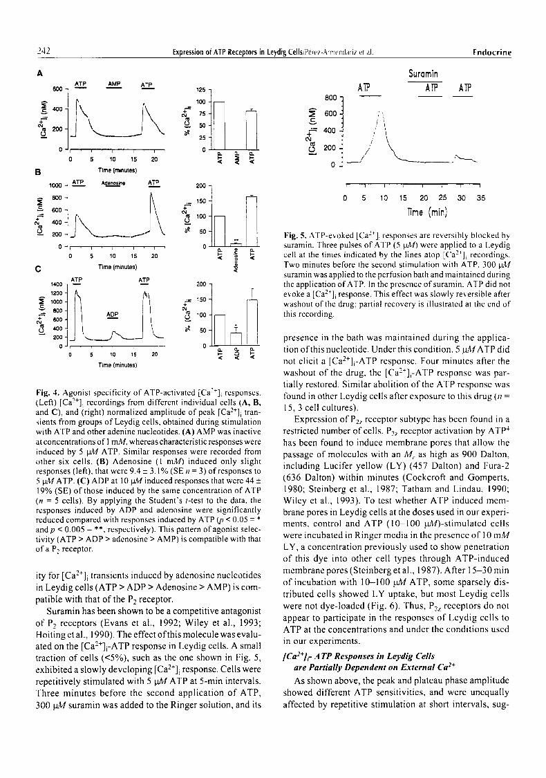
Another characteristic of receptor-mediated responses is that they display agonist selectivity to molecules with similar structures. In order to determine whether the responses induced by ATP in Leydig cells were mediated by activation of Pt or P2 purinergic receptors, the effects o f adenosine, adenosine monophosphate (AMP) and aden- osine diphosphate (ADP) on [Ca2+]i recordings were com- pared to those of ATP (Fig. 4). AMP had no effect even at concentrations as high as 1 mM(Fig. 4A). Adenosine (1 rnM) induced only slight increases in [Ca2+]i, the amplitudes of which were 9.4% of the responses induced by 5 gag ATP (Fig. 4B). ADP (10 pM) also induced small [Ca2+]i tran- sients with amplitudes that were 44% of those induced by
l 0 gMATP (Fig. 4C). Thus, the pattern ofagonis t selectiv-
242 Expression of ATP Receptors in Leydig Cells/Perez-Armendariz eta[. Endocrine
A
o
6O0
40O
+ ( M
{~ 200- o_
C
e-
+
ATP AMP ATP
i i 1 i
5 10 15 20
Time (minutes)
ATP Adenosine ATP
~ 150 +
j , \ ,oo
. . . . o
S 10 15 20 ~ "-
Time (minutes) I
1000
8OO
600
400
200
0
ATP 1 4 0 0 -
1200 - ; 1000- 800- 600 - 400- 200- ~ .
0 - ,
5
125
1 0 0 8 0 0
+ 75 ~ 600
r 50 ,-~ 400 25
0 r 200 < < < 0
ATP _
+ c q
j " v _ _ - - J .< so
0
Time (minutes)
Fig. 4. Agonist specificity of ATP-activated [Ca2+]i responses. (Left) [Ca2+]i recordings from different individual cells (A, B, and C), and (right) normalized amplitude of peak [Ca2+]i tran- sients from groups of Leydig cells, obtained during stimulation with ATP and other adenine nucleotides. (A) AMP was inactive at concentrations of 1 mM, whereas characteristic responses were induced by 5 pM ATP. Similar responses were recorded from other six cells. (B) Adenosine (1 mM) induced only slight responses (left), that were 9.4 _+ 3.1% (SEn = 3) of responses to 5 pA,/ATP. (C) ADP at 10 pM induced responses that were 44 _+ 19% (SE) of those induced by the same concentration of ATP (n -- 5 cells). By applying the Student's t-test to the data, the responses induced by ADP and adenosine were significantly reduced compared with responses induced by ATP (p < 0.05 -- * and p < 0.005 = **, respectively). This pattern of agonist selec- tivity (ATP > ADP > adenosine > AMP) is compatible with that of a P2 receptor.
ity for [Ca2+]i transients induced by adenosine nucleotides in Leydig cells (ATP > ADP > Adenosine > AMP) is com- patible with that of the P2 receptor.
Suramin has been shown to be a competitive antagonist of P2 receptors (Evans et al., 1992; Wiley et al., 1993; Hoiting et al., 1990). The effect of this molecule was evalu- ated on the [Ca2+]i-ATP response in Leydig cells. A small traction of cells (<5%), such as the one shown in Fig. 5, exhibited a slowly developing [Ca2+]i response. Cells were repetitively stimulated with 5 ~ ATP at 5-min intervals. Three minutes before the second application of ATP, 300 NI4 suramin was added to the Ringer solution, and its
A'rP
Suramin
ATP ATP
t/'/"/ ' L , ~ . . . _
1 I [ i I I 1 I
o s lo is 2o 25 ao as
]]me (rain)
Fig. 5. ATP-e~.oked [Ca2*]i responses are reversibly blocked by suramin. Three pulses of ATP (5 Wig) we.e applied to a Leydig cell at the times indicated by the lines atop [Ca2+]i recordings. Two minutes before the second stimulation with ATP, 300 ~.L,tr suramin was applied to the perfusion bath and maintained during the application of ATP. In the presence ofsuramin, ATP did not evoke a [Ca2+]i response. This effect was slowly reversible after washout of the drug; partial recovery is illustrated at the end of tiffs recording.
presence in the bath was maintained during the applica- tion of this nucleotide. Under this condition, 5 gMATP did not elicit a [Ca2+]i-ATP response. Four minutes after the washout of the drug, the [Ca2§ response was par- tially restored. Similar abolition of the ATP response was lbund in other Leydig cells after exposure to this drug (n = 15, 3 cell cultures).
Expression of P2z receptor subtype has been found in a restricted number of cells. P2z receptor activation by ATP 4 has been found to induce membrane pores that allow the passage of molecules with an M,. as high as 900 Dalton, including Lucifer yellow (LY) (457 Dalton) and Fura-2 (636 Dalton) within minutes (Cockcroft and Gomperts, 1980; Steinberg et al., 1987; Tatham and Lindau, 1990; Wiley et al., 1993). To test whether ATP induced mem- brane pores in Leydig cells at the doses used in out" experi- ments, control and ATP (10-100 laM)-stirnulated cells were incubated in Ringer media in the presence of 10 mM LY, a concentration previously used to show penetration of this dye into other cell types through ATP-induced membrane pores (Steinberg et al., 1987). After 15-30 min of incubation with 10-100 gM ATP, some sparsely dis- tributed cells showed LY uptake, but most Leydig cells were not dye-loaded (Fig. 6). Thus, P2z receptors do not appear to participate in the responses of Leydig cells to ATP at the concentrations and under the conditions used in our experiments.
[Ca2+]c A TP Responses in Leydig Cells are Partially Dependent oll External Ca :+
As shown above, the peak and plateau phase amplitude showed different ATP sensitivities, and were unequally affected by repetitive stimulation at short intervals, sug-
Vol. 4, No. 3 Expression of ATP Receplors in Leydig Cells/Perez-Armendariz et al. 243
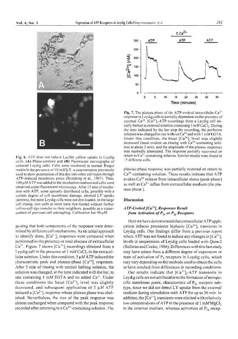
Fig. 6. ATP does not induce Lucifer yellow uptake in Leydig cells. (A) Phase-contrast and (B) fluorescent micrographs of cultured Leydig cells. Cells were incubated in normal Ringer media in the presence of 10 mM LY, a concentration previously used to show penetration of this dye into other cell types through ATP-induced membrane pores (Steinberg et al., 1987). Then, 100 him ATP was added to the incubation medium and cells were observed under fluorescent microscopy. After 15 min of incuba- tion with ATP, some sparsely distributed cells, possibly with a certain degree of cell membrane damage, showed LY uptake (arrows), but most Leydig cells were not dye-loaded. In the large cell clump, two cells at most were dye-loaded without further cell-to-cell dye transfer to their neighbors, possibly as a conse- quence of previous cell uncoupling. Calibration bar 40 p.M.
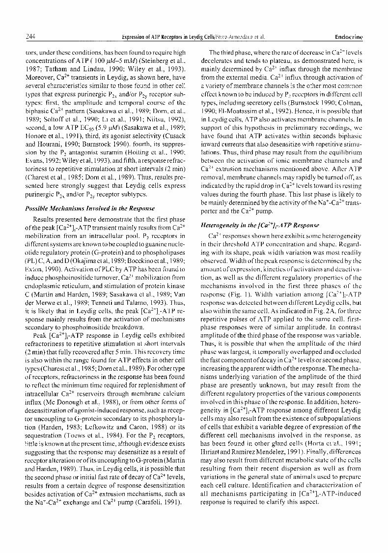
gesting that both components of the response were deter- mined by different cell mechanisms. As an initial approach to identify them, [Ca2+]i responses were compared when performed in the presence or total absence of extracellular Ca 2+. Figure 7 shows [Ca2+]i recordings obtained from a Leydig cell in the presence of I mM CaCI 2 in the extracel- lular solution. Under this condition, 5 btMATP induced the characteristic peak and plateau-phase [Ca2+]i responses. After 5 min of rinsing with normal bathing solution, the solution was changed, at the time indicated with the bar, to one containing 1 mM EGTA and no added Ca 2+. Under these condi t ions the basal [Ca2+]i level was slightly decreased, and subsequent application of 5 btM ATP induced a [Ca2+]i response whose plateau phase was abol- ished. Nevertheless, the size of the peak response was ahnost unchanged when compared with the peak response recorded after returning to a Ca>-containing solution. The
300 ATP
0 Ca 2+
ATP ATP
r v
, , - - , -T +
04
(.5
250
200
150
100
50
0 ,
0
i i ~ i i i i
5 10 15 20 25 30 35
Time (minutes)
Fig. 7. The plateau phase of the ATP-evoked intracellular Ca 2+ response in Leydig cells is partially dependent on the presence of external Ca 2+. [Ca2+]i-ATP recordings from a Leydig cell ini- tially bathed in external solution containing 1 mMCaCl 2. During the time indicated by the bar atop the recording, the perfusion solution was changed to one without Ca 2+ and with 1 mMEGTA. Under this condition, the basal [Ca2+]i level was slightly decreased (most evident on rinsing with Ca2+-containing solu- tion at about 3 min), and the amplitude of the plateau responses was markedly attenuated. The response partially recovered on return to Ca>-containing solution. Similar results were found in 15 different cells.
plateau-phase response was partially restored on return to Ca2+-containing solution. These results indicate that ATP evokes Ca 2+ release from intracellular stores (peak phase) as well as Ca 2+ influx from extracellular medium (the pla- teau phase ).
Discussion
A TP-Evoked [Ca2+]i Responses Result from Activation of e2x or P& Receptors
Here we have demonstrated that extracellular ATP appli- cation induces prominent biphasic [Ca2+]i transients in Leydig cells. Our findings differ from a previous report where ATP was not found to induce any changes in [Ca2+]i levels in suspensions of Leydig cells loaded with Quin-2 (Sullivan and Cooke, 1986). Differences with this last study may have arisen from a different degree of expression or state of activation of P2 receptors in Leydig cells, which may vary depending on the methods used to obtain the cells or have resulted from differences in recording conditions.
Our results indicate that [Ca2+]i-ATP transients in Leydig cells are not attributable to the formation ofnonspe- cific membrane pores, characteristic of P2z receptor sub- type, since we did not detect LY uptake from the external medium during stimulation with ATP for up to 30 min. In addition, the [Ca2+]i transients were elicited with relatively low concentrations o f ATP in the presence of 1 mM MgC12 in the external medium, whereas activation of P2z recep-
244 Expression of AIP Receptors in Leydig Cells/Perez-Armendariz et at. Endocrine
tors, under these conditions, has been found to require high concentrations ofATP ( 100 gM-5 mM) (Steinberg et al., 1987; Tatham and Lindau, 1990; Wiley et al., 1993). Moreover, Ca 2+ transients in Leydig, as shown here, have several characteristics similar to those found in other cell types that express purinergic P2x and/or P2y receptor sub- types: first, the amplitude and temporal course of the biphasic Ca 2+ pattern (Sasakawa et al., 1989; Dorn, et al., 1989; Soltoff et al., 1990; Li et al., 1991; Niitsu, 1992), second, a low ATP ECs0 (5.9 ILg'/) (Sasakawa et al., 1989; Honore et al., 1991 ), third, its agonist selectivity (Cusack and Hourani, 1990; Burnstock 1990), fourth, its suppres- sion by the P2 antagonist suramin (Hoiting et al., 1990; Evans, 1992; Wiley et al, 1993), and fifth, a response refrac- toriness to repetitive stimulation at short intervals (2 min) (Charest et al., 1985; Dorn et al., 1989). Thus, results pre- sented here strongly suggest that Leydig cells express purinergic P2• and/or P2y receptor subtypes.
Possible Mechanisms Involved in the Response
Results presented here demonstrate that the first phase of the peak [Ca2+]i-ATP transient mainly results from Ca 2+ mobilization from an intracellular pool. P2 receptors in different systems are known to be coupled to guanine nucle- otide regulatory protein (G-protein) and to phospholipases (PL) C, A, and D (Okajima et al, 1989; Bocckino et al., 1989; Exton, 1990). Activation of PLC by ATP has been found to induce phosphoinositide turnover, Ca 2+ mobilization from endoplasmic reticulum, and stimulation of protein kinase C (Martin and Harden, 1989; Sasakawa et al., 1989; Van der Merwe et al., 1989; Tenneti and Talamo, 1993). Thus, it is likely that in Leydig cells, the peak [Ca2+]i-ATP re- sponse mainly results from the activation of mechanisms secondary to phosphoinositide breakdown.
Peak [Ca2+]i-ATP response in Leydig cells exhibited refractoriness to repetitive stimulation at short intervals (2 min) that fully recovered after 5 rain. This recovery time is also within the range found for ATP effects in other cell types (Charest et al., 1985; Dorn et al., 1989). For other type of receptors, refractoriness in the response has been found to reflect the minimum time required for replenishment of intracellular Ca 2+ reservoirs through membrane calcium influx (Mc Donough et al., 1988), or from other forms of desensitization ofagonist-induced response, such as recep- tor uncoupling to G-protein secondary to its phosphoryla- tion (Harden, 1983; Lefkowitz and Caron, 1988) or its sequestration (Toews et al., 1984). For the P2 receptors, little is known at the present time, although evidence exists suggesting that the response may desensitize as a result of receptor alteration or of its uncoupling to G-protein (Martin and Harden, 1989). Thus, in Leydig cells, it is possible that the second phase or initial fast rate of decay of Ca 2+ levels, results from a certain degree of response desensitization besides activation of Ca 2+ extrusion mechanisms, such as the Na+-Ca 2+ exchange and Ca 2+ pump (Carafoli, 1991).
The third phase, where the rate of decrease in Ca 2+ levels decelerates and tends to plateau, as demonstrated here, is mainly determined by Ca 2+ influx through the membrane from the external media. Ca 2+ influx through activation of a variety of membrane channels is the other most common effect known to be induced by P2 receptors in different cell types, including secretory cells (Burnstock 1990; Colman, 1990; El-Moatassim et al., 1992). Hence, it is possible that in Leydig cells, ATP also activates membrane channels. In support of this hypothesis in preliminary recordings, we have found that ATP activates within seconds biphasic inward currents that also desensitize with repetitive stimu- lations. Thus, third phase may result from the equilibrium between the activation of ionic membrane channels and Ca 2+ extrusion mechanisms mentioned above. After ATP removal, membrane channels may rapidly be turned off, as indicated by the rapid drop in Ca 2+ levels toward its resting values during the fourth phase. This last phase is likely to be mainly determined by the activity of the Na+-Ca 2+ trans- porter and the Ca 2. pump.
Heterogeneity in the [Ca2+]i-A TP Response
Ca 2+ responses shown here exhibit some heterogeneity in their threshold ATP concentration and shape. Regard- ing with its shape, peak width variation was most readily observed. Width of the peak response is determined by the amount of expression, kinetics of activation and deactiva- tion, as well as the different regulatory properties of the mechanisms involved in the first three phases of the response (Fig. 1). Width variation among [Ca2+]i-ATP response was detected between different Leydig cells, but also within the same cell. As indicated in Fig. 2A, for three repetitive pulses of ATP applied to the same cell, first- phase responses were of similar amplitude. In contrast amplitude of the third phase of the response was variable. Thus, it is possible that when the amplitude of the third phase was largest, it temporally overlapped and occluded the fast component of decay in Ca 2+ levels or second phase, increasing the apparent width of the response. The mecha- nisms underlying variation of the amplitude of the third phase are presently unknown, but may result from the different regulatory properties of the various components involved in this phase of the response. In addition, hetero- geneity in [Ca2+]i-ATP response among different Leydig cells may also result from the existence of subpopulations of cells that exhibit a variable degree of expression of the different cell mechanisms involved in the response, as has been found in other gland cells (Horta et'al., 1991; Hiriart and Ramirez Mendelez, 1991 ). Finally, differences may also result from different metabolic state of the cells resulting from their recent dispersion as well as from variations in the general state of animals used to prepare each cell culture. Identification and characterization of all mechanisms participating in [Ca2+]i-ATP-induced response is required to clarify this aspect.
Vol. 4, No. 3 Expression of ATP Receptors in Leydig Cells/Perez-Armendariz eta[. 245
Possible Sources of A TP Release in the Interstitial Space of the Testis
Results presented here suggest that ATP may function as either neurotransmitter or neuromodulator at nerve termi- nals found in the interstitial space of the testis. ATP released from purinergic and sympathetic terminals has been pro- posed as the primary source of ATP for the different cell types that express P2 receptor activity (Burnstock, 1972, 1978). In the testis, evidence for both indirect and direct innervation of Leydig cells has been found in various spe- cies, including cells from mammalian testis (see Introduc- tion). In human testis, ultrastructural studies suggest that in addition to the cholinergic (type I) and adrenergic (type II) terminals, there is a third group of nonadrenergic, non- cholinergic nerve endings (Prince, 1992). However, mor- phological studies have not yet addressed whether ATP is present at some of these types of terminals. Interestingly, the nonadrenergic and noncholinergic type of nerve termi- nals have also been found in other parts of the male repro- ductive system, such as vas deferens and epididymis (Gu et al., 1983), where there is also evidence that ATP activates P2 receptors (Friel, 1988).
Other possible sources of ATP in the testicular intersti- tial space may include ATP released from secretory gran- ules of mast cells (Uvnas, 1974), which represent a second cell type coexisting with Leydig cells in this space (Gaytan et al., 1992). Alternatively or in addition, ATP may be locally released from a population of cells that are periodi- cally renewed, as is suggested by the presence of macro- phages, which also normally coexist with Leydig cells in this space (Raburn et al., 1993).
Possible Biological Consequences of External Stimulation with A TP in Leydig Cells
P2 receptors are ubiquitously expressed in a great variety of cells as indicated in the Introduction. However, only recently in a few of them, their functional consequences have been identified. In secretory cells, P2 receptor activa- tion has been found to induce [Ca2+]i transients and ionic membrane fluxes in different primary exocrine (Soltoff et al., 1990; Sasaki and Gallacher, 1992; Vincent, 1992) and endocrine (Sugiyama, 1971 ; Loubatieres-Mariani et al., 1979; Sasakawa et al., 1989; Geschwind et al., 1989) peptidergic cells, as well as in derived cell lines (Geschwind et al., 1989; Okajima et al., 1989; Li et al,, 1991 ; Sela et al., 1991). In some of them, where they have been studied, these changes were found associated with an increase in fluid and/or peptide secretion. Moreover, in adrenocortical cells, ATP has also been found to enhance steroidogenesis (Niitsu, 1992). Here it is demonstrated that [Ca2+]i in Leydig cells is finely regulated by ATP, and that large transients could be elicited by low ATP concentrations. Since ste- roidogenesis is a Ca2+-dependent process (Hall, 1988), it is possible that activation ofP 2 receptors stimulates steroido- genesis and testosterone release. Although such a function
of ATP was not detected in a previous report (Sullivan and Cooke, 1986), in that work [Ca2+]i-ATP changes were also not detected, and consequently, the negative evidence does not yet rule out such a possibility. Independently of the possible stimulatory role of ATP on steroidogenesis, expression of P2 receptors in Leydig cells may also be rel- evant for other cell functions (see Introduction), such as for pH regulation (Puceat et al., 1993) and control of cell growth (Fang and Wu, 1993). Future experiments will address these possible roles of ATP in testicular function.
Local stimulation of Leydig cells is expected in situ if ATP is released from any of the possible sources discussed above. Nevertheless, important amplification of Ca z+ sig- nals is expected to occur by transferring the elevated Ca 2+ from one cell to its neighbors through their numerous gap junction channels formed with Cx43 (Risley et al., 1992; Varanda and Campos de Carvahlo, 1994; Perez-Armen- dariz et al., 1994, 1995, 1996), which are known to be perthe- able to Ca 2+ (Christ et al., 1992). In preliminary experiments, local stimulation with ATP to one cell of a clump, through a high-resistance patch pipet, induced synchronous changes in [Ca2+]i in the other cells, suggesting its transfer through gap junctions. If future experiments confirm these observa- tions, an extensive modulatory role of ATP might be expected in the possible cell functions mentioned before.
Methods
Experimental Preparation
As an initial step to study ATP effects on Leydig cells, we used a rapid nonenzymatic technique lbr dissociation of interstitial cells slightly modified from the one described by Kawa in 1987, which we had previously characterized (P~rez-Armendariz et al., 1996). Owing to its briefiless, it is likely that this method favors unaltered receptor expres- sion in cell membranes. Briefly, testes were dissected from adult CDI mice and the tunica albuginea was removed. Three testes were placed in 10 mL of Dulbeco's Modified Eagle's Medium (DM EM) in a 50-mL tube and were gently shaken for 4 min at room temperature until the seminifer- ous tubules became slightly loose, but not separated. The resulting exudate was enriched in Ieydig cells. Cells were washed three times by centrifugation. The final pellet from a number of testis was suspended in a small volume of DMEM to adjust a density of 10 6 cells/mL. Then, I mL of cell suspension was plated in glass coverslips and main- tained in DMEM with 10% fetal bovine serum for 10-18 h in 95% O2 and 5% CO., at 34~ before use.
Cell Identification and Functional State
Using the dispersion technique mentioned before, we had previously found a significant enrichment of Leydig cells, as indicated by detection of 70-84% 3 13-hydroxy steroid dehydrogenase-positive cells, and a significant increase in testosterone production in cell cultures main- tained for 8, 24, and 36 h, in the presence of lutenizing,
246 Expression of ATP Receptors in Leydig Cells/Perez-Armendariz et al. Endocr ine
human chorionic gonadotropin hormones or dibutiryl ciclic AMP (Perez-Armendariz et al., 1996). In the present article, ATP studies were done in cells cultured for 10-18 h, a period of time where we have confidence about their survival and functional integrity. For [Ca2+]i recordings, Leydig cells were further selected by identifying their characteristic abundant cytoplasmic cholesterol droplets, visualized as birefringent bodies under phase-contrast optics (Christen- sen, 1975; Kawa, 1987). [Ca2+]i recordings were obtained within 2 h after cells were exposed to a Kreb's Ringer medium. Within this period of time, cells had been found to maintain stable resting membrane potential and channel properties (Perez-Armendariz et al., 1994). [Ca2+]i record- ings were obtained within 1 11 after dye loading and 40 rain after initiation of fluorescence excitation, conditions under which cells are able to keep stable [Ca2+]i basal levels.
Cell Loading
To load Leydig cells, the acetoxymethyl ester form of Fura-2 (Fura-2/AM; Molecular Probes, Eugene, OR) was dissolved in dimethyl sulfoxide (DMSO) to form a l-raM stock solution. Leydig cells plated on coverslips were removed from culture media and incubated in a normal Ringer solution containing 5 btM Fura-2/AM without Ca 2+ with a final DMSO concentration of 0.05% for 40 rain at room temperature. Cells were washed by transferring the coverslips to a dish containing normal Ringer solution, where they were gently shaken 5 10 times. This step was repeated three times, to eliminate detritus and nonattached remaining cells. After the rinsing period, coverslips were transferred to a recording chamber mounted on an inverted microscope equipped with an intensified CCD Camera for Ca 2+ measurements.
ICa2+]i Measurements
In each field, Leydig cells were epi-illuminated alter- nate[y at 340 and 380 nm using a computer-controlled filter wheel (Sutter Instruments, Novato, CA) containing appro- priate excitation optical filters that was synchronized with the recording camera. The fluorescence emitted by single Leydig cells in each field was selected; images were acquired at emission wavelengths >500 nm using an intensified CCD camera (Quantex) and were stored for further analysis using the Image 1 AT/FL hardware/software package (Uni- versal Imaging, Media, PA) on a Dell System 325 com- puter equipped with an optical disk drive for mass storage.
Fluorescence measurements were corrected for back- ground fluorescence and dark camera current. Four video frames at each wavelength were averaged, obtaining an over- all time resolution of 2 s for each pair of images at alternating wavelengths. The resulting images were ratioed (340/380) pixel by pixel to produce ratio images, and [Ca2+]i was calculated by interpolation into a lookup table constructed by imaging Fura-2-free acid in the presence of known Ca 2+ concentrations using the same microscope objective.
Experimental Ca 2+ Recording Conditions
The recording chamber (0.5-mL vol) was connected to a gravity perfusion system. Recordings under basal condi- tions were obtained when cells were bathed with a normal saline solution that was continuously perfusing the bath. When changes in solution were applied, the media in the recording chamber were exchanged completely within l 0 seconds. Experiments were carried out at room tempera- ture (22~ in an external Ringer solution with the follow- ing composition: 145 mMNaCI, 5 mM KC1, 1 mM CaC12, 1 mM MgC12, 2 mM NaHCO 3, 10 mM HEPES, pH 7.36, unless otherwise indicated.
Dye Experiments
Leydig cells were incubated for 10 min in dye solution containing 5 mg of LY/mL of normal Ringer solution (~10 mM, 0.5% w/v), in the presence or in the absence of 10 and 100 bO4 ATP. After this time, cells were rinsed several times in normal external solution, dye uptake was visualized in an inverted microscope using conventional fluorescence microscopy with Xenon illumination and FITC filters, and the percentage of cells taking up dye was determined.
Acknowledgments
This work is in memorial of Sir E. Alejandro Perez Armendariz and Lic. Enrique Perez Hernfindez, who encouraged us during the development of this project.
We thank G. Coello and A. Escalante (Unidad de Computo, Instituto de Fisiotogia C61ular, Universidad Nacional Autonoma de M6xico) for their assistance and to D. Page for scientific discussion. Dr. A. Nadal's research at Albert Einstein College of Medicine was covered by a fel- lowship from the Ministry of Education and Science from Spain. A. Nadal and E. Fuentes are presently at the Depart- ment of Physiology, Division of Biomedical Science, King's College, London, UK. This project has been sup- ported by NIH grants DK38529 and NS07512.
References
Baumgarten, H. G. and Holstein, A. F. (1968). Z. ZeH/brsch. 91, 402-410.
Beyer, E. C. and Steinberg, T. H. (1991). J. Biol. Chem. 266, 7971 7974.
Bocckino, S. B., Blackmore, P. F., and Exton J. H. (1989). J. Biol. Chem. 264, 8847 8856.
Burnstock, G. (1972). Pharmacol. Rev. 24, 509-581. Burnstock, G. (1978). In: Cell Membrane Receptors for Drugs and
Hormones. A multidisciplinary approach. Straub, R. W. and Bolis, g. (eds.). Raven, New York, pp. 107-118.
Burnstock, G. (1990). In: Biological Actions of Extracellular A TP. Dubyak, G. R. and Fedan, J. S. (eds.). Ann. NYAcad. Sci. 603, 1-t 7.
Carafoli, E. (1991). Ann. Rev. Physiol. 53, 531-547. Charest, R., Blackmore, P. F., and Exton, J. H. (1985). J. Biol. Chem.
260, 15,789-15,794.
Vol. 4, No. 3 Expression of ATP Receptors in teydig Cells/Perez-Amlendariz et al. 247
Christensen, A. K. (1975). In: Handbook qll~hvsiology, vol. 5. Greep, R. O., Astrwood. E. g., Hahninton, D. W., and Geiger, S. (eds.). American Physiological Society, Washington DC 5, pp. 57 94.
Colman, R. W. (1990). FASEBJ. 4, 1425-1435. Cockcroft, S. and Gomperts, B. D. (1980). Bioch. J. 188, 789 798. Cusack, N. J. and Hourani, S. M. O. (1990). In: Biologicalactions
o['extracelhdar ATP. Dubyak, G. R. and Fedan, J. S. (eds.). ..Inn. NYAead. &'i. 603, 172-181.
CIlrist, O. J.. Moreno, A. P., Melman, A., and Spray, D. C. (1992). Am. J. Physiol. 263, C373-C383.
Dorn, C. C., Rice, W. R., and Singleton, F. M. (1989). Br. J. Pharmacol. 97, 163-170.
EI-Moatassim, C., Dornand, J., and Mani, J. C. (1992). Biochim. Bioph.vs. Acta 1134, 31-45.
Evans, R. J., Derkach. V., and Surprenant, A. (1992). Nature 357, 503-505.
gxton, J. H. (1990). In: Biological actions o['extracellular A TP. Dubyak, G. R. and Fedan. J. S. (eds.). Ann. NYAcad. Sci. 6113, 246-254.
Fang, W. G. and Wu, B. Q. (1993). Clin. Exp. Metastasis 11,33(>336.
Friel, D. D. (1988). J. Physiol. 401,361-380.
(iaytan, F., Aceitero, J., Lucena, C., Aguilar, E., Pinilla, L., Ga.nelo, P., and Bellido, C. (1992). J. Androl. 13, 387-397.
Geschwind, J, F.. Hiriart. M., Glennon, M. C., Najafi, H., Cordkey, B. E., Matschinsky, F. M.. and Prentki. M. (1989). Biochim. Bioph.vs. Acta 1012, 107 115.
(,iresik, E. W. (1973). Gen. Comp. Endocrinol. 21,210 213.
(iordon, J. L. (1986). Biochem. J. 233, 309 319.
Liu, .I.. Polak. J. M., Probert, 1,.. Islam, K. N., Marangos, P. J., Mina. S.. Adrian, T. E.. Mc Gregor, G. P.. O'Shaughnessy, D. J., and Bloom. S. R. (1983). J. Urol. 130, 38(7-391.
Hall, P. F. (1988). In: Theph.vsiologvol)'eproduction. Knobil. E. and Neill, J. (eds). Ra,.en, New York, pp. 975-998.
Harden, T. K. (1983). Pharmacol. Rev. 35, 5-32. Hiriarl, M. and Ramirez Medelez, M. C. (1991). Endocrinology
128, 3193-3198. Hodson, N. (1970). In: The Testis. vol. 1. Johnson, A. D., Gomes,
W. R., and Vandermark, N. L. (eds.). Academic, New York, pp. 47 99.
Hoiting, B., Molleman, A., Nelemans, A., and Den-Hertog, A. (1990). Eur. J. Pharmacol. 181, 127-13 I.
Honore, E., Fournier, F., Collin, T., Nargeot, J., and Guilbauh, P. (I99l). PJlugers. Arch. 418, 447-452.
Horta, J., Hiriart, M.. and Cota, G. (1991). Am. J. Physiol. 261, C865-C867.
Kawa, K. (1987). J. Physiol. Lond. 393, 647~66. L.i, G., Milani, D., Dunne, M. J., Pralong, W. F., Theler, J. M.,
Petersen, O. H., and Wollheim. C. B. (1991). J. Biol. Chem. 266, 3449-3457.
t_etkowitz, R. J. and Caron, M. G. (1988). J. Biol. Chem. 263, 4993- 4996.
Loubatieres-Mariani, M. M., Chapal, J., Lignon, F., and Valette, G. (1979). Eur. J. Pharmacol. 59, 277-286.
McDonough, P. M., Eubanks, J. H., and Brown, J. H. (1988). Biochem. J. 249, 135-141.
Martin, M. W. and Harden, Y. K. (1989). J. Biol. Chem. 264, 19,535 19,539.
Nadal, A., Fuentes, E., Spray. D. C., Bennett, M. V. L., and Perez-Armendariz, E. M. (1993). Biophys. J. 64, A327, 446a.
Niitsu, A. (1992). Jpn. J. Pharmacol. 60, 269 274. Okajima, F., Sato, K., and Kondo, Y. (1989). FEBSLett. 253, 132-136. Perez-Armendariz, E. M., Romano, M., Luna, J., Bennett, M. V. L.,
and Moreno, A. (1994). Am. J. Physiol. 267, C570-C58(). Perez-Armendariz, E. M., Romano, M., Luna, J., Talavera, D.,
Moreno, A. P., and Bennett, M. V. L., (1995). In: Intercellular communication through gap junctions. Kanno, Y., Kataoka, K., Shiba, Y., Shibata, Y., and Shimazu, T. (eds.). Elsevier Science B. V., Amsterdam. Prog. in Cell Res. 4, 413-417.
Perez-Armendariz, E. M., Luna, J., Miranda, C., Talavera, D., and Romano, M. C. (1996). Endocrine 4(2), 1-10.
Prince, F. P. (1992). Cell Tissue Res. 269, 383-390. Puceat, M., Clement-Chomienne, O., Yerzic, A., and Vassort, G.
(1993). Am. J. Physiol. 264, H310-9. Raburn, D. J., Coquel in, A., Reinharl, A. J., and Hutson, J. C. (1993).
d. Repord. Immunol. 24, 139 15 I. Risley, M. S., Tan, 1. P., Roy, C., and Saez, J. C. (1992). J. Cell Sci.
103, 81 96. Sasaki, T. and Gallacher, D. V. (1992). J. Pttvsiol. Lond. 447, 103 118. Sasakawa, N., Nakaki, T., Yamamoto, S., and Kato, R. (1989). J.
Neurochem. 52, 441-447.
Sela, D., Ram, E., and Atlas, D. (1991).J. Biol. Chem. 266~ 17,990~ 17,994.
Soltoff, S. P., MacMillian, M. K., Lechleiter, J. D., Cantley, k. C., and Talamo, B. R. (1990). In: Biologicalactions ofextracellularA TP. Dubyak, G. R. and Fedan, J. S. Ann. NYAcad. Sci. 603, 76-90.
Steinberg, Y. H., Newman, A. S., Swanson, J. A., and Silverstein, S. C. (1987). J. Biol. Chem. 262, 8824-8888.
Sugiyama, K. (1971). Jpn. J. Pharmacol. 21,531 539. Sullivan, M. H. F. and Cooke, B. A. (1986). Biochem. J. 236, 45-51. Tatham, P. E. R. and Lindau, M. (1990). J. Gen. Physiol. 95,459M76. Tenneti, L. and Talamo, B. R. (1993). Biochem. J. 295, 255-261. Yoews, M. L., Waldo, G. L., Harden, T. K., and Perkins, J. E. (1984).
J. Biol. Chem. 259, 11,844-11,850. Unsicker, K. (1973). Z ZellJ'orsch. 146, 123-138. Uvnas, B. (1974). L!l'e Sci. 14, 2355-2366. van der Merwe, P. A., Wakefield, I. K., Fine, J., Millar, R. P., and
Davidson, J. S. (1989). FEBS. Lett. 243, 333-336. Varanda, W. A., Campos de Carvahlo, A. C. (1994). Am. J. Physiol.
267, C563-C569. Vincent, P. (1992). J. Physiol. Lond. 449, 313-331. Wiley, J. S., Chen, R., and Jamieson, G. P. (1993). Arch. Biochem.
Bioph.vs. 305, 544~0.


![Increased Rate of Adenosine Triphosphate …...(CANCER RESEARCH 55, 4352-4360, October 1, 1995] Increased Rate of Adenosine Triphosphate-dependent Etoposide (VP-16) Efflux in a Murine](https://img.pdfslide.net/doc/110x75/5e7e8d68c5d0407f2447f2a9/increased-rate-of-adenosine-triphosphate-cancer-research-55-4352-4360-october.jpg)