Embed Size (px)
Citation preview
Biochemistry @ Copyright 1970 b y the American Chemical Society Volume 9, Number 20 September 29,1970
Adenosine Triphosphate and Inosine Triphosphate Dependent Conformational Changes of Adenosine Diphosphate-G-Actin*
Joel J. West
ABSTRACT: Ultraviolet absorbance changes occur when actin-bound nucleotide is changed from ADP to ATP or ITP. These absorbance changes may reflect a conformational change in actin when the nature of the bound nucleotide
A lthough the validity of the sliding filament hypothesis as a model accounting for the events in muscle contraction appears to be well established (Huxley, 1960) the mechanism by which the energy of ATP is utilized to produce filament displacement is not understood. While it is generally believed that some shortening or change in shape of the myosin cross bridge consequent to ATP hydrolysis accounts for the movement of myosin past the actin filaments, the possibility remains that this movement is due to limited shortening of the actin filaments (Huxley, 1969). It has been suggested that the basis of flagellar contraction depends on an ATP- induced conformational shortening of flagellin, the flagellar analog of muscle actin (Silvester and Holwill, 1965). Conse- quently, evidence that the actin molecule changes in confor- mation and, hence, possibly shortens when ATP is substituted for bound ADP would be of interest. That such a conforma- tional change of actin leading to muscle contraction occurs has been suggested (Laki, 1969), but direct supporting evidence has been lacking. Higashi and Oosawa (1965) concluded from their spectral studies that there is no appre- ciable difference in the spectra of G-actin containing bound ATP, ADP, or ITP. No evidence of conformational differ- ences between ATP and ADP-G-actin was detected in optical rotation studies (Nagy, 1966), although the ultraviolet absorbance changes which resulted from addition of EDTA to G-actin solutions differed depending on whether the actin
* From the Division of Neurology, Duke University Medical Center, and The Center for Cerebral Vascular Research, Veterans Administra- tion Hospital, Durham, North Carolina 27706. Receiued April 27, 1970. Supported by National Institutes of Health Grant 5 ROI NS 08534-02 and a Veterans Administration Hospital Clinical Investigatorship.
is changed, Such a conformational change in actin may provide an explanation of how the chemical energy of adenosine triphosphate is utilized in order to produce muscle contraction.
bound nucleotide was ATP, ADP, or ITP (West et a[., 1967). Actin exists in a variety of states: native G-actin with or without bound nucleotide, denatured actin in dimer form, aggregated denatured actin (Lewis et a[., 1963), and polym- erized actin. The proportion of each of these changes with time or after nucleotide addition. Since the conformation of actin in each of these states most likely differs from the others, comparison of one actin preparation with another utilizing procedures such as ultracentrifugation or optical rotatory dispersion might not readily detect small conforma- tional changes due to the effect of bound nucleotide. This communication presents evidence, based on changes in ultraviolet absorbance when ATP or ITP is added to ADP- G-actin, that the conformation of G-actin does change when ATP or ITP is substituted for actin-bound ADP.
Materials
An acetone-dried powder was prepared from rabbit mus- cle by the method of Feuer et al. (1948) and stored at -10". Actin was extracted from this powder with 0.2 mM ATP in 1 mM Tris-HC1 (pH 8.0) at 0" (Drabikowski and Gergely, 1962) and purified by polymerization first with 0.1 M KCl and 0.1 mM MgClz and, subsequently, with 0.6 mM MgClz (Martonosi, 1962). The F-actin was precipitated by ultracentrifugation for 70 min at 150,OOOg between each polymerization step (Mommaerts, 1951). ADP-G-actin solutions prepared from ATP-free F-actin pellets (West et al., 1967) by homogenization in solutions containing 50 PM ADP and 1 mM Tris-HC1 (pH 8.0) were clarified by centrifugation at 100,OOOg for 15 min. ITP-G-actin was prepared by addition of 1 mM ITP to ADP-G-actin solutions.
B I O C H E M I S T R Y , VOL. 9, N O . 20, 1970 3847
W E S T
The ITP-G-actin was then clarified by centrifugation at 100,OOOg for 10 min prior to Dowex treatment. In a few preparations, a 2-hr dialysis step was inserted before clarifica- tion without evident change in the characteristics of the re- sulting ITP-G-actin. In certain studies, ITP-G-actin was prepared directly from clarified Dowex-treated ADP-G- actin solutions by addition of 0.2 mM ITP. ATP-G-actin solutions were prepared from F-actin pellets by homogeniza- tion with 1 mM ATP in 1 mM Tris-HC1 (pH 8.0) and dial- yzed for 3 days at 2" against a 100-fold excess of 0.4 mM ATP in 1 mM Tris-HC1 (pH 8.0) prior to clarification at 100,OOOg for 20-60 min. All preparations were treated with Dowex 1-X2 (Cl) as previously described (West, 1970) to re- move free nucleotide just prior to studies and the desired concentration of free nucleotide was readded. The solutions were maintained at pH 8.0.
ADP-G-actin solutions were felt not to contain either ATP-G-actin or myokinase since there was no significant increase in viscosity during a prolonged interval at 0" after addition of 1 mM ADP, 0.1 M KC1 and 1 mM MgClz (West, 1970). ATP-G-actin solutions appeared to be free of tropo- myosin since their viscosity was in the same range as that found by Drabikowski and Gergely (1962) for ATP-G-actin solutions containing no tropomyosin. AMP deaminase was apparently absent since G-actin solutions containing 0.1 mM AMP developed no absorbance changes for a prolonged period at 25" (Lowey and Luck, 1969). ITP-G-actin solutions appeared to be largely free of adenine nucleotide since the ab- sorbance of perchloric acid extracts of ITP-G-actin had virtually the same 249 mp/259 mp ratio as the added ITP.
Methods
Difference spectra were recorded with a Durrum PGS spectrophotometer. Matched cells with path length of 1 cm, 0.2 cm, 0.1 cm, or two split compartment cells with chamber path length of 0.44 cm (Yankeelov, 1963) were utilized. Protein and buffer were placed in separate compart- ments of the sample and reference cells and a base line was determined prior to addition of perturbant. Nucleotide, EDTA, or salt ordinarily was added at high concentrations with a microliter pipet. The same pipet used for perturbant additions to the sample actin and the reference buffer was also used to add buffer to the reference actin to maintain equal protein concentrations in sample and reference cells. In some studies, to avoid additions of high concentrations of perturbant, low concentrations of the perturbant were added to one side of split compartment cells in place of buffer and the two compartments of the sample cell were mixed with each other after the base line was determined. In such studies, 1,000 ml was delivered to both sample compart- ments with the aid of a micrometer-driven pipet. Virtually identical results were obtained whether the perturbant was added at low or high dilution. Cells usually were maintained in thermostatted cell compartments at 0.5 =t 0.4" or at 4.5 =I= 0.4". In studies of absorbance changes due to actin polymerization, difference spectra were corrected for light scattering by extend- ing a double logarithmic plot of the difference spectrum from 650 to 320 mp into the ultraviolet region (Leach and Scheraga, 1960). Difference spectra were obtained at several different slit widths to ensure reproducibility and minimal contribution of stray light. At 230 mp and lower wavelengths, this proved
difficult since the absorbance was sufficiently high to require slit widths of 0.2 mm or greater and good reproducibility was not always obtained when wider slit widths were em- ployed. In addition, multiple experiments were performed under a variety of conditions and the results illustrated represent typical changes. When two difference spectra were subtracted from each other, scans were run at slow rates to permit accurate definition of wavelength and to reduce effect of background noise, and the curves were carefully matched with respect to wavelength. Scans employing ADP-G-actin as a reference were initially obtained rapidly to define the shape of the curve before significant spontaneous denaturation of the actin in the reference cell could occur. Slower scans to define the shape of the smaller peaks were then obtained.
Actin concentration was calculated from the molar con- centration of bound nucleotide. This was determined by measuring the absorbance at 259 mp of an 0.6 M perchloric acid extract of an aliquot of the G-actin solutions which had previously been exposed to Dowex 1 for 4 min to remove free nucleotide. An extinction coefficient of 14.9 X 10⁸/mole at 257 mp was employed for ATP (Pabst, 1956), or of 10.9 X 10a/mole at 250 mp for ITP at pH 1. This was checked with the concentration of the same solution calculated from the absorbance at 280 mp using an extinction of 11.08 (West et al., 1967). Agreement within 10% was nearly always obtained. Nucleotide was always added to maintain stability of the G-actin solutions following the final Dowex 1 treat- ment. Nevertheless, some spontaneous denaturation of the actin occurred so that the active protein concentrations were somewhat less at the time of perturbant addition than those determined at the start of the study. In the case of ATP-G- actin such spontaneous denaturation appeared to be minor during the interval of the studies, but more appreciable denaturation occurred in ITP-G-actin and especially ADP- G-actin solutions. The extent of this spontaneous denaturation was determined by measuring the rate of absorbance change at 256 mp using either bovine serum albumin as a reference or, in the studies employing EDTA to denature actin, the sample actin, once completely denatured by EDTA, was used as a reference to measure the rate of decrease in absor- bance of the reference actin, assuming that there were no further changes in absorbance of the sample cell. During a 15-hr period these changes followed first-order kinetics and, hence, could be extrapolated back to the time of the final Dowex 1 treatment. The rate of actin denaturation appeared reproducible for a given free nucleotide concentra- tion and, hence, estimates of actin denaturation could be made for all studies. The actin concentrations listed were corrected for spontaneous denaturation by this technique.
Results
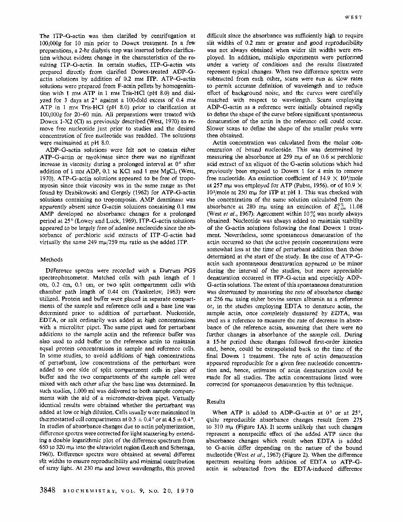
When ATP is added to ADP-G-actin at 0" or at 25", quite reproducible absorbance changes result from 275 to 310 mp (Figure 1A). It seems unlikely that such changes represent a nonspecific effect of the added ATP since the absorbance changes which result when EDTA is added to G-actin differ depending on the nature of the bound nucleotide (West et al., 1967) (Figure 2). When the difference spectrum resulting from addition of EDTA to ATP-G- actin is subtracted from the EDTA-induced difference
3848 B I O C H E M I S T R Y , V O L . 9, N O . 20 , 1 9 7 0
C O N F O R M A T I O N A L C H A N G E O F A D P - G - A C T I N
0.010
A I
ci 0 Q
'-'-I
Ybwlength, m+
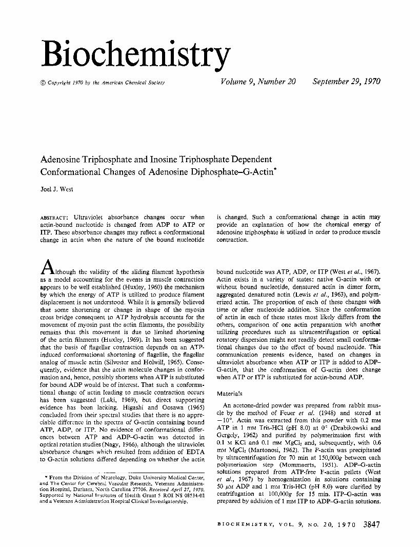
FIGURE 1 : The absorbance changes resulting from addition of ATP to ADP-G-actin compared to the absorbance difference resulting from subtraction of the EDTA-induced difference spectrum of ATP- G-actin from that of ADP-G-actin at 0". Absorbance changes below 240 mp were detected with a path length of 1 mm. (A) Absorbance changes after addition of 50 p~ ATP to ADP-G-actin 50 p~ in 1 I n M Tris-HC1 (pH 9.0) containing 50 p~ ADP in a cell with a path length of 0.44 cm. (B) The absorbance difference result- ing from subtraction of the difference spectra induced by addition of 1 mM EDTA at 0' to ATP-G-actin from the EDTA-induced difference spectrum of ADP-G-actin. The ATP-G-actin concen- tration was 13.6 p~ in 1 mM Tris-HC1 (pH 8.0) containing 5 pM ATP. The ADP-G-actin concentration was 13.6 p~ in 1 mM Tris- HCI (pH 8.0) containing 10 WM ADP in a cell with a 1-cm path length.
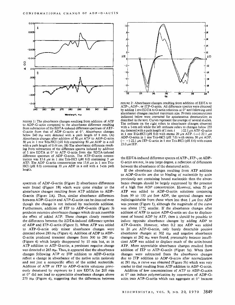
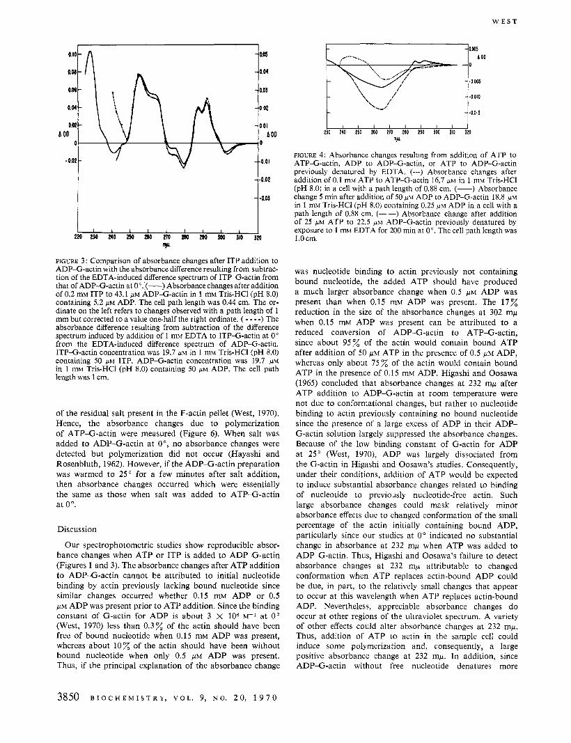
spectrum of ADP-G-actin (Figure 2) absorbance differences were found (Figure 1B) which were quite similar to the absorbance changes resulting from ATP addition to ADP- G-actin (Figure 1A). Thus, similar absorbance differences between ADP-G-actin and ATP-G-actin can be detected even though the change is not induced by nucleotide addition. Furthermore, addition of ITP to ADP-G-actin (Figure 3) produces extensive absorbance changes which do not resemble the effect of added ATP. These changes closely resemble the differences between the EDTA-induced difference spectra of ADP and ITP-G-actin (Figure 3). When ATP was added to ATP-G-actin only minor absorbance changes were detected above 280 mp (Figure 4). Addition of ADP to ADP- G-actin produced transient changes from 300 to 280 mp (Figure 4) which largely disappeared by 10 min but, as in ATP addition to ATP-G-actin, a persistant negative change was detected at 265 mp. Thus, it appeared that the absorbance changes following ATP or ITP addition to ADP-G-actin reflect a change in absorbance of the native actin molecule and not just a nonspecific effect of the added nucleotide. Addition of ATP or ITP to ADP-G-actin solutions previ- ously denatured by exposure to 1 mM EDTA for 200 min at 0" did not lead to appreciable absorbance changes above 275 mp (Figure 4)) suggesting that the differences between
I I I 240 260 2W 300
I \ I I 220
w
FIGURE 2: Absorbance changes resulting from addition of EDTA to ATP-, ADP-, or ITP-G-actin. All difference spectra were obtained by adding 1 mM EDTA to G-actin solutions at 0" and following until absorbance changes reached maximum size. Protein concentrations indicated below were corrected for spontaneous denaturation as described in the text. Curves represent the average of several studies. The ordinate on the right refers to absorbance changes observed with a 1-cm cell while the left ordinate refers to changes below 255 mp detectedlwith a path length of 1 mm. (-) 22.1 ,UM ATP-G-actin in 1 mM Tris-HC1 (pH 8.0) with excess 20 p~ ATP. (----) 22.1 p~ ADP-G-actin in 1 mM Tris-HC1 (pH 7.0) with excess 50 p~ ADP. (- -) 22.1 p t ~ ITP-G-actin in 1 mM Tris-HC1 (pH 8.0) with excess 23.0 pM ITP.
the EDTA-induced difference spectra of ATP-, ITP-, or ADP- G-actin are not, in any large degree, a reflection of differences between the absorbance of the denatured actin.
If the absorbance changes resulting from ATP addition to ADP-G-actin are due to binding of nucleotide by actin previously not containing bound nucleotide then the absor- bance changes should be largely suppressed by the presence of a high free ADP concentration. However, when 50 p~ ATP was added to ADP-G-actin solutions containing from 50 to 150 p~ free ADP, the spectral changes were indistinguishable from those when less than 1 p~ free ADP was present (Figure l), although the magnitude of the curve was about 17% smaller. If the absorbance changes after addition of ATP to native ADP-G-actin are due to displace- ment of bound ADP by ATP, then it should be possible to induce opposite absorbance changes by adding ADP to ATP-G-actin. However, when 0.2 mM ADP was added to 20 /.LM ATP-G-actin, only barely detectable positive absorbance changes at 302 mp and negative absorbance changes at 292 mp were found, presumably because insuffi- cient ADP was added to displace much of the actin-bound ATP. More appreciable absorbance changes resulted from addition of ITP to ATP-G-actin (Figure 5a). When these changes were subtracted from the absorbance changes due to ITP addition to ADP-G-actin after normalization at 281 mp, a curve was obtained (Figure 5b) which was very similar to that resulting from ATP addition to ADP-G-actin.
Addition of low concentrations of ATP to ADP-G-actin at 0" can induce polymerization by conversion of ADP-G- actin into ATP-G-actin which can aggregate at 0" because
B I O C H E M I S T R Y , VOL. 9, N O . 20 , 1 9 7 0 3849
{ 0.05
-0.03
w E S T
o.Mt ' I A. 0.M
0.04
0.02
0
-
- -
A OD
- 0.02 -4.01 -
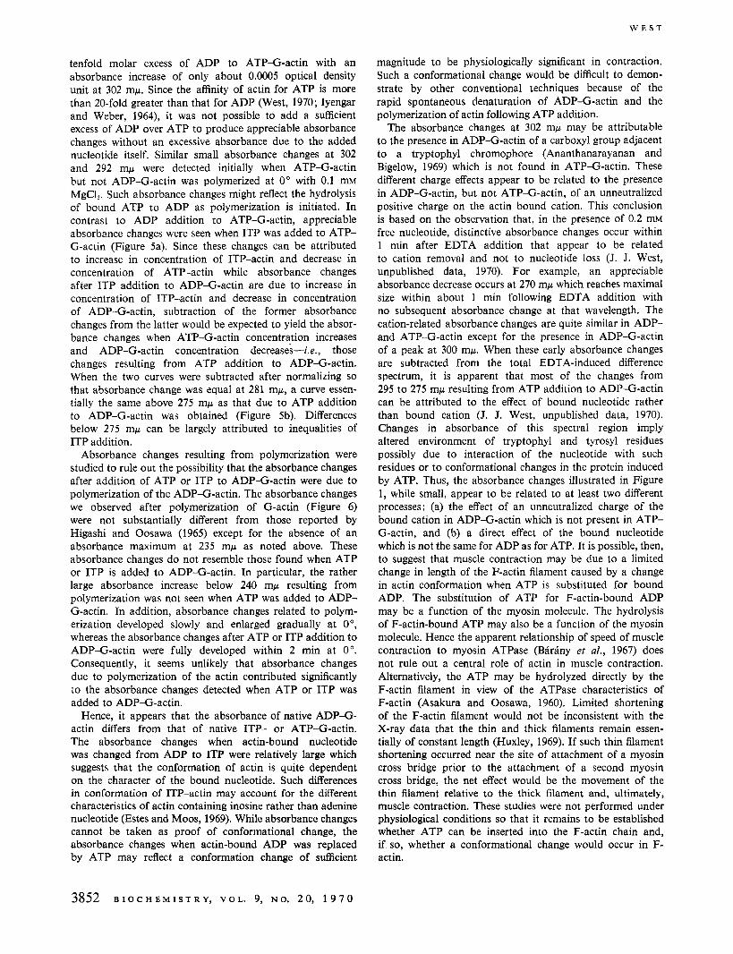
of the residual salt present in the F-actin pellet (West, 1970). Hence, the absorbance changes due t o polymerization of ATP-G-actin were measured (Figure 6). When salt was added to ADP-G-actin a t O " , no absorbance changes were detected but polymerization did not occur (Hayashi and Rosenbluth, 1962). However, if the ADP-G-actin preparation was warmed to 25" for a few minutes after salt addition, then absorbance changes occurred which were essentially the same as those when salt was added to ATP-G-actin at 0".
Discussion
Our spectrophotometric studies show reproducible absor- bance changes when ATP or ITP is added to ADP-G-actin (Figures 1 and 3). The absorbance changes after ATP addition to ADP-G-actin cannot be attributed to initial nucleotide binding by actin previously lacking bound nucleotide since similar changes occurred whether 0.15 mM ADP or 0.5 p~ ADP was present prior to ATP addition. Since the binding constant of G-actin for ADP is about 3 X 10⁶ ~ - 1 a t 0" (West, 1970) less than 0.3% of the actin should have been free of bound nucleotide when 0.15 mM ADP was present, whereas about 10% of the actin should have been without bound nucleotide when only 0.5 p~ ADP was present. Thus, if the principal explanation of the absorbance change
230 240 250 260 270 280 290 300 310 320 mfi
FIGURE 4: Absorbance changes resulting from addition of ATP to ATP-G-actin, ADP to ADP-G-actin, or ATP to ADP-G-actin previously denatured by EDTA. (---) Absorbance changes after addition of 0.1 mM ATP to ATP-G-actin 16.7 p~ in 1 mM Tris-HC1 (pH 8.0) in a cell with a path length of 0.88 cm. (-) Absorbance change 5 min after addition of 50 p~ ADP to ADP-G-actin 18.8 p~ in 1 mM Tris-HC1 (pH 8.0) containing 0.25 IJM ADP in a cell with a path length of 0.88 cm. (- -) Absorbance change after addition of 25 p~ ATP to 22.5 ~ IM ADP-G-actin previously denatured by exposure to 1 mM EDTA for 200 min at 0". The cell path length was 1 .O cm.
was nucleotide binding to actin previously not containing bound nucleotide, the added ATP should have produced a much larger absorbance change when 0.5 p~ ADP was present than when 0.15 mM ADP was present. The 17% reduction in the size of the absorbance changes at 302 mp when 0.15 mM ADP was present can be attributed to a reduced conversion of ADP-G-actin to ATP-G-actin, since about 95% of the actin would contain bound ATP after addition of 50 p~ ATP in the presence of 0.5 p~ ADP, whereas only about 75% of the actin would contain bound ATP in the presence of 0.15 mM ADP. Higashi and Oosawa (1965) concluded that absorbance changes at 232 mp after ATP addition to ADP-G-actin at room temperature were not due to conformational changes, but rather to nucleotide binding to actin previously containing no bound nucleotide since the presence of a large excess of ADP in their ADP- G-actin solution largely suppressed the absorbance changes. Because of the low binding constant of G-actin for ADP at 25" (West, 1970), ADP was largely dissociated from the G-actin in Higashi and Oosawa's studies. Consequently, under their conditions, addition of ATP would be expected to induce substantial absorbance changes related to binding of nucleotide to previously nucleotide-free actin. Such large absorbance changes could mask relatively minor absorbance effects due to changed conformation of the small percentage of the actin initially containing bound ADP, particularly since our studies at 0" indicated no substantial change in absorbance at 232 mp when ATP was added to ADP-G-actin. Thus, Higashi and Oosawa's failure to detect absorbance changes a t 232 mp attributable to changed conformation when ATP replaces actin-bound ADP could be due, in part, to the relatively small changes that appear to occur at this wavelength when ATP replaces actin-bound ADP. Nevertheless, appreciable absorbance changes do occur at other regions of the ultraviolet spectrum. A variety of other effects could alter absorbance changes a t 232 mp. Thus, addition of ATP to actin in the sample cell could induce some polymerization and, consequently, a large positive absorbance change at 232 mp. In addition, since ADP-G-actin without free nucleotide denatures more
3850 B I O C H E M I S T R Y , V O L . 9, N O . 2 0 , 1 9 7 0
C O N F O R M A T I O N A L C H A N G E O F A D P - G - A C T I N
nyl
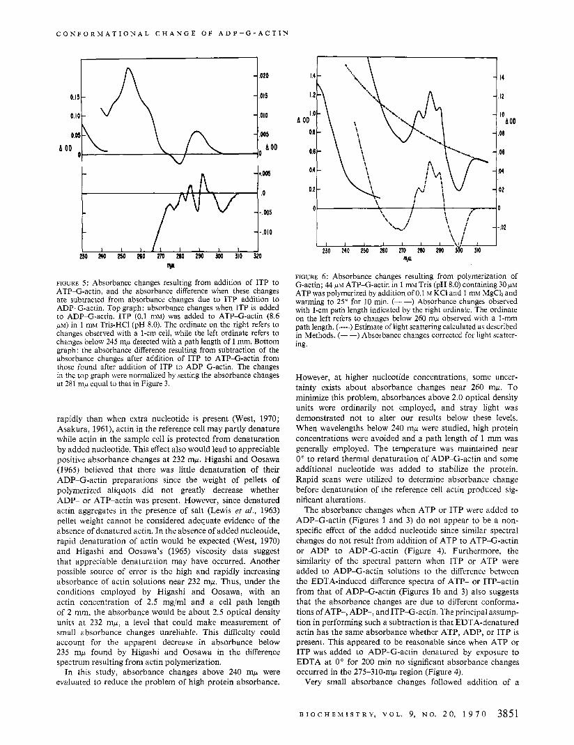
FIGURE 5: Absorbance changes resulting from addition of ITP to ATP-G-actin, and the absorbance difference when these changes are subtracted from absorbance changes due to ITP addition to ADP-G-actin. Top graph: absorbance changes when ITP is added to ADP-G-actin. ITP (0.1 mM) was added to ATP-G-actin (8.6 p ~ ) in 1 mM Tris-HC1 (pH 8.0). The ordinate on the right refers to changes observed with a 1-cm cell, while the left ordinate refers to changes below 245 mp detected with a path length of 1 mm. Bottom graph: the absorbance difference resulting from subtraction of the absorbance changes after addition of ITP to ATP-G-actin from those found after addition of ITP to ADP-G-actin. The changes in the top graph were normalized by setting the absorbance changes at 281 m,u equal to that in Figure 3.
rapidly than when extra nucleotide is present (West, 1970; Asakura, 1961), actin in the reference cell may partly denature while actin in the sample cell is protected from denaturation by added nucleotide. This effect also would lead to appreciable positive absorbance changes a t 232 mp. Higashi and Oosawa (1965) believed that there was little denaturation of their ADP-G-actin preparations since the weight of pellets of polymerized aliquots did not greatly decrease whether ADP- or ATP-actin was present. However, since denatured actin aggregates in the presence of salt (Lewis et al., 1963) pellet weight cannot be considered adequate evidence of the absence of denatured actin. In the absence of added nucleotide, rapid denaturation of actin would be expected (West, 1970) and Higashi and Oosawa's (1965) viscosity data suggest that appreciable denaturation may have occurred. Another possible source of error is the high and rapidly increasing absorbance of actin solutions near 232 mp. Thus, under the conditions employed by Higashi and Oosawa, with an actin concentration of 2.5 mg/ml and a cell path length of 2 mm, the absorbance would be about 2.5 optical density units at 232 my, a level that could make measurement of small absorbance changes unreliable. This difficulty could account for the apparent decrease in absorbance below 235 mp found by Higashi and Oosawa in the difference spectrum resulting from actin polymerization.
In this study, absorbance changes above 240 mp were evaluated to reduce the problem of high protein absorbance.
- .I2
- . IO
.06
.08
.M
.02
A OD - - -
-
w FIGURE 6: Absorbance changes resulting from polymerization of G-actin; 44 PM ATP-G-actin in 1 mM Tris (pH 8.0) containing 30 p~ ATP was polymerized by addition of 0.1 M KCI and 1 mM MgCh and warming to 25" for 10 min. (- -) Absorbance changes observed with 1-cm path length indicated by the right ordinate. The ordinate on the left refers to changes below 260 mp observed with a I-mm path length. (.---.) Estimate of light scattering calculated as described in Methods. (--) Absorbance changes corrected for light scatter- ing.
However, at higher nucleotide concentrations, some uncer- tainty exists about absorbance changes near 260 mp. To minimize this problem, absorbances above 2.0 optical density units were ordinarily not employed, and stray light was demonstrated not to alter our results below these levels. When wavelengths below 240 mp were studied, high protein concentrations were avoided and a path length of 1 mm was generally employed. The temperature was maintained near 0" to retard thermal denaturation of ADP-G-actin and some additional nucleotide was added to stabilize the protein. Rapid scans were utilized to determine absorbance change before denaturation of the reference cell actin produced sig- nificant alterations.
The absorbance changes when ATP or ITP were added to ADP-G-actin (Figures 1 and 3) do not appear to be a non- specific effect of the added nucleotide since similar spectral changes do not result from addition of ATP to ATP-G-actin or ADP to ADP-G-actin (Figure 4). Furthermore, the similarity of the spectral pattern when ITP or ATP were added to ADP-G-actin solutions to the difference between the EDTA-induced difference spectra of ATP- or ITP-actin from that of ADP-G-actin (Figures l b and 3) also suggests that the absorbance changes are due to different conforma- tions of ATP-, ADP-, and ITP-G-actin. The principal assump- tion in performing such a subtraction is that EDTA-denatured actin has the same absorbance whether ATP, ADP, or ITP is present. This appeared to be reasonable since when ATP or ITP was added to ADP-G-actin denatured by exposure to EDTA at 0' for 200 min no significant absorbance changes occurred in the 275-310-mp region (Figure 4).
Very small absorbance changes followed addition of a
B I O C H E M I S T R Y , V O L . 9, N O . 2 0 , 1 9 7 0 3851
W E S T
tenfold molar excess of ADP to ATP-G-actin with an absorbance increase of only about 0.0005 optical density unit at 302 mp. Since the affinity of actin for ATP is more than 20-fold greater than that for ADP (West, 1970; Iyengar and Weber, 1964), it was not possible to add a sufficient excess of ADP over ATP to produce appreciable absorbance changes without an excessive absorbance due to the added nucleotide itself. Similar small absorbance changes at 302 and 292 rnp were detected initially when ATP-G-actin but not ADP-G-actin was polymerized at 0" with 0.1 mM MgCl?. Such absorbance changes might reflect the hydrolysis of bound ATP to ADP as polymerization is initiated. In contrast to ADP addition to ATP-G-actin, appreciable absorbance changes were seen when ITP was added to ATP- G-actin (Figure 5a). Since these changes can be attributed to increase in concentration of ITP-actin and decrease in concentration of ATP-actin while absorbance changes after ITP addition to ADP-G-actin are due to increase in concentration of ITP-actin and decrease in concentration of ADP-G-actin, subtraction of the former absorbance changes from the latter would be expected to yield the absor- bance changes when ATP-G-actin concentration increases and ADP-G-actin concentration decreases-Le., those changes resulting from ATP addition to ADP-G-actin. When the two curves were subtracted after normalizing so that absorbance change was equal at 281 mp, a curve essen- tially the same above 275 mp as that due to ATP addition to ADP-G-actin was obtained (Figure 5b). Differences below 275 mp can be largely attributed to inequalities of ITP addition.
Absorbance changes resulting from polymerization were studied to rule out the possibility that the absorbance changes after addition of ATP or ITP to ADP-G-actin were due to polymerization of the ADP-G-actin. The absorbance changes we observed after polymerization of G-actin (Figure 6) were not substantially different from those reported by Higashi and Oosawa (1965) except for the absence of an absorbance maximum at 235 mp as noted above. These absorbance changes do not resemble those found when ATP or ITP is added to ADP-G-actin. In particular, the rather large absorbance increase below 240 mw resulting from polymerization was not seen when ATP was added to ADP- G-actin. In addition, absorbance changes related to polym- erization developed slowly and enlarged gradually at O", whereas the absorbance changes after ATP or ITP addition to ADP-G-actin were fully developed within 2 min at 0". Consequently, it seems unlikely that absorbance changes due to polymerization of the actin contributed significantly to the absorbance changes detected when ATP or ITP was added to ADP-G-actin.
Hence, it appears that the absorbance of native ADP-G- actin differs from that of native ITP- or ATP-G-actin. The absorbance changes when actin-bound nucleotide was changed from ADP to ITP were relatively large which suggests that the conformation of actin is quite dependent on the character of the bound nucleotide. Such differences in conformation of ITP-actin may account for the different characteristics of actin containing inosine rather than adenine nucleotide (Estes and Moos, 1969). While absorbance changes cannot be taken as proof of conformational change, the absorbance changes when actin-bound ADP was replaced by ATP may reflect a conformation change of sufficient
magnitude to be physiologically significant in contraction. Such a conformational change would be difficult to demon- strate by other conventional techniques because of the rapid spontaneous denaturation of ADP-G-actin and the polymerization of actin following ATP addition.
The absorbance changes at 302 mp may be attributable to the presence in ADP-G-actin of a carboxyl group adjacent to a tryptophyl chromophore (Ananthanarayanan and Bigelow, 1969) which is not found in ATP-G-actin. These different charge effects appear to be related to the presence in ADP-G-actin, but not ATP-G-actin, of an unneutralized positive charge on the actin bound cation. This conclusion is based on the observation that, in the presence of 0.2 mM free nucleotide, distinctive absorbance changes occur within 1 min after EDTA addition that appear to be related to cation removal and not to nucleotide loss (J. J. West, unpublished data, 1970). For example, an appreciable absorbance decrease occurs at 270 mp which reaches maximal size within about 1 min following EDTA addition with no subsequent absorbance change at that wavelength. The cation-related absorbance changes are quite similar in ADP- and ATP-G-actin except for the presence in ADP-G-actin of a peak at 300 mp. When these early absorbance changes are subtracted from the total EDTA-induced difference spectrum, it is apparent that most of the changes from 295 to 275 mp resulting from ATP addition to ADP-G-actin can be attributed to the effect of bound nucleotide rather than bound cation (J. J. West, unpublished data, 1970). Changes in absorbance of this spectral region imply altered environment of tryptophyl and tyrosyl residues possibly due to interaction of the nucleotide with such residues or to conformational changes in the protein induced by ATP. Thus, the absorbance changes illustrated in Figure 1, while small, appear to be related to at least two different processes: (a) the effect of an unneutralized charge of the bound cation in ADP-G-actin which is not present in ATP- G-actin, and (b) a direct effect of the bound nucleotide which is not the same for ADP as for ATP. It is possible, then, to suggest that muscle contraction may be due to a limited change in length of the F-actin filament caused by a change in actin conformation when ATP is substituted for bound ADP. The substitution of ATP for F-actin-bound ADP may be a function of the myosin molecule. The hydrolysis of F-actin-bound ATP may also be a function of the myosin molecule. Hence the apparent relationship of speed of muscle contraction to myosin ATPase (BEirBny et al., 1967) does not rule out a central role of actin in muscle contraction. Alternatively, the ATP may be hydrolyzed directly by the F-actin filament in view of the ATPase characteristics of F-actin (Asakura and Oosawa, 1960). Limited shortening of the F-actin filament would not be inconsistent with the X-ray data that the thin and thick filaments remain essen- tially of constant length (Huxley, 1969). If such thin filament shortening occurred near the site of attachment of a myosin cross bridge prior to the attachment of a second myosin cross bridge, the net effect would be the movement of the thin filament relative to the thick filament and, ultimately, muscle contraction. These studies were not performed under physiological conditions so that it remains to be established whether ATP can be inserted into the F-actin chain and, if so, whether a conformational change would occur in F- actin.
3852 B I O C H E M I S T R Y , VOL. 9, N O . 20 , 1 9 7 0
C O N F O R M A T I O N A L C H A N G E O F A D P - G - A C T I N
Acknowledgments
I thank Ronald G. Greene, of the Department of Bio- chemistry, Duke University, for his valuable suggestions, and Floyd Dorrity for his expert technical assistance.
References
Ananthanarayanan, V. S., and Bigelow, C. C. (1969), Bio-
Asakura, S. (1961), Arch. Biochem. Biophys. 92,140. Asakura, S., and Oosawa, F. (1960), Arch. Biochem. Biophys.
87,273. BBrBny, M., Conover, T., Schliselfeld, L., Gaetjens, E.,
and Goffart, M. (1967), Eur. J. Biochem. 2,156. Drabikowski, W., and Gergely, J. (1962), J. Biol. Chem.
237,3412. Estes, J. E., and Moos, C. (1969), Arch. Biochm. Biophys.
132,388. Feuer, G., Molnar, F., Pettko, E., and Straub, F. (1948),
Acta Physiol. Acad. Sci. Hung. 1,150. Hayashi, T., and Rosenbluth, R. (1964), in Biochemistry of
Muscle Contraction, Gergely, J., Ed., Boston, Mass.,
chemistry 8,3723.
Little, Brown, p 180. Higashi, S., and Oosawa, F. (1965), J. Mol. Biol. 12,
843. Huxley, H. E. (1960), in The Cell, Vol. IV, Mirsky, B., Ed.,
New York, N. Y. , Academic, p 365. Huxley, H. E. (1969), Science 164,1356. Iyengar, M. R., and Weber, H. H. (1964), Biochim. Biophys.
Laki, K . (1969),Physio~, Chem. Pltys. I , 237. Leach, S. J., and Scheraga, H. A. (1960), J. Amer. Chem. Soc.
Lewis, M., Maruyama, K., Carroll, W., Kominz, D., and
Lowey, S., and Luck, S. (1969), Biochemistry 8,3195. Martonosi, A. (1962), J . Biol. Chem. 237,2795. Mommaerts, W. F. (1951), J. Biol. Chem. 188,559. Nagy, B. (1966), Biochim. Biophys. Acta 115,498. Pabst Laboratories (1956), Circular OR-10. Silvester, N., and Holwill, M. (1965), Nature 205,665. West, J. J. (1970), Biochemistry 9,1239. West, J. J., Nagy, B., and Gergely, J. (1967), Biochem. Biophys.
Yankeelov, J. A., Jr. (1963), Anal. Biochem. 6,287.
Acta 86, 543.
82,4790.
Laki, K. (1963), Biochemistry 2,34.
Res. Commun. 29,611.
B I O C H E M I S T R Y , VOL. 9, N O . 20, 1 9 7 0 3853


![Increased Rate of Adenosine Triphosphate …...(CANCER RESEARCH 55, 4352-4360, October 1, 1995] Increased Rate of Adenosine Triphosphate-dependent Etoposide (VP-16) Efflux in a Murine](https://img.pdfslide.net/doc/110x75/5e7e8d68c5d0407f2447f2a9/increased-rate-of-adenosine-triphosphate-cancer-research-55-4352-4360-october.jpg)