Embed Size (px)
Citation preview
290 BIOCHIMICA ET BIOPHYSICA ACTA
BBA 3954
A D E N O S I N E T R I P H O S P H A T E - I N D U C E D CONTRACTION OF
R A T - L I V E R M I T O C H O N D R I A AND
S Y N T H E S I S OF M I T O C H O N D R I A L P H O S P H O L I P I D S
LECH WOJTCZAK, PAULINA W L O D A W E R ANO JOZEF ZBOROWSKI
Department of Biodtemistry, Nencki Institute of Experimental Biology, Warsaw (Poland)
(Received September 28th, 1962)
SUMMARY
The incorporation of a-glycero[32P]phosphate into mitochondrial phospholipids was investigated during swelling and contraction of rat-liver mitochondria.
The extent of the incorporation increased considerably during ATP-induced contraction of swollen mitochondria. The chief labelled phospholipid was tentatively identified as phosphatidic acid.
Evidence is presented supporting the view that the increased incorporation of ~,-glycerophosphate into the mitochondrial phospholipids is in some way related to mitochondrial contraction. This is based on the following results:
I. The time course of ~-glycerophosphate incorporation paralled the time course of mitochondrial contraction.
2. There was no increased incorporation, if mitochondria were protected against swelling by serum albumin and if, consequently, no contraction occurred upon the addition of ATP.
3- Inhibitors of mitochondrial contraction usually decreased the extent of a-glycerophosphate incorporation.
4. The incorporation was significantly depressed, when a-glycerophosphate was added to the contracted mitochondria.
The possible role of phospholipids in mitochondrial contraction is discussed.
INTRODUCTION
Mitochondria isolated from a variety of mammalian tissues undergo morphological changes, known as swelling, when placed in isotonic saline or sucrose media. This process, which consists of penetration of water into the mitochondria, can be acceler- ated by low concentrations of thyroxine, long chain fat ty acids, calcium ions, phlorizin, inorganic phosphate and a number of other substances. LEHNINGER AND REMMERT 1 isolated from sonically disrupted liver mitochondria a substance which was a potent swelling-inducing factor. This substance was identified as a mixture of non-esterified long chain fa t ty acids 2. It was found 2 that spontaneous swelling of mitochondria, as well as swelling induced by Ca ~+ and thyroxine, are accompanied
Abbreviat ions: GP, c¢-glycerophosphate; I'CMB, p-chloromercuribenzoate.
Biochim. Biophys..4cta, 7 ° (I963) 290-305
MITOCHONDRIAL CONTRACTION AND PHOSPHOLIPID SYNTHESIS 2 9 I
by an increase in the content of non-esterified fat ty acids, presumably resulting from hydrolytic splitting of mitochondrial lipids.
Swelling of mitochondria can be reversed by the addition of ATP 8,~; the presence of Mg s+ or Mn 2+ is usually necessary for the maximum effect. This process, which consists of extrusion of water from mitochondria, is known as shrinkage or "con- traction". I t was reported previouslyL that a considerable part of the non-esterified fa t ty acids which either accumulated during the swelling of mitochondria or were added to the medium, disappeared during the contraction. A part of the added [t'C]- oleic acid was found to be incorporated into mitochondrial phospholipids. Two striking features of this process were observed: (a) the incorporation of [l'C]oleic acid into phospholipids was inhibited by sucrose which is known to inhibit the ATP- induced contraction of mitochondria 6; (b) both the incorporation and the contraction had a similar time course, being highest during the first few minutes following the addition of ATP. This led us to assume that the synthesis of phospholipids might be in some way related to the contraction of the mitochondria.
In the present s tudy the biosynthesis of phospholipids during the swelling and contraction of mitochondria has been further investigated by the use of [nP]GP.
MATERIAL AND METHODS
Rat-liver mitochondria were isolated as described by LEHNINGER ~ al. s. Mito- chondria derived from one liver (about 5 g wet wt.) were suspended in 3 ml of 0.25 M sucrose. I ml of the suspension contained about 4 ° mg protein.
Swelling and contraction of mitochondria were followed opticaily s at 520 m/L in x8 × I5o-mm test tubes, using a Coleman "Junior" Spectrophotometer.
Experiments with [14C]oleic acid, including the extraction and fractionation of the lipids, were carried out as described previously 2.
In experiments with [3*P]GP the basic medium was o.I25 M KCl-o.oz M Tr is - HCI buffer (pH 7-4) and the swelling agent was usually sodium oleate or sometimes inorganic phosphate (pH 7.4). 0.2 ml of mitochondrial suspension was added per IO ml of the medium and the incubation was carried out at 2o °. After zo rain, o.I ml of 0.25 M ATP was added and the incubation was continued for another 15 min. Spectra- photometer readings were taken every 5 min. The reaction was stopped by the ad- dition of trichloroacetic acid to a final concentration of 8 % and the tubes were cen- trifuged. The precipitate was washed several times with 5 % trichloroacetic acid and then with water.
To the washed precipitate 2 ml of methanol followed by 4 ml of chloroform were added and the samples were left overnight at room temperature. The extract was filtered and the residue was extracted 4 times with chloroform - methanol (2: I, v/v) at 60 ° . The extracts were combined and the solvent was evaporated in vacua almost to dryness. To the resulting residue a few drops of water were added and the lipids were re-extracted with petroleum e t h e r - chloroform (2:I, v/v). The extract was dehydrated over NasSO,, filtered and made up to a known volume.
Radioactivity of a,p was measured in suitable aliquots of the lipid extract by use of a Geiger-Miiller counter with a thin mica end-window and the radioactivity of 14C with a "Micromil" window counter (Nuclear Chicago CO. ).
Column chromatography of phospholipids was carried out on silicic acid (Mal-
Biochim. Biophys. Acta, 7 ° (I963) 290-305
292 L. WOJTCZAK, P. WLODAWER, J. ZBOROWSKI
linckrodt, Ioo mesh). Non-phospholipids were removed by chloroform, and phospho- lipids were fractionated by elution with increasing concentrations of methanol in chloroform.
Paper chromatography of phospholipids was carried out on commercial silicic acid-impregnated papeY (Schleicher and Schfill) using the diisobutyl ketone - acetic a c i d - water (4 ° :30: 7, v/v) system of MARINETTI AND STOTZ 6 and the diisobutyl k e t o n e - formic a c i d - water (4o:15:2, v/v) system of THIELE AND WOBER 7. The chromatograms were analyzed in "Actigraph I I " chromatographic strip scanner with a "Micromil" window (Nuclear Chicago Co.) and autoradiograph3~ was carried out on usual X-ray photographic plates. Phospholipids were detected by staining with Rhodamine 6G and viewed under an ultraviolet lamp while wet 8.
Mild alkaline hydrolysis of phospholipids was performed according to DAWSON *. Products of the hydrolysis were chromatographed on Whatman No. I filter paper using the following solvent systems: A, ethanol - acetic acid - water 1° (83 : I : 16, v/v) ; B, acetic acid - ethyl acetate - water 11 (3:3 : I, v/v) ; C, phenol saturated with o.I % NH 3 in waterS; D, Cellosolve - methyl ethyl ketone - 3 N NH 3 (7 : 2:3, v/v) saturated with boric acid1*; E, bu t ano l - IO % trichloroacetic acid ° (62:38, v/v). Descending chromatography was used for pheno l -NH z and ascending chromatography for.all other solvent systems. The paper was washed before use with 1% EDTA for solvents A and B and with 2 N acetic acid for the other systems. The spots were located by spraying with the acid molybdate reagent of HANES AND ISHERWOOD TM and irra- diation with ultraviolet light 1..
Reagents of analytical grade, and distilled and deionized water were used through- out. The puri ty of ATP appeared to be of special importance. Among several samples of ATP tested the highest contraction of mitochondria was achieved by using ATP (crystalline disodium salt) from Pabst Laboratories, Milwaukee, Wisc., and only this preparation was used in the present investigation.
[~P]GP was synthesized as described by KENNEDY 15. I t contained 9 ° % of DL-a-glycerophosphate and only IO % as the fl-form.
Carrier-free [32P]phosphoric acid was obtained from the Institute of Nuclear Research in Warsaw and [I-l*C]oleic acid from California Corporation for Biochemical Research, Los Angeles, Calif.
DL-,,-Lecithin (synthetic), phosphatidylethanolamine (from brain) and phos- phatidyl-L-serine used as reference substances in the chromatography of phospholipids were obtained from Nutritional Biochemicals Co., Cleveland, Ohio.
RESULTS
In the first series of experiments mitochondria were incubated in the presence of [i-l~C]oleic acid as the swelling agent and of either =-glycerophosphate, phospho- choline, phosphoethanolamine or phosphoserine. After 2o rain of incubation, ATP and MgC12 were added and the incubation was continued for another I5 rain. The reaction was stopped and the incorporation of 14C into mitochondrial phospholipids was measured as described previously ~. I t was found (Table I, Expt. I) that the in- corporation was strongly stimulated by GP but not by the other phosphoric esters tested. However, the presence of GP plus either phosphocholine or phosphoethanol- amine resulted in a further increase of the incorporation (Table I, Expt. 2).
BiocMm. Biopkys. Acta, 7 ° (I963) 290-305
MITOCHONDRIAL CONTRACTION AND PHOSPHOLIPID SYNTHESIS 293
These results suggested that GP labelled in the phosphate moiety could be a useful tool in studying the biosynthesis of phospholipids in mitochondria during their swelling and contraction. This was confirmed by experiments in which ~P]GP has been found to be incorporated into phospholipids at a rate greatly exceeding that of the incorporation of either 8*Pl or [nP]ATP.
T A B L E I
EFFECT OF VARIOUS PHOSPHORIC ESTERS ON THE INCORPORATION OF [I-14C]OLEIC ACID INTO MITOCHONDRIAL PHOSPHOLIPIDS
Incuba t ion mix tu re consisted of 35 ml of o.125 M K C l - o . o 2 M T r i s - H C l (pH 7.4), ]54 mFmoles sodium oleate conta ining IO ~ counts / ra in [I-14C~ oleic acid, and o.25 ml of mi tochondr ia suspension; o ther addi t ions as indicated. The mix ture was incuba ted 20 rain a t 20 °, t h e n ATP and MgCl 2 were added to t he final concent ra t ions of 0.005 M and 0.o03 M respectively, and t h e incubat ion
was cont inued for ano ther 15 rain.
Totadradioo, ait~y A dditio~ ol'the pk~photipid
Iractios~ (co=~s/min)
Exp t . I None 8 ooo DL-~-Glycerophosphate (xo/~moles) 13 350 Phosphochol ine (io/~moles) 6 50o Phosphoe thano lamine (xo #moles) 8 ooo Phosphoser ine (IO #,moles) 6 95o
None 4 050 DL-~t-Glycerophosphate ( io ~umoles) 8 i oo DL-~-Glycerophosphate
(io s/moles) + phosphochol ine (IO/*moles) I I 200 DL-~-Glycerophosphate
(IO #*moles) + phosphoe thano lamine (IO/~moles) i I 6oo nL-~-Glycerophosphate
(io/~moles) + phosphoser ine (Io/~moles) 5 30o
E x p t . 2
Incorporation of [~P]GP into mitochondrial phospholipids The effect of ATP on the incorporation of glycerophosphate into mitochondrial
phospholipids is shown in Table II, and the effect of CoA and oleate in Table III. It is evident that the addition of ATP to the swollen mitochondria greatly increased the incorporation (see also Fig. I). A stimulatory effect of CoA and of oleate was also observed (Table III, Expt. 2). However, it should be noted that in some cases the addition of oleate did not affect the incorporation. This fact might be due to the presence of a somewhat larger amount of endogenous non-esterified fatty acids in some samples of mitochondria. Oleate at higher concentrations was even inhibitor, (Table III, Expt. I).
As can be seen in Table III, the mitochondrial phospholipids contained only a very small part of the [mP]GP present in the medium. Therefore, it appeared necessary to use [nP]GP of a high specific activity (lO T counts/min/~mole) in sub- sequent experiments.
Fig. I shows the time course of the swelling and contraction of mitochondria as well as of [UP]GP incorporation into mitochondrial phospholipids. It is evident that the swelling of mitochondria (as visualized by the fall in light absorbancy) was ac- companied by only a small incorporation of label into the phospholipids. The addition of ATP induced mitochondria to contract (as shown by the increase in light ab-
Biochim. Biophys. Acta, 7 ° (x963) 29o-305
294 L. WOJTCZAK, P. WLODAWER, J. ZBOROWSKI
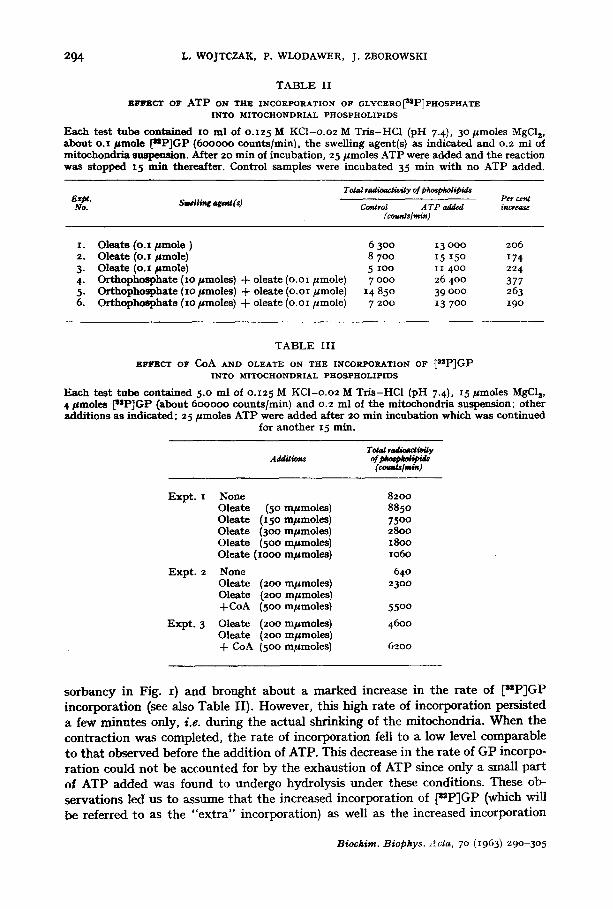
TABLE II
EFFECT OF ATP oN THE INCORPORATION OF GLYCERO~2P]PHOSPHATE INTO MITOCHONDRIAL PHOSPHOLIPIDS
Each test tube contained I0 ml of 0.125 M KCl-o.o2 M Tris-HC1 (pH 7-4), 30/,moles MgClz, about o.I #*mole [uP]GP (6ooooo counts/mitt), the swelling agent(s) as indicated and o.2 ml of mitochondris suspension. After 2o rain of incubation, 25 #*moles ATP were added and the reaction was stopped t 5 rain thereafter. Control samples were incubated 35 min with no ATP added.
Total ,adioaaivily of phospholipid, s Ezpt. Swelling agog(s) Per cent No. Control A TP added increaae
( counSs lmin )
1. Oleate (o.x #*mole ) 6 300 13 ooo 206 2. Oleate (o.1 #*mole) 8 700 15 15o 174 3. Ole~te (o.x #*mole) 5 ioo i i 40o 224 4. Orthophosphate (xo #*moles) + olcate (o.oi #*mole) 7 ooo 26 40o 377 5- Orthopho~hate (io/tmoles) + oleate (O.OI //mole) 14 850 39 ooo 263 6. Orthophosphate (i0 #*moles) + oleate (o.oi pmole) 7 200 13 700 I9o
TABLE III
EFFECT OF CoA AND OLEATE ON THE INCORPORATION OF ~zP]GP INTO MITOCHONDRIAL PHOSPHOLIPIDS
Each test tube contained 5.0 ml of o.125 M KCI-o.o2 M Tris-HCl (pH 7-4), I5 #*moles I~I~C]|, 4 #*moles [ssP]GP (about 600000 counts/min) and 0.2 m] of the mitochondria suspension; other additions as indicated; 25 #*moles ATP were added after 20 rain incubation which was continued
for another x5 min.
Tot~/mdbma/t~y
(cot~,slmi.)
Expt. 1 None 8200 Oleate (50 mpmoles) 8850 Oleate (150 m#*moles) 7500 Oleate (3oo mpmoles) 2800 Oleate (500 m#*moles) 18oo Ole~te (1000 m#*moles) 1060
Expt. 2 None 640 Oleate (2oo m#*moles) 23o0 Oleate (2oo mpmoles) +CoA (500 mpmoles) 5500
Expt. 3 Oleate (200 mpmoles) 4600 Olcate (200 mpmoles) + CoA (500 m#*moles) 6200
so rbancy in Fig. I) and brought abou t a marked increase in the rate of [stP]GP incorpora t ion (see also Table II). However, this high rate of incorporat ion persisted a few minu te s only, i.e. dur ing the ac tua l shr inking of the mitochondria . W h e n the con t rac t ion was completed, the rate of incorpora t ion fell to a low level comparable to t ha t observed before the addi t ion of ATP. This decrease in the rate of GP incorpo- ra t ion could no t be accounted for b y the exhaust ion of ATP since only a small par t of ATP added was found to undergo hydrolysis unde r these condit ions. These ob- servat ions led us to assume tha t the increased incorporat ion of [~P]GP (which will be referred to as the "ex t ra" incorporat ion) as well as the increased incorporat ion
Biochim. Biophys. Acta, 7 ° (1963) 290-305
MITOCHONDRIAL CONTRACTION AND PHOSPHOLIPID SYNTHESIS 295
of [z*C]oleic acid' into mitochondrial phospholipids might be in some way related to the process of mitochondrial contraction. The aim of further experiments was to check this hypothesis.
CE
EO.6 o
< 0.2
/ I
I J
s ~ .o ,~"
-" J" TATP ~o 2b 3b ' "
Min
12000
t -
4000
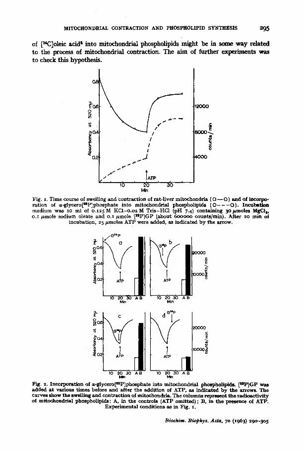
Fig. z. Time course of swelling and contract ion of rat- l iver mitochondria (O- -O) and of incorpo- ra t ion of u-glycero[SSP]phosphste into mitochondrial phospholipids ( O - - - O ) . Incubat ion medium was xo ml of o.x25 M KCI -o .o2M Tr i s -HCl (pH 7.4) containing 5opmoles MgCls, o.z/=mole sodium oleate and o.I /=mole ~uP]GP (about 6oooo0 counts/rain). After 2o rain of
incubation, 25/~mo|es ATP were added, as indicated by the arrow.
E L °
10 2 0 3 0 A B 10 2 0 3 0 A g Min Min
Fig. 2. Incorporation of =-8tycero~sP]phospbate into mitochondria3 phmpholipids. [uP]GP was added a t various t imes before and after the addit ion of ATP, as indicated by the arrows. The curves show the swelling and contract ion of mitochondria. The columns represent the radioactivi ty of mitochondrial phospholipids: A, in the controls (ATP omit ted); B, in the presence of ATP.
Experimental conditions as in Fig. I.
Biochim. Biophys. Acta, 7 ° (][963) 29o-3o 5
296 L. WOJTCZAK, P, WLODAWER, J. ZBOROWSKI
In the following experiments, [s2P]GP was added to the incubation mixture at various times before and after the addition of ATP. In the controls ATP was omitted. The results are shown in Fig. 2. I t is evident that in all cases when [z~P]GP was added before ATP, there was a highly significant "ex t ra" incorporation. This was observed even if the addition of [s*P]GP preceded that of ATP by as little as 3o sec. On the other hand, the incorporation of [a2P]GP into the phospholipids did not appreciably exceed tha t in the control when [mP]GP was added 5 min after ATP, i.e. to the contracted mitochondria. In this case the presence of ATP had no effect on the in- corporation.
Contraction of mitochondria induced by ATP has been reported to be inhibited by sucrose 5, glucose and other Saccharides is, by azide s, by s o m e i o n s 17, and by glutathione 4.
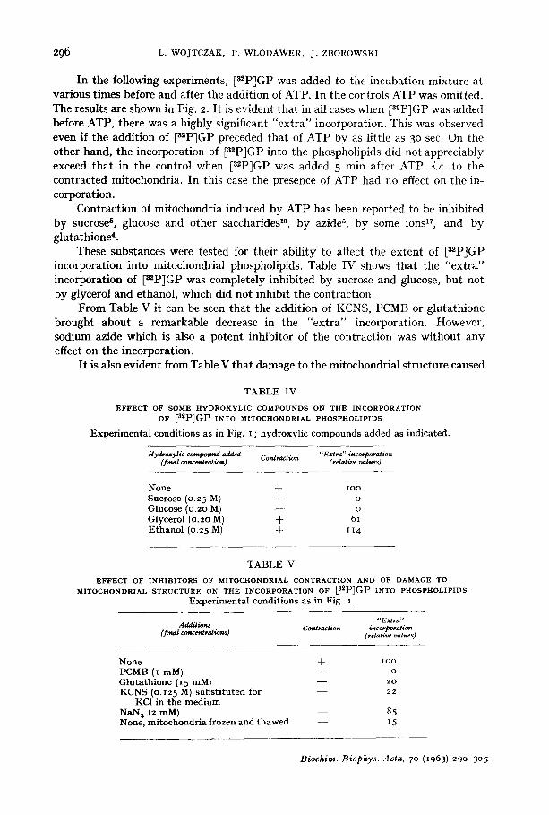
These substances were tested for their ability to affect the extent of [a*P]GP incorporation into mitochondrial phospholipids. Table IV shows that the "ex t ra" incorporation of [szP]GP was completely inhibited by sucrose and glucose, but not by glycerol and ethanol, which did not inhibit the contraction.
From Table V it can be seen that the addition of KCNS, PCMB or glutathione brought about a remarkable decrease in the "ex t ra" incorporation. However, sodium azide which is also a potent inhibitor of the contraction was without any effect on the incorporation.
I t is also evident from Table V that damage to the mitochondrial structure caused
T A B L E IV
E F F E C T O F S O M E H Y D R O X Y ' L I C C O M P O U N D S O N T H E I N C O R P O R A T I O N
OF [azP]GP INtO MITOCHONDRIAL PHOSPHOLIPIDS
E x p e r i m e n t a l cond i t ions as in Fig. i ; h y d r o x y l i c c o m p o u n d s added as ind ica ted .
Hydroxylic compound added Contraction "Extra" incorporation (final concc~ra2ion) (relative valtcs)
None + lOO Sucrose (0.25 M) - - o Glucose (0.20 M) - - o Glycerol (0.20 M) + 6 i E t h a n o l (0.25 M) + I t 4
T A B L E V
E F F E C T O F I N H I B I T O R S O F M I T O C H O N D R I A L C O N T R A C T I O N A N D O F D A M A G E T O
M I T O C H O N D R I A L S T R U C T U R E O N T H E I N C O R P O R A T I O N O F [aZP]GP I N T O P H O S P I t O L I P I D S
E x p e r i m e n t a l cond i t ions as in Fig. i .
" E x t r a " Additions Contraction incorporation
(final concoUrations) (relative vatues)
None + ioo PCMB (I mM) - - o G l u t a t h i o n e (15 mM~ - - 2o KCNS (0.I25 M) s u b s t i t u t e d for - - 22
KC1 in the m e d i u m NaN a (2 mM) - - 8.5 None, m i t o c h o n d r i a frozen and t h a w e d - - 15
Biochim. Biophys. Acta, 7 ° (I963) 290-30.5
MITOCHONDRIAL CONTRACTION AND PHOSPHOLIPID SYNTHESIS 297
by repeated freezing and thawing markedly decreased the incorporation. Such mito- chondria showed a very slight if any swelling and contraction as measured optically.
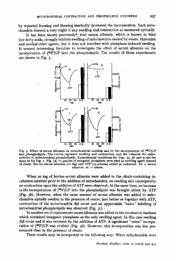
It has been shown previously z, that serum albumin, which is known to bind free fatty acids, strongly inhibits swelling of mitochondria caused by oleate, thyroxine and seveYal ~other agents, but it does not interfere with phosphate-induced swelling. It seemed interesting therefore to investigate the effect of serum albumin on the incorporation of [s=P]GP into the phospholipids. The results of these experiments are shown in Fig. 3.
~.( j a . ~ . ~ b
A!P
~oA
~o 2o ao A B 6 ~ a ~ As Min Min
Fig. 3- Effec t of s e r u m a l b u m i n on mi tochondr i a l swel l ing a n d on t h e incorpora t ion of ~=P]GP in to phosphol ip ids . T h e cu rves r ep resen t swell ing and cont rac t ion , a n d t h e c o l u m n s t h e radio- a c t i v i t y of mi tochondr i a l phosphol ip ids . E x p e r i m e n t a l condi t ions for Figs. 3a, 3b a n d 3c are t he s a m e as for Fig. z ; Fig. 3d, Io p m o l e s of inorganic p h o s p h a t e were used as swell ing agen t i n s t ead of oleate. Bov ine s e r u m a l b u m i n (2o rag) and A T P (z5 pmoles) added as indica ted . SA = s e r u m
a l b u m i n ; ol. = oleate.
When 20 nag of bovine-serum albumin were added to the oleate-containing in- cubation mixture prior to the addition of mitochondria, no swelling and consequently no contraction upon the addition of ATP were observed. At the same time, no increase in the incorporation of [szP]GP into the phospholipids was brought about by ATP (Fig. 3b). However, when the same amount of serum albumin was added to mito- chondria already swollen in the presence of oleate, just before or together with ATP, contraction of the mitochondria did occur and an appreciable "extra" labelling of mitochondrial phospholipids was observed (Fig. 3c).,
In another set of experiments serum albumin was added to the incubation medium which contained inorganic phosphate as the only swelling agent. In this case swelling did occur and it was reversed by the addition of ATP. A significant "extra" incorpo- ration of [nP]GP was evident (Fig. 3d). However, this incorporation was less pro- nounced than in the presence of oleate.
These results may be interpreted in the following way: When mitochondria were
Biochim. Biophys. Acta, 70 (1963) 29o-3o 5
298 L. WOJTCZAK, P. WLODAWER, J. ZBOROWSKI
protected against swelling by serum albumin and consequently ATP did not cause any contraction, the incorporation of [uP]GP was equal to that observed in the absence of ATP. Hence the addition of ATP, if not followed by contraction of mito- chondria, did not bring about any "ext ra" incorporation of [~P]GP into the phos- pholipids. On the other hand, serum albumin did not affect the extent of incorporation if added to the swollen mitochondria, i.e. when ATP produced a normal contraction. The less pronounced "ext ra" incorporation found in mitochondria which underwent phosphate-induced swelling might be due to lack of fa t ty acids necessary for the synthesis of phospholipids.
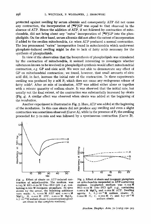
In view of the observation that the biosynthesis of phospholipids was stimulated by the contraction of mitochondria, it seemed interesting to investigate whether substances known to be involved in phospholipid synthesis would affect mitochondrial contraction, e.g. GP and oleic acid. We were not able to demonstrate any effect of GP on mitochondrial contraction; we found, however, that small amounts of oleic acid did, in fact, increase the initial rate of the contraction. In these experiments swelling was produced by 2 mM Pi which does not cause any endogenous release of fa t ty acids *. After 20 min of incubation, ATP was added either alone or together with a minute quanti ty of sodium oleate. It was observed that the initial rate, but usually not the final extent, of the contraction was substantially increased by oleate (Fig. 4). A similar effect was observed when oleate was added at the beginning of the incubation.
Another experiment is illustrated in Fig. 5. Here, ATP was added at the beginning of the incubation. In this case oleate did not produce any swelling and even a slight contraction was sometimes observed (Curve A), while in the presence of Pi the swelling proceeded for 5-1o rain and was followed by a spontaneous contraction (Curve B).
a~
o.4 E ~ - B o oo A ~0 .3
ga2 t
!o., t t lb 2b ab 4b 5'o
Min
Fig. 4- Egec t of oleate on ATl '- induced con- t ract ion of mitochondria . The medium was o.T2 5 M KCl-o .o2 M Tr i s -HC1 (pH 7.4), con- ta ining o.oo2 M inorganic phosphate . At t ime shown by the arrow, the following addit ions were made: A, o.oo2 M ATP and o.oo12 M MgC12; l:t, o .oo2M ATI', o . o o I 2 M MgC12, 6.7" i o .7 M sodium oleate {concentrations given
are those in the complete medium).
°•-0• . . ~ - - . . . . . . . . . . . . . . . . . . A
[o i. I L !
Min
Fig. 5. Eftect of oleate and inorganic phospha te on mi tochondr ia suspended in ATP-containing medium. Incuba t ion medium was o .125M KCl -o .o2 M T r i s - H C l {pH 7.4), containing o.oo12 M MgClz and o.oo2 M ATP. Swelling agents: A, 6. 7 .IO -~ M sodium oleate; B, o . o o 2 M I'l; C, o . o o 2 M I~i and 6.7"xo-~M
sodium o|eate.
Biochim. Biophys. Acta, 7 ° (I963) 290-305
MITOCHONDI~AL. CO~TRACTION AND PHOSPHOLIPID SYNTHESIS 299
There was no swelling, however, if both Pt and oleate were present in the medium (Curve C).
This observation may be interpreted as follows: ATP prevents swelling only if a small quantity of free fatty acid is already present. The spontaneous contraction of mitochondria swollen in the presence of Pt might be due to a release of endogenous fatty acids during the ageing of mitochondria.
I t might be expected that PPt would interfere with biosynthesis of phospholipids by shifting to the left the equilibrium of acyl-CoA formation according to the equation:
fatty acid + CoA + ATP ~ acyl-CoA + AMP + PPt.
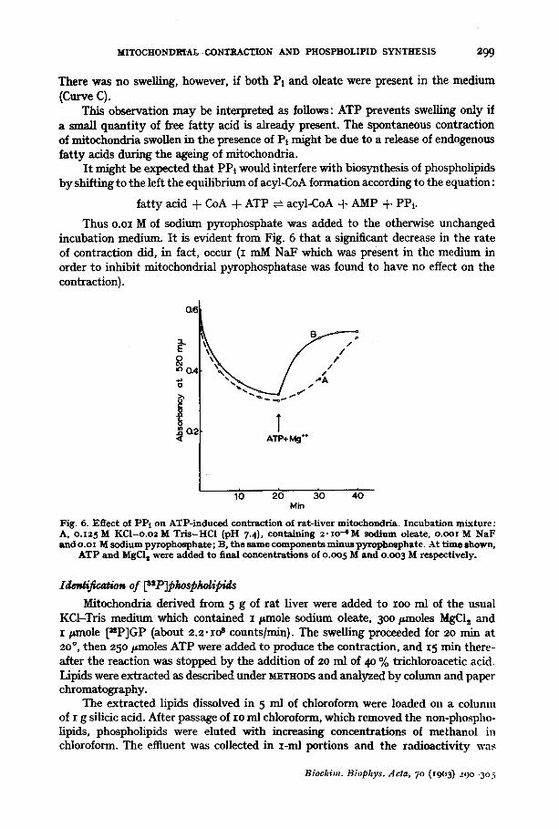
Thus o.oi M of sodium pyrophosphate was added to the otherwise unchanged incubation medium. It is evident from Fig. 6 that a significant decrease in the rate of contraction did, in fact, occur (I mM NaF which was present in the medium in order to inhibit mitochondrial pyrophosphatase was found to have no effect on the contraction).
0.6
0 (M m,O.4
/ / l" / .6,
~ ~ ~, o ~ J
I ATP+ Mg ""
ib 2b ~o 4'o Min
Fig. 6. Effect of PPI on ATP-induced contraction of rat-liver mitochondria. Incubation mixture: A, o.I25 M KCI-o.o2 M Tr is -HCl (pH 7-4), containing z .xo-~M sodium oleate, o.ool M NaF and o.ox M sodium pyrophosphate; B, the same components minus pyrophosphate. At time shown,
ATP and MgCI t were added to final concentrations of o.oo 5 M and o.oo 3 M respectively.
Mitochondria derived from 5 g of rat liver were added to IOO m/of the usual KCI-Tris medium which contained I/anole sodium oleate, 3oo/anoles MgCI 2 and I pmole [~P]GP (about 2.2-lO s counts/min). The swelling proceeded for zo rain at 2o °, then 25o/~moles ATP were added to produce the contraction, and 15 rain there- after the reaction was stopped by the addition of zo ml of 4o % trichloroacetic acid. Lipids were extracted as described under M~THODS and analyzed by column and paper chromatography.
The extracted lipids dissolved in 5 ml of chloroform were loaded on a column of I g silicic acid. After passage of Io ml chloroform, which removed the non-phospho- lipids, phospholipids were eluted with increasing concentrations of methanol in chloroform. The effluent was collected in I-ml portions and the radioactivity was
Biochim. Biophys. Acta, 7 ° (x9o3) zgo -3o5
300 L. WOJTCZAK, P. WLODAWER, J . ZBOROWSKI
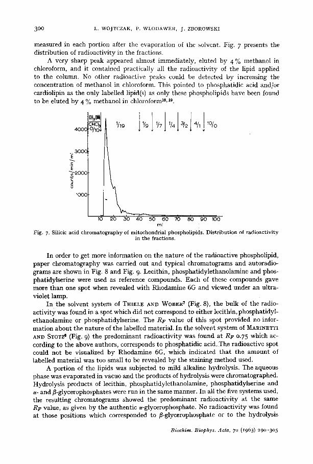
measured in each portion after the evaporation of the solvent. Fig. 7 presents the distribution of radioactivity in the fractions.
A very sharp peak appeared almost immediately, eluted by 4 To methanol in chloroform, and it contained practically all the radioactivity of the lipid applied to the column. No other radioactive peaks could be detected by increasing the concentration of methanol in chloroform. This pointed to phosphatidic acid and/or cardiolipin as the only labelled lipid(s) as only these phospholipids have been found to be eluted by 4 % methanol in chloroform Is, 19.
°'" ' l J l l J 4000 J 1/9 1/7 1/4 3/2
3000
E
5 8
1000
10 20 30 40 50 60 70 80 90 100 ml
Fig. 7. Silicic acid chromatography of mitochondrial phospholipids. Distribution of radioactivity in the fractions.
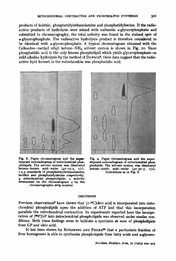
In order to get more information on the nature of the radioactive phospholipid, paper chromatography was carried out and typical chromatograms and autoradio- grams are shown in Fig. 8 and Fig. 9. Lecithin, phosphatidylethanolamine and phos- phatidylserine were used as reference compounds. Each of these compounds gave more than one spot when revealed with Rhodamine 6G and viewed under an ultra- violet lamp.
In the solvent system of THIELE AND WOBER 7 (Fig. 8), the bulk of the radio- activity was found in a spot which did not correspond to either lecithin, phosphatidyl- ethanolamine or phosphatidylserine. The RF value of this spot provided no infor- mation about the nature of the labelled material. In the solvent system of MARINETTI ANn STOTZ e (Fig. 9) the predominant radioactivity was found at RF 0-75 which ac- cording to the above authors, corresponds to phosphatidic acid. The radioactive spot could not be visualized by Rhodamine 6G, which indicated that the amount of labelled material was too small to be revea.led by the staining method used.
A portion of the lipids was subjected to mild alkaline hydrolysis. The aqueous phase was evaporated in vacuo and the products of hydrolysis were chromatographed. Hydrolysis products of lecithin, phosphatidylethanolamine, phosphatidylserine and c¢- and fl-glycerophosphates were run in the same manner. In all the five systems used, the resulting chromatograms showed the predominant radioactivity at the same RF value, as given by the authentic ~-glycerophosphate. No radioactivity was found at those positions which corresponded to fl-glycerophosphate or to the hydrolysis
Biochim. Biophys. Acta, 7o (1963) 29o-3o5
MITOCHONDRIAL CONTRACTION AND PHOSPHOLIPID SYNTHESIS 30I
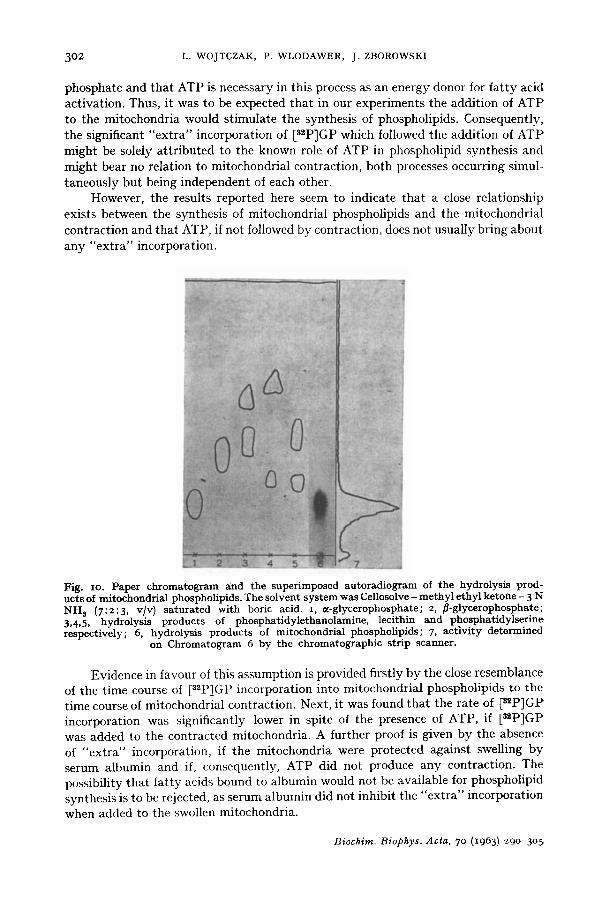
products of lecithin, phosphatidylethanolamine and phosphatidylserine. If the radio- active products of hydrolysis were mixed with authentic ~-glycerophosphate and submitted to chromatography, the total activity was found in the stained spot of ~-glycerophosphate. The radioactive hydrolysis product is therefore considered to be identical with ~-glycerophosphate. A typical chromatogram obtained with the Cellosolve-methyi ethyl ketone-NH s solvent system is shown in Fig. IO. Since phosphatidic acid is the only known phospholipid which yields glycerophosphate on mild alkaline hydrolysis by the method of DAWSON °, these data suggest that the radio- active lipid formed in the mitochondria was phosphatidic acid.
Fig. 8. Paper chromatogram and the super- imposed autoradiogram of mitochondrial phos- pholipids. The solvent system was diisobutyl ketone-formic acid-water (4o:I5:2, v/v). x,2,3, standards of phosphatidylethanolamine, lecithin and phosphatidylserine respectively; 4, mitochondrial phospholipids; 5, activity determined on the chromatogram 4 by the
chromatographic strip scanner.
Fig. 9- Paper chromatogram arid the super- imposed autoradiogram of mitochondrial phos- pholipids. The solvent system was diisobutyl ketone-acetic acid-water (4o:5o:7, v]v).
Indications as in Fig. 8.
DISCUSSION
Previous observations s have shown that [x-l*C]oleic acid is incorporated into mito- chondrial phospholipids upon the addition of ATP ~nd that this incorporation parallels the mitochondrial contraction. In experiments reported here the incorpo- ration of [~P]GP into mitochondrial phospholipids was observed under silDii~r con- ditions. Both these findings seem to indicate a synthesis de novo of phospholipids from GP and oleic acid.
It has been shown by KORNBEltG AND PRICER I° that a particulate fraction of liver homogenate is able to synthesize phosphohpids from fatty acids and ~-glycero-
Biochim. Biophys. Acta, 7 ° (x965) 29o-505
302 L. WOJTCZAK, P. WLODAWER, J. ZBOROWSKI
phosphate and that ATP is necessary in this process as an energy donor for fa t ty acid activation. Thus, it was to be expected that in our experiments the addition of ATP to the mitochondria would stimulate the synthesis of phospholipids. Consequently, the significant "ex t ra" incorporation of [3~P]GP which followed the addition of ATP might be solely at tr ibuted to the known role of ATP in phospholipid synthesis and might bear no relation to mitochondrial contraction, both processes occurring simul- taneously but being independent of each other.
However, the results reported here seem to indicate that a close relationship exists between the synthesis of mitochondrial phospholipids and the mitochondrial contraction and that ATP, if not followed by contraction, does not usually bring about any "ext ra" incorporation.
Fig. lO. Paper chromatogram and the ~superimposed autoradiogram of the hydrolysis prod- ucts of mitochondrial phospholipids. The solvent system was Cellosolve - methyl ethyl ketone - 3 N NH 3 (7:2:3, v/v) sa turated with boric acid. i, ~-glycerophosphate; 2, fl-glycerophosphate; 3,4,5, hydrolysis products of phosphatidylethanoiamine, lecithin and phosphatidyiserine respectively; 6, hydrolysis products of mitochondrial phospholipids; 7, act ivi ty d e t e r m i n e d
o n Chromatogram 6 by the chromatographic s t r i p s c a n n e r .
Evidence in favour of this assumption is provided firstly by the close resemblance of the time course of [3*P]GP incorporation into mitochondrial phospholipids to the time course of mitochondrial contraction. Next, it was found that the rate of [32P]GP incorporation was significantly lower in spite of the presence of ATP, if [3*P]GP was added to the contracted mitochondria. A further proof is given by the absence of "ext ra" incorporation, if the mitochondria were protected against swelling by serum albumin and if, consequently, ATP did not produce any contraction. The possibility that fat ty acids bound to albumin would not be available for phospholipid synthesis is to be rejected, as serum albumin did not inhibit the "ex t ra" incorporation when added to the swollen mitochondria.
Biochim. Biophys. Acta, 7 ° (1963) 290-305
MITOCHONDRIAL CONTRACTION AND PHOSPHOLIPID SYNTHESIS 3 0 3
Other evidence suggesting the coupling of phospholipid synthesis with mito- chondrial contraction is provided by the fact that substances which inhibit contraction depress the extent of [32P]GP incorporation. (The only exception was azide which did not affect [szp]GP incorporation although it strongly inhibited the contraction. No explanation for this can be given as yet.) Furthermore, substances which might be expected to interfere with the mechanism of phospholipid synthesis, such as PCMB and PPb were found to decrease or even completely to inhibit the contraction of mito- chondria. On the other hand, small amounts of oleate increased the rate of contraction of mitochondria swollen in the presence of Pl, presumably by supplying the fatty acid moiety for the synthesis of the phospholipid molecule. An increase in the rate of mitochondrial contraction brought about by thyroxine has also been observed by LEHNINGER 21. It seems reasonable to attribute this effect of thyroxine likewise to the action of fatty acids, as thyroxine has been found to stimulate the endogenous release of free fatty acids from mitochondria 2.
Thus, one may conclude that the biosynthesis of some mitochondrial phospho- lipids is an obligatory prerequisite for mitochondrial contraction, even if it is not solely responsible for it.
On the basis of column and paper chromatography and analysis of the hydrolysis products, there is good reason to assume that phosphatidic acid accounts for almost all the radioactivity of the mitochondrial phospholipids formed in the presence of [szP]GP. Neither lecithin nor kephalins were found to be labelled. This does not seem surprising since, according to KENNEDY 2z phosphatidic acid is the chief intermediate product in the synthesis of phospholipids and since only phosphatidic acid, cardiolipin and inositol phosphatides contain the phosphate moiety derived from ~-glycero- phosphate. However, phosphatidic acid was likewise the main labelled phospholipid component, when [14C]oleic acid was used as tracer 2. This seems to indicate that phosphatidic acid formed during mitochondrial contraction is not only an intermediate product in the synthesis of other phospholipids, but may play an important, although still obscure, role in the mechanism of mitochondrial contraction.
One can only speculate on the possible role of phospholipid synthesis in mito- chondrial contraction. Three posibilities seem worth considering:
I. Swelling of mitochondria is presumably accompanied by some structural change in mitochondrial membranes. As lipoprotein complexes are the main con- stituents of mitochondrial membranes, it is possible that splitting of some phospho- lipids may be partly responsible for these changes. Thus, the resynthesis of these phospholipids may serve to reconstitute the damaged membranes. It is known 23 that the Initochondrial membrane is negatively charged, and it has been suggested 24 that negatively charged groups, possibly those of phospholipids, may control the transport across the membrane. Phosphatidic acid is one of the most negatively charged phospholipids and therefore its role in controlling the transport of molecules through mitochondrial membrane may be of particular importance.
2. Synthesis of phospholipids may be related to changes in the structure of some mitochondrial proteins. The view that the shrinkage of mitochondria may be due to some contractile proteins has been put forward by LEI.ININGER ~ . Furthermore, J U D A H ~ has observed phosphorylation of some mitochondrial proteins during the contraction induced by ATP. However, nothing is known about the relationship of lipids to contractile proteins.
Biochim. Biophys. Acta, 7 ° [30~'3) "Oo 305
30 4 L. WOJTCZAK, P. WLODAWER, J. ZBOROWSKI
3- Synthesis of phospholipids may be related to active transport of water and of other substances across mitochondrial membranes. The extrusion of water from the mitochondria may bear some resemblance to the active transport of ions through membranes or it may be accompanied by active transport of some other molecules. If this were the case, the stimulation of phospholipid synthesis in response to mito- chondria[ contraction would be similar to the "phospholipid effect" observed by HoKm ef ./.~-so.
Many data have been accumulated in recent years which point to phosphatidic acid as a metabolically active substance involved in the transport of various organic and inorganic compounds through cell membranes sv-33. On the basis of numerous studies, HOKIN AND Homlq a~ assume that the exchange of the phosphate moiety in phosphatidic acid may be part of a carrier mechanism in the transport of various molecules across membranes. They presented a scheme a° whereby phosphatidic acid, which is presumably part of the lipoprotein complexes in the membranes, functions as a carrier for ions through the membrane. The scheme is based on the combined actions of diglyceride kinase and phosphatidic acid phosphatase which result not only in the synthesis and breakdown of the phosphatidic acid but also in ATPase activity. Furthermore, the ATPase activity has been shown to be closely related to the action of the sodium-potassium pumps in both nerve ~ and erythrocyte as membranes. Thus, the utilization of ATP for the synthesis of phosphatidic acid could represent the input of e'nergy for transport.
In the present investigation a sh-nilar biochemical response was observed on contraction of the mitochondria as has been observed by others for the stimulation of the transport of various molecules across membranes. One may suppose, therefore, that phosphatidic acid formed during the contraction is involved in the mechanism of the active extrusion of water from the mitochondria.
ACKNOWLEDGEMENTS
The authors wish to thank Professor W. NIEMIERKO for his keen interest in this research and Mrs. M. B~DNAP, EK for her valuable technical assistance.
The chromatographic strip scanner (Nuclear Chicago Co.) used in this investi- gation was a gift from the Rockefeller Foundation.
R E F E R E N C E S
l A. L. LBHNmOER AND L. F. R.EMMERT, J. Biol. Chem., 234 (1959) 1459. s L. WOJTCZAK AND A. L. LEHNINOER, Biochim. Biophys.Acta, 5z (I961) 44 z. S A. L. LKI~ININGBR, B. L. RAY AND M. SCHNgIDKR, jr. B i o ~ $ . Biochem. Cytol., 5 (x959) 97. • A. L. LImNINGER, J. Biol. Chem., 234 11959) 2465. 5 A. L. LgHNINOIm, f . Biol. Chem., 234 (I959) z i 85 . 6 G. V. MARtNEX'rt AND E. STOTZ, Biochim. Biophys. A a a , 2 i (I956) i68. v O. W. TRmLE ANY W . W o g B g , Z. Physiol. Chem., 326 (I96I) 89. s G. V. MAmxm~l'rt, J . ERBLAND AND J. KOCHEN, Fe.d~ration Proc., I6 (t957) 837- 9 R. M. C. DAWSON, Biochim. Biophys Aaa, z 4 (x934) 374.
10 S. CongN AND D. SCOTT, S ~ , 111 (t950) 2890. It G. WYATT, T. L0t'~OHEED AND S. WYATT, f . Gen. Physiol., 39 i1956) 853. Is D. C. MORTINER, Can. J. Chem., 30 (I952) 653. ta C. S. HANgs AND F. A. ISHERWOOD, Nature, t 64 (I949) I io7 . 1, R . S. B A N v u g s g t AND B. AXgLROD, f . Biol. Chem., I93 (I951) 4o5 • is E. P . ]KENNgDY, ~v. Biol. Chem., 2o i (1953) 399.
Biochim. Biophys. Aaa, 70 (I963) 19o-3o5
MITOCHONDRIAL CONTRACTION AND PHOSPHOLIPID SYNTHESIS ~05
1, A. L. LEHNINGER, f . Bioclun~. (Tokyo), 49 (I96I) 553- t7 A. L. LEHmNOBR, B ~ k i m . BuTpky$. Ac~, 48 {1961) 324.
G. HOBSCHSR AND B. CLARK, Biochim. B ~ h y s . Acta, 4 x (196o) 45. 1, L. W. WHmCLDOS, J. L i ~ Res., x (I96O) 439. so A. KORX~SBRG ASD W. E. PnXCBR, Jr., J. Am. Chem. Soc., 74 (I95Z) x617. st A. L. LEHmNGSa, B~him. B ~ h y s . Acre, 37 (196°) 387 • m E. P. KENNEDY, F ~ Proc., 16 (1957) 847. a H. PAXn.Y, I~. PACgER HND H. *P. SCHWHN, J. Biophys. Bioci~m. CytoL, 7 (196°) 589 • s4 j . M. MACHINIST, M. L. DAS, F. L. CRANE AND E. E. JACOBS, Biochem. Biophys. Res. Commun.,
6 (196z) 475. • x A. L. LEtlNINGER, F e c / m , ~ Procl, x9 (x96o) 952. s, j . D. JUVAH, Naha, e, I87 (I960) 5o6.
M. R. HOKIN, L. E. HOKIN, M. SAlZlCRAN, A. V. SCHALLY AND B. U. ZIMMERMANN, J . B id . Clam., z33 (x958~ 8xx.
Is M. R. I-IoKxN, B. G. BENX~EY aND L. E. I"IoxzN, J. Biol. Cl~m., 235 (I958) 814- "* L. D. EC, OMAN ann L: E. HOKIN, J . Biol. C~m., 235 (196o) 2569.
L. E. HoxtN HND M. R. I'IoxxN, J. Ges. Physiol., 44 (196o) 61. • x L. E. HoKxN AND A. L. SHBRWtN, J. Physiol. ILo'ad~m), 135 {I957) 18. n L. E. I'-IOKIN ANI~, M. R. HOi~IN, J.: Biol. Chem., 233 ~1958)805. aa A. M. WoovxN, Bioch~m. J . , 8z (I962) 9. as L. E, HOKIN AND M. R. HOKIN, IVa~v$, 18 9 (196I) 836. aS j . C. Sgou, Bioc, kim. Biopkys. Ac,~, 23 (I957) 394-
R. L. POST, C. R. MERRIT, C. R. KINSOLVIN Q AND C. D. AJ.BRIOHT, J . Biol. C ~ . ° 235 (1960) 1796.
Bicchim. Bi~opkys. Ac.~, 7o (1963) z9o-3o5


![Increased Rate of Adenosine Triphosphate …...(CANCER RESEARCH 55, 4352-4360, October 1, 1995] Increased Rate of Adenosine Triphosphate-dependent Etoposide (VP-16) Efflux in a Murine](https://img.pdfslide.net/doc/110x75/5e7e8d68c5d0407f2447f2a9/increased-rate-of-adenosine-triphosphate-cancer-research-55-4352-4360-october.jpg)