Embed Size (px)
Citation preview
Adrenal gland
Dr Heyam AwadFRCPath
Slide notes-A.K
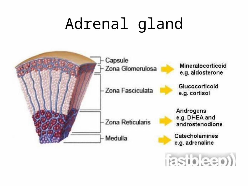
As we all know the adrenal glands are made up of a cortex and a medulla
There are 3 zones in the cortex; Glomerusa secreting aldosterone, Fasciculata secreting cortisol and reticularis secreting androgens
The medulla secrets catecholaminesTo begin with, the diseases of the cortex
could be related to:1)Hypersecretion2)Undersecretion3)Mass effectin the cortex the mass effect is not very
prominent, usually it is from an incidental finding or from hormone-secreting pathologies
Adrenal gland
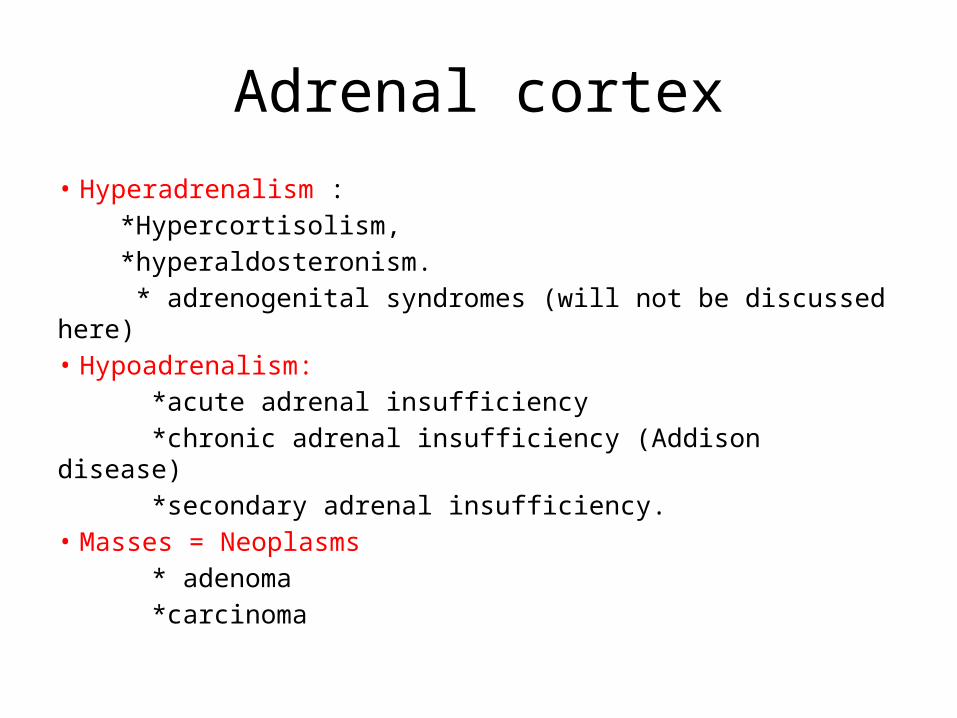
Adrenal cortex
• Hyperadrenalism : *Hypercortisolism, *hyperaldosteronism. * adrenogenital syndromes (will not be discussed here)• Hypoadrenalism: *acute adrenal insufficiency *chronic adrenal insufficiency (Addison disease) *secondary adrenal insufficiency.• Masses = Neoplasms * adenoma *carcinoma
Hypercortisolism (Cushing Syndrome)
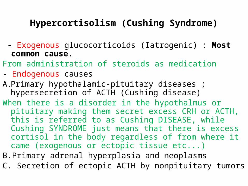
- Exogenous glucocorticoids (Iatrogenic) : Most common cause.From administration of steroids as medication- Endogenous causesA. Primary hypothalamic-pituitary diseases ; hypersecretion of ACTH
(Cushing disease) When there is a disorder in the hypothalmus or
pituitary making them secret excess CRH or ACTH, this is referred to as Cushing DISEASE, while Cushing SYNDROME just means that there is excess cortisol in the body regardless of from where it came (exogenous or ectopic tissue etc...)
B.Primary adrenal hyperplasia and neoplasms C. Secretion of ectopic ACTH by nonpituitary tumors
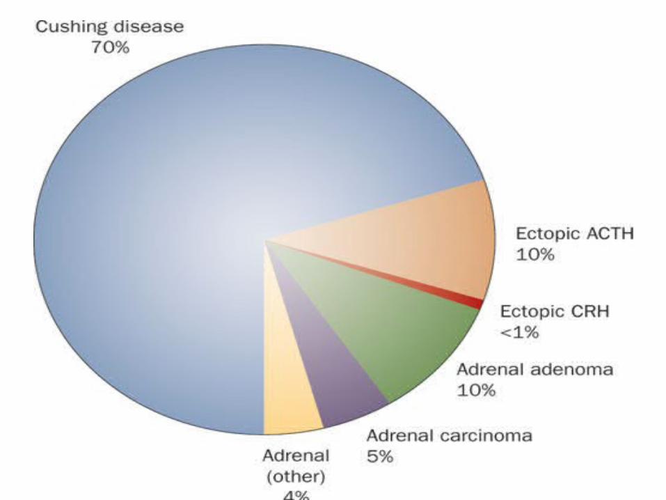
1)HYPOTHALAMIC- PITUITARY CAUSESCUSHING DISEASE
-70% of cases of spontaneous, endogenous Cushing syndrome are due to Cushing disease.
- Occurs most frequently during young adulthood (the 20s and 30s)
- mainly affecting women.
CUSHING DISEASE
Causes?majority: pituitary gland contains an ACTH-producing
adenoma
- In the remaining patients, the anterior pituitary contains areas of corticotroph cell hyperplasia which may be:
a. Primaryb. or, less commonly, secondary to CRH producing tumor
IMPORTANT NOTE: ACTH regulates cortisol and androgens, it does NOT affect aldosterone secretion in any way nor is it affected by it.
**extra note: the pituitary negative feedback for ACTH is from cortisol, androgens do not effect the release of ACTH but are affected by it
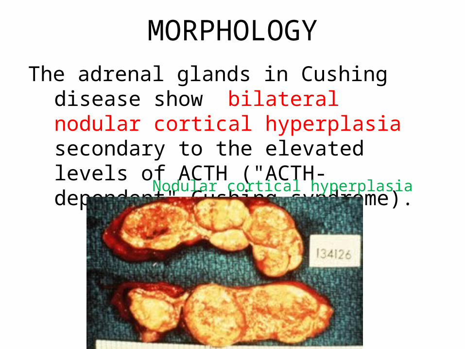
MORPHOLOGYThe adrenal glands in Cushing disease show bilateral
nodular cortical hyperplasia secondary to the elevated levels of ACTH ("ACTH-dependent" Cushing syndrome).
Nodular cortical hyperplasia
2)PRIMARY ADRENAL HYPERPLASIA AND NEOPLASMS
- 10% to 20% of cases of endogenous Cushing syndrome- ACTH-independent Cushing syndrome, or adrenal
Cushing syndrome ; low serum levels of ACTH - Caused by adrenal adenoma or carcinoma.- primary hyperplasia can cause it but is very rare.
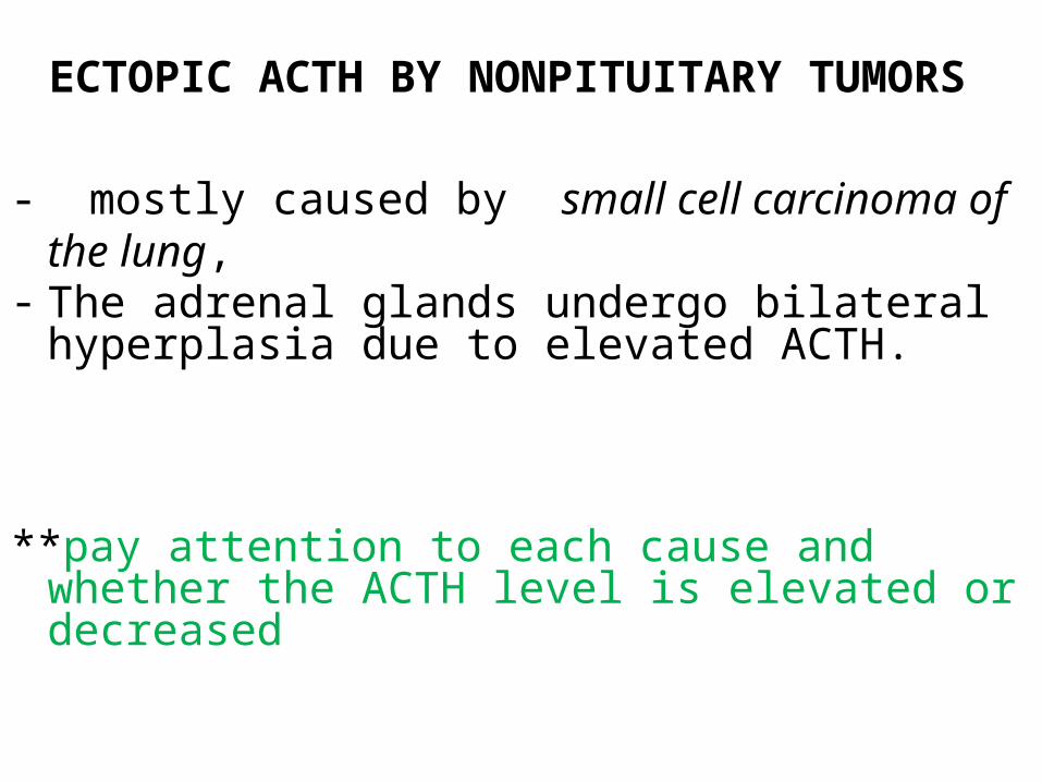
ECTOPIC ACTH BY NONPITUITARY TUMORS
- mostly caused by small cell carcinoma of the lung, - The adrenal glands undergo bilateral hyperplasia due to
elevated ACTH.
**pay attention to each cause and whether the ACTH level is elevated or decreased
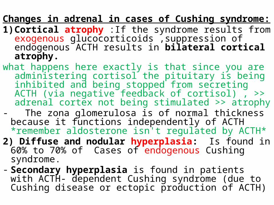
Changes in adrenal in cases of Cushing syndrome:1) Cortical atrophy :If the syndrome results from exogenous
glucocorticoids ,suppression of endogenous ACTH results in bilateral cortical atrophy.
what happens here exactly is that since you are administering cortisol the pituitary is being inhibited and being stopped from secreting ACTH (via negative feedback of cortisol) , >> adrenal cortex not being stimulated >> atrophy
- The zona glomerulosa is of normal thickness because it functions independently of ACTH *remember aldosterone isn't regulated by ACTH*
2) Diffuse and nodular hyperplasia: Is found in 60% to 70% of Cases of endogenous Cushing syndrome.
- Secondary hyperplasia is found in patients with ACTH- dependent Cushing syndrome (due to Cushing disease or ectopic production of ACTH)
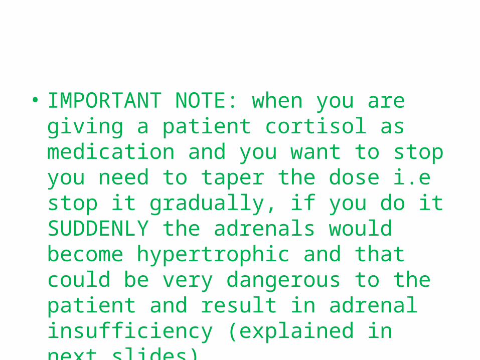
• IMPORTANT NOTE: when you are giving a patient cortisol as medication and you want to stop you need to taper the dose i.e stop it gradually, if you do it SUDDENLY the adrenals would become hypertrophic and that could be very dangerous to the patient and result in adrenal insufficiency (explained in next slides)
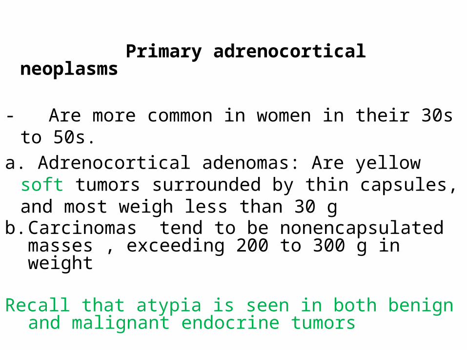
Primary adrenocortical neoplasms
- Are more common in women in their 30s to 50s. a. Adrenocortical adenomas: Are yellow soft tumors
surrounded by thin capsules, and most weigh less than 30 g
b. Carcinomas tend to be nonencapsulated masses , exceeding 200 to 300 g in weight
Recall that atypia is seen in both benign and malignant endocrine tumors
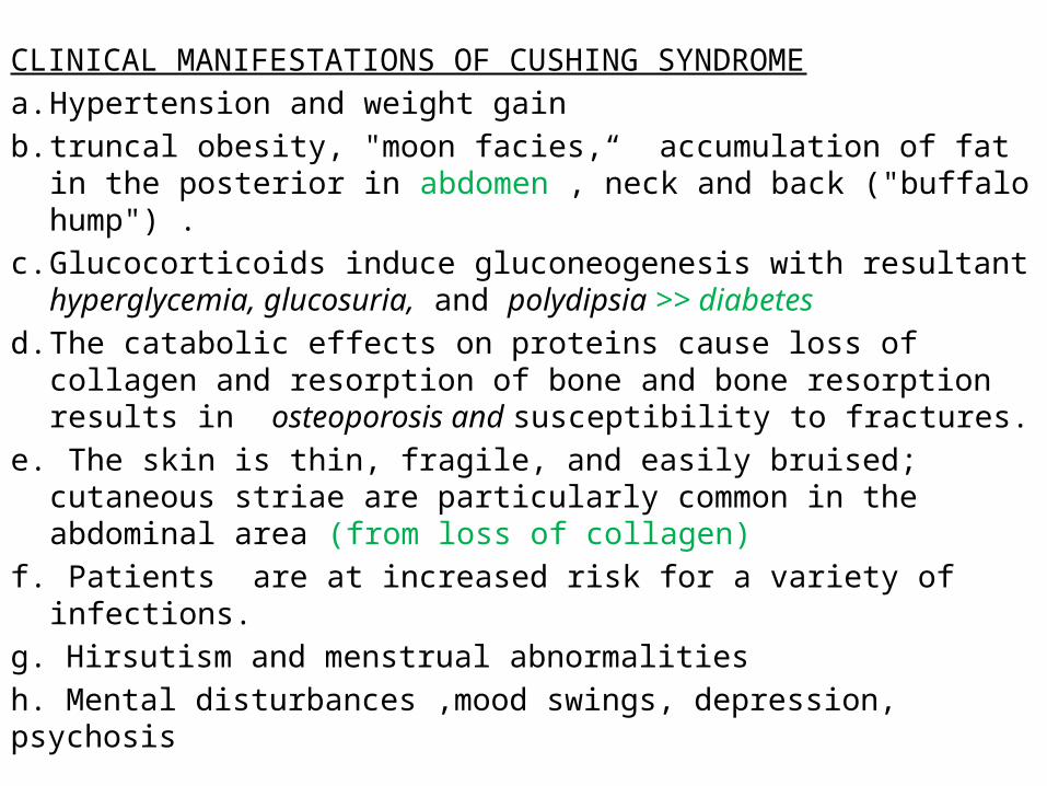
CLINICAL MANIFESTATIONS OF CUSHING SYNDROMEa. Hypertension and weight gain b. truncal obesity, "moon facies,“ accumulation of fat in the posterior in
abdomen , neck and back ("buffalo hump") .c. Glucocorticoids induce gluconeogenesis with resultant
hyperglycemia, glucosuria, and polydipsia >> diabetesd. The catabolic effects on proteins cause loss of collagen and
resorption of bone and bone resorption results in osteoporosis and susceptibility to fractures.
e. The skin is thin, fragile, and easily bruised; cutaneous striae are particularly common in the abdominal area (from loss of collagen)
f. Patients are at increased risk for a variety of infections. g. Hirsutism and menstrual abnormalities h. Mental disturbances ,mood swings, depression, psychosis
Clinical features
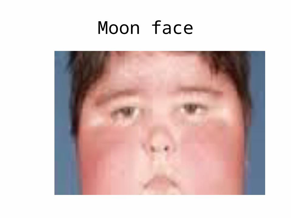
Moon face
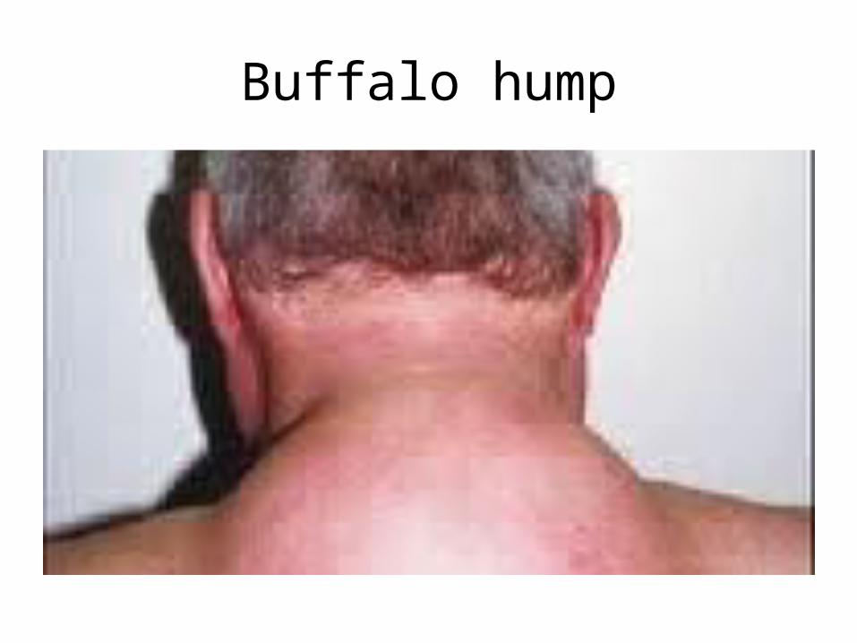
Buffalo hump

buffalo
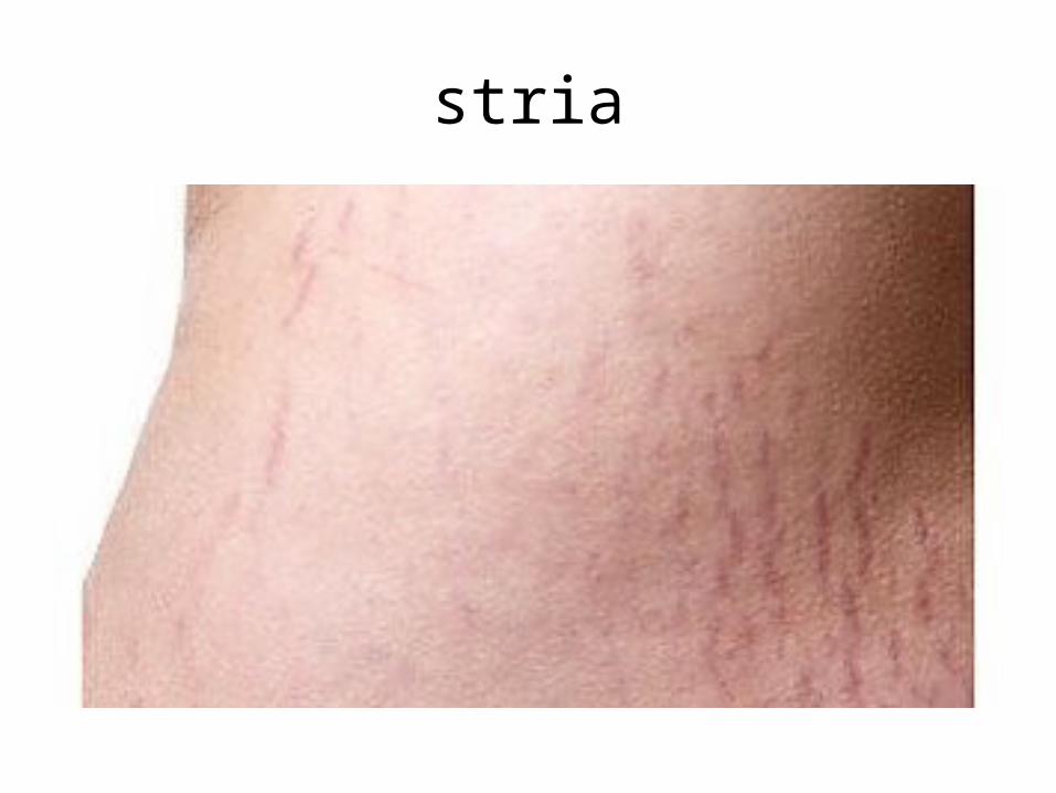
stria
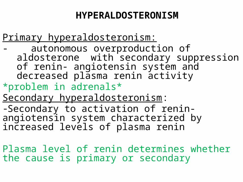
HYPERALDOSTERONISM
Primary hyperaldosteronism:- autonomous overproduction of aldosterone with
secondary suppression of renin- angiotensin system and decreased plasma renin activity
*problem in adrenals*Secondary hyperaldosteronism:-Secondary to activation of renin-angiotensin system characterized by increased levels of plasma renin
Plasma level of renin determines whether the cause is primary or secondary
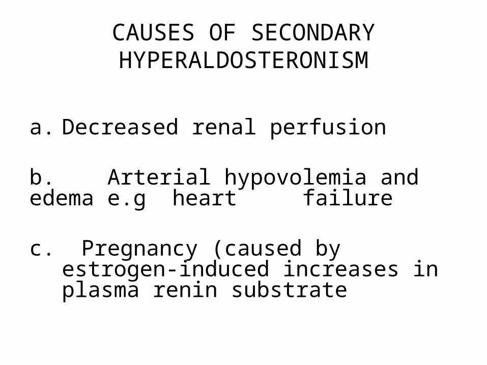
CAUSES OF SECONDARY HYPERALDOSTERONISM
a. Decreased renal perfusion
b. Arterial hypovolemia and edema e.g heart failure c. Pregnancy (caused by estrogen-induced increases
in plasma renin substrate
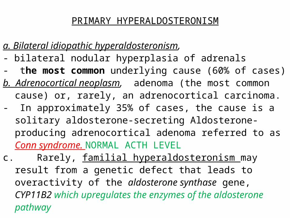
PRIMARY HYPERALDOSTERONISM
a. Bilateral idiopathic hyperaldosteronism, - bilateral nodular hyperplasia of adrenals - the most common underlying cause (60% of cases)b. Adrenocortical neoplasm, adenoma (the most common cause) or,
rarely, an adrenocortical carcinoma.- In approximately 35% of cases, the cause is a solitary aldosterone-
secreting Aldosterone-producing adrenocortical adenoma referred to as Conn syndrome. NORMAL ACTH LEVEL
c. Rarely, familial hyperaldosteronism may result from a genetic defect that leads to overactivity of the aldosterone synthase gene, CYP11B2 which upregulates the enzymes of the aldosterone pathway
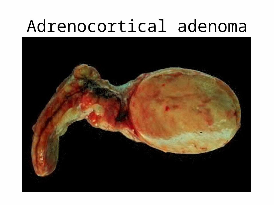
Adrenocortical adenoma
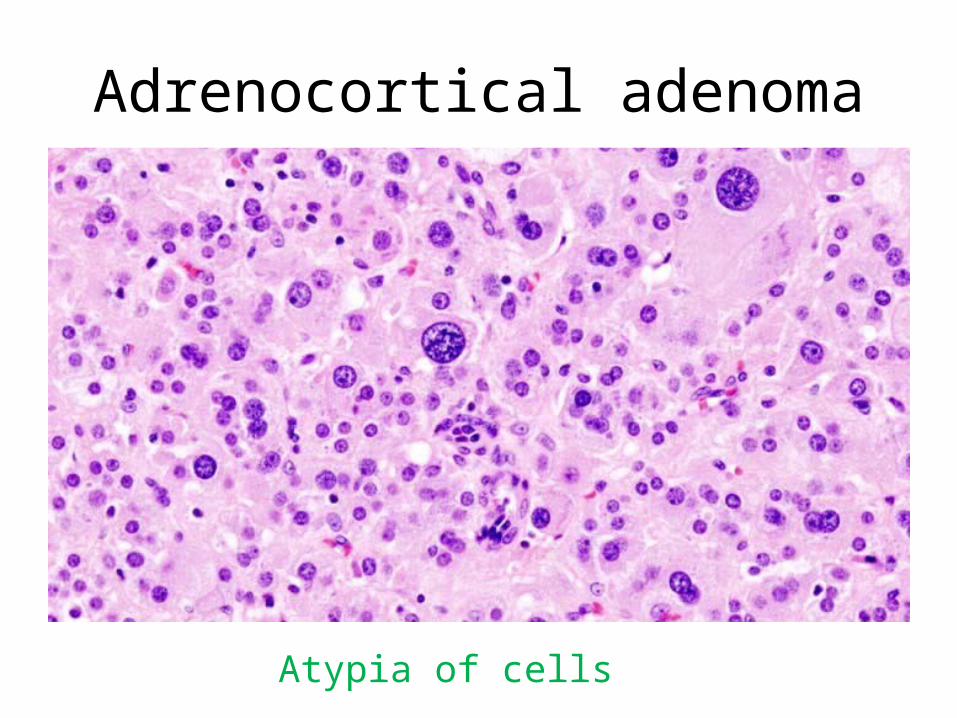
Adrenocortical adenoma
Atypia of cells
CLINICAL FEATURES OF HYPERALDOSTERONISM
The clinical hallmark is hypertension- Hyperaldosteronism may be the most common cause of secondary
hypertension Note: if a young person comes to you with hypertension
then the cause is most probably secondary and can be from hypertaldosteronism but it is rare
- Hypokalemia results from renal potassium wasting and, can cause neuromuscular manifestations, including weakness, paresthesias,, and occasionally frank tetany.
- Parasthesia = abnormal sensation, typically tingling or pricking (“pins and needles”), caused chiefly by pressure on or damage to peripheral nerves.
- Tetany = intermittent muscular spasms.
ADRENAL INSUFFICIENCYnot enough hormones being produced by the
adrenals
Causes could be primary (acute or chronic) or secondary
Acute Adrenocortical Insufficiency : causesa. Crisis in patients with chronic adrenocortical
insufficiency precipitated by stress b. In patients maintained on exogenous
corticosteroids .. Sudden withdrwal, or stressc. Massive adrenal hemorrhage. Must lose at
least 90% of gland’s function
Massive adrenal hemorrhage may destroy enough of the adrenal cortex to cause acute
adrenocortical insufficiency.- This condition may occur :1. In patients maintained on anticoagulant therapy2. Patients suffering from sepsis : a condition known as
the Waterhouse-Friderichsen syndrome - Sepsis due to: Neisseria meningitidis (most
common cause) ,Pseudomonas spp., , and Haemophilus influenzae
- Underlying cause??? unclear but probably involves endotoxin-induced vascular injury .
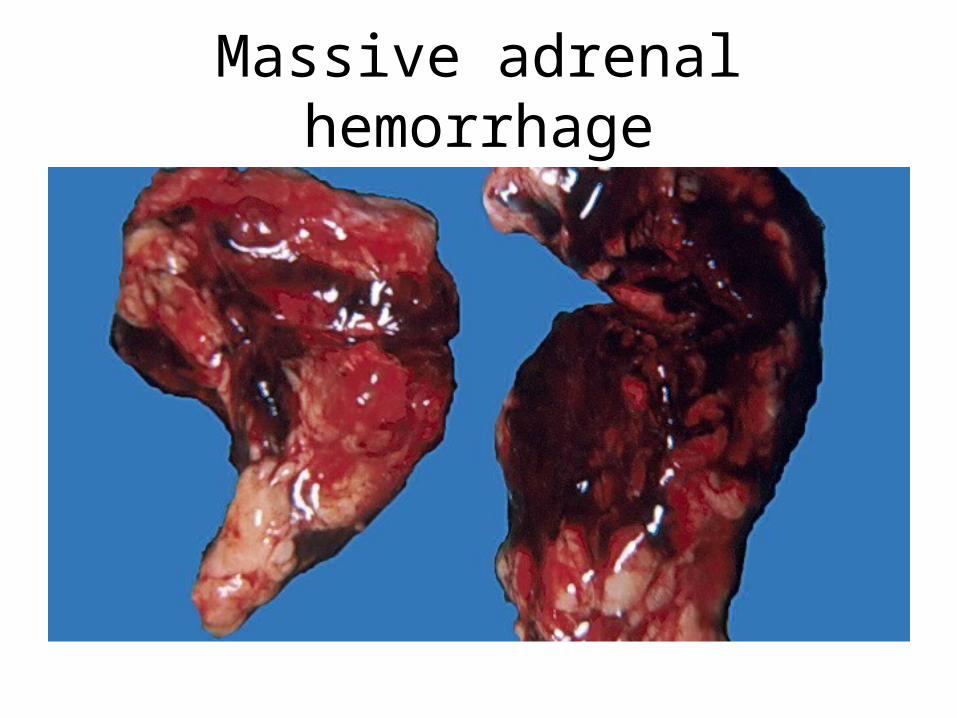
Massive adrenal hemorrhage
primary chronic adrenocortical insufficiency (Addison disease):
-uncommon disorder resulting from progressive destruction of the adrenal cortex. ALL adrenal hormones are affected (even aldosterone)Causes:- Autoimmune adrenalitis. (Most common right now)- Infections (before it was the most common, due
to TB) - Metastatic tumors- Basically anything that destroys the gland
ADDISON DISEASE
1. Autoimmune adrenalitis - 60% to 70% of Addison disease cases and is the most
common cause of primary adrenal insufficiency in developed countries.
- There is autoimmune destruction of steroid-producing cells, and autoantibodies to several key steroidogenic enzymes have been detected in affected patients
Addison disease
2. Infections,: Tuberculosis and Fungal infections- Tuberculous adrenalitis, which once accounted for as many
as 90% of cases of Addison disease, has become less common with the advent of anti-tuberculosis therapy
- Disseminated infections caused by Histoplasma capsulatum and Coccidioides immitis also may result in chronic adrenocortical insufficiency.
- Patients with AIDS are at risk for the development of adrenal insufficiency from several infectious (cytomegalovirus and TB) and noninfectious (Kaposi sarcoma) .
ADDISON DISEASE
3- Metastatic neoplasms involving the adrenals:
Carcinomas of the lung and breast are the most common primary sources.
Secondary adrenocortical insufficiency
Hypothalamic- pituitary diseases (aldosterone not affected *reminder*) including:
• Metastasis• Infection.• Infarction• Irradiation• Can be part of panhypopituitarism.• Again basically anything that can
destroy the hypothalmus or pituitary
Clinical features of adrenal insufficiency- Clinical manifestations of adrenocortical insufficiency do not appear
until at least 90% of the adrenal cortex has been compromised. a. progressive weakness and easy fatigability, the less
hormones secreted the more weakness there isb. Gastrointestinal disturbances are common and include anorexia,
nausea, vomiting, weight loss, and diarrhea c. In patients with primary adrenal disease, increased levels of
ACTH precursor hormone stimulate melanocytes, with resultant hyperpigmentation of the skin and mucosal surfaces: The face, axillae, nipples, areolae, and perineum are mainly affected
Note: hyperpigmentation is not seen in patients with secondary adrenocortical insufficiency.
So if the adrenals are damaged like in primary hypoadrenalism, no cortisol is negatively inhibiting the secretion of CRH and ACTH, therefore more ACTH is being produced, if you recall we have one long polypeptide chain precursor containing ACTH, MSH, and two unimportant thingies, if we keep producing ACTH, we will also be producing MSH which is responsible for the dark colour of the skin, that is why patients with Addison’s disease are dark coloured and hyperpigmented in certain areas. For secondary hypoadrenalism the ACTH level is low so the synthesis of MSH is low too that’s why we do not see hyperpigmentation there.
d. Decreased aldosterone in primary hypoadrenalism results in potassium retention and sodium loss , with consequent - hyperkalemia, hyponatremia, volume depletion, and hypotension,
- In secondary hypoadrenalism is characterized by deficient cortisol and androgen output but normal or near-normal aldosterone synthesis. Why?
- I’m not gonna write why, you should be able to answer that by now :P
Adrenal medulla
• Chromaffin cells… derived from the neural crest.
• Secrete catecholamines.• Most important disease: neoplasms.• ONLY PCC is going to be discussed
here, it is a tumor that could be benign or malignant, the majority are benign 90%
TUMORS OF THE ADRENAL MEDULLAPheochromocytoma - give rise to a surgically correctable form of hypertension.
(since they secret epinephrine and norepinephrine)
- Pheochromocytomas usually subscribe to "rule of 10s": a. 10% of pheochromocytomas are extraadrenal, called
paragangliomas, i.e they occur in other tissues b. 10% of adrenal pheochromocytomas are bilateral; this
proportion may rise to 50% in cases that are associated with familial syndromes.
To explain more about the previous point, if you find a tumor that is bilateral, i.e both glands are affected, and you want to determine the genetic cause, there is a 50% chance that this is a familial case. (for the 10% that is bilateral)
If you notice there is a trend that familial cases are usually bi-lateral because what is the probability of both adrenals for example being hit with a mutation sporadically, but if you have a genetic mutation then wouldn’t all your cells be at a higher risk of developing a tumor? **not the doctors words**
c. 10% of adrenal pheochromocytomas are malignant, - Frank malignancy is somewhat more common in tumors arising in
extraadrenal sites.- So if you have an extra-adrenal PCC it is more likely to
be malignant
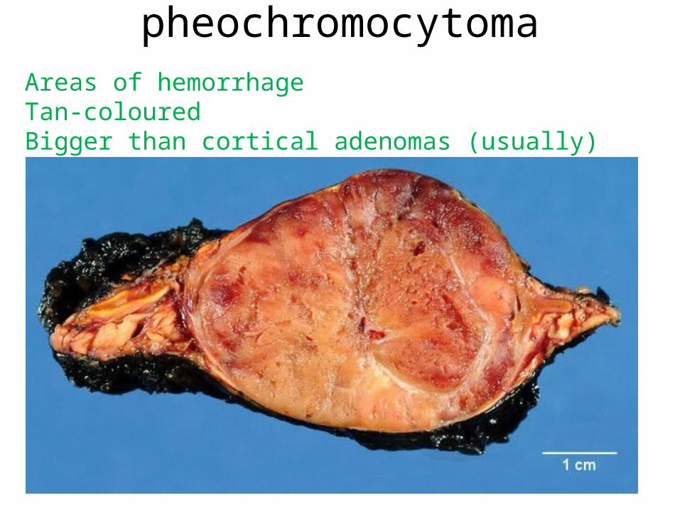
pheochromocytomaAreas of hemorrhageTan-colouredBigger than cortical adenomas (usually)
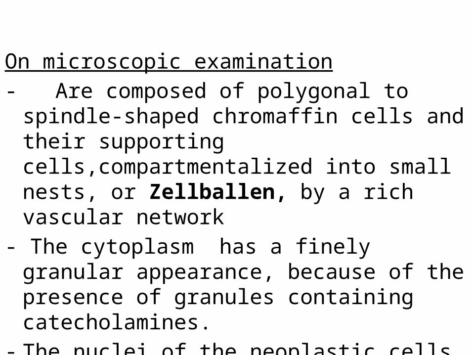
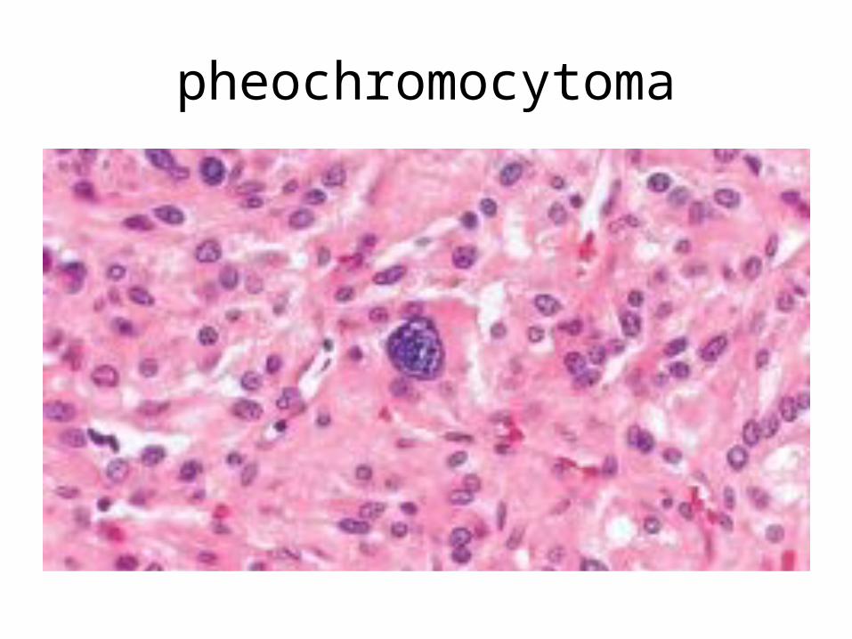
On microscopic examination- Are composed of polygonal to spindle-shaped chromaffin
cells and their supporting cells,compartmentalized into small nests, or Zellballen, by a rich vascular network
- The cytoplasm has a finely granular appearance, because of the presence of granules containing catecholamines.
- The nuclei of the neoplastic cells are often pleomorphic- Anaplasia of cells (not necessarily a
feature of malignancy)- **PCC could secret ectopic hormones
pheochromocytoma
Pheochromocytoma..
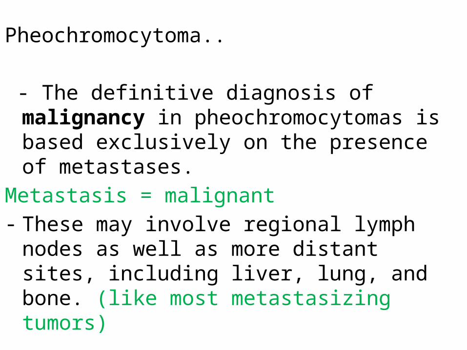
- The definitive diagnosis of malignancy in pheochromocytomas is based exclusively on the presence of metastases.
Metastasis = malignant- These may involve regional lymph nodes as well as more
distant sites, including liver, lung, and bone. (like most metastasizing tumors)
Clinical Features - The predominant clinical manifestation is hypertension
- Sudden cardiac death may occur, probably secondary to catecholamine-induced myocardial irritability and ventricular arrhythmias.
- In some cases, pheochromocytomas secrete hormones such as ACTH and somatostatin.
- The laboratory diagnosis of pheochromocytoma is based on demonstration of increased urinary excretion of free catecholamines and their metabolites, such as vanillylmandelic acid and metanephrines.