Embed Size (px)
Citation preview
Vision Development & Rehabilitation Volume 5, Issue 1 • March 201919
AR
TIC
LEAdvanced Neuro-Optometric Diagnostic Tests for Mild Traumatic Brain Injury/Concussion: A Narrative Review, Proposed Techniques and ProtocolsKenneth J. Ciuffreda, OD, PhD1
Barry Tannen, OD, FAAO, FCOVD1
Naveen K. Yadav, BS Optom, MS, PhD2
Diana P. Ludlam, BA, COVT1
1 SUNY College of Optometry, Brain Injury Research Unit, New York, New York
2 Western University of Health Sciences, College of Optometry, Ponoma, California
AbstrActA range of visual deficits and related visual symptoms are common in those afflicted with mild traumatic brain injury (mTBI/concussion). Several basic neuro-optometric, diagnostic test protocols have been proposed over the past decade. However, none have specifically addressed and focused upon an advanced level of care. Thus, a comprehensive set of advanced, diagnostic vision tests of a sensory and motor nature is proposed, with all having a clinical and scientific rationale. These tests have been used by the authors for many years, with good success and providing important clinical insights into this population.
IntroductIonOne of the newest and most exciting areas
of optometry is the care of vision problems in patients with traumatic brain injury (TBI), especially of the mild type (mTBI/concussion)1, including both diagnostic and therapeutic aspects (i.e., “neuro-optometry”). While the present paper will only focus on the former aspect, the related clinical ramifications of the latter are obvious and manifold.1
The conventional, optometric, diagnostic armamentarium for the medical condition of mTBI/concussion is ever growing, as new techniques and tests are being introduced (e.g., the Tannen test2), and older ones are being better understood and hence incorporated more frequently into patient care (e.g., binasal occlusion, yoked prisms).1 This evolution is positive for the field of neuro-optometry.
There have been several basic clinical and laboratory diagnostic protocols proposed over the years.3-5 These developments and other factors (e.g., the sports concussion crisis) have lead to tremendous growth in this area of brain injury within the neuro-optometric domain.1
One area of diagnostic, clinical vision testing that is still evolving and gradually being used more frequently is what may be considered “advanced” testing in mTBI/concussion. It is growing as technology advances, and as
Correspondence regarding this article should be emailed to Kenneth J. Ciuffreda, OD, PhD, at [email protected]. All state ments are the authors’ personal opinions and may not reflect the opinions of the College of Optometrists in Vision Development, Vision Development & Rehabilitation or any institu tion or organization to which the authors may be affiliated. Permission to use reprints of this article must be obtained from the editor. Copyright 2019 College of Optometrists in Vision Development. VDR is indexed in the Directory of Open Access Journals. Online access is available at covd.org. https://doi.org/10.31707/VDR2019.5.1.p19
Ciuffreda K, Tannen, B, Yadav, NK Ludlam D. Advanced neuro-optometric diagnostic tests for mild traumatic brain injury/concussion: A narrative review, proposed techniques and protocols. Vision Dev & Rehab 2019;5(1):19-30.
Keywords: advanced diagnostic testing, concussion, mild traumatic brain injury,
mTBI, neuro-optometry, vision problems, vision
20Vision Development & Rehabilitation Volume 5, Issue 1 • March 2019
The electro-retinogram (ERG) is an objective, quantitative technique that examines dynamic responsivity of the retinal elements.7 Using the non-pattern “flash” ERG, the initial a-wave response reflects global photoreceptor activity, whereas the subsequent b-wave response reflects global bipolar and Mueller cell activity, with critical parameters being their amplitude and latency. In contrast, when using the “pattern” ERG, the response primarily reflects global ganglion cell activity. The pattern ERG is sensitive to retinal defocus effects, as well as age, in both cases reducing response amplitude. Thus, in the latter case with a patterned target, careful pre-testing refraction is mandated for the presence of clear retinal imagery. Fortunately, over the normal range of pupil diameters across all ages, there is no effect on the response. Since many with mTBI manifest retinal/optic nerve diseases in much greater proportion than found in the normal population,8 such as glaucoma and optic nerve damage, the pattern ERG is probably the more important of the two approaches. Furthermore, the newer multifocal pattern ERG technology allows the clinician to assess small, “local” regions of the retina. For example, over 100 different areas throughout the central 50 degrees of the retina can be evaluated quantitatively and simultaneously following a single test presentation.7 Only one study9
used electro-retinography to assess retinal function in patients with mTBI (n=50). No abnormalities were uncovered. However, this may be due to using the flash versus pattern ERG. Further studies are thus warranted using conventional and multi-focal pattern ERGs in this population, especially where combined ocular injury and head trauma have been documented.
Optical coherence tomography (OCT) is an objective, non-invasive, rapid, direct, simple, and quantitative high resolution imaging technique to view the internal ocular structures, especially of the retina.10 It uses the principle of low-coherence interferometry. The
the procedures become automated, such as the case with hand-held, objective dynamic pupillometry.6 Advanced diagnostic testing can be extremely beneficial, especially to the more experienced neuro-optometrist, and others, to provide better diagnostic capability, hence in turn leading to more targeted and efficacious therapeutic interventions1. While such “advanced” testing has been used for years in the non-TBI population, it still remains relatively infrequently used in those with mTBI/TBI.
Thus, the purpose of the present narrative review is to briefly discuss the area of vision problems in mTBI/concussion, and furthermore to propose a set of “advanced” diagnostic tests and procedures of a sensory and motor nature that can be employed by the neuro-optometrist and others. Incorporating these tests and protocols will broaden the diagnostic spectrum and improve clinical care for the visually-symptomatic patient with mTBI/concussion. This paper reflects the views and perspectives of the authors based on their years of clinical and research experience in the area.
There are several “advanced” diagnostic, sensory tests that may be beneficial in the mTBI/concussion population. The first four are objective in nature and assess processing in the early visual system pathways. In contrast, the other tests are of a subjective, psychophysical nature and assess the visual pathways over a range of relatively early and late processing levels.
table 1: Advanced diagnostic sensory tests
• electro-retinography• opticalcoherencetomography• b-scanultrasound• visual-evokedpotential• darkadaptation• glaresensitivity• intuitivecolorimetry• perceptualtesting
21Vision Development & Rehabilitation Volume 5, Issue 1 • March 2019
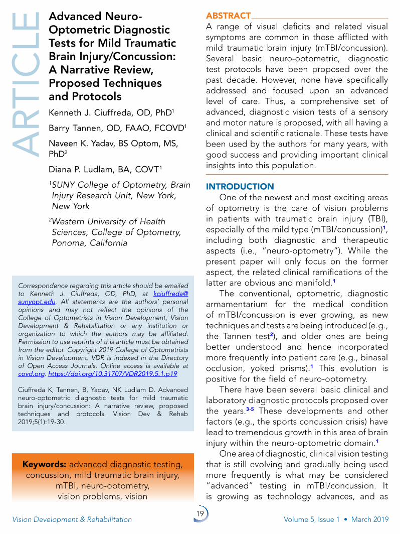
technique provides a two-dimensional, cross-section of the globe (Figure 1). It has been helpful diagnostically in a variety of ocular conditions, such as glaucoma, macular edema, and rod and cone disturbances.11 Individuals with mTBI/TBI have a high frequency of occurrence of such ocular problems,8 as mentioned earlier, and thus the inclusion of OCT would likely prove beneficial in many cases. For example, it has been used to follow the progression of retrograde degeneration of the nerve fiber layer in blunt eye trauma,12 which frequently occurs in conjunction with head trauma, to assess retinal dysfunction and the effect of its physical disruption.13 Lastly, the OCT was proposed by us years ago as one of the key, “targeted”, objective techniques to use in the diagnosis of ocular-based, vision problems in mTBI.4
B-scan ultrasonography is a non-invasive, rapid, and simple technique to visualize and
examine the internal structures of the entire eye.14 It provides a two-dimensional, cross-section of the globe. This is especially critical when such information is not available by other procedures/techniques, such as with the presence of a dense and large corneal or lenticular opacity. This technique employs high-frequency, ultrasound waves (e.g., 10 Mhz) that reflect, or “echo”, off the internal structures of the eye (e.g., lens, retina). This information is then used to reconstruct a two-dimensional, ocular image of relatively high resolution. This technique is of particular relevance to the present paper for detection of vitreal (e.g., posterior vitreal detachment, vitreal hemorrhage), retinal (e.g., retinal tear, retinal detachment), and optic nerve (e.g., papilledema) problems, which are common in the general mTBI population,8 and in fact occur with a much greater frequency than in the general population (5-10x greater), especially in our warfighters.1 In fact, B-scan ultrasonography has been advocated for use in civilian survivors of improvised explosive devices (IEDs), where ocular (and head) injury is prevalent due to the extreme and rapid shockwave forces, as well as to the presence of flying debris.15
The visual-evoked potential (VEP) is an objective, quantitative technique that assess-es globally the dynamic responsivity of the visual cortex over the central visual field.16 Only the “pattern” VEP will be discussed, as it is most relevant to the mTBI/concussion population. Furthermore, it exhibits much less variability than the non-pattern, “flash” VEP, and hence is more useful in the clinic. Similar to the earlier pattern ERG discussion, careful refraction is mandated prior to testing to maximize responsivity, especially as regards to its amplitude. The pattern VEP has two main aspects to its waveform that are important clinically. The initial N1/P75 wave reflects general visual cortical responsivity, whereas the subsequent P1/P100 wave reflects activity of the dorsal striate visual cortex of the
Figure 1: Optical coherence tomography scans on day 7 (A), 28 (B), 49 (C), and 77 (D) after injury. There is progressive thinning of the macular region.
22Vision Development & Rehabilitation Volume 5, Issue 1 • March 2019
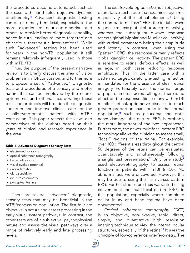
middle occipital gyrus. The pattern-reversal VEP has been used in several recent studies in the mTBI population. First, an optimal stimulus and test protocol was developed.17 It was found that the mean VEP amplitude of the mTBI population was smaller than found in the normal cohort. Then, two important, objectively-based, cortical, vision biomarkers were discovered. First, with marked reduction in mean test field luminance (~0.75 cd/m2), the average latency in those with mTBI was consistently and significantly more delayed than found in the normal cohort.18 Second, in those having mTBI and visual motion sensitivity (VMS),19 the addition of binasal occluders (BNO) to the patient’s spectacles produced a significant and consistent enhancement in amplitude as compared to the baseline VEP19
(Figure 2). In an early study using the pattern VEP, abnormal and variable waveforms were found in approximately 75% of the mTBI clinical population.9 Thus, the pattern VEP could prove invaluable to the neuro-optometrist, and others (e.g., the neuro-ophthalmologist), to document objectively the presence of mTBI/concussion with VMS, especially in those cases where the history and symptoms
leading up to the referral are vague, unclear, and/or incomplete, for example as may be the case in a young child. Lastly, use of the newer multifocal VEP approach allows one to assess small, “local” regions of the patient’s visual field and correlated visual cortex.16 Thus, the combined use of a multifocal pattern ERG and VEP provides a detailed, quantitative “roadmap” of functional abnormalities in the early visual system neural pathways.
Dark adaptation refers to the eye’s ability to detect progressively lower intensities of light over time, following a full bleaching of the retinal photopigments.20 It reflects both cone (initial photopic) and rod (later scotopic) responsivity, with the rods exhibiting overall greater influence over the 30 or so minute test period. The total response exhibits a range of sensitivity of 7 or more log units in the typical normal case. It also exhibits a cortical influence, at least in mTBI.20 This technique has been used to study macular problems and retinitis pigmentosa, and other retinal dysfunctions.21 Most interestingly, it has been consistently found that many with mTBI (50-100%)20,21 exhibited elevated visual thresholds; that is, they needed more light to detect the test stimulus than was found in normals. At first glance, this may seem to be paradoxical, since most of this population (50% or more) also exhibit photosensitivity.22 It has been hypothesized that in this subset of photosensitive mTBI patients, a cortical adaptive gain mechanism is active, which effectively functions to attenuate all levels of light to, in turn, reduce the sensation of photosensitivity at photopic levels. Thus, an overall elevated threshold is present.20 Such testing may help the clinician better understand light and dark adaptation/basic visual perception in this popu lation. Furthermore, the results are consistent with the finding that approximately 50% of patients with mTBI and photosensitivity later exhibit some degree of neuro-perceptual gain adaptation, and thus report reduced photosensitivity over time (e.g., one year or
Figure 2: Group mean VEP amplitude as a function of test condition (with or without BNO) in normals and in those with mTBI and VMS.
23Vision Development & Rehabilitation Volume 5, Issue 1 • March 2019
more).22 A hand-held device to measure dark adaptation exists for the clinician.20
Glare sensitivity refers to the adverse effect of scattered light on a visual task (e.g., visual acuity),23 for example as found in some patients with cataracts but otherwise being normal. Light scatter reduces retinal-image contrast, thus resulting in a “washed-out” perceptual effect. There are two categories of glare:23 (1) the “discomfort” variety in which a visual task is made slower, more difficult, or “taxing”, but is not reduced in visual performance level (e.g., visual acuity remains constant), with addition of a glare source; and, (2) the “disability” variety in which addition of a glare source reduces visual performance (e.g., visual acuity decreases with glare). Unfortunately, the terms “glare” and “glare sensitivity” are frequently, and we and others believe incorrectly, used interchangeably with the common phenomenon of “photosensitivity/light sensitivity” in the mTBI population, and elsewhere.24 So, is glare sensitivity per se really a problem in this subset of mTBI patients? Upon consultation with several experts and literature sources in the field, the answer is likely “not”, or at least it is not very common, as a separate and distinct entity, assuming absence of cataracts or other media light scattering problems. However, both careful clinical testing and laboratory studies should be performed for clarification
and insight. Clinically, testing is simple and direct.23 Essentially, a standardized visual task, such as Snellen visual acuity, is assessed with and without a controlled glare source. Any substantial worsening of visual performance with the presence of glare would confirm the diagnosis of disability glare. A hand-held device is available for clinical testing (www.marco.com). Neurologically, discomfort glare in normals has recently been found to be associated with the cortical phenomenon of response “hyperexcitability” in three brain regions:23 the cunei, lingual gyri, and superior parietal lobes. Interestingly, this increased hyper-type of brain response has also been documented in other possibly-related neuro logical conditions (e.g., migraine).23 Further brain imaging studies are needed to disentangle this complex situation in both the normal and mTBI populations.
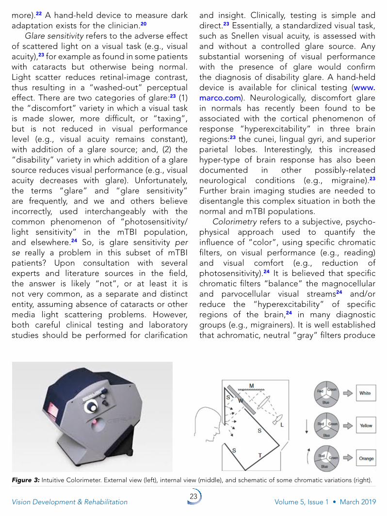
Colorimetry refers to a subjective, psycho-physical approach used to quantify the influence of “color”, using specific chromatic filters, on visual performance (e.g., reading) and visual comfort (e.g., reduction of photosensitivity).24 It is believed that specific chromatic filters “balance” the magnocellular and parvocellular visual streams24 and/or reduce the “hyperexcitability” of specific regions of the brain,24 in many diagnostic groups (e.g., migrainers). It is well established that achromatic, neutral “gray” filters produce
Figure 3: Intuitive Colorimeter. External view (left), internal view (middle), and schematic of some chromatic variations (right).
24Vision Development & Rehabilitation Volume 5, Issue 1 • March 2019
relief from photosensitivity in visually-normal individuals.24 These same filters can also be beneficial in those with mTBI/concussion and photosensitivity, but specific chromatic filters may have even more dramatic relief21,24,25 such as Omega-3 (purplish-red) and NL-41 (rose). While there are a range of diagnostic tests to assess for the presence of photosensitivity and to quantify its magnitude,24 an intriguing development has been the Intuitive Colorimeter.26 This device allows for a very precise, optimal chromatic tint to be found using independently-controlled variations in light hue, saturation, and brightness, while viewing and reading standardized texts (Figure 3). Use of the Intuitive Colorimeter and its prescribed tints has been found to decrease visual discomfort and enhance reading ability in some patients with mTBI/concussion.24-26 Further detailed testing (e.g., a clinical trial) is warranted to demonstrate the instrument’s diagnostic robustness in the mTBI/concussion population.
Perceptual testing refers to the assessment of the higher-level, neuro-sensory, visuo-per cep tual aspects of the visual system.27 It includes such areas as: visual discrimination, visual memory, spatial relations, form constancy, sequential memory, visual figure ground, and visual closure.27-28 These are common areas of perceptual dysfunction in the mTBI population.27 For example, an individual with a “figure-ground” deficit would have difficulty finding an object that is well above their detection visual threshold (i.e., visual acuity level) embedded in an array of objects. This might include locating a milk carton in the refrigerator or a pencil on a crowded desktop. A common test used by the neuro-optometrist, and others, is the Test of Visual Perceptual Skills (TVPS).28 This is a topic deserving more attention in both the clinic and research laboratory. The underlying neurology is beyond the scope of the present paper.
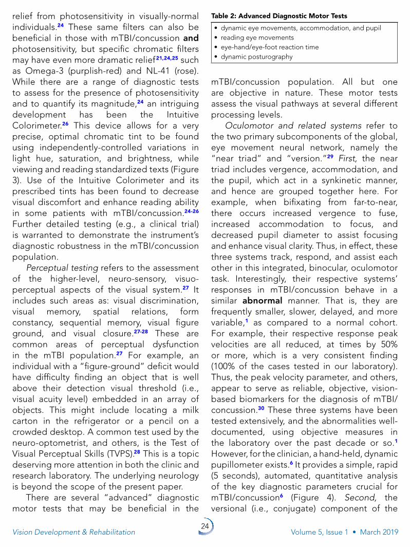
There are several “advanced” diagnostic motor tests that may be beneficial in the
mTBI/concussion population. All but one are objective in nature. These motor tests assess the visual pathways at several different processing levels.
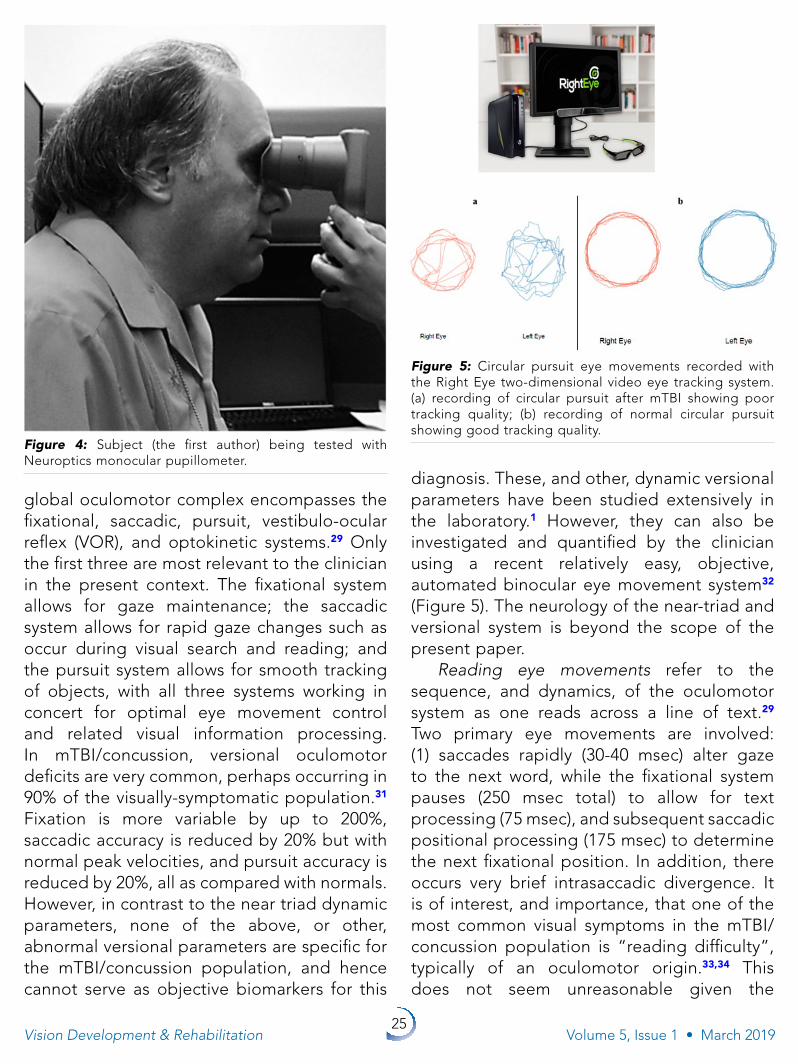
Oculomotor and related systems refer to the two primary subcomponents of the global, eye movement neural network, namely the “near triad” and “version.”29 First, the near triad includes vergence, accommodation, and the pupil, which act in a synkinetic manner, and hence are grouped together here. For example, when bifixating from far-to-near, there occurs increased vergence to fuse, increased accommodation to focus, and decreased pupil diameter to assist focusing and enhance visual clarity. Thus, in effect, these three systems track, respond, and assist each other in this integrated, binocular, oculomotor task. Interestingly, their respective systems’ responses in mTBI/concussion behave in a similar abnormal manner. That is, they are frequently smaller, slower, delayed, and more variable,1 as compared to a normal cohort. For example, their respective response peak velocities are all reduced, at times by 50% or more, which is a very consistent finding (100% of the cases tested in our laboratory). Thus, the peak velocity parameter, and others, appear to serve as reliable, objective, vision-based biomarkers for the diagnosis of mTBI/concussion.30 These three systems have been tested extensively, and the abnormalities well-documented, using objective measures in the laboratory over the past decade or so.1 However, for the clinician, a hand-held, dynamic pupillometer exists.6 It provides a simple, rapid (5 seconds), automated, quantitative analysis of the key diagnostic parameters crucial for mTBI/concussion6 (Figure 4). Second, the versional (i.e., conjugate) component of the
table 2: Advanced diagnostic Motor tests
• dynamiceyemovements,accommodation,andpupil• readingeyemovements• eye-hand/eye-footreactiontime• dynamicposturography
25Vision Development & Rehabilitation Volume 5, Issue 1 • March 2019
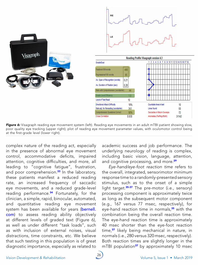
global oculomotor complex encompasses the fixational, saccadic, pursuit, vestibulo-ocular reflex (VOR), and optokinetic systems.29 Only the first three are most relevant to the clinician in the present context. The fixational system allows for gaze maintenance; the saccadic system allows for rapid gaze changes such as occur during visual search and reading; and the pursuit system allows for smooth tracking of objects, with all three systems working in concert for optimal eye movement control and related visual information processing. In mTBI/concussion, versional oculomotor deficits are very common, perhaps occurring in 90% of the visually-symptomatic population.31 Fixation is more variable by up to 200%, saccadic accuracy is reduced by 20% but with normal peak velocities, and pursuit accuracy is reduced by 20%, all as compared with normals. However, in contrast to the near triad dynamic parameters, none of the above, or other, abnormal versional parameters are specific for the mTBI/concussion population, and hence cannot serve as objective biomarkers for this
diagnosis. These, and other, dynamic versional parameters have been studied extensively in the laboratory.1 However, they can also be investigated and quantified by the clinician using a recent relatively easy, objective, automated binocular eye movement system32 (Figure 5). The neurology of the near-triad and versional system is beyond the scope of the present paper.
Reading eye movements refer to the sequence, and dynamics, of the oculomotor system as one reads across a line of text.29 Two primary eye movements are involved: (1) saccades rapidly (30-40 msec) alter gaze to the next word, while the fixational system pauses (250 msec total) to allow for text processing (75 msec), and subsequent saccadic positional processing (175 msec) to determine the next fixational position. In addition, there occurs very brief intrasaccadic divergence. It is of interest, and importance, that one of the most common visual symptoms in the mTBI/concussion population is “reading difficulty”, typically of an oculomotor origin.33,34 This does not seem unreasonable given the
Figure 4: Subject (the first author) being tested with Neuroptics monocular pupillometer.
Figure 5: Circular pursuit eye movements recorded with the Right Eye two-dimensional video eye tracking system. (a) recording of circular pursuit after mTBI showing poor tracking quality; (b) recording of normal circular pursuit showing good tracking quality.
26Vision Development & Rehabilitation Volume 5, Issue 1 • March 2019
complex nature of the reading act, especially in the presence of abnormal eye movement control, accommodative deficits, impaired attention, cognitive difficulties, and more, all leading to “cognitive fatigue”, frustration, and poor comprehension.33 In the laboratory, these patients manifest a reduced reading rate, an increased frequency of saccadic eye movements, and a reduced grade-level reading performance.34 Fortunately, for the clinician, a simple, rapid, binocular, automated, and quantitative reading eye movement system has been available for years (bernell.com) to assess reading ability objectively at different levels of graded text (Figure 6), as well as under different “task loads”, such as with inclusion of external noises, visual distractions, time constraints, etc. We believe that such testing in this population is of great diagnostic importance, especially as related to
academic success and job performance. The underlying neurology of reading is complex, including basic vision, language, attention, and cognitive processing, and more.29
Eye-hand/eye-foot reaction time refers to the overall, integrated, sensorimotor minimum response time to a randomly-presented sensory stimulus, such as to the onset of a simple light target.35-37 The pre-motor (i.e., sensory) processing component is approximately twice as long as the subsequent motor component (e.g., 167 versus 77 msec, respectively), for eye-hand reaction time in normals,35 with the combination being the overall reaction time. The eye-hand reaction time is approximately 40 msec shorter than the eye-foot reaction time,37 likely being mechanical in nature, in normals (i.e., 280 versus 320 msec, respectively). Both reaction times are slightly longer in the mTBI population37 by approximately 10 msec
Figure 6: Visagraph reading eye movement system (left). Reading eye movements in an adult mTBI patient showing slow, poor quality eye tracking (upper right); plot of reading eye movement parameter values, with oculomotor control being at the first-grade level (lower right).
27Vision Development & Rehabilitation Volume 5, Issue 1 • March 2019
on average; however, it can be up to 50 msec longer, with these delays occurring at the pre-motor level. This 10 msec delay likely causes no adverse vocational, sports, or driving effects. However, of interest, these delays can be up to 100-150 msec in the moderate TBI population,37 which may bear adverse consequences. These reaction times have been assessed in the laboratory.35-37 However, a system designed for the physical therapist is available to the optometrist, and others, that can be used with some practice.36,37 The underlying neurology is beyond the scope of this paper.
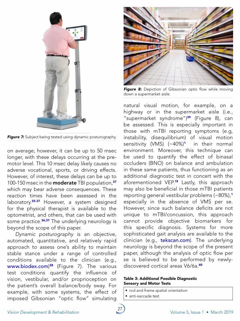
Dynamic posturography is an objective, automated, quantitative, and relatively rapid approach to assess one’s ability to maintain stable stance under a range of controlled conditions available to the clinician (e.g., www.biodex.com)38 (Figure 7). The various test conditions quantify the influence of vision, vestibular, and/or proprioception on the patient’s overall balance/body sway. For example, with some systems, the effect of imposed Gibsonian “optic flow” simulating
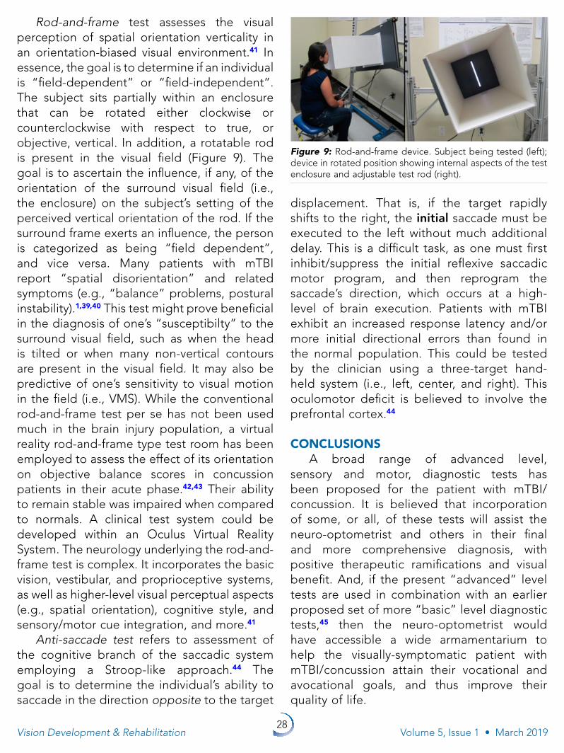
natural visual motion, for example, on a highway or in the supermarket aisle (i.e., “supermarket syndrome”)39 (Figure 8), can be assessed. This is especially important in those with mTBI reporting symptoms (e.g, instability, disequilibrium) of visual motion sensitivity (VMS) (~40%)1 in their normal environment. Moreover, this technique can be used to quantify the effect of binasal occluders (BNO) on balance and ambulation in these same patients, thus functioning as an additional diagnostic test in concert with the aforementioned VEP.19 Lastly, this approach may also be beneficial in those mTBI patients reporting general vestibular problems (~80%),1 especially in the absence of VMS per se. However, since such balance deficits are not unique to mTBI/concussion, this approach cannot provide objective biomarkers for this specific diagnosis. Systems for more sophisticated gait analysis are available to the clinician (e.g., tekscan.com). The underlying neurology is beyond the scope of the present paper, although the analysis of optic flow per se is believed to be performed by newly-discovered cortical areas V6/6a.40
table 3: Additional Possible diagnostic sensory and Motor tests
• rodandframespatialorientation• anti-saccadetest
Figure 7: Subject being tested using dynamic posturography.
Figure 8: Depiction of Gibsonian optic flow while moving down a supermarket aisle.
28Vision Development & Rehabilitation Volume 5, Issue 1 • March 2019
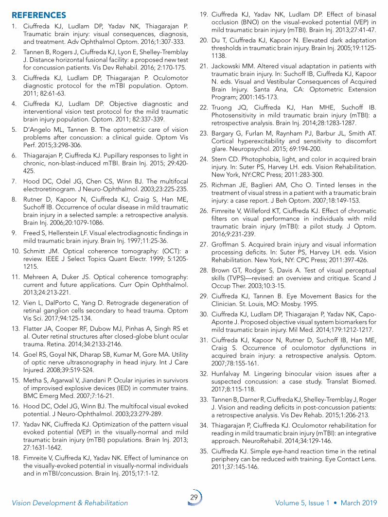
Rod-and-frame test assesses the visual perception of spatial orientation verticality in an orientation-biased visual environment.41 In essence, the goal is to determine if an individual is “field-dependent” or “field-independent”. The subject sits partially within an enclosure that can be rotated either clockwise or counterclockwise with respect to true, or objective, vertical. In addition, a rotatable rod is present in the visual field (Figure 9). The goal is to ascertain the influence, if any, of the orientation of the surround visual field (i.e., the enclosure) on the subject’s setting of the perceived vertical orientation of the rod. If the surround frame exerts an influence, the person is categorized as being “field dependent”, and vice versa. Many patients with mTBI report “spatial disorientation” and related symptoms (e.g., “balance” problems, postural instability).1,39,40 This test might prove beneficial in the diagnosis of one’s “susceptibilty” to the surround visual field, such as when the head is tilted or when many non-vertical contours are present in the visual field. It may also be predictive of one’s sensitivity to visual motion in the field (i.e., VMS). While the conventional rod-and-frame test per se has not been used much in the brain injury population, a virtual reality rod-and-frame type test room has been employed to assess the effect of its orientation on objective balance scores in concussion patients in their acute phase.42,43 Their ability to remain stable was impaired when compared to normals. A clinical test system could be developed within an Oculus Virtual Reality System. The neurology underlying the rod-and-frame test is complex. It incorporates the basic vision, vestibular, and proprioceptive systems, as well as higher-level visual perceptual aspects (e.g., spatial orientation), cognitive style, and sensory/motor cue integration, and more.41
Anti-saccade test refers to assessment of the cognitive branch of the saccadic system employing a Stroop-like approach.44 The goal is to determine the individual’s ability to saccade in the direction opposite to the target
displacement. That is, if the target rapidly shifts to the right, the initial saccade must be executed to the left without much additional delay. This is a difficult task, as one must first inhibit/suppress the initial reflexive saccadic motor program, and then reprogram the saccade’s direction, which occurs at a high-level of brain execution. Patients with mTBI exhibit an increased response latency and/or more initial directional errors than found in the normal population. This could be tested by the clinician using a three-target hand-held system (i.e., left, center, and right). This oculomotor deficit is believed to involve the prefrontal cortex.44
conclusIons A broad range of advanced level,
sensory and motor, diagnostic tests has been proposed for the patient with mTBI/concussion. It is believed that incorporation of some, or all, of these tests will assist the neuro-optometrist and others in their final and more comprehensive diagnosis, with positive therapeutic ramifications and visual benefit. And, if the present “advanced” level tests are used in combination with an earlier proposed set of more “basic” level diagnostic tests,45 then the neuro-optometrist would have accessible a wide armamentarium to help the visually-symptomatic patient with mTBI/concussion attain their vocational and avocational goals, and thus improve their quality of life.
Figure 9: Rod-and-frame device. Subject being tested (left); device in rotated position showing internal aspects of the test enclosure and adjustable test rod (right).
29Vision Development & Rehabilitation Volume 5, Issue 1 • March 2019
references1. Ciuffreda KJ, Ludlam DP, Yadav NK, Thiagarajan P.
Traumatic brain injury: visual consequences, diagnosis, and treatment. Adv Ophthalmol Optom. 2016;1:307-333.
2. Tannen B, Rogers J, Ciuffreda KJ, Lyon E, Shelley-Tremblay J. Distance horizontal fusional facility: a proposed new test for concussion patients. Vis Dev Rehabil. 2016; 2:170-175.
3. Ciuffreda KJ, Ludlam DP, Thiagarajan P. Oculomotor diagnostic protocol for the mTBI population. Optom. 2011; 82:61-63.
4. Ciuffreda KJ, Ludlam DP. Objective diagnostic and interventional vision test protocol for the mild traumatic brain injury population. Optom. 2011; 82:337-339.
5. D’Angelo ML, Tannen B. The optometric care of vision problems after concussion: a clinical guide. Optom Vis Perf. 2015;3:298-306.
6. Thiagarajan P, Ciuffreda KJ. Pupillary responses to light in chronic, non-blast-induced mTBI. Brain Inj. 2015; 29:420-425.
7. Hood DC, Odel JG, Chen CS, Winn BJ. The multifocal electroretinogram. J Neuro-Ophthalmol. 2003;23:225-235.
8. Rutner D, Kapoor N, Ciuffreda KJ, Craig S, Han ME, Suchoff IB. Occurrence of ocular disease in mild traumatic brain injury in a selected sample: a retrospective analysis. Brain Inj. 2006;20:1079-1086.
9. Freed S, Hellerstein LF. Visual electrodiagnostic findings in mild traumatic brain injury. Brain Inj. 1997;11:25-36.
10. Schmitt JM. Optical coherence tomography: (OCT): a review. IEEE J Select Topics Quant Electr. 1999; 5:1205-1215.
11. Mehreen A, Duker JS. Optical coherence tomography: current and future applications. Curr Opin Ophthalmol. 2013;24:213-221.
12. Vien L, DalPorto C, Yang D. Retrograde degeneration of retinal ganglion cells secondary to head trauma. Optom Vis Sci. 2017;94:125-134.
13. Flatter JA, Cooper RF, Dubow MJ, Pinhas A, Singh RS et al. Outer retinal structures after closed-globe blunt ocular trauma. Retina. 2014;34:2133-2146.
14. Goel RS, Goyal NK, Dharap SB, Kumar M, Gore MA. Utility of optic nerve ultrasonography in head injury. Int J Care Injured. 2008;39:519-524.
15. Metha S, Agarwal V, Jiandani P. Ocular injuries in survivors of improvised explosive devices (IED) in commuter trains. BMC Emerg Med. 2007;7:16-21.
16. Hood DC, Odel JG, Winn BJ. The multifocal visual evoked potential. J Neuro-Ophthalmol. 2003;23:279-289.
17. Yadav NK, Ciuffreda KJ. Optimization of the pattern visual evoked potential (VEP) in the visually-normal and mild traumatic brain injury (mTBI) populations. Brain Inj. 2013; 27:1631-1642.
18. Fimreite V, Ciuffreda KJ, Yadav NK. Effect of luminance on the visually-evoked potential in visually-normal individuals and in mTBI/concussion. Brain Inj. 2015;17:1-12.
19. Ciuffreda KJ, Yadav NK, Ludlam DP. Effect of binasal occlusion (BNO) on the visual-evoked potential (VEP) in mild traumatic brain injury (mTBI). Brain Inj. 2013;27:41-47.
20. Du T, Ciuffreda KJ, Kapoor N. Elevated dark adaptation thresholds in traumatic brain injury. Brain Inj. 2005;19:1125-1138.
21. Jackowski MM. Altered visual adaptation in patients with traumatic brain injury. In: Suchoff IB, Ciuffreda KJ, Kapoor N. eds. Visual and Vestibular Consequences of Acquired Brain Injury. Santa Ana, CA: Optometric Extension Program; 2001:145-173.
22. Truong JQ, Ciuffreda KJ, Han MHE, Suchoff IB. Photosensitivity in mild traumatic brain injury (mTBI): a retrospective analysis. Brain Inj. 2014;28:1283-1287.
23. Bargary G, Furlan M, Raynham PJ, Barbur JL, Smith AT. Cortical hyperexcitability and sensitivity to discomfort glare. Neuropsychol. 2015; 69:194-200.
24. Stern CD. Photophobia, light, and color in acquired brain injury. In: Suter PS, Harvey LH. eds. Vision Rehabilitation. New York, NY:CRC Press; 2011:283-300.
25. Richman JE, Baglieri AM, Cho O. Tinted lenses in the treatment of visual stress in a patient with a traumatic brain injury: a case report. J Beh Optom. 2007;18:149-153.
26. Fimreite V, Willeford KT, Ciuffreda KJ. Effect of chromatic filters on visual performance in individuals with mild traumatic brain injury (mTBI): a pilot study. J Optom. 2016;9:231-239.
27. Groffman S. Acquired brain injury and visual information processing deficits. In: Suter PS, Harvey LH. eds. Vision Rehabilitation. New York, NY: CPC Press; 2011:397-426.
28. Brown GT, Rodger S, Davis A. Test of visual perceptual skills (TVPS)—revised: an overview and critique. Scand J Occup Ther. 2003;10:3-15.
29. Ciuffreda KJ, Tannen B. Eye Movement Basics for the Clinician. St. Louis, MO: Mosby. 1995.
30. Ciuffreda KJ, Ludlam DP, Thiagarajan P, Yadav NK, Capo-Aponte J. Proposed objective visual system biomarkers for mild traumatic brain injury. Mil Med. 2014;179:1212-1217.
31. Ciuffreda KJ, Kapoor N, Rutner D, Suchoff IB, Han ME, Craig S. Occurrence of oculomotor dysfunctions in acquired brain injury: a retrospective analysis. Optom. 2007;78:155-161.
32. Hunfalvay M. Lingering binocular vision issues after a suspected concussion: a case study. Translat Biomed. 2017;8:115-118.
33. Tannen B, Darner R, Ciuffreda KJ, Shelley-Tremblay J, Roger J. Vision and reading deficits in post-concussion patients: a retrospective analysis. Vis Dev Rehab. 2015;1:206-213.
34. Thiagarajan P, Ciuffreda KJ. Oculomotor rehabilitation for reading in mild traumatic brain injury (mTBI): an integrative approach. NeuroRehabil. 2014;34:129-146.
35. Ciuffreda KJ. Simple eye-hand reaction time in the retinal periphery can be reduced with training. Eye Contact Lens. 2011;37:145-146.
30Vision Development & Rehabilitation Volume 5, Issue 1 • March 2019
36. Gould JA, Ciuffreda KJ, Arthur B, Yadav NK. Retinal defocus and eye dominance effect on eye-hand reaction time. Optom Vis Perf. 2013;1:129-136.
37. Gould JA, Ciuffreda KJ, Yadav NK, Thiagarajan P, Arthur B. The effect of retinal defocus on simple eye-hand and eye-foot reaction time in traumatic brain injury. Brain Inj. 2013; 27:1643-1648.
38. Keshner EA, Kenyon RV. Postural and spatial orientation driven by virtual reality. Stud Health Technol Inform. 2009;145:209-228.
39. Ciuffreda KJ. Visual vertigo syndrome: clinical demonstration and diagnostic tool. Clin Eye Vis Care. 1999;11:41-44.
40. Ciuffreda KJ, Yadav NK, Ludlam DP. Binasal occlusion (BNO), visual motion sensitivity (VMS), and the visually-evoked potential (VEP) in mild traumatic brain injury and traumatic brain injury (mTBI/TBI). Brain Sci. 2017;7,98;doi:10.3390/brainsci7080098.
41. Witkin HA, Asch SE. Studies in space orientation: IV. Further experiments on perception of the upright with displaced visual fields. J Exp Psychol. 1948;38:762-782.
42. Slobounov S, Slobounov E, Newell K. Application of virtual reality graphics in assessment of concussion. Cyber Psychol Beh. 2006;9:188-191.
43. Teel EF, Slobounov SM. Validation of a virtual reality balance module for use in clinical concussion assessment and management. Clin J Sport Med. 2015;25:144-148.
44. Kraus M, Little DM, Donnell AJ, Reilly JL, Simonian N, Sweeney JA. Oculomotor function in chronic traumatic brain injury. Cog Beh Neurol. 2007;20:170-178.
45. Ciuffreda KJ, Tannen B, Ludlam DP, Yadav NK. Basic neuro-optometric diagnostic vision test battery for mild traumatic brain injury (mTBI)/concussion: A narrative review, perspective, and proposed techniques and protocols. Vis Dev Rehab, 2018; 4:157-169.
CORRESPONDING AUTHOR BIOGRAPHY:Kenneth J. Ciuffreda, OD, PhDNew York, New York
Kenneth J. Ciuffreda received his B.S in biology from Seton Hall University in 1969, his O.D. from the Massachusetts College of Optometry in 1973, and his Ph.D. degree in physiological optics from
the University of California/School Optometry at Berkeley in 1977. He has been a faculty member at the SUNY/State College of Optometry in New York City since 1979, where he is presently a Distinguished Teaching Professor. He has also had adjunct appointments for many years at Rutgers/ The State University of New Jersey, as well as at the New Jersey Institute of Technology, both in the department of biomedical engineering. He also helped establish a school of optometry in Harbin, China. He has conducted research in many areas: amblyopia, strabismus, reading, myopia, eye movements, accommodation, bioengineering applications to optometry, and more recently with an emphasis in the area of acquired brain injury, both the diagnostic and therapeutic aspects. His goal has been the use of objective recording techniques in the diagnosis and treatment of neurological and ocular conditions. He holds two patents, and has received many awards and honors from the AAO, AOA, NORA, COVD, and various state optometric associations and colleges. He has authored over 400 research papers/chapters, and 10 books. His hobbies are playing jazz guitar and enjoying the visual aspects of art.