Embed Size (px)
Citation preview
Advances in Radiation Oncology
Regional lymph node irradiation in breast cancer may worsen lung damage in COVID-19 positive patients
--Manuscript Draft--
Manuscript Number: ADVANCESRADONC-D-20-00122R1
Article Type: Teaching Case
Section/Category: COVID-19
Corresponding Author: Yazid Belkacemi, MD PHDCHU Henri MondorCréteil, FRANCE
First Author: Noemie Grellier
Order of Authors: Noemie Grellier
Asma Hadhri
Jerome Bendavid
Marie Adou
Alix Demory
Sarah Bouchereau
Wissal Hassani
Marie-Laure Herve
Mathilde Mahé
Laurianne Colson-Durand
Yazid Belkacemi, MD PHD
Abstract: Radiation-induced interstitial pneumonitis (IP) is a rare after breast radiotherapy. Usingmodern techniques, IP risk has been significantly reduced. However, dose delivered tothe ipsilateral lung, co-morbidities and virus infection could increase this riskparticularly in patients infected by COVID-19 during RT. We report here evidence of aclear correlation between dose distribution in the ipsilateral lung and the COVID-19lung damage shown in CT scan. Thus, it seems of paramount importance to point outthe risk of severe IP during nodal RT for breast cancer in patients who became infectedby COVID-19 during the period of their treatment. High caution for indications andvolume definition is recommended when RT cannot be delayed in high risk patients.
Powered by Editorial Manager® and ProduXion Manager® from Aries Systems Corporation
1
Regional lymph node irradiation in breast cancer may worsen
lung damage in COVID-19 positive patients
Running title: Breast radiotherapy in COVID-19 positive patiens
Noémie Grellier1,*, Asma Hadhri1, Jerome Bendavid1, Marie Adou1, Alix Demory1, Sarah Bouchereau1,
Wissal Hassani1, Marie-Laure Hervé1, Laurianne Colson-Durand1, Yazid Belkacemi, MD, PhD 1,*
1APHP; Henri Mondor Breast Center and Department of Radiation Oncology. INSERM U955 Team 21.
University Paris-Est Creteil (UPEC). France
*Association of Radiotherapy and Oncology of the Mediterranean area (AROME; www.aromecancer.org) &
TransAtlantic Radiation Oncology Network (TRONE)
#Correspondence to:
Prof. Yazid Belkacemi MD, PhD
Université Paris Est Créteil
CHU Henri Mondor, 51 Av Mal De Lattre de Tassigny
Créteil 94000, France
Tel: +33 1 49 81 45 22
Fax: +33 1 49 81 25 89
Email: [email protected]
Conflict of interest: None
Financial support: None
Title Page (WITH Author Details)
Radiation-induced interstitial pneumonitis (IP) is a rare but possibly severe toxicity after
radiotherapy (RT) in patients with co-morbidities treated for breast cancer. Minimal symptoms
are generally seen in the majority of patients and are resolved with short-term steroid
administration. Since the introduction of treatment planning based on three-dimensional
conformal RT (3D CRT), IP risk has been significantly reduced with the application of rigorous
lung constraints. With the use of modern techniques and hypofractionation, less than 2% of
patients have experienced IP symptoms after a median follow-up of 15 months with ipsilateral
lung V30 as the most relevant dosimetric predictor for IP risk (1, 2). However, dose delivered
to the ipsilateral lung, co-morbidities and virus infection could change the outcome of patients
infected by COVID-19 during RT.
In this report, we describe a 73-year-old woman with severe COVID-19 pneumonia infection
diagnosed during RT for breast cancer. The main comorbidities were obesity (BMI 40 kg/m2),
hypertension and thrombosis following surgery for knee prosthesis.
The patient had right breast conservative surgery with sentinel node biopsy (SLNB). The final
pathology report suggested invasive carcinoma, grade II, without lymphovascular invasion,
without DCIS component, hormone receptor positive and HER2 negative. The surgical margins
were clear. One of the three nodes removed was involved by macro metastasis and extracapsular
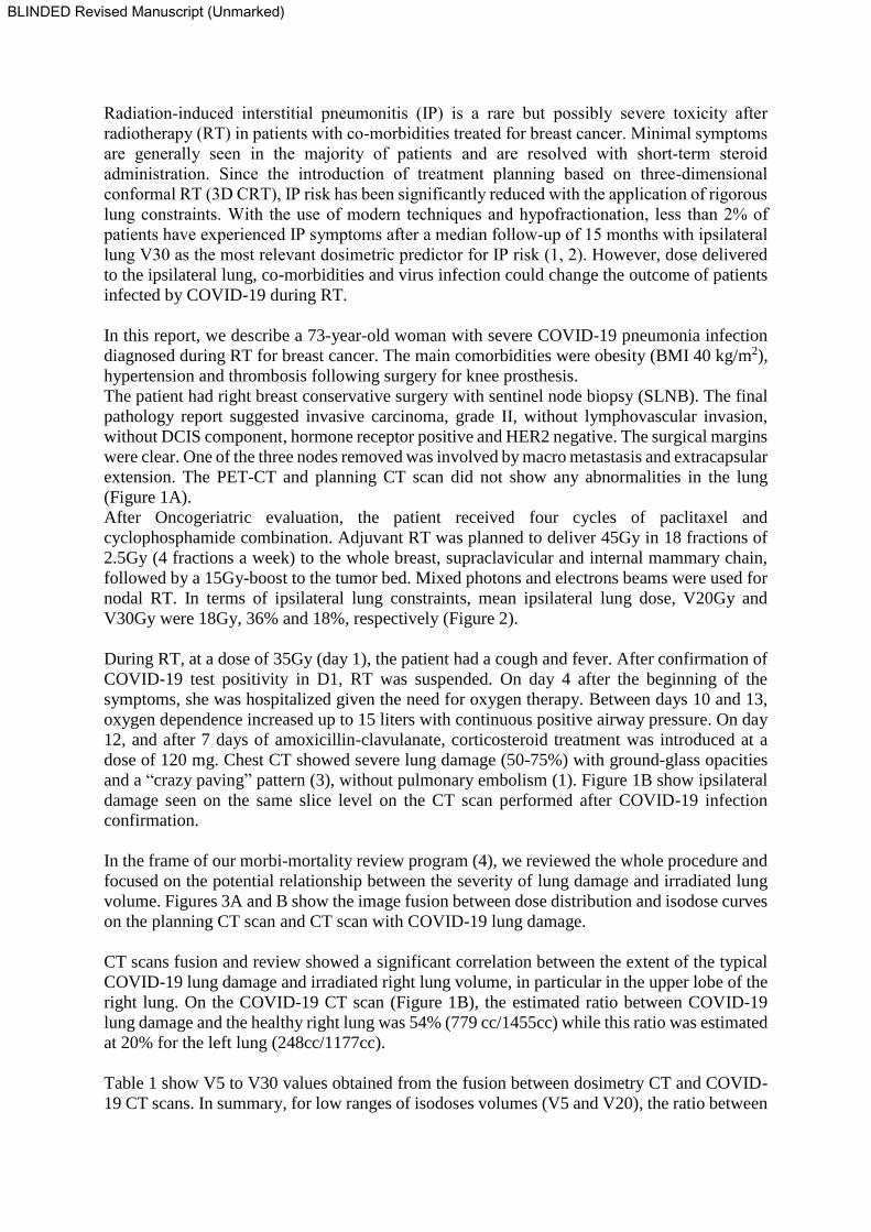
extension. The PET-CT and planning CT scan did not show any abnormalities in the lung
(Figure 1A).
After Oncogeriatric evaluation, the patient received four cycles of paclitaxel and
cyclophosphamide combination. Adjuvant RT was planned to deliver 45Gy in 18 fractions of
2.5Gy (4 fractions a week) to the whole breast, supraclavicular and internal mammary chain,
followed by a 15Gy-boost to the tumor bed. Mixed photons and electrons beams were used for
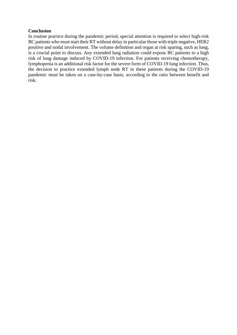
nodal RT. In terms of ipsilateral lung constraints, mean ipsilateral lung dose, V20Gy and
V30Gy were 18Gy, 36% and 18%, respectively (Figure 2).
During RT, at a dose of 35Gy (day 1), the patient had a cough and fever. After confirmation of
COVID-19 test positivity in D1, RT was suspended. On day 4 after the beginning of the
symptoms, she was hospitalized given the need for oxygen therapy. Between days 10 and 13,
oxygen dependence increased up to 15 liters with continuous positive airway pressure. On day
12, and after 7 days of amoxicillin-clavulanate, corticosteroid treatment was introduced at a
dose of 120 mg. Chest CT showed severe lung damage (50-75%) with ground-glass opacities
and a “crazy paving” pattern (3), without pulmonary embolism (1). Figure 1B show ipsilateral
damage seen on the same slice level on the CT scan performed after COVID-19 infection
confirmation.
In the frame of our morbi-mortality review program (4), we reviewed the whole procedure and
focused on the potential relationship between the severity of lung damage and irradiated lung
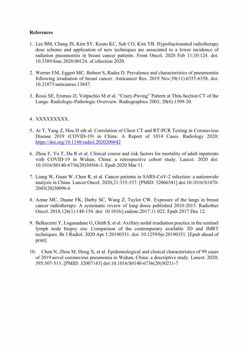
volume. Figures 3A and B show the image fusion between dose distribution and isodose curves
on the planning CT scan and CT scan with COVID-19 lung damage.
CT scans fusion and review showed a significant correlation between the extent of the typical
COVID-19 lung damage and irradiated right lung volume, in particular in the upper lobe of the
right lung. On the COVID-19 CT scan (Figure 1B), the estimated ratio between COVID-19
lung damage and the healthy right lung was 54% (779 cc/1455cc) while this ratio was estimated
at 20% for the left lung (248cc/1177cc).
Table 1 show V5 to V30 values obtained from the fusion between dosimetry CT and COVID-
19 CT scans. In summary, for low ranges of isodoses volumes (V5 and V20), the ratio between
BLINDED Revised Manuscript (Unmarked)

the damaged and the healthy lungs as delineated on the COVID-19 CT scan ranged between
54% and 59%. Furthermore, Figures 3A and B shows the projection of V5 to V30 isodose lines
on the healthy lung delineated on the simulation CT scan and on the lung lesions induced by
COVID-19 as seen on the COVID-19 CT diagnosis. The majority of the lung images are
covered isodoses lines of V10 to V20.
Discussion
In daily practice, patients undergoing RT may be infected with COVID-19 at any time during
treatment. Their identification is crucial not only to avoid contamination of staff and other
patients, but also to carefully monitor their state of health with the risk of respiratory distress.
Among the risk factors reported in the literature, the irradiated lung volume is not studied. On
the other hand, it is known that 20-40% of the patients develop unilateral CT scan COVID-19
lung damage, while both lungs could be concerned in the other patients (5).
In breast cancer RT, the lung volume is greater when regional nodal irradiation (RNI) is
indicated. In the present case, as shown in Figure 3B, COVID-19 induced lung damage is
predominant in the irradiated areas while limited images are seen in the left lung. This is
correlated to the right lung volume coverage by isodose lines as showed in the comparison
presented Figure 4.
Given the lack of data concerning the impact of RT on the severity of COVID-19 lung damage,
it seems important to remain extremely careful in cases receiving RT that partly involve the
lungs, such as whole breast cancer +/- nodal areas that include additional lung volume (6, 7).
Thus, it seems of paramount importance to point out the risk of severe PI during RT for breast
cancer in patients who became infected by COVID-19 during the period of their treatment.
High caution for indications and volume definition is recommended when RT cannot be delayed
in high risk patients. In RNI cases, IMRT (which increase only low doses), breathing adaptation
and prone/lateral positioning may help to reduce ipsilateral lung exposure (8, 9). However,
prone/lateral position can not allow adequate nodal coverage. Moreover, hypofractionation was
reported to reduce lung dose irradiation (1). Of note, our patient had moderate hypofractionated
schedule due to her age and the distance between her home and our department.
It is known that COVID-19 disproportionally harms elderly persons and those with comorbid
conditions (10). Moreover, in early reports from China, patients with cancer who acquired
COVID-19 had a higher risk for significant morbidity, including requirements for ventilatory
support or death with a hazard ratio of 3.56 (7). On the other hand, it is known that several
parameters may influence incidence and severity of either radiation induced IP and COVID-19
severe acute respiratory syndrome. For this patient, unfortunately several risk factors for severe
COVID-19 pneumonia, such as age >70 years, obesity, hypertension, cancer and chemotherapy
(with preexisting lymphopenia), were reported in her disease and personal history. She is still
in an oxygen dependence situation with 15 liters between D10 and D24.
Thus, in patients with BC undergoing RT, the utility of intervention must be weighed against
the risk for inadvertent COVID-19 exposure in the health care system, especially during the
initial weeks of the pandemic, when the risk for viral dissemination cannot be quantified and
remains largely unknown.
Conclusion
In routine practice during the pandemic period, special attention is required to select high-risk
BC patients who must start their RT without delay in particular those with triple negative, HER2
positive and nodal involvement. The volume definition and organ at risk sparing, such as lung,
is a crucial point to discuss. Any extended lung radiation could expose BC patients to a high
risk of lung damage induced by COVID-19 infection. For patients receiving chemotherapy,
lymphopenia is an additional risk factor for the severe form of COVID-19 lung infection. Thus,
the decision to practice extended lymph node RT in these patients during the COVID-19
pandemic must be taken on a case-by-case basis, according to the ratio between benefit and
risk.
References
1. Lee BM, Chang JS, Kim SY, Keum KC, Suh CO, Kim YB. Hypofractionated radiotherapy
dose scheme and application of new techniques are associated to a lower incidence of
radiation pneumonitis in breast cancer patients. Front Oncol. 2020 Feb 11;10:124. doi:
10.3389/fonc.2020.00124. eCollection 2020.
2. Werner EM, Eggert MC, Bohnet S, Rades D. Prevalence and characteristics of pneumonitis
following irradiation of breast cancer. Anticancer Res. 2019 Nov;39(11):6355-6358. doi:
10.21873/anticanres.13847.
3. Rossi SE, Ersmus JJ, Volpachio M et al. “Crazy-Paving” Pattern at Thin-Section CT of the
Lungs: Radiologic-Pathologic Overview. Radiographics 2003; 20(6):1509-20.
4. XXXXXXXXX.
5. Ai T, Yang Z, Hou H etb al. Correlation of Chest CT and RT-PCR Testing in Coronavirus
Disease 2019 (COVID-19) in China: A Report of 1014 Cases. Radiology 2020;
https://doi.org/10.1148/radiol.2020200642
6. Zhou F, Yu T, Du R et al. Clinical course and risk factors for mortality of adult inpatients
with COVID-19 in Wuhan, China: a retrospective cohort study. Lancet. 2020 doi:
10.1016/S0140-6736(20)30566-3. Epub 2020 Mar 11.
7. Liang W, Guan W, Chen R, et al. Cancer patients in SARS-CoV-2 infection: a nationwide
analysis in China. Lancet Oncol. 2020;21:335-337. [PMID: 32066541] doi:10.1016/S1470-
2045(20)30096-6
8. Aznar MC, Duane FK, Darby SC, Wang Z, Taylor CW. Exposure of the lungs in breast
cancer radiotherapy: A systematic review of lung doses published 2010-2015. Radiother
Oncol. 2018;126(1):148-154. doi: 10.1016/j.radonc.2017.11.022. Epub 2017 Dec 12.
9. Belkacemi Y, Loganadane G, Ghith S, et al. Axillary nodal irradiation practice in the sentinel
lymph node biopsy era: Comparison of the contemporary available 3D and IMRT
techniques. Br J Radiol. 2020 Apr 1:20190351. doi: 10.1259/bjr.20190351. [Epub ahead of
print]
10. Chen N, Zhou M, Dong X, et al. Epidemiological and clinical characteristics of 99 cases
of 2019 novel coronavirus pneumonia in Wuhan, China: a descriptive study. Lancet. 2020;
395:507-513. [PMID: 32007143] doi:10.1016/S0140-6736(20)30211-7
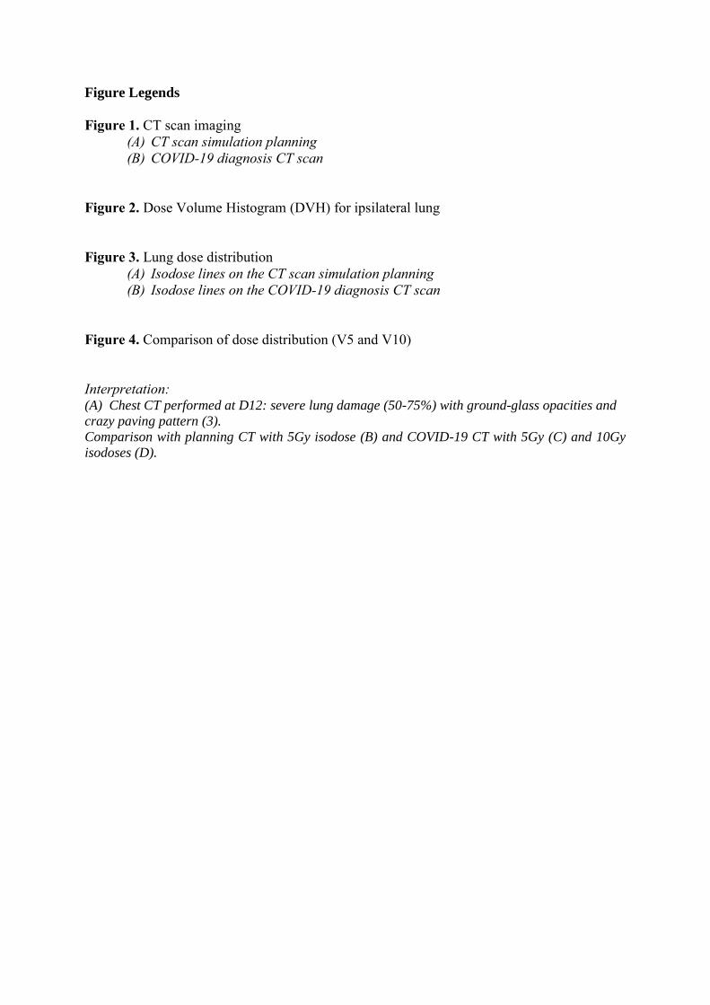
Figure Legends
Figure 1. CT scan imaging
(A) CT scan simulation planning
(B) COVID-19 diagnosis CT scan
Figure 2. Dose Volume Histogram (DVH) for ipsilateral lung
Figure 3. Lung dose distribution
(A) Isodose lines on the CT scan simulation planning
(B) Isodose lines on the COVID-19 diagnosis CT scan
Figure 4. Comparison of dose distribution (V5 and V10)
Interpretation: (A) Chest CT performed at D12: severe lung damage (50-75%) with ground-glass opacities and
crazy paving pattern (3).
Comparison with planning CT with 5Gy isodose (B) and COVID-19 CT with 5Gy (C) and 10Gy
isodoses (D).
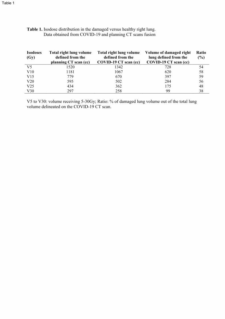
Table 1. Isodose distribution in the damaged versus healthy right lung.
Data obtained from COVID-19 and planning CT scans fusion
V5 to V30: volume receiving 5-30Gy; Ratio: % of damaged lung volume out of the total lung
volume delineated on the COVID-19 CT scan.
Isodoses
(Gy)
Total right lung volume
defined from the
planning CT scan (cc)
Total right lung volume
defined from the
COVID-19 CT scan (cc)
Volume of damaged right
lung defined from the
COVID-19 CT scan (cc)
Ratio
(%)
V5
V10
V15
V20
V25
V30
1520
1181
779
595
434
297
1342
1067
670
502
362
258
728
620
397
284
175
99
54
58
59
56
48
38
Table 1
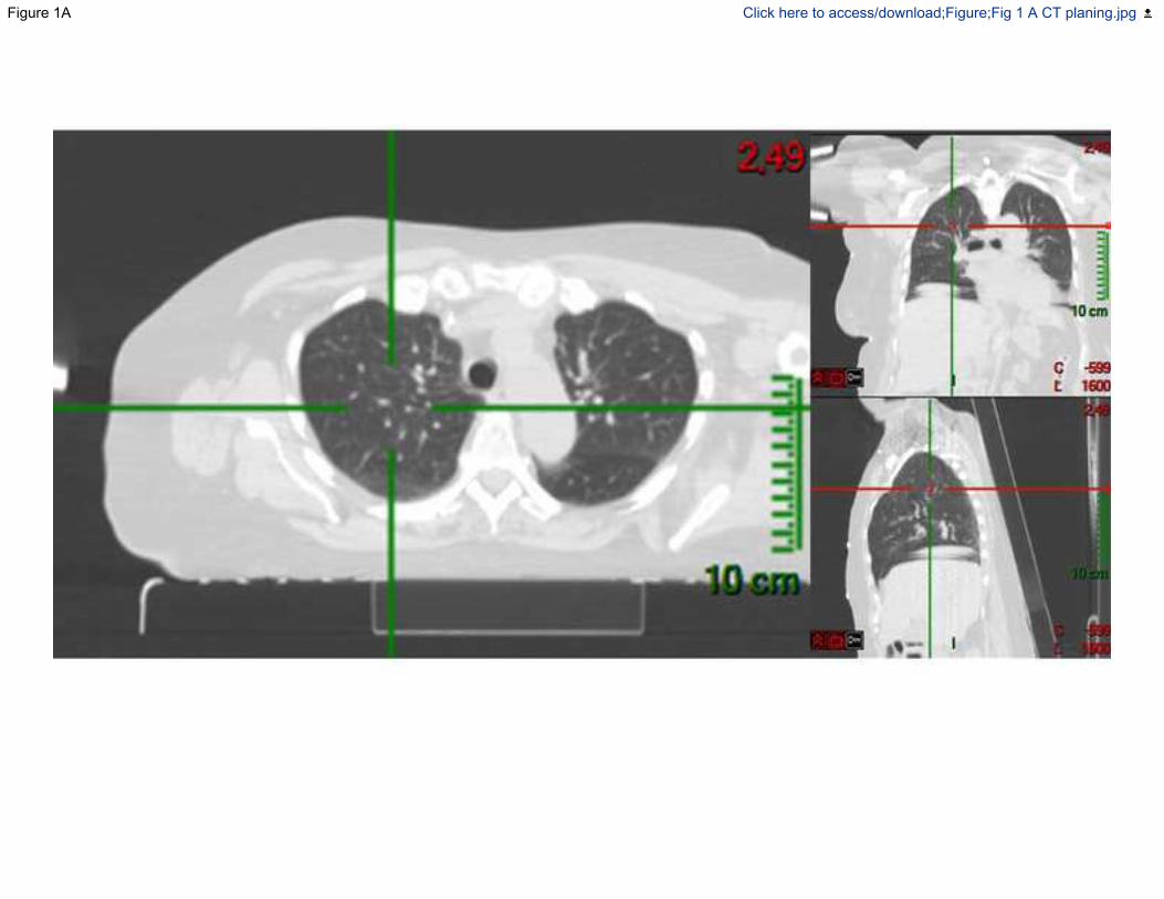
Figure 1A Click here to access/download;Figure;Fig 1 A CT planing.jpg
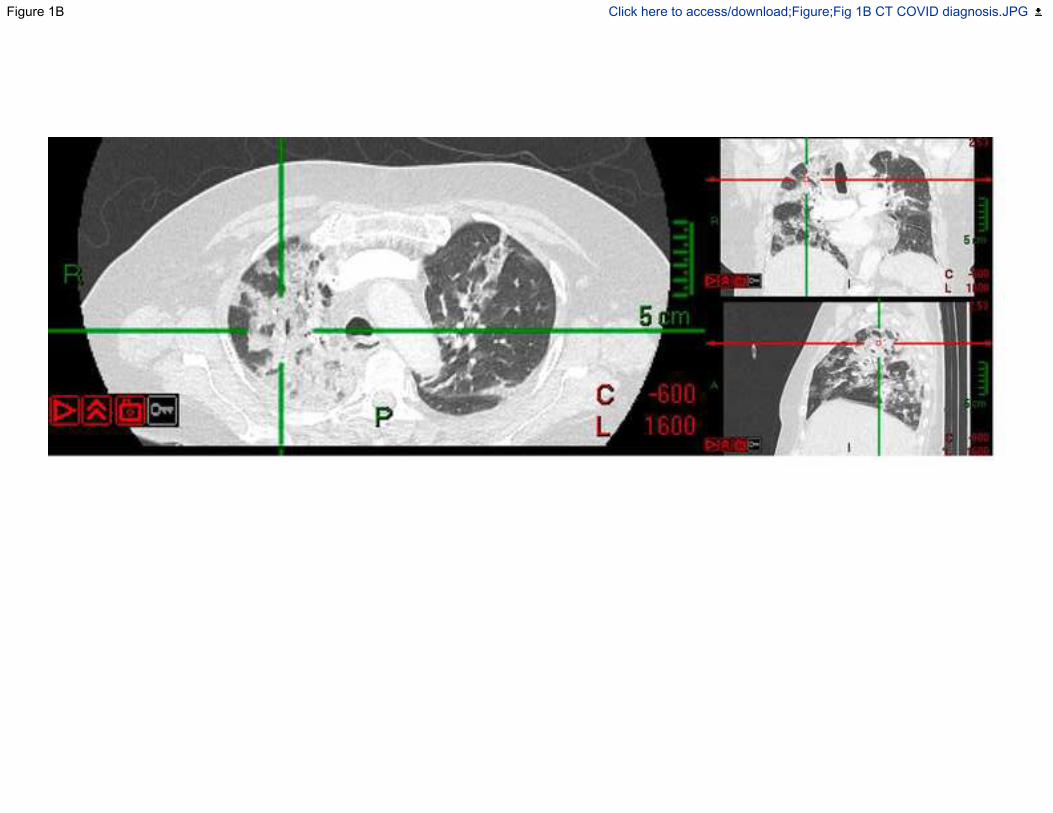
Figure 1B Click here to access/download;Figure;Fig 1B CT COVID diagnosis.JPG
Figure 2 Click here to access/download;Figure;Fig 2 DVH ipsilateral lung.jpg
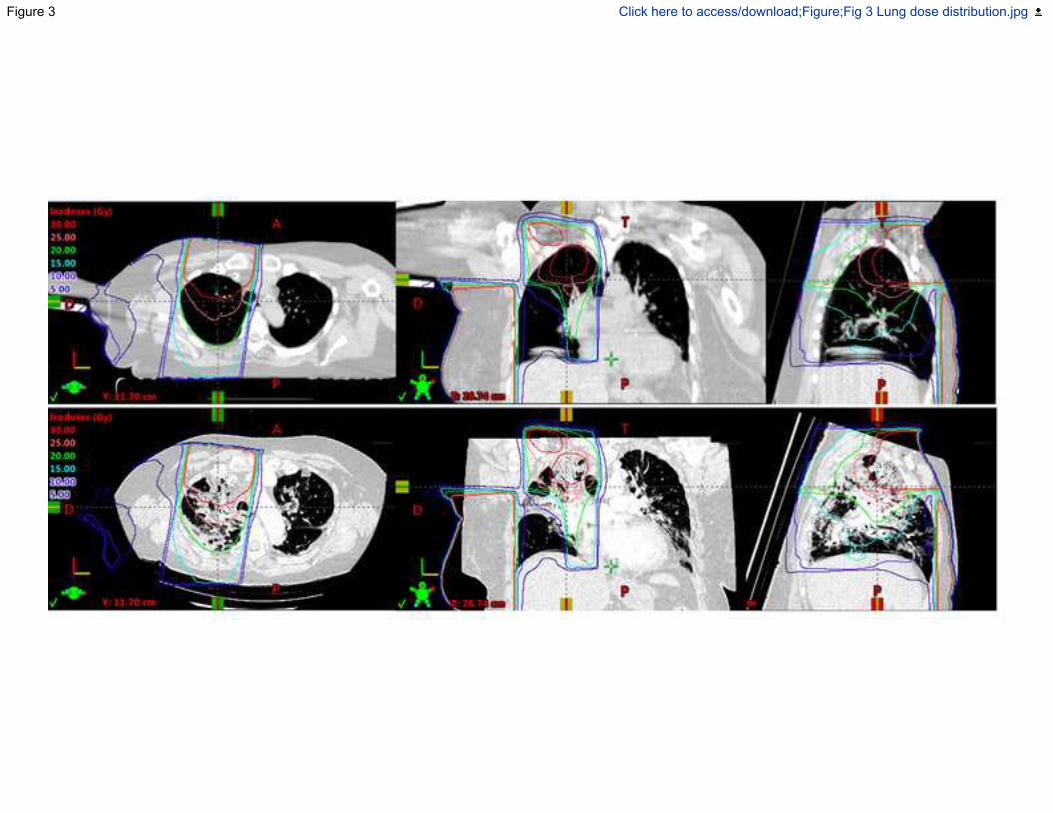
Figure 3 Click here to access/download;Figure;Fig 3 Lung dose distribution.jpg
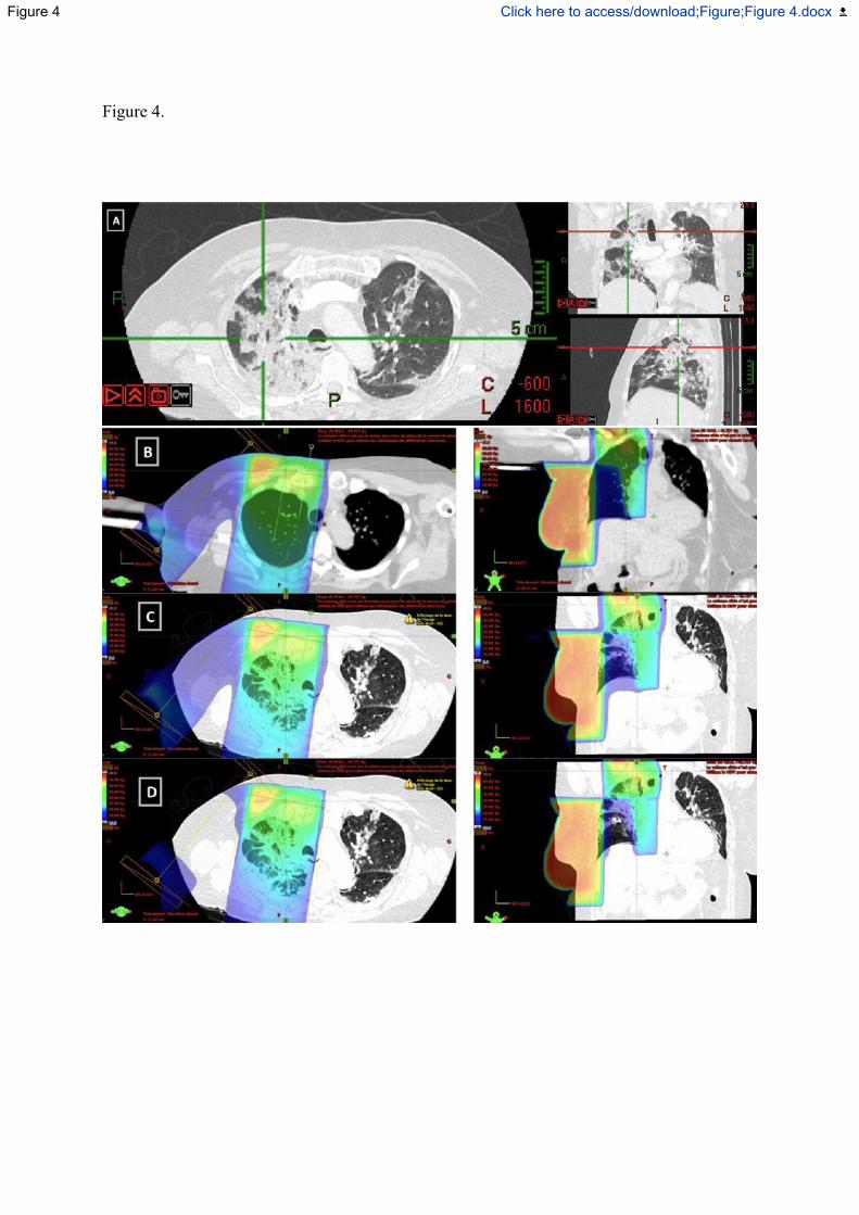
Figure 4.
Figure 4 Click here to access/download;Figure;Figure 4.docx