Embed Size (px)
Citation preview
African Journal of Neurological Sciences 2010 - Vol. 29, No 1
http://ajns.paans.org 1
African Journal of Neurological Sciences 2010 - Vol. 29, No 1
Sommaire / Table of ContentsCLINICAL STUDIES / ETUDES CLINIQUES...................................................................................................3
AUDITING THE ATTITUDE AND KNOWLEDGE OF PARENTS OF CHILDREN WITH FEBRILE SEIZURES................................................................................................................................................... 3COMMUNITY REINTEGRATION AMONG STROKE SURVIVORS IN OSUN, SOUTHWESTERN NIGERIA...................................................................................................................................................... 9CRYPTOCOCCAL MENINGITIS ASSOCIATED HIV INFECTION IN THE DONKA NATIONAL HOSPITAL IN CONAKRY (GUINEA)............................................................................................................................ 17EPIDEMIOLOGIE DES TRAUMATISMES CRANIO-ENCEPHALIQUES A PARAKOU (BENIN)...............25EVALUATION DU TRAITEMENT DES EPILEPTIQUES ADULTES HOSPITALISES DANS LE SERVICE DE NEUROLOGIE DU CHU DE YOPOUGON A ABIDJAN - COTE D’IVOIRE.........................................34OUTCOME AFTER ACUTE TRAUMATIC SUBDURAL HAEMATOMA IN KENYA: A SINGLE-CENTRE EXPERIENCE............................................................................................................................................ 38PRISE EN CHARGE DE L’HEMATOME EXTRADURAL A DAKAR. A PROPOS DE 40 CAS...................47
CASE REPORT / CAS CLINIQUE..................................................................................................................57ANOREXIE MENTALE ET MYELINOLYSE CENTROPONTINE ET EXTRAPONTINE.............................57CASE REPORT: CALCIFIED CHRONIC SUBDURAL HAEMATOMA IN AN ELDERLY NIGERIAN.........66IMAGERIE DE LA MALADIE DE CREUTZFELDT JACOB SPORADIQUE...............................................70TRAUMATISME VERTEBRO-MEDULLAIRE COMPLIQUANT UNE SPONDYLARTHRITE ANKYLOSANTE........................................................................................................................................ 73
INFORMATIONS............................................................................................................................................ 7919th PAANS CONGRESS IN TRIPOLI......................................................................................................79
http://ajns.paans.org 2
African Journal of Neurological Sciences 2010 - Vol. 29, No 1
CLINICAL STUDIES / ETUDES CLINIQUESAUDITING THE ATTITUDE AND KNOWLEDGE OF PARENTS OF CHILDREN WITH FEBRILE
SEIZURES
EXAMEN DE L’ATTITUDE ET DES CONNAISSANCES DES PARENTS D’ENFANTS PRESENTANT DES CONVULSIONS FEBRILES
ZEGLAM Adel M. 1
AL-HMADI Suad 1
BESHISH Asaad 1
1. Department of Paediatrics, Al-Khadra Teaching Hospital, Tripoli, Libya
E-Mail Contact - ZEGLAM Adel M. : zeglama (at) yahoo (dot) com
Mots-clés:Keywords: febrile seizures, Tripoli, Libya, knowledge, attitude, Al-Khadra hospital
ABSTRACT
Introduction Febrile seizures are the most common seizure disorder in children. Most studies on the knowledge, attitude and practice towards children with febrile seizures have been taken in western countries. Little is known on the knowledge, attitude and practice of mothers regarding febrile seizures in Tripoli, Libya.
AimsMost parents witnessing their child’s first febrile seizures find it a frightening experience and a significant number think that their child is going to die. In their panic parent’s initial reaction are often inappropriate. It is recommended that parents should be taught how to cope with the recurrence by explaining the pathophysiology of the condition and give written information about the condition and its managements. The aim of the audit is to assess the attitude and knowledge of parents of children with febrile seizures before and after the introduction of the information leaflets regarding febrile seizures.
Method The parents of all children in the study period (2007 & 2008) admitted to Paediatric ward Al-Khadra hospital Tripoli, Libya with diagnostic code for febrile seizures were interviewed. A well prepared questionnaire was completed by parents of each child admitted with diagnostic code for febrile seizures. Total number of children admitted to the Pediatric ward in 2007 was 1506 of which 126 were diagnosed as Febrile Seizures (7.9%), and were selected randomly. Total number of children admitted to the Pediatric ward in 2008 were 1849 of which 113 were diagnosed as Febrile Seizures (6.1%), and were selected randomly. 11 out of 113 had recurrent episodes of febrile seizures. An organized and comparative prospective study was done to obtain the results before and after the introduction of the information leaflet.
Results The population sample was generally a mixture of urban and rural civilians with average level of education and employment. The majority of febrile seizure cases that were admitted to our department in 2007 & 2008 were found to be of equal sex incidence. The peak age for febrile seizures in patients that were included in this study were found to be between 5-12 months representing (44%) in 2007 & (29%) in 2008. The attitude of mothers before and after the introduction of the information leaflet did not change significantly. Their practice, however, were still inappropriate.
Conclusion Parents of children being treated for febrile seizures had a variety of different answers when asked to describe their action when their child had a fit. Our study suggests clues regarding parent’s anxiety and fear when witnessing a seizure. These include: Lack of Knowledge about the disease and lack of education regarding first aid and basic life support. This study is limited by the relatively small sample size. The study results do however describe prevalent views of the disease and provide indications regarding the connection between education, knowledge, and attitude of parents of children with febrile seizures.
3
African Journal of Neurological Sciences 2010 - Vol. 29, No 1
INTRODUCTIONFebrile seizures (FS) are generally considered to be benign, occurring in 2-5% of all children (1). FS are the most common seizures disorder in childhood occurring between 6 months to 6 years, with a peak incidence at 18 months in industrialized countries (1) (11) (15). The definition of FS has been a subject of debate for many years. The two definitions of febrile seizures currently operational in the UK were published by the NIH and by the ILAE .NIH defines FS as” an event in infancy or childhood usually occurring between three months and five years of age, associated with fever but without evidence of intracranial infection or defined cause for seizure” (9) (10).ILAE defines FS as “a seizure occurring in childhood after one month of age, associated with a febrile illness not caused by an infection of the CNS, without previous neonatal seizures or previous unprovoked seizures ,and not meeting criteria for other acute symptomatic seizures” (6)(7).
Very little is known about the incidence of febrile seizures in Libya. A hospital based study was conducted at Al-Khadra hospital, Tripoli Libya in 2004 revealed a rate of 7.5% (17).
Most parents faced for the first time with a febrile seizure in their child, may believe that the child is dying (3).
Febrile seizures provoke an anxiety in most of the parents and relatives. High level of anxiety is more often found in parents with little or no knowledge on febrile seizures and with a low level of education (4). Adequate provision of information seemed to reduce this anxiety (2, 16).
METHODOLOGYThe parents of all children in the study period (2007 & 2008) admitted to Paediatric ward Al-Khadra hospital Tripoli, Libya with diagnostic code for febrile seizures were interviewed with the aim of evaluating the knowledge, attitude and practice of mothers regarding febrile seizures. A well prepared questionnaire was completed by parents of each child admitted with diagnostic code for febrile seizures. Total number of children admitted to the Pediatric ward in 2007 was 1506 of which 126 were diagnosed as Febrile Seizures (7.9%), and were selected randomly. Total number of children admitted to the Pediatric ward in 2008 was 1849 of which 113 were diagnosed as Febrile Seizures (6.1%), and were selected randomly. An organized and comparative prospective study was done to obtain the results before and after the introduction of the information leaflet.
Libya is the fourth largest country in Africa. It lies on the north coast of Africa, on the Mediterranean Sea, and it is bounded by Tunisia, Algeria, Egypt, Sudan, Chad and Niger. Tripoli is the Capital of Libya. The adult literacy rate in 2003 was 91% and 81% for males and females respectively.
The study took place in Al-Khadra hospital which is one of the main teaching hospitals housing a total of 640 beds. The Paediatric ward deals with emergency and routine admissions and secondary referral work from other hospitals and specialties. In 2008 the total number of children seen in the A/E department was 75000. This study is the first in Libya to identify and evaluate the knowledge, attitude and practice of mothers regarding febrile seizures.
The following exclusion criteria were used: child who has fever due CNS pathology, child who has history of neonatal seizures, child who has history of afebrile seizures, child who has previous history of neurological abnormality by examination or by developmental history, acutely ill child and any child whose age is less than 6 months or more than 6 years. The research aims and methods are explained to the parents involved in this study. A questionnaire was used to collect information from parents and includes questions related to knowledge, practice and attitude of mothers regarding febrile seizures. Sociodemographic data were also included in the questionnaire. Data analysis was performed using the SPSS 16 for Window statistical software package. Approval for the study was obtained from the Hospital Ethical committee. The purpose of the study was explained to all parents and written informed consent was also obtained.
Sample Description
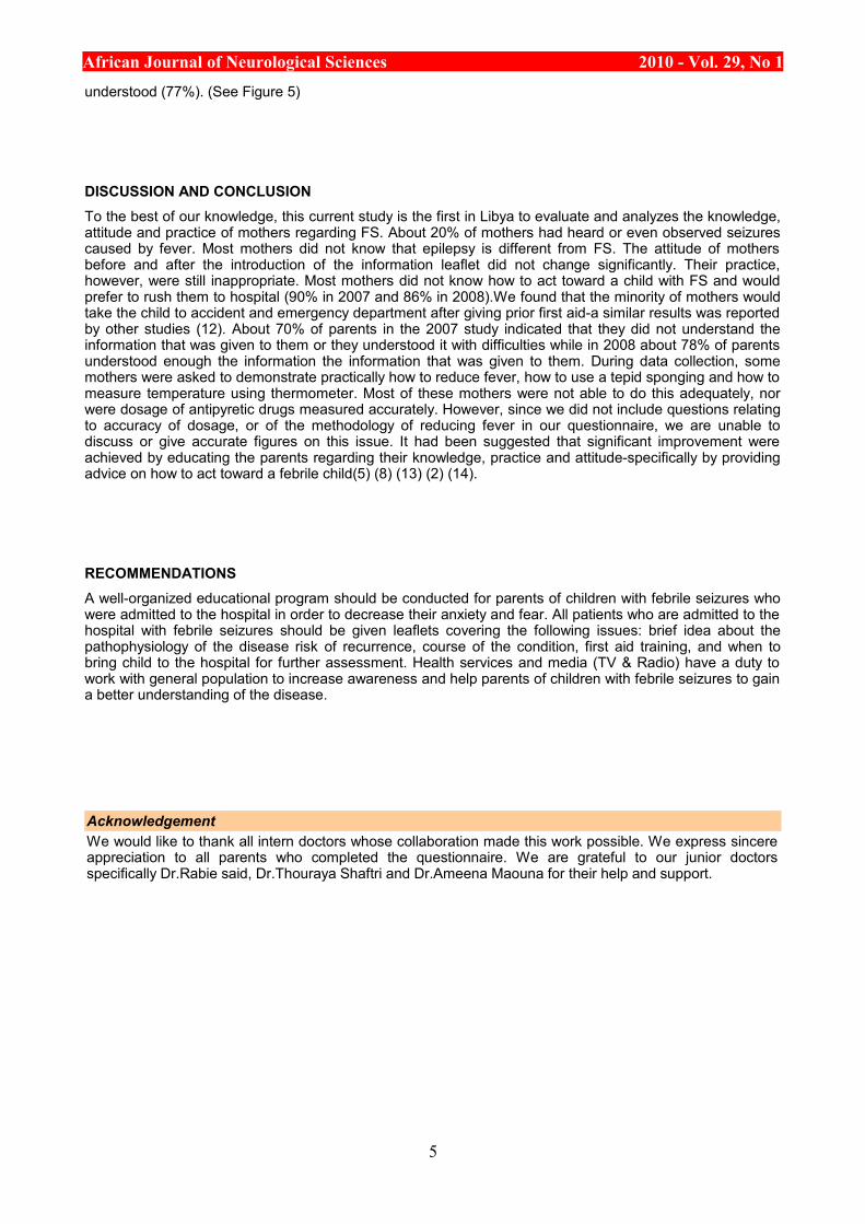
Parents who witnessed the febrile seizure had rushed the child to the hospital as the first aid of management with a small percentage tried to cool the child or did both. (See Figure 1)
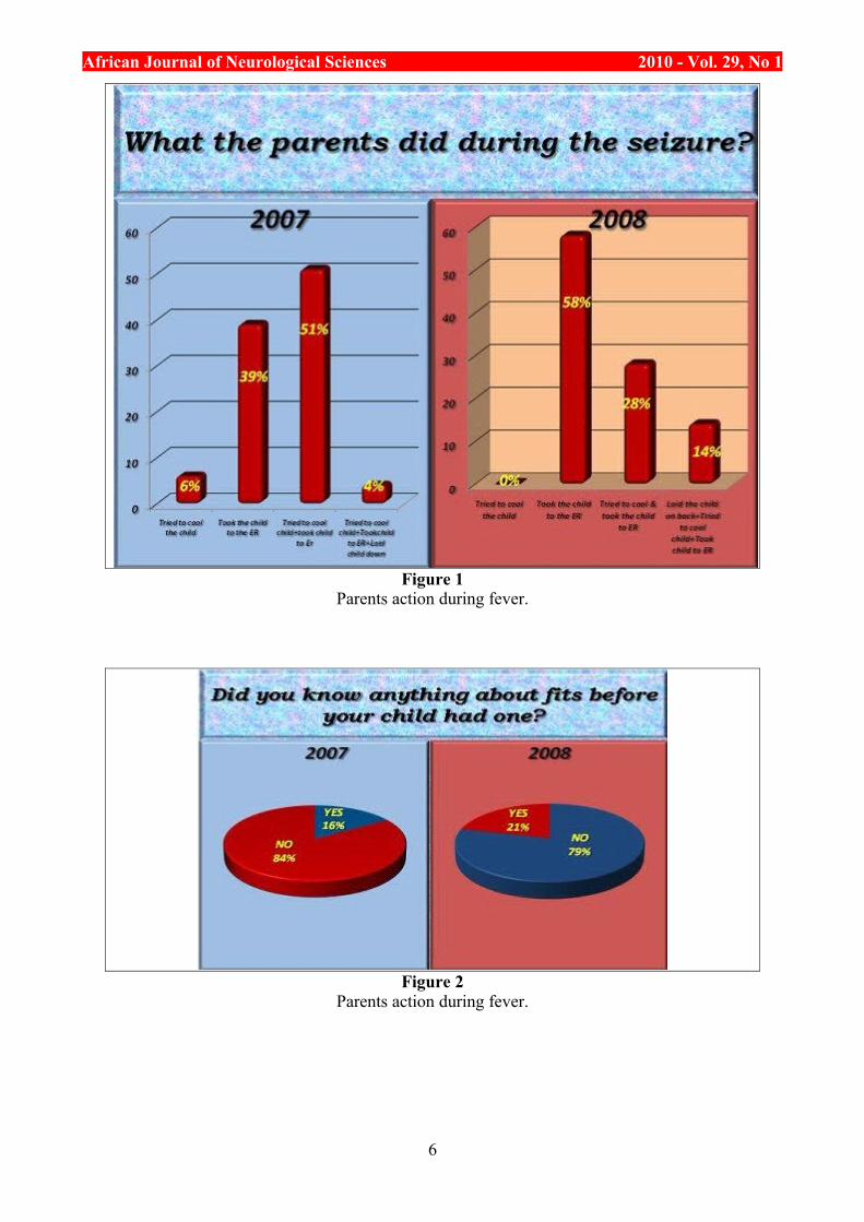
Just a minority of parent in this study knew some information about seizures before their child had one. (See Figure 2)
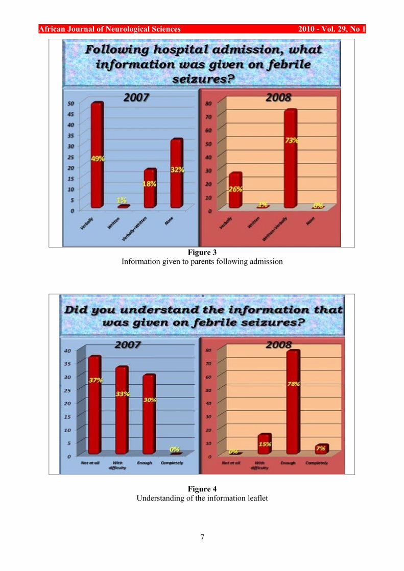
The 2007 study showed that the information given to the child’s parents was mainly verbally, while in 2008 (73%) of parents received written & verbal information together. (See Figure 3)
About 70% of parents in the 2007 study indicated that they did not understand the information that was given to them or they understood it with difficulties while in 2008 about 78% of parents understood enough the information the information that was given to them. (See Figure 4)
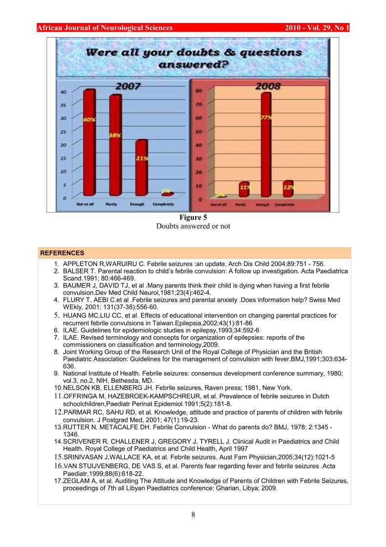
When reviewing the parents after the information given it was found that in the 2007 study most parent’s doubts were not at all cleared out (78%) while in the 2008 study just enough doubts were cleared &
4
African Journal of Neurological Sciences 2010 - Vol. 29, No 1understood (77%). (See Figure 5)
DISCUSSION AND CONCLUSIONTo the best of our knowledge, this current study is the first in Libya to evaluate and analyzes the knowledge, attitude and practice of mothers regarding FS. About 20% of mothers had heard or even observed seizures caused by fever. Most mothers did not know that epilepsy is different from FS. The attitude of mothers before and after the introduction of the information leaflet did not change significantly. Their practice, however, were still inappropriate. Most mothers did not know how to act toward a child with FS and would prefer to rush them to hospital (90% in 2007 and 86% in 2008).We found that the minority of mothers would take the child to accident and emergency department after giving prior first aid-a similar results was reported by other studies (12). About 70% of parents in the 2007 study indicated that they did not understand the information that was given to them or they understood it with difficulties while in 2008 about 78% of parents understood enough the information the information that was given to them. During data collection, some mothers were asked to demonstrate practically how to reduce fever, how to use a tepid sponging and how to measure temperature using thermometer. Most of these mothers were not able to do this adequately, nor were dosage of antipyretic drugs measured accurately. However, since we did not include questions relating to accuracy of dosage, or of the methodology of reducing fever in our questionnaire, we are unable to discuss or give accurate figures on this issue. It had been suggested that significant improvement were achieved by educating the parents regarding their knowledge, practice and attitude-specifically by providing advice on how to act toward a febrile child(5) (8) (13) (2) (14).
RECOMMENDATIONSA well-organized educational program should be conducted for parents of children with febrile seizures who were admitted to the hospital in order to decrease their anxiety and fear. All patients who are admitted to the hospital with febrile seizures should be given leaflets covering the following issues: brief idea about the pathophysiology of the disease risk of recurrence, course of the condition, first aid training, and when to bring child to the hospital for further assessment. Health services and media (TV & Radio) have a duty to work with general population to increase awareness and help parents of children with febrile seizures to gain a better understanding of the disease.
AcknowledgementWe would like to thank all intern doctors whose collaboration made this work possible. We express sincere appreciation to all parents who completed the questionnaire. We are grateful to our junior doctors specifically Dr.Rabie said, Dr.Thouraya Shaftri and Dr.Ameena Maouna for their help and support.
5
African Journal of Neurological Sciences 2010 - Vol. 29, No 1
Figure 1Parents action during fever.
Figure 2Parents action during fever.
6
African Journal of Neurological Sciences 2010 - Vol. 29, No 1
Figure 3Information given to parents following admission
Figure 4Understanding of the information leaflet
7
African Journal of Neurological Sciences 2010 - Vol. 29, No 1
Figure 5Doubts answered or not
REFERENCES1. APPLETON R,WARUIRU C. Febrile seizures :an update, Arch Dis Child 2004;89:751 - 756.2. BALSER T. Parental reaction to child’s febrile convulsion: A follow up investigation. Acta Paediatrica
Scand.1991; 80:466-469.3. BAUMER J, DAVID TJ, et al .Many parents think their child is dying when having a first febrile
convulsion.Dev Med Child Neurol,1981;23(4):462-4.4. FLURY T, AEBI C.et al .Febrile seizures and parental anxiety .Does information help? Swiss Med
WEkly, 2001; 131(37-38):556-60.5. HUANG MC,LIU CC, et al. Effects of educational intervention on changing parental practices for
recurrent febrile convulsions in Taiwan.Epilepsia,2002;43(1):81-866. ILAE. Guidelines for epidemiologic studies in epilepsy,1993;34:592-67. ILAE. Revised terminology and concepts for organization of epilepsies: reports of the
commissioners on classification and terminology,2009.8. Joint Working Group of the Research Unit of the Royal College of Physician and the British
Paediatric Association: Guidelines for the management of convulsion with fever.BMJ,1991;303:634-636.
9. National Institute of Health. Febrile seizures: consensus development conference summary, 1980; vol.3, no.2, NIH, Bethesda, MD.
10.NELSON KB, ELLENBERG JH. Febrile seizures, Raven press; 1981, New York.11.OFFRINGA M, HAZEBROEK-KAMPSCHREUR, et al. Prevalence of febrile seizures in Dutch
schoolchildren,Paediatr Perinat Epidemiol.1991;5(2):181-8.12.PARMAR RC, SAHU RD, et al. Knowledge, attitude and practice of parents of children with febrile
convulsion. J Postgrad Med, 2001; 47(1):19-23.13.RUTTER N, METACALFE DH. Febrile Convulsion - What do parents do? BMJ, 1978; 2:1345 -
1346.14.SCRIVENER R, CHALLENER J, GREGORY J, TYRELL J. Clinical Audit in Paediatrics and Child
Health. Royal College of Paediatrics and Child Health, April 199715.SRINIVASAN J,WALLACE KA, et al. Febrile seizures. Aust Fam Physician,2005;34(12):1021-516.VAN STUIJVENBERG, DE VAS S, et al. Parents fear regarding fever and febrile seizures .Acta
Paediatr,1999;88(6):618-22.17.ZEGLAM A, et al. Auditing The Attitude and Knowledge of Parents of Children with Febrile Seizures,
proceedings of 7th all Libyan Paediatrics conference: Gharian, Libya; 2009.
8
African Journal of Neurological Sciences 2010 - Vol. 29, No 1
CLINICAL STUDIE / ETUDE CLINIQUECOMMUNITY REINTEGRATION AMONG STROKE SURVIVORS IN OSUN, SOUTHWESTERN NIGERIA.
REINTEGRATION COMMUNAUTAIRE POST-ACCIDENT VASCULAIRE CEREBRAL A OSUN, SOUTHWESTERN NIGERIA.
OBEMBE Adebimpe Olayinka 1
JOHNSON Olubusola Esther 1
FASUYI Theresa Funmilayo 1
1. Department of Medical Rehabilitation, Faculty of Basic Medical Sciences, College of Health Sciences, Obafemi Awolowo University, Ile-Ife, Osun State, Nigeria
E-Mail Contact - OBEMBE Adebimpe Olayinka : bimpy248 (at) yahoo (dot) com
Keywords: stroke, community, physiotherapy, rehabilitation
ABSTRACTBackground Stroke is a major neurological problem and a leading cause of disability in the elderly in Nigeria. The incidence is increasing due to increasing risk factors, but many stroke victims now survive because of improved medical care. These survivors become community-dwellers after inpatient rehabilitation.
Aims To assess community reintegration among stroke survivors and factors associated with it.
Methods Cross-sectional survey study of patients who had survived six months or more after a stroke. Participants consisted of stroke patients attending the outpatient physiotherapy clinics of four selected government owned hospitals in Osun state, south-west Nigeria. Community reintegration was assessed using the Reintegration to Normal Living Index (RNLI) and walking ability was assessed using the Functional Ambulatory Categories (FAC).
Results A total of 64 patients (43 men and 21 women, mean age 58.80¡À 10.31 years) participated in this study. The mean RNLI was 63.8¡À14.3 for all the participants. Forty eight participants (75%) had slight disability (Score=2) and 16 participants (25%) had moderate disability (Score =3) using Modified Rankin Scale (mRS). Age, sex, physiotherapy duration, number of stroke occurrence and walking ability, were not associated with community reintegration. Post-stroke duration however had a significant association with community reintegration.
ConclusionA significant proportion of chronic stroke survivors attending the selected outpatient clinics have mild to moderate level of reintegration and the longer the post stroke duration, the better the satisfaction with community reintegration.
INTRODUCTIONStroke is the leading cause of disability among adults and frequently results in impaired mobility (11). It is a major public health problem with long-term physical, emotional, and relational consequences (15). A major component of the management of stroke is aimed at facilitating functional independence and community reintegration (16). One of the most important elements of stroke rehabilitation, and likely the most underestimated area, is community reintegration. Reintegration to normal living has been defined as the reorganization of physical, psychologic and social characteristics so that the individual can resume well-adjusted living after incapacitating illness or trauma (25). Specifically, the term community integration is used
9
African Journal of Neurological Sciences 2010 - Vol. 29, No 1to refer to re-establishing, to the degree possible, previously-existing roles and relationships, creating substitute new ones, and assisting people in making these changes (6). Failure to mobilize adequate support in the community can potentially negate the best efforts and results of stroke rehabilitation. Issues revolving around community and role integration can potentially have a profound impact on quality of life of the stroke survivor (3). For many people, the reintegration into community life marks the end point of their rehabilitation (14).
Regardless of regions in the world, the concept associated with re-integration to normal pattern of social and community life is one of key ideas in rehabilitation. Numerous international rehabilitation scholars have identified the importance of social and community integration to the well-being of people with disabilities (4,13,23).
Stroke is an important cause of morbidity and mortality in Africans. It is responsible for 0.9 to 4% of total admissions to hospitals and 0.5 to 45% of neurological admissions. In Nigeria, the incidence and prevalence of stroke have not been established. However, a report (19) from a Stroke Registry in Ibadan gave the annual incidence of stroke in Nigerians as 26 per 100 000 populations. A study (18) reported that frequency of stroke in hospital populations has varied from 0.9% to 4.0%, whereas among neurological admissions, stroke accounted for 0.5% to 45 % (17). Another study (24) reported the current prevalence of stroke in Nigeria as 1-14 per 1000.
Community reintegration has been studied in stroke in some countries (6, 21). Apart from studies on functional outcome (8) and quality of life (2, 7, 20) there has been to the best of our knowledge, no study on community reintegration of stroke survivors in Nigeria. Therefore the aims of this study were to determine the level of reintegration in community-dwelling individuals with stroke and to examine its relationship with the following selected variables; age, sex, post stroke duration, number of stroke occurrence, duration of physiotherapy and walking ability.
METHODS
ParticipantsThe participants in the study were individuals who had a stroke 6 or more months previously recruited in the outpatient physiotherapy clinics of selected hospitals in the south-west of Nigeria. Participants for this study were community-dwelling stroke survivors recruited by purposive selection.
Four government-owned hospitals in Osun State, Nigeria were selected for this study. Osun state is located in the south western part of the country. The study was approved by the Ethics and Research Committee of the Obafemi Awolowo University Teaching Hospitals Complex, Ile-Ife. All potential participants were screened and had to fulfill the following criteria: first or second episode of unilateral stroke with hemiparesis, were ambulatory before stroke, were independent in ambulation with or without mobility /walking aid or were partially dependent on wheelchair for mobility, and were living at home. All potential participants received verbal and written information about the purpose and procedure of the study before giving informed written consent to participate in the study.
Exclusion criteria for this study include: history of other neurological pathology, conditions affecting balance, dementia or impaired vision. People with severe musculoskeletal conditions affecting the lower limbs were also excluded from the study.
Medical records were obtained to confirm the diagnosis of stroke from the information provided by the physician through the relevant medical and imaging results. Other information collected include; age, sex, duration of stroke, physiotherapy duration, side affected by the stroke, type of stroke and number of stroke occurrence.
Outcome measuresThe main outcome measure for this study was the Reintegration to Normal Living Index. This is an 11-item scale that covers areas such as participation in recreational and social activities, movement within the community, and how comfortable the individual is in his or her role in the family and with other relationships. It can be completed by either a patient or a significant other (25). The RNL Index is made up of 11 declarative statements (e.g. I move around my community as I feel necessary), including the following domains: indoor, community and distance mobility, self care, daily activity (work and school) recreational and social activities, general coping skills, family role(s), personal relationships, and presentation of self to others (4). A 4-point categorical scale (1-4: where 1=does not describe my situation, 2=describe my situation a little 3=describe my situation a lot, 4=fully describe my situation) was used (21) and yielded total values ranging from 11 to 44, with higher scores indicating better perception of reintegration.
The modified Rankin Scale (mRS) is widely used to assess global outcome after stroke. The scale describes 6 grades of disability after a stroke. Score of 5 denotes severe disability, bedridden; and score of 0 denotes no symptoms at all (24).
Functional Ambulation Categories (FAC) was used to assess the walking ability of the stroke survivors. The
10
African Journal of Neurological Sciences 2010 - Vol. 29, No 1
FAC instrument is designed to provide information on the level of physical support needed by subjects in order to ambulate safely. It has been found to be reliable and valid in classifying hemiplegic gait (10). This instrument distinguishes among 6 levels of mobility ranging from dependence to independence. For the purpose of this study, FAC scores of 3 to 5 were used to recruite participants for this study. This is because participants in these categories do not require physical assistance from a therapist or carer.
Data AnalysisData were analyzed using the Statistical Package for Social Sciences (SPSS) version 16 program. Participants’ clinical and socio demographic characteristics were analyzed descriptively in terms of mean, standard deviations, frequencies and percentages. The variables analyzed are: age, sex, post stroke duration, number of stroke occurrence, duration of physiotherapy, walking ability and side of stroke.
Pearson’s correlation coefficient (r) was used to determine the association between the reintegration to normal living index scores and the other variables. The measure of strength of association was defined by the value of the correlation coefficients obtained (.00-.25= little or no relationship, .25-.50 =fair relationship, .50-.75 =moderate relationship, > .75 =good to excellent relationship) (22).
RESULTS
Participant CharacteristicsThe ages of the participants ranged from 31 to 80 years (mean 58.80¡À 10.31). Forty three (67.2%) of the 64 participants were males while 21(32.8%) were females. Twenty nine had left sided hemiparesis. Nineteen used a mobility aid (wheel chair n=5, walking stick/ quad cane n=14). Thirty six (56.3%) of the participants had ischaemic stroke and the others had haemorrhagic stroke. Twelve participants have had 2 episodes of stroke and the others have had only one. Forty eight participants (75%) had slight disability (i.e., mRS score of 2) and 16 participants (25%) had moderate disability (i.e., mRS score of 3) using the Modified Rankin Scale (mRS). The mean RNLI score was 63.8¡À14.3.
There were no significant differences between the RNLI scores of men and women (62.2¡À13.2 and 65.4¡À12.1), participants with ischaemic stroke (66.2¡À11.6) and those with haemorrhagic stroke (64.4¡À14.2) and between participants with left side affected (63.6¡À11.4) and those with the right side affected (62.5¡À14.2) (Table 1).
Association between RNLI scores and other variablesThere was a significantly moderate association between reintegration level and post stroke duration (P<0.05). There was no statistically significant association between the RNLI scores and these variables; age, sex, number of stroke occurrence, physiotherapy duration and walking ability (Table 2).
Association among the other variablesThere were statistically significant associations between the number of stroke occurrence and age (P<0.01), post stroke duration and physiotherapy duration (P<0.01), post stroke duration and number of stroke occurrence (P<0.05), physiotherapy duration and number of stroke occurrence (P<0.05) (Table 3). Post stroke duration and physiotherapy duration displayed an excellent relationship, while relationships between the others were fair.
Reintegration to Normal Living Index domainThe frequency of the scores obtained in each domain of the reintegration to normal living index (RNLI) is shown in table 4.
Indoor mobilityTwenty two (34.4%) participants did not have any form of limitation with mobility indoors. Twenty (31.3%) could not move around their homes as they feel is necessary and 8 (12.5%) reported they were a moderately satisfied with how they were able to move around their homes.
Community mobilityTwenty (31.3%) participants reported severe loss of all forms of mobility in the community. Eleven (17.2%) of the participants were not satisfied with how they move around the community as they feel is necessary.
Distance MobilityTwelve (18.8%) participants were able to take trips out of town as they feel is necessary. Seven (10.9%) stroke survivors reported moderate loss of distance mobility and were not fully satisfied with mobility that involves taking a trip out of town.
Self-carePoor satisfaction with how self-care needs were met was reported in 9 (14.5%) stroke survivors. Twenty
11
African Journal of Neurological Sciences 2010 - Vol. 29, No 1(32.3%) reported moderate satisfaction, while 11 (17.7%) expressed full satisfaction.
Daily activity (work)Twenty five (39.7%) stroke survivors reported that they did not spend most of their days occupied in a work activity that is necessary or important to them. Seven (11.1%) reported full level of satisfaction with their daily activities.
Recreational activitiesFour (6.3%) participants experienced full satisfaction with their participation in recreational activities. Twenty nine (46%) reported some level of satisfaction and 12 (19%) reported the least level of satisfaction.
Social activitiesNine (14.3%) participants reported that they were satisfied with their participation in recreational activities. Sixteen (25.4%) were not satisfied with their participation in recreational activities at all.
Family roleHigh level of satisfaction was reported in 14 (22.2%) participants for this domain. The lowest level of satisfaction was reported in 12.7% (8) of the participants and 21 (33.3%) participants expressed moderate level of satisfaction with roles in the family that met their needs and those of other family members.
Personal relationshipsThis domain showed the least number of participants (6) with the lowest level of satisfaction with their personal relationships. Seventeen (27%) participants were moderately comfortable with their personal relationships, while 14 (22%) were fully satisfied.
Presentation of self to othersTwelve (19%) participants were fully satisfied with this domain of the RNL index. Twenty eight (44.4%) stroke survivors expressed moderate satisfaction with how comfortable they are with themselves in the company of others. Seven (11.1%) stroke survivors reported no satisfaction.
General coping skillsFourteen (22.6%) participants were not in any way satisfied with how they can deal with life’s events as they happen. Eight (12.9%) reported moderate satisfaction in this domain, while 14 (22.6%) reported the least level of satisfaction.
DISCUSSIONThe result of this study shows that none of the community-dwelling people with stroke that participated in this study was fully satisfied (RNLI score 100) with their reintegration to normal living. Most of the participant (75%) had mild to moderate deficits (RNLI score 60-99). The mean RNLI score of the participants is lower than that of the findings of previous studies (21,25) and it is contrary to the results of a study by Carter et al (5) who reported that more than half of their participants were fully satisfied with their reintegration into the community and the study by Hoffmann et al (9) who reported successful reintegration into the community for most of their participants.
The findings from this study also revealed that men had less satisfaction with their community reintegration than women. This is consistent with the findings of Pang et al (21) who reported that men had less satisfaction with community reintegration, though the difference was not statistically significant.
The findings of the present study showed that more participants (34.4%) expressed full satisfaction with indoor mobility than any other domain and this is closely followed by satisfaction with community mobility with 20 (31.24%) participants. These findings are in agreement with that of Hoffmann et al (9) who reported most participants were able to move around their home and community. But it is contrary to their findings that most participants were satisfied with their personal relationships. In our study, more participants (48.8%) reported the least level of satisfaction with the distance mobility domain than in any other domain and this is followed by the daily activity domain (39.7%). This is in agreement with the findings of Hoffmann et al (9), who reported that participants expressed the most difficulty with taking trips out of town. They also reported in their study that the ability to fill one’s day with necessary or important activities was also of some concern to most participants in their study. Nigeria is a developing country with various economic and developmental challenges. One of these is making homes and buildings in public places accessible to people with disabilities. This may be a strong factor in the low score obtained in the distance mobility domain. A dominant finding in the study by Hoffmann et al was that social support and peer mentoring were invaluable. In our environment, the culture encourages extended family networks and kinships, so isolation of people with disabilities is not common. People encourage their relatives with disabilities to live with them in their homes or make provision for other members of the family to live with them if the person with the disability does not want to or is unable to leave his/her home.
12
African Journal of Neurological Sciences 2010 - Vol. 29, No 1Similarly, the mean of reintegration to normal living of stroke survivors with the RNL index was 2.40+ 2.5 which falls between the level II and III of RNL index meaning (describes my situation a little and describes my situation a lot respectively). This implies that the stroke survivors that were assessed in this study were not fully reintegrated into their respective community. This may be due to the fact that most outpatient stroke rehabilitation programs in this environment do not incorporate community ambulation and reintegration. Doing this will go a long way in improving the health status of the community-dwelling stroke survivors and the ability to live a satisfied and independent life in the community. Also it is common in our environment that people, who have a form of disability, are usually denied of their participation in social activities.
The findings of this study show a strong association between community reintegration and post stroke duration. Though this study found no association between walking ability and community reintegration, a study has established that patients with a long duration of stroke are better at walking and have better tolerance of body pain (1). This study was done in an outpatient stroke unit, and thus was done on patients in the post-acute stage. It thus indicates that walking is better with longer post stroke duration. The reasons for the finding in this current study may be similar to this, that those that have longer duration of stroke are better at walking and therefore have a higher level of satisfaction with community reintegration. A longer post-stroke duration allows more time for patients to cope with their disabilities (12).
The result of this study also shows a significant association between age and number of stroke occurrence. This is in line with a study in our country that shows that the frequency of stroke increases with advancing age (24).
In our environment, stroke rehabilitation is presently largely focused on the motor function of the stroke survivor. Programs incooperating reintegration into the community should be included in the rehabilitative intervention. Limitations of the findings in this study include a lack of generalisability to people who require physical assistant to walk, as all participants in this study can ambulate independently, with or without a cane or are dependent on wheelchair for mobility. Thus, patients with severe strokes are not likely to have been included. Also because of the pre-selected nature of the study, only those stroke survivors who attend the physiotherapy outpatient clinics of the selected hospitals were included in the study.
CONCLUSIONSGenerally, despite the ability to move about independently, all the community-dwelling stroke survivors were not fully satisfied with their level of reintegration into the community and the longer the post stroke duration, the better the satisfaction with community reintegration.
ACKNOWLEDGEMENTSThe authors are grateful to all the stroke survivors who took part in the study and the physiotherapists in the selected hospitals for their support.
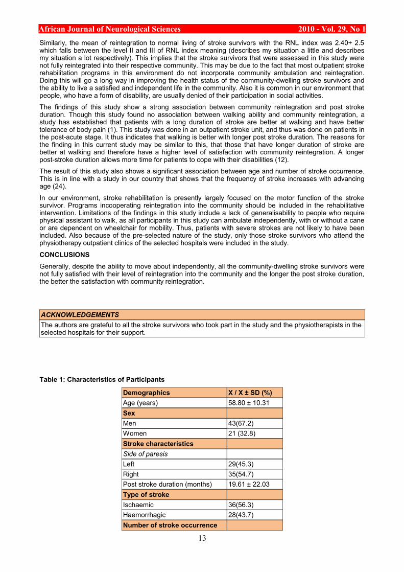
Table 1: Characteristics of Participants
Demographics X / X ± SD (%)Age (years) 58.80 ± 10.31SexMen 43(67.2)Women 21 (32.8)Stroke characteristicsSide of paresisLeft 29(45.3)Right 35(54.7)Post stroke duration (months) 19.61 ± 22.03Type of strokeIschaemic 36(56.3)Haemorrhagic 28(43.7)Number of stroke occurrence
13
African Journal of Neurological Sciences 2010 - Vol. 29, No 1
Once 52(81.2)Twice 12(18.8)MobilityWalking stick 14(21.9)Wheel chair 5(7.8)MeasuresWalking ability 3.06 ± 1.53RNLI ScoresTotal score 63.8 ± 14.3Male 62.2 ± 13.2Female 65.4 ± 12.1Ischaemic stroke 66.2 ± 11.6Haemorrhagic stroke 64.4 ± 14.2Left side affectation 63.6 ± 11.4Right side affectation 62.5 ± 14.2
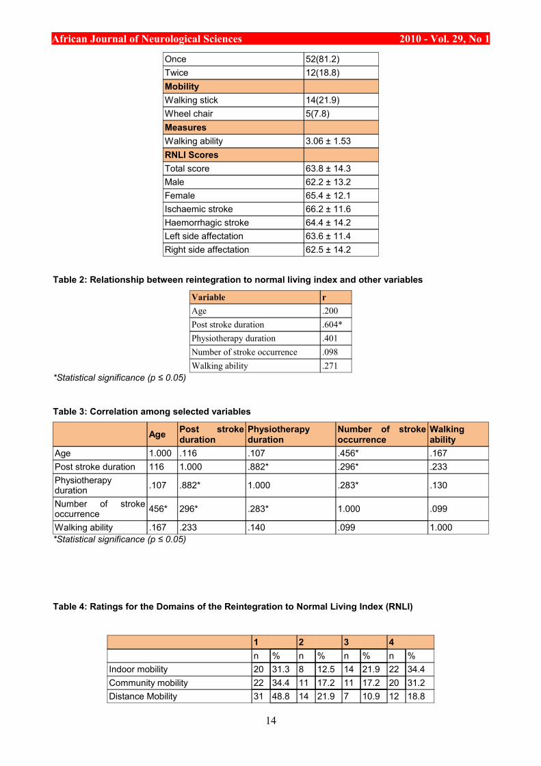
Table 2: Relationship between reintegration to normal living index and other variables
Variable rAge .200Post stroke duration .604*Physiotherapy duration .401Number of stroke occurrence .098Walking ability .271
*Statistical significance (p ≤ 0.05)
Table 3: Correlation among selected variables
Age Post stroke duration
Physiotherapy duration
Number of stroke occurrence
Walking ability
Age 1.000 .116 .107 .456* .167Post stroke duration 116 1.000 .882* .296* .233Physiotherapy duration .107 .882* 1.000 .283* .130
Number of stroke occurrence 456* 296* .283* 1.000 .099
Walking ability .167 .233 .140 .099 1.000*Statistical significance (p ≤ 0.05)
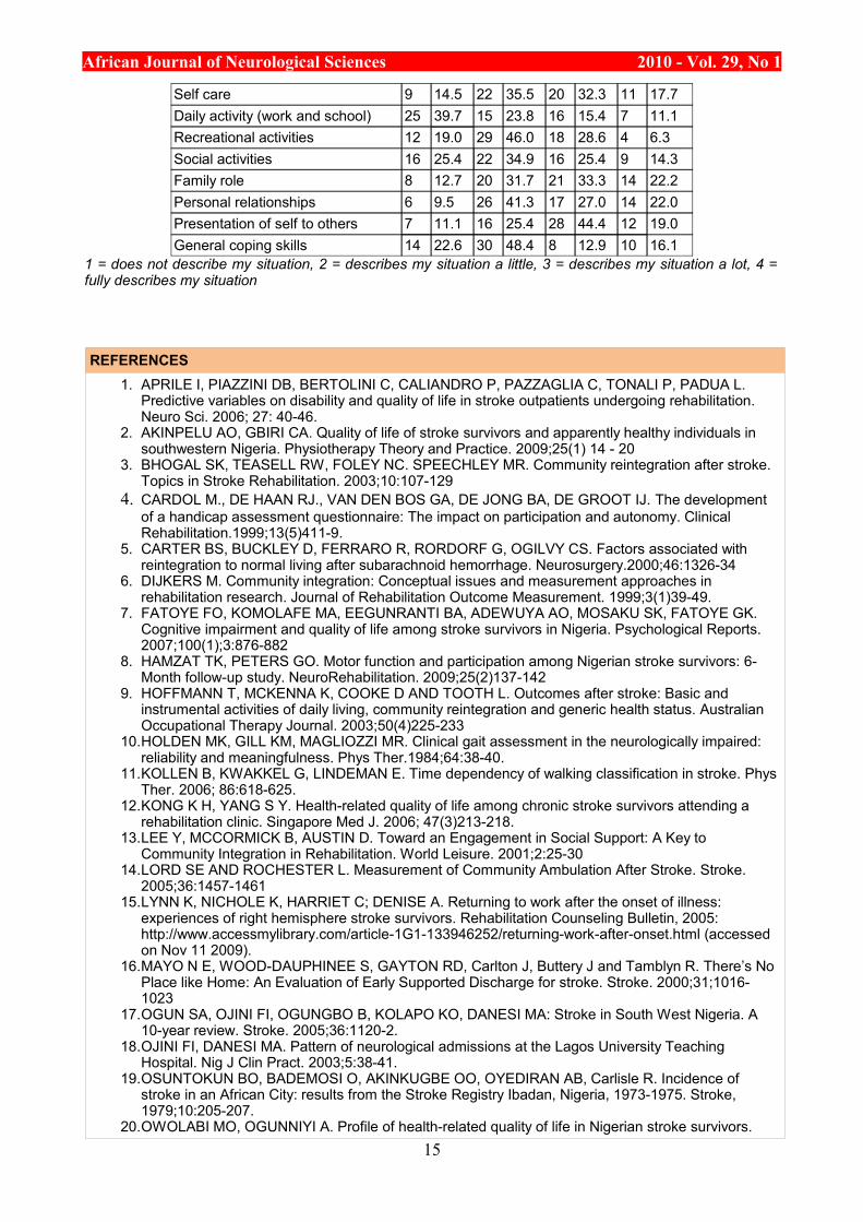
Table 4: Ratings for the Domains of the Reintegration to Normal Living Index (RNLI)
1 2 3 4n % n % n % n %
Indoor mobility 20 31.3 8 12.5 14 21.9 22 34.4Community mobility 22 34.4 11 17.2 11 17.2 20 31.2Distance Mobility 31 48.8 14 21.9 7 10.9 12 18.8
14
African Journal of Neurological Sciences 2010 - Vol. 29, No 1
Self care 9 14.5 22 35.5 20 32.3 11 17.7Daily activity (work and school) 25 39.7 15 23.8 16 15.4 7 11.1Recreational activities 12 19.0 29 46.0 18 28.6 4 6.3Social activities 16 25.4 22 34.9 16 25.4 9 14.3Family role 8 12.7 20 31.7 21 33.3 14 22.2Personal relationships 6 9.5 26 41.3 17 27.0 14 22.0Presentation of self to others 7 11.1 16 25.4 28 44.4 12 19.0General coping skills 14 22.6 30 48.4 8 12.9 10 16.1
1 = does not describe my situation, 2 = describes my situation a little, 3 = describes my situation a lot, 4 = fully describes my situation
REFERENCES1. APRILE I, PIAZZINI DB, BERTOLINI C, CALIANDRO P, PAZZAGLIA C, TONALI P, PADUA L.
Predictive variables on disability and quality of life in stroke outpatients undergoing rehabilitation. Neuro Sci. 2006; 27: 40-46.
2. AKINPELU AO, GBIRI CA. Quality of life of stroke survivors and apparently healthy individuals in southwestern Nigeria. Physiotherapy Theory and Practice. 2009;25(1) 14 - 20
3. BHOGAL SK, TEASELL RW, FOLEY NC. SPEECHLEY MR. Community reintegration after stroke. Topics in Stroke Rehabilitation. 2003;10:107-129
4. CARDOL M., DE HAAN RJ., VAN DEN BOS GA, DE JONG BA, DE GROOT IJ. The development of a handicap assessment questionnaire: The impact on participation and autonomy. Clinical Rehabilitation.1999;13(5)411-9.
5. CARTER BS, BUCKLEY D, FERRARO R, RORDORF G, OGILVY CS. Factors associated with reintegration to normal living after subarachnoid hemorrhage. Neurosurgery.2000;46:1326-34
6. DIJKERS M. Community integration: Conceptual issues and measurement approaches in rehabilitation research. Journal of Rehabilitation Outcome Measurement. 1999;3(1)39-49.
7. FATOYE FO, KOMOLAFE MA, EEGUNRANTI BA, ADEWUYA AO, MOSAKU SK, FATOYE GK. Cognitive impairment and quality of life among stroke survivors in Nigeria. Psychological Reports. 2007;100(1);3:876-882
8. HAMZAT TK, PETERS GO. Motor function and participation among Nigerian stroke survivors: 6-Month follow-up study. NeuroRehabilitation. 2009;25(2)137-142
9. HOFFMANN T, MCKENNA K, COOKE D AND TOOTH L. Outcomes after stroke: Basic and instrumental activities of daily living, community reintegration and generic health status. Australian Occupational Therapy Journal. 2003;50(4)225-233
10.HOLDEN MK, GILL KM, MAGLIOZZI MR. Clinical gait assessment in the neurologically impaired: reliability and meaningfulness. Phys Ther.1984;64:38-40.
11.KOLLEN B, KWAKKEL G, LINDEMAN E. Time dependency of walking classification in stroke. Phys Ther. 2006; 86:618-625.
12.KONG K H, YANG S Y. Health-related quality of life among chronic stroke survivors attending a rehabilitation clinic. Singapore Med J. 2006; 47(3)213-218.
13.LEE Y, MCCORMICK B, AUSTIN D. Toward an Engagement in Social Support: A Key to Community Integration in Rehabilitation. World Leisure. 2001;2:25-30
14.LORD SE AND ROCHESTER L. Measurement of Community Ambulation After Stroke. Stroke. 2005;36:1457-1461
15.LYNN K, NICHOLE K, HARRIET C; DENISE A. Returning to work after the onset of illness: experiences of right hemisphere stroke survivors. Rehabilitation Counseling Bulletin, 2005: http://www.accessmylibrary.com/article-1G1-133946252/returning-work-after-onset.html (accessed on Nov 11 2009).
16.MAYO N E, WOOD-DAUPHINEE S, GAYTON RD, Carlton J, Buttery J and Tamblyn R. There’s No Place like Home: An Evaluation of Early Supported Discharge for stroke. Stroke. 2000;31;1016-1023
17.OGUN SA, OJINI FI, OGUNGBO B, KOLAPO KO, DANESI MA: Stroke in South West Nigeria. A 10-year review. Stroke. 2005;36:1120-2.
18.OJINI FI, DANESI MA. Pattern of neurological admissions at the Lagos University Teaching Hospital. Nig J Clin Pract. 2003;5:38-41.
19.OSUNTOKUN BO, BADEMOSI O, AKINKUGBE OO, OYEDIRAN AB, Carlisle R. Incidence of stroke in an African City: results from the Stroke Registry Ibadan, Nigeria, 1973-1975. Stroke, 1979;10:205-207.
20.OWOLABI MO, OGUNNIYI A. Profile of health-related quality of life in Nigerian stroke survivors. 15

African Journal of Neurological Sciences 2010 - Vol. 29, No 1
European Journal of Neurology. 2008;16(1)54-6221.PANG MYC, ENG JJ, MILLER WC .Determinants of satisfaction with community Reintegration in
older Adults with Chronic Stroke: Role of Balance Self-Efficacy. Physical Therapy. 2007;87:282-291.22.PORTNEY LG, WATKINS MP. Foundations of Clinical Research: Applications to Clinical Practice.
3rd edition. Upper Saddle River, NJ: Prentice Hall, 2009; 523-538.23.TRIGG R., WOOD, VA., HEWER, R. L. Social reintegration after stroke: The first stages in the
development of the Subjective Index of Physical and Social Outcome (SIPSO). Clinical Rehabilitation.1999; 13: 341-353.
24.WAHAB KW. The burden of stroke in Nigeria. International Journal of Stroke. 2008;3:290-29225.WILSON JTL, HAREENDRAN A, HENDRY A, POTTER J, BONE I, MUIR KW. Reliability of the
modified Rankin Scale across multiple raters: benefits of a structured interview. Stroke. 2005; 36: 777-781
26.WOOD-DAUPHINEE SL, OPZOOMER A, WILLIAMS JI, MARCHAND B, SPITZER WO. Assessment of global function: the Reintegration to Normal Living Index. Arch Phys Med Rehabil. 1988;69:583-590.
16
African Journal of Neurological Sciences 2010 - Vol. 29, No 1
CLINICAL STUDIE / ETUDE CLINIQUECRYPTOCOCCAL MENINGITIS ASSOCIATED HIV INFECTION IN THE DONKA NATIONAL HOSPITAL
IN CONAKRY (GUINEA)
MENINGITE A CRYPTOCOQUES AU COURS DE L’INFECTION PAR LE VIH A L’HOPITAL NATIONAL DONKA DE CONAKRY (GUINEE)
CHERIF Mahamoud Sama 1
MAGASSOUBA N`faly 2
CAMARA Facely 3
BALDÉ Ousmane 1
DIALLO Alpha Amadou Sanck 4
DIAKITÉ Mandjou 2
CISSE Amara 5
SHUAIBU Mohammed Nasir 6
HELEGBE Gideon Kofi 6
1. Service de maladies infectieuses et tropicales de l’hôpital national Donka2. Laboratoire de parasitologie- mycologie de l’hôpital national Donka3. Service de Pédiatrie de l’hôpital national Donka4. Service de médecine interne de l’hôpital national Donka5. Service de neurologie de l’hôpital national Ignace Deen6. Institute of Tropical Medicine (NEKKEN); Nagasaki University, Japan
E-Mail Contact - CHERIF Mahamoud Sama :
Mots-clés: Cryptococcose méningée - Cryptococcus neoformans - Guinée - SIDA- VIHKeywords: AIDS- Cryptococcal Meningitis - Guinea - HIV
ABSTRACTBackgroundCryptococcal meningitis (CM) is an infection of the brain parenchyma and subarachnoid space by the encapsulated saprophyte yeast organisms such as Cryptococcus neoformans. Over the last twenty years, HIV has created a large and severely immune compromisized population in whom C. neoformans is a dangerous opportunistic infection. In Guinea, the prevalence of CM is unknown. We hypothesized that the occurrence of CM correlates with AIDS/ HIV prevalence.
MethodThis retrospective observational study was carried out at the national Hospital of Conakry (Guinea) between 2001 and 2002. We describe here the epidemiological and clinical and biological characteristics of CM disease in our national hospital.
ResultsOur data show that, 28.6 % of HIV patients with neurological symptoms had Cryptococcus neoformans in their CSF by using Indian ink staining. The median age was 36±3 years and sex ratio (M/F) was 1.8. The major complaints were fever and cephalgia, giddiness while the major complications were altered consciousness and hemiplegia. CSF was clear with low level of glucose and higher level of albumin. The means of lymphocytes in CSF was 8±2/mm3.
ConclusionThis data therefore becomes relevant in not only focusing of neurological symptoms associated with HIV to
17
African Journal of Neurological Sciences 2010 - Vol. 29, No 1
be toxoplasmosis but the possibility of C neoformans in these patients; particularly when they present symptoms such as headaches, giddiness and sniff neck etc. This can easily be carried out with Indian ink staining technique.
RESUMECryptococcose méningée (CM) est une infection du parenchyme cérébral et de l’espace sous arachnoïdien par Cryptococcus neoformans. Durant les vingt dernières années, la CM a connue une flambée grâce à l`avènement du VIH/SIDA, et devenant ainsi, une des infections opportunistes dangereuse chez les immunodéprimés. En Guinée, sa fréquence n’est pas encore connue et elle pourrait être en corrélation avec la séroprévalence du VIH.
MéthodeCette étude rétrospective a été effectuée à l’Hôpital National de Conakry, portant sur les patients admis entre 2001 et 2002. L’objectif était de décrire les caractéristiques épidémiologiques, cliniques et biologiques de la CM.
RésultatsL’examen direct du liquide céphalorachidien à l’encre de Chine avait montré que, 28.6 % de patients infectés par le VIH admis avec des symptômes neurologiques, avait le C neoformans. L’âge médian était 36±3 ans et le sex ratio (M/F) était 1.8. Les plaintes majeures étaient la fièvre et céphalées et vertiges tandis que les complications majeures étaient le trouble de la conscience (coma) et l’hémiplégie. La majorité des patients avaient le LCR clair avec une basse glycorhachie, une hyperalbuminorhachie et le nombre moyen de lymphocytes dans le LCR était 8±2/mm3.
ConclusionA la lumière de ces résultats, il apparait utile de rechercher en plus de la toxoplasmose, la présence du C neoformans chez tout patient infecté par le VIH et particulièrement présentant des symptômes neurologiques comme céphalées, vertiges, raideur de la nuque etc. Cette recherche peut être réalisée par la coloration du LCR à l’encre de chine.
INTRODUCTIONCryptococcal meningitis (CM) is a common opportunistic infection in acquired immune deficiency syndrome (AIDS ) patients, particularly in Southeast Asia and Africa[6,10,11,15]. It is AIDS defining illness in patients with late-stage Human Immunodeficiency Virus (HIV) infection, particularly in Southeast Asia and Southern and East Africa.[16,17,26]. Over the last 20 years, HIV infection pandemic has created a large and severely immune compromized population, with cryptococcal meningitis also occurring in patients with immunosupression. In parts of sub-Saharan Africa with the highest HIV prevalence, cryptococcal meningitis is now the leading cause of community-acquired meningitis, ahead of Streptococcus pneumoniae and Neisseria meningitidis[ 4,12,14,26]. Frequency of CM associated HIV infections varies from 2 to 35% according to the areas [9]. In the USA, about 7% [4,9]has been reported. In France, 88% of CM cases was associated with HIV infection [8,13]. While in Africa the range of frequency varies between 13 and 35% [5,8,11], 5.4% in Tunisia [18,19], 3,25% in Burkina Faso [21] and 5,4% in Ivory Coast [3]. In Uganda, the incidence of cryptococcal disease in patients with CD4 counts <200 cells/ml was estimated at 10.3 cases per 100 person years of follow-up [9]. In Thailand, cryptococcosis accounted for 19% of AIDS-defining illnesses between 1994 and 1998[19]. It seems most likely the high incidence of cryptococcal meningitis in parts of Africa and Asia reflects differences in exposure rather than host susceptibility or cryptococcal strain virulence, although no studies have addressed this issue [5,6,8,20]. Meningitis is the most frequent manifestation of cryptococcosis. Infection of the subarachnoid space is accompanied by involvement of the brain parenchyma, and therefore the term meningo-encephalitis may be more appropriate [11, 17,26].
Mortality from HIV associated cryptococcal meningitis remains high (10-30%), even in developed countries, because of the inadequacy of current antifungal drugs. In cohorts of HIV-infected patients from sub-Saharan Africa, cryptococcal meningitis has accounted for 13-44% of all deaths[26].
In Guinea, HIV prevalence increase from 1.7% in 1995, to 2.8% in 2002 [23] and the burden of CM is not yet clearly documented. We hypothesized that the frequency of CM associated HIV infection is correlated to HIV prevalence. In this study we describe the epidemiological characteristic, clinical presentation and CSF findings of CM in the national hospital of Conakry.
18
African Journal of Neurological Sciences 2010 - Vol. 29, No 1
MATERIALS AND METHODSThis retrospective observational study was carried out at the national Hospital of Conakry (Guinea) between 2001 and 2002. Seventy (70) HIV-infected patients were enrolled in the study. These patients underwent lumbar puncture as part of the routine work-up for a suspected neurological disease. The specimens of cerebrospinal fluid (CSF) from adult Human Immunodeficiency Virus-positive were taken for further mycological study.
Cryptococcal meningitis was diagnosed based on one of the following: (i) a positive India ink CSF result only, (ii) a specific cryptococcal CSF antigen test, (iii) or a positive CSF culture for Cryptococcus neoformans, has not realized.
Patients’ demographic data and clinical symptoms were collated from interviews of the patients. Physical findings, CSF examination (parameters such as glucose, albumin) outcome were analyzed. Informed consent was obtained from the patients enrolled in the study after ethical approval by the national ethics committee Statistical analysis: Statistical analysis was done using the statistical software ’EPI info version 0.6’ (CDC Atlanta, USA). Descriptive analysis consisted of mean with standard deviation and range for various parameters. Frequency of various clinical and laboratory findings are expressed as percentage.
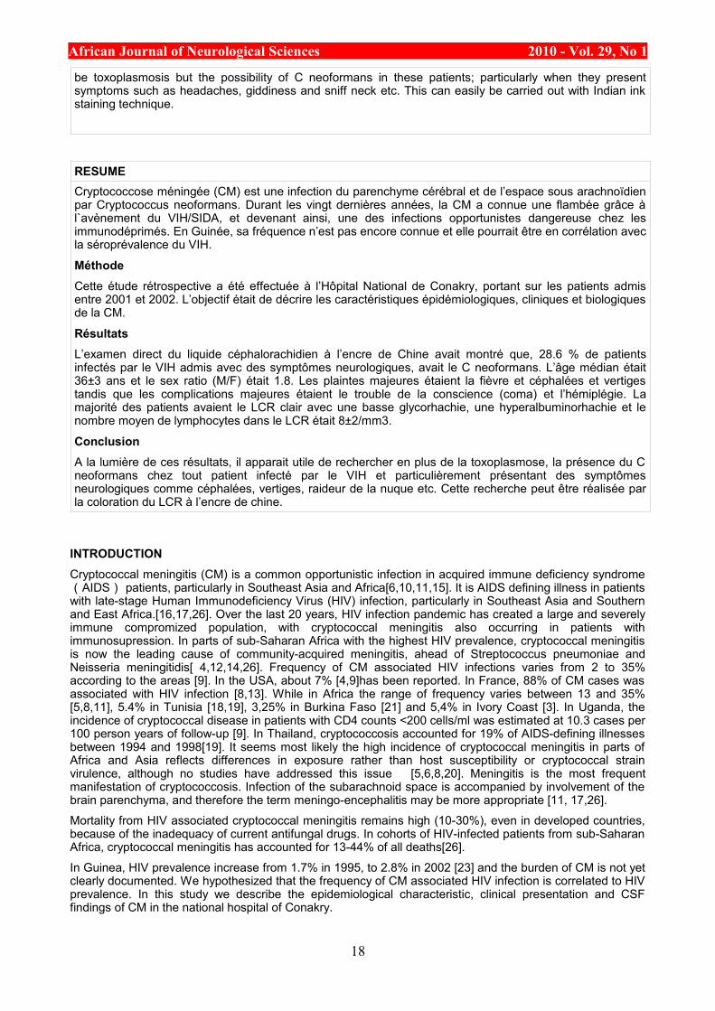
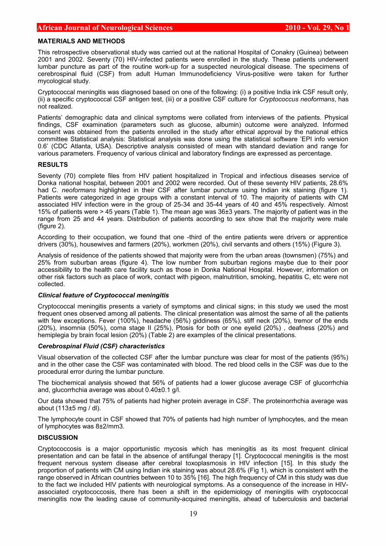
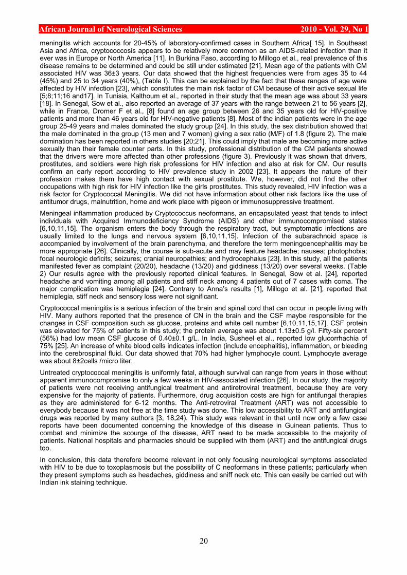
RESULTSSeventy (70) complete files from HIV patient hospitalized in Tropical and infectious diseases service of Donka national hospital, between 2001 and 2002 were recorded. Out of these seventy HIV patients, 28.6% had C. neoformans highlighted in their CSF after lumbar puncture using Indian ink staining (figure 1). Patients were categorized in age groups with a constant interval of 10. The majority of patients with CM associated HIV infection were in the group of 25-34 and 35-44 years of 40 and 45% respectively. Almost 15% of patients were > 45 years (Table 1). The mean age was 36±3 years. The majority of patient was in the range from 25 and 44 years. Distribution of patients according to sex show that the majority were male (figure 2).
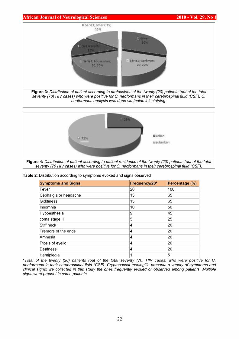
According to their occupation, we found that one -third of the entire patients were drivers or apprentice drivers (30%), housewives and farmers (20%), workmen (20%), civil servants and others (15%) (Figure 3).
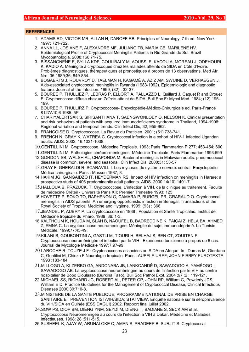
Analysis of residence of the patients showed that majority were from the urban areas (townsmen) (75%) and 25% from suburban areas (figure 4). The low number from suburban regions maybe due to their poor accessibility to the health care facility such as those in Donka National Hospital. However, information on other risk factors such as place of work, contact with pigeon, malnutrition, smoking, hepatitis C, etc were not collected.
Clinical feature of Cryptococcal meningitisCryptococcal meningitis presents a variety of symptoms and clinical signs; in this study we used the most frequent ones observed among all patients. The clinical presentation was almost the same of all the patients with few exceptions. Fever (100%), headache (56%) giddiness (65%), stiff neck (20%), tremor of the ends (20%), insomnia (50%), coma stage II (25%), Ptosis for both or one eyelid (20%) , deafness (20%) and hemiplegia by brain focal lesion (20%) (Table 2) are examples of the clinical presentations.
Cerebrospinal Fluid (CSF) characteristicsVisual observation of the collected CSF after the lumbar puncture was clear for most of the patients (95%) and in the other case the CSF was contaminated with blood. The red blood cells in the CSF was due to the procedural error during the lumbar puncture.
The biochemical analysis showed that 56% of patients had a lower glucose average CSF of glucorrhchia and, glucorrhchia average was about 0.40±0.1 g/l.
Our data showed that 75% of patients had higher protein average in CSF. The proteinorrhchia average was about (113±5 mg / dl).
The lymphocyte count in CSF showed that 70% of patients had high number of lymphocytes, and the mean of lymphocytes was 8±2/mm3.
DISCUSSIONCryptococcosis is a major opportunistic mycosis which has meningitis as its most frequent clinical presentation and can be fatal in the absence of antifungal therapy [1]. Cryptococcal meningitis is the most frequent nervous system disease after cerebral toxoplasmosis in HIV infection [15]. In this study the proportion of patients with CM using Indian ink staining was about 28.6% (Fig 1), which is consistent with the range observed in African countries between 10 to 35% [16]. The high frequency of CM in this study was due to the fact we included HIV patients with neurological symptoms. As a consequence of the increase in HIV-associated cryptococcosis, there has been a shift in the epidemiology of meningitis with cryptococcal meningitis now the leading cause of community-acquired meningitis, ahead of tuberculosis and bacterial
19
African Journal of Neurological Sciences 2010 - Vol. 29, No 1meningitis which accounts for 20-45% of laboratory-confirmed cases in Southern Africa[ 15]. In Southeast Asia and Africa, cryptococcosis appears to be relatively more common as an AIDS-related infection than it ever was in Europe or North America [11]. In Burkina Faso, according to Millogo et al., real prevalence of this disease remains to be determined and could be still under estimated [21]. Mean age of the patients with CM associated HIV was 36±3 years. Our data showed that the highest frequencies were from ages 35 to 44 (45%) and 25 to 34 years (40%), (Table I). This can be explained by the fact that these ranges of age were affected by HIV infection [23], which constitutes the main risk factor of CM because of their active sexual life [5;8;11;16 and17]. In Tunisia, Kalthoum et al., reported in their study that the mean age was about 33 years [18]. In Senegal, Sow et al., also reported an average of 37 years with the range between 21 to 56 years [2], while in France, Dromer F et al., [8] found an age group between 26 and 35 years old for HIV-positive patients and more than 46 years old for HIV-negative patients [8]. Most of the indian patients were in the age group 25-49 years and males dominated the study group [24]. In this study, the sex distribution showed that the male dominated in the group (13 men and 7 women) giving a sex ratio (M/F) of 1.8 (figure 2). The male domination has been reported in others studies [20;21]. This could imply that male are becoming more active sexually than their female counter parts. In this study, professional distribution of the CM patients showed that the drivers were more affected than other professions (figure 3). Previously it was shown that drivers, prostitutes, and soldiers were high risk professions for HIV infection and also at risk for CM. Our results confirm an early report according to HIV prevalence study in 2002 [23]. It appears the nature of their profession makes them have high contact with sexual prostitute. We, however, did not find the other occupations with high risk for HIV infection like the girls prostitutes. This study revealed, HIV infection was a risk factor for Cryptococcal Meningitis. We did not have information about other risk factors like the use of antitumor drugs, malnutrition, home and work place with pigeon or immunosuppressive treatment.
Meningeal inflammation produced by Cryptococcus neoformans, an encapsulated yeast that tends to infect individuals with Acquired Immunodeficiency Syndrome (AIDS) and other immunocompromised states [6,10,11,15]. The organism enters the body through the respiratory tract, but symptomatic infections are usually limited to the lungs and nervous system [6,10,11,15]. Infection of the subarachnoid space is accompanied by involvement of the brain parenchyma, and therefore the term meningoencephalitis may be more appropriate [26]. Clinically, the course is sub-acute and may feature headache; nausea; photophobia; focal neurologic deficits; seizures; cranial neuropathies; and hydrocephalus [23]. In this study, all the patients manifested fever as complaint (20/20), headache (13/20) and giddiness (13/20) over several weeks. (Table 2) Our results agree with the previously reported clinical features. In Senegal, Sow et al. [24], reported headache and vomiting among all patients and stiff neck among 4 patients out of 7 cases with coma. The major complication was hemiplegia [24]. Contrary to Anna’s results [1], Millogo et al. [21], reported that hemiplegia, stiff neck and sensory loss were not significant.
Cryptococcal meningitis is a serious infection of the brain and spinal cord that can occur in people living with HIV. Many authors reported that the presence of CN in the brain and the CSF maybe responsible for the changes in CSF composition such as glucose, proteins and white cell number [6,10,11,15,17]. CSF protein was elevated for 75% of patients in this study; the protein average was about 1.13±0.5 g/l. Fifty-six percent (56%) had low mean CSF glucose of 0.40±0.1 g/L. In India, Susheel et al., reported low glucorrhachia of 75% [25]. An increase of white blood cells indicates infection (include encephalitis), inflammation, or bleeding into the cerebrospinal fluid. Our data showed that 70% had higher lymphocyte count. Lymphocyte average was about 8±2cells /micro liter.
Untreated cryptococcal meningitis is uniformly fatal, although survival can range from years in those without apparent immunocompromise to only a few weeks in HIV-associated infection [26]. In our study, the majority of patients were not receiving antifungical treatment and antiretroviral treatment, because they are very expensive for the majority of patients. Furthermore, drug acquisition costs are high for antifungal therapies as they are administered for 6-12 months. The Anti-retroviral Treatment (ART) was not accessible to everybody because it was not free at the time study was done. This low accessibility to ART and antifungical drugs was reported by many authors [3, 18,24). This study was relevant in that until now only a few case reports have been documented concerning the knowledge of this disease in Guinean patients. Thus to combat and minimize the scourge of the disease, ART need to be made accessible to the majority of patients. National hospitals and pharmacies should be supplied with them (ART) and the antifungical drugs too.
In conclusion, this data therefore become relevant in not only focusing neurological symptoms associated with HIV to be due to toxoplasmosis but the possibility of C neoformans in these patients; particularly when they present symptoms such as headaches, giddiness and sniff neck etc. This can easily be carried out with Indian ink staining technique.
20
African Journal of Neurological Sciences 2010 - Vol. 29, No 1
Acknowledgements:We also appreciated the assistance of Doctors CAMARA Lansana Mady; CAMARA Sine, Benourou DIABATE, Alpha KONE, Moumie BARRY and CONDE Namoudou; Prof CISSE Mohamed, and Mr. Boamah Daniel, for criticisms and suggestions to improve the contents of this work.
Figure 1: Frequency of CM associated HIV positive. This data represents 28.6% (20) of the seventy (70) HIV positive patients with neurological symptoms and positivity for C neoformans analysis in their
cerebrospinal fluid (CSF). C neoformans analysis was done via Indian ink staining.
Table 1: Distribution of patient according to age.
Age group(years) Frequency %25-34 8 4035-44 9 4545-54 2 1055 -64 1 5Total 20 100
This data represents 28.6% (20) of the seventy (70) HIV positive patients with neurological symptoms and positivity for C neoformans analysis in their cerebro spinal fluid (CSF). C neoformans analysis was done via Indian ink staining.
Figure 2: Distribution of patient according to sex. Within the 28.6%(20) of the seventy (70) HIV positive patients that were positive for C. neoformans analysis in their cerebrospinal fluid (CSF).
21
African Journal of Neurological Sciences 2010 - Vol. 29, No 1
Figure 3: Distribution of patient according to professions of the twenty (20) patients (out of the total seventy (70) HIV cases) who were positive for C. neoformans in their cerebrospinal fluid (CSF); C.
neoformans analysis was done via Indian ink staining.
Figure 4: Distribution of patient according to patient residence of the twenty (20) patients (out of the total seventy (70 HIV cases) who were positive for C. neoformans in their cerebrospinal fluid (CSF).
Table 2: Distribution according to symptoms evoked and signs observed
Symptoms and Signs Frequency/20* Percentage (%)Fever 20 100Céphalgia or headache 13 65Giddiness 13 65Insomnia 10 50Hypoesthesia 9 45coma stage II 5 25Stiff neck 4 20Tremors of the ends 4 20Amnesia 4 20Ptosis of eyelid 4 20Deafness 4 20Hemiplegia 1 5
*Total of the twenty (20) patients (out of the total seventy (70) HIV cases) who were positive for C. neoformans in their cerebrospinal fluid (CSF). Cryptococcal meningitis presents a variety of symptoms and clinical signs; we collected in this study the ones frequently evoked or observed among patients. Multiple signs were present in some patients
22
African Journal of Neurological Sciences 2010 - Vol. 29, No 1
REFERENCES1. ADAMS RD, VICTOR MR, ALLAN H, DAROFF RB. Principles of Neurology, 7 th ed. New York
1997; 721-722.2. ANNA LL, JOSIANE F, ALEXANDRE MF, JULIANO TB, MARIA CB, MARILENE HV.
Epidemiological Profile of Cryptococcal Meningitis Patients in Rio Grande do Sul. Brazil Mycopathologia. 2008;166:71-75.
3. BISSANGNENE E, SYLLA KDF, COULIBALY M, AOUSSI E, KACOU A, MOREAU J, ODEHOURI K, KADIO A. Méningite à cryptocoques chez les malades atteints de SIDA en Côte d’Ivoire. Problèmes diagnostiques, thérapeutiques et pronostiques à propos de 13 observations. Med Afr Nre. 36.1989;36; 849-854.
4. BOGAERTS J, ROUVROY D, TAELMAN H, KAGAMÉ A, AZIZ AM, SWUINE D, VERHAEGEN J. Aids-associated cryptococcal meningitis in Rwanda (1983-1992). Epidemiologic and diagnostic feature. Journal of the Infection: 1999; (32) : 32-37.
5. BOUREE P, THULLIEZ P, LEBRAS P, ELLORT A, PALLAZZO L, Quillard J, Caquet R and Drouet E. Cryptococcose diffuse chez un Zaïrois atteint de SIDA, Bull Soc Fr Mycol Med. 1984; (12):195-199.
6. BOUREE P, THULLIRZ P. Cryptococcose- Encyclopédie-Médico-Chirurgicale ed. Paris-France 8127A10;6 1985. 5P
7. CHARIYALERTSAK S, SIRISANTHANA T, SAENGWONLOEY O, NELSON K. Clinical presentation and risk behaviors of patients with acquired immunodeficiency syndrome in Thailand, 1994-1998: Regional variation and temporal trends. Clin Infect Dis, 32, 955-962.
8. FRANCOISE D. Cryptococcose. La Revue du Praticien. 2001; (51):738-741.9. FRENCH N, GRAY K, WATREA C. Cryptococcal infection in a cohort of HIV-1 infected Ugandan
adults. AIDS. 2002; 16:1031-1038.10.GENTILLINI M. Cryptococcose. Médecine Tropicale. 1993; Paris Flammarion P 277; 453-454; 60011.GENTILLINI M. Pathologies cérébro-meningées. Médecine Tropicale. Paris Flammarion.1993:59912.GORDON SB, WALSH AL, CHAPONDA M. Bacterial meningitis in Malawian adults: pneumococcal
disease is common, severe, and seasonal. Clin Infect Dis. 2000;31: 53-5713.GRAY F, GHERALDI R, SCARAVILL I. Les mycoses du système nerveux central. Encyclopédie
Médico-chirurgicale. Paris : Masson 1987; 8.14.HAKIM JG, GANGAIDZO IT, HEYDERMAN RS. Impact of HIV infection on meningitis in Harare: a
prospective study of 406 predominantly adult patients. AIDS. 2000;14(10):1401-7.15.HALLOUA B, PRAZUCK. T. Cryptococcose. L’infection à VIH, de la clinique au traitement. Faculté
de médecine Créteil - Université Paris XII; Premier Trimestre 1993: 12516.HOVETTE P, SOKO TO, RAPHENON G, CAMARA P, BURGEL PR, GRARAUD O. Cryptococcal
meningitis in AIDS patients: An emerging opportunistic infection in Senegal. Transactions of the Royal Society of Tropical Medicine and Hygiene. 1999; (93) : 368.
17.JEANDEL P, AUBRY P. La cryptococcose en 1988 ; Population et Santé Tropicales. Institut de Médecine tropicale du Pharo. 1989 ;36: 1-3.
18.KALTHOUM K, HOUDA M, SLAH B, NAOUFEL B, BADREDDINE K, FAIÇAl Z, HELA BA, AHMED Z, EMNA C. La cryptococcose neuroméningée: Méningite du sujet immunodéprimé. La Tunisie Médicale. 1999;77:45-49.
19.KILANI B, GOUBONTINI A, GASTLI M, TIOURI H, BELHAJ S, BEN CT, ZOUITEN F. Cryptococcose neuroméningée et infection par le VIH : Expérience tunisienne à propos de 6 cas. Journal de Mycologie Médicale 1997;7:97-99.
20.LAROCHE R. TOUZE J F : Cryptococcoses associées au SIDA en Afrique. In : Dumas M, Giordano C, Gentilini M, Chieze F Neurologie tropicale. Paris : AUPELF-UREF; JOHN EIBBEY EUROTEXTE. 1993 ;183-184
21.MILLOGO A, KI-ZERBO GA, ANDONABA JB, LANKOANDÉ D, SAWADOGO A, YAMÉOGO I, SAWADOGO AB. La cryptococcose neuroméningée au cours de l’infection par le VIH au centre hospitalier de Bobo Dioulasso (Burkina Faso). Bull Soc Pathol Exot, 2004 ;97 :2 : 119-121.
22.MICHAEL SS, RICHARD JG, ROBERT AL, PETER GP, JOHN RP, William G, Powderly JDS, William E D. Practice Guidelines for the Management of Cryptococcal Disease, Clinical Infectious Diseases 2000;30:710-8.
23.MINISTERE DE LA SANTE PUBLIQUE; PROGRAMME NATIONAL DE PRISE EN CHARGE SANITAIRE ET PREVENTION IST/VIH/SIDA; STATVIEW. Enquête nationale sur la séroprévalence du VIH/SIDA en Guinée (ESSIDAGUI) 2002. Rapport final juillet 2002.
24.SOW PS, DIOP BM, DIENG YNM, SEYDI M, DIENG T, BADIANE S, SECK AM et al. Cryptococcose Neuroméningée au cours de l’infection à VIH à Dakar. Médecine et Maladies Infectieuses. 1998; 28 :511-515.
25.SUSHEEL K, AJAY W, ARUNALOKE C, AMAN S, PRADEEP B, SURJIT S. Cryptococcal
23

African Journal of Neurological Sciences 2010 - Vol. 29, No 1
Meningitis in HIV infected, Experience from North Indian tertiary center. Neurology India. 2008;56:444-449.
26.TIHANA B, THOMAS S. Cryptococcal meningitis. British Medical Bulletin. 2004; 72: 99-118.
24
African Journal of Neurological Sciences 2010 - Vol. 29, No 1
CLINICAL STUDIES / ETUDES CLINIQUESEPIDEMIOLOGIE DES TRAUMATISMES CRANIO-ENCEPHALIQUES A PARAKOU (BENIN)
EPIDEMIOLOGY OF TRAUMATIC BRAIN INJURIES IN PARAKOU (BENIN).
FATIGBA Olatoundji Holden 1
PADONOU Jidjoho 2
1. Unité de Neurochirurgie, Centre Hospitalier Départemental et Universitaire de Parakou, Bénin2. Clinique Universitaire de Traumatologie, d’Orthopédie et de Chirurgie Réparatrice du CNHU de
Cotonou, BP 386 Cotonou, Bénin
E-Mail Contact - FATIGBA Olatoundji Holden : ftg_holden (at) yahoo (dot) fr
Mots-clés: Traumatisme crânio-encéphalique, Accident de la circulation, Epidémiologie.Keywords: Traumatic brain injury, Road crash, Epidemiology
RESUMEIntroduction Les traumatismes crânio-encéphaliques (TCE) constituent une cause importante de mortalité. C’est problème majeur de santé publique.
Objectif La présente étude avait pour objectif d’établir l’épidémiologie des (TCE) au CHD-Borgou du Bénin. Méthode : Il s’agissait d’une étude prospective menée du 1er janvier 2008 au 31 Décembre 2009 au CHD-Borgou. Tous les TCE hospitalisés ont été suivis de l’admission à la sortie.
Résultat 755 (31.9%) TCE ont été hospitalisés. Ces patients se répartissaient en 642 (85%) hommes et 113 (15%) femmes. L’âge moyen était 28,6 ans ± 15,4. Les accidents de la circulation représentaient 87,7% des mécanismes. Une circonstance aggravante a été retrouvée dans 134 cas (20,3%) avec une prédominance de l’éthylisme 61,2%. Les accidents de la voie publique impliquaient 43(6,5%) automobilistes, 492 (74,3%) motocyclistes, 8 (1,2%) cyclistes et 119 (18%) piétons. 482 (63,8%) patients présentaient un TCE léger, 102 (13,5%) un TCE modéré et 171 (22,7%) un TCE grave. Le TCE était pur chez 135 (20,3%) patients. 414 patients ont bénéficié d’une radiographie du crâne et 85 d’un scanner cérébral. 47 (11,4%) patients présentaient une fracture de la voûte, 38 (9,2%) une embarrure et 7 (1,7%) une pneumocéphalie. La contusion cérébrale (30,6%) était la lésion scanographique prédominante suivie des collections péri cérébrales (28,3%). La durée moyenne d’hospitalisation était de 7,25 jours. Une guérison sans séquelle a été observée chez 674 (89,2%) patients. La mortalité était de 8,5 %.
Conclusion Les traumatismes crânio-encéphaliques sont une préoccupation majeure au CHD-Borgou ; la prévention et la stratégie de prise en charge doivent être améliorées.
SUMMARYIntroduction Traumatic brain injury (TBI) is the leading cause of death for traumatic injury. It is an major public health problem.
Objective This study aimed to investigate the epidemiology of (TBI) in CHD-Borgou (Benin).
Method It was prospective study. A total of TBI from admission to output was collected during the period January 1, 2008 to December 31, 2009.
Result Of the 755 (31.9%) total TBI admitted, males were 642 (85%) and females 113 (15%). 87,7% cases resulted from road traffic accident. The mean age was 28.6 years ± 15.4. An risk factor was found in 134(20.3%) cases and Alcohol use was 61,2%. Road crash concerned 492 (74,3%) motorcyclists, 8 (1.2%) cyclist, 119 (18%) pedestrians and 43 (6.5%) motor vehicle occupants. The distribution of head injury
25

African Journal of Neurological Sciences 2010 - Vol. 29, No 1
severity, on the basis of Glasgow Coma Scale scores, was mild in 482 (63,8%), moderate in 102 (13.5%), and severe in 171 (22.7%) for all cases. TBI was lone in 135 (20.3%) cases. The skull X-rays performed for 414 patients and cerebral CT-Scan for 85. Skull x-ray showed 47 (11.4%) fissure fracture, 38 (9.2%) depressive fracture and 7 (1.7%) pneumocephalus. The most observed brain lesions were the cerebral contusion (30,6%) and hematoma (28.3%).The mean duration of hospitalization was 7.25 days. 674 (89.2%) patients had good recovery. Mortality was 8.5%.
Conclusion __ The TBI are a major problem in CHD-Borgou; Prevention of road traffic injuries and improved emergency care and health facility-based treatment is needed.
INTRODUCTIONLes traumatismes crânio-encéphaliques constituent un motif fréquent de consultation au CHD-Borgou du Bénin. Les lésions observées sont variables, la gravité dépendant du mécanisme et des circonstances de survenue. La mise en place depuis 2008 d’une unité de neurochirurgie dans ce centre hospitalier vise à améliorer la prise en charge globale de cette affection. Pour parvenir à une telle performance, il était nécessaire dans une première démarche d’établir une cartographie quantitative et qualitative des traumatismes crânio-encéphaliques à Parakou. La présente étude avait pour objectif de recenser les TCE admis au CHD-Borgou de Parakou, d’établir le profil socioprofessionnel et les attitudes à risques des personnes impliquées puis d’étudier les aspects diagnostics et évolutifs.
METHODEIl s’agissait d’une étude prospective menée sur une période de deux ans entre le 1er janvier 2008 et le 31 Décembre 2009 au CHD-Borgou. Ce Centre hospitalier situé dans le département de Borgou-Alibori est l’hôpital de référence de la région septentrionale du bénin. Tous les cas de TCE admis aux urgences, hospitalisés au centre hospitalier ont fait l’object d’une enquête grâce à une fiche de collecte de données. Ils ont été suivis depuis leur admission jusqu’à la sortie quelque soit la durée du séjour hospitalier. Les patients présentant des plaintes légères et sans notion de perte de connaissance initiale ont été traités en ambulatoire. L’état de conscience était évalué en fonction de l’échelle des comas de Glasgow. Le diagnostic de TCE reposait sur l’anamnèse, les signes locaux touchant l’extrémité céphalique, l’examen neurologique et dans certains cas les données de l’imagerie.
Les données collectées concernaient le sexe, l’âge, le mécanisme et les circonstances de survenue du traumatisme, la profession, le moyen de locomotion impliqué pour les traumatisés par accident de la circulation, les signes d’examen clinique, les associations lésionnelles, les données d’examens complémentaires d’imagerie et l’évolution. Les patients décédés dès l’admission et avant toute prise en charge thérapeutique n’ont pas été retenus. L’exploitation, la saisie et l’analyse des données ont été réalisées par les logiciels Word 2007 et Excel 2007, Epi-Info 3.5 .Le test de corrélation utilisée était le Khi2 de Yates avec p inférieur à 5% considéré comme significatif.
RESULTATDurant la période d’étude, 1229 patients, victimes d’un traumatisme crânio-encéphalique ont été recensés. Ils représentaient 13.5% des 9095 consultants durant la même période. 474 (38.6%) cas de TCE ont été traité en ambulatoire et 755 (61.4%) hospitalisés ; ces derniers représentaient 31.9% des hospitalisations.
En fonction du sexe, les patients hospitalisés se répartissaient en 642 (85%) hommes et 113 (15%) femmes. Le sexe ratio H/F était 5,6. L’âge moyen des patients était 28,6 ans ± 15,4 avec des extrêmes de 1 et 97 ans. La tranche d’âge 20-40 ans était de 55,2 %. La répartition des patients en fonction de l’âge a été rapportée à la figure 1. La distribution des patients en fonction de leur occupation a été rapportée dans le tableau I. Parmi les mécanismes en cause, les accidents de la circulation concernaient 662 (87,7%) patients. La distribution des mécanismes des TCE a été rapportée dans le tableau II. Parmi les 662 victimes d’accident de la circulation, une circonstance aggravante a été retrouvée dans 134 cas (20,3%). L’éthylisme aigu était la circonstance la plus rapportée (82 cas ; 12,4%) et constituait 61,2% de toutes les circonstances aggravantes. La distribution des circonstances aggravantes a été rapportée au tableau III. Les accidents de la voie publique impliquaient 43(6,5%) automobilistes, 492 (74,3%) motocyclistes, 8 (1,2%) et 119 (18%) piétons. Le délai moyen d’admission était de 8 h avec des extrêmes de 10 min et 20 jours. Les moyens d’admission étaient une ambulance dans 173 cas (22,9%), les sapeurs-pompiers dans 349 cas (46,2%) et des moyens individuels dans 233 cas (30,9%). En fonction de l’échelle de coma de Glasgow (GCS), 482 (63,8%) patients présentaient un TCE léger (15≤ GCS ≤13), 102 (13,5%) un TCE modéré (12≤ GCS ≤9) et 171 (22,7%) un TCE grave (GCS ≤ 8). De point de vue hémodynamique, 647 (85,7%) patients étaient stables et 108 (14,3%) étaient instables. Un séjour en réanimation ou unité de soins intensifs était observé chez 301 (39,9%) patients et 454 (60,1%) patients ont été directement admis en hospitalisation. Les signes d’examen objectivés chez les patients ont été rapportés dans le tableau IV. Le TCE était pur chez 135 (20,3%) patients et associé à d’autres lésions chez 602 (79,7%). Il s’agissait de fracture des membres 123cas (16,3%), traumatisme facial 183 cas (24,2%), traumatisme thoracique 50 cas (6,6%), traumatisme abdominal 15 cas (2,0%), traumatisme du bassin 15 cas (2,0%) et un traumatisme rachidien 27 cas (3,6%). Les lésions des
26
African Journal of Neurological Sciences 2010 - Vol. 29, No 1parties molles étaient présentes chez 189 (25%) patients. Des examens d’imagerie ont été réalisés chez 499 patients ; Parmi les 414 patients ayant bénéficié d’une radiographie standard du crâne, 47 (11,4%) présentaient une fracture de la voûte, 38 (9,2%) une embarrure et 7 (1,7%) une pneumocéphalie. Une lésion osseuse y compris de la base du crâne a été observée chez 16 (18,8%) patients parmi les 85 ayant bénéficié d’un scanner cérébral. La répartition des lésions observées scanner cérébrale chez 85 patients ont été rapportée au tableau V. Les figures 2, 3, 4, 5 illustrent quelques lésions observées à l’imagerie. Une prise en charge neurochirurgicale a été réalisée chez 52 (6,9%) patients. Elle consistait en un redressement d’embarrure (32,7% ; n=17), une évacuation d’hématome sous dural (21,2% ; n=11) ou extra dural (11,5% ; n=6), une réparation de plaie crânio-encéphalique (19,2% ; n=10) et une réparation de brèche ostéoméningée (15,4% ; n=8).
La durée moyenne d’hospitalisation était de 7,25 jours ± 8,68 avec des extrêmes de 1 et 90 jours. Une guérison sans séquelle a été observée chez 674 (89,2%) patients. Soixante et quatre (8,5 %) patients sont décédés dans notre étude. La tranche d’âge des 21-30 ans représentaient 37,5%. 33 patients (51,6%) sont décédés le jour de l’admission, 15 (23,4%) dans les 72 heures et 16 (25,0%), 72 heures après l’admission (χ2 = 0,515 et p= 0,473 la différence est statistiquement non significative). Parmi les patients décédés, 48 (75,0%) patients avaient un TCE grave ; 10 (15,6%) un TCE modéré et 6 (9,4%) un TCE léger (χ 2 = 64,855 et p = 0,000000 ; la différence est statistiquement significative). Cette mortalité était de 51,2% parmi les patients ayant une anomalie pupillaire et de 11,6% parmi ceux qui n’en avaient pas (χ 2=37,89 et p=0,000000 ; la différence est statistiquement significative).
COMMENTAIRESL’âge moyen des patients dans notre étude était de 28,6 ans avec une nette prédominance masculine. La tranche d’âge 20-40 ans représentaient 55,2% des cas tout mécanisme confondu. Ces données confortent les multiples études existantes (2, 4,9) à propos des TCE où les sujets jeunes étaient les plus concernés et la prédominance masculine établie. La principale cause de ces TCE reste les accidents de la circulation (6, 7, 14). La fréquence des TCE dans notre étude (31,9% des hospitalisations) est sujette à caution et ne reflète certainement pas la réalité. Nos données étaient exclusivement hospitalières n’incluant que les patients du CHD-Borgou et excluaient les patients décédés dès admission ou directement admis à la morgue. L’absence d’un standard ou d’une uniformisation des méthodes de collecte des données concernant l’épidémiologie des TCE (11) participent également à ce biais.
Des circonstances aggravantes étaient observées dans 20,3% des cas parmi les traumatisés suite à un accident de la circulation ; l’éthylisme occupait le 1er rang 61,2%. Il n’existe pas de d’études à Parakou montrant l’impact de ces circonstances mais les mesures préventives et répressives prises dans les pays développés pour réduire l’incidence des TCE ont fait leur preuve (1, 3, 8). Elles doivent servir de modèle dans notre pays. Ces mesures pourraient également contribuer à réduire les associations lésionnelles observées dans notre étude.
Parmi les TCE par accident de la circulation, les motocyclistes étaient impliqués dans 74,3% des cas. Ce taux est semblable à ceux rapportés par Chiu (3) 64,5% à Taïwan et Suriyawongpaisal (13) 72 % en Thaïlande. Les engins à deux roues motorisés constituent le moyen de locomotion le plus utilisés à Parakou. Ces usagers au profil socioprofessionnel très varié ne sont pas toujours respectueux des règles de la circulation et sont sans casque de protection.
En fonction du score de Glasgow les TCE légers 63,8% prédominaient mais la fréquence des TCE grave 22,7% est préoccupante ; fréquence supérieure à celle rapportée par Sidibé (12) 16,70%, inférieure à celles observées par Bahloul (1) 29,1% et Coulibaly (5) 37,5%. Bien au-delà de la fréquence, les TCE graves posent un problème de management face à la légèreté du plateau technique du département d’Urgence et d’Anesthésie Réanimation du CHD-Borgou.
Bien que l’apport de la TDM dans la prise en charge des TCE soit clairement établi (5, 7, 12) notre étude souffre d’un faible taux de réalisation de cet examen chez nos patients. Ceci est le fait de la non disponibilité à temps plein du scanner dans notre ville. A cette difficulté technique s’ajoutait les coûts d’examen encore élevés pour la population locale majoritairement sans revenu mensuel garanti ou fixe. La contusion cérébrale était la lésion intracérébrale la plus observée 30,6% suivie des collections péridurales 28,3% devant l’œdème cérébral 14,1% et les hémorragies sous arachnoïdiennes 13%. Nos observations scanographiques sont ainsi opposées à celles rapportées de nombreux auteurs où l’hémorragie sous arachnoïdienne et l’œdème cérébral sont les lésions prédominantes (1, 5, 7, 12) mais superposables aux résultats rapportés par Emejulu (7). Nos constations peuvent être le fait d’une réalisation différée du scanner cérébral chez ces patients pour des motifs déjà évoqués. Une lésion osseuse était observée parmi 20,5% des patients ayant bénéficié d’une radiographie standard du crâne, Chiu (3) et Sidibé (12) ont respectivement rapporté des fréquences de 14,6% et 24,8%.
La mortalité hospitalière dans notre étude était de 8,5%, fréquence bien inférieure à celle rapportée par Emejulu (7) 19.1% Coulibaly et al (5) 30%, Bahloul (1) 38%. La tranche d’âge 21-30 ans, prédominante dans notre étude représentait 37,5% des décès mais nous n’avons pu établir une corrélation lésionnelle entre l’âge de décès et cette mortalité. La mortalité rapportée dans ce travail masque la gravité des lésions puisque
27
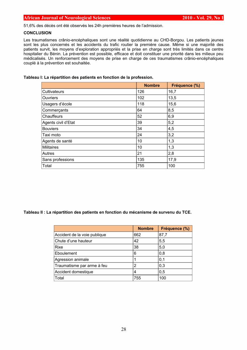
African Journal of Neurological Sciences 2010 - Vol. 29, No 151,6% des décès ont été observés les 24h premières heures de l’admission.
CONCLUSIONLes traumatismes crânio-encéphaliques sont une réalité quotidienne au CHD-Borgou. Les patients jeunes sont les plus concernés et les accidents du trafic routier la première cause. Même si une majorité des patients survit, les moyens d’exploration appropriés et la prise en charge sont très limités dans ce centre hospitalier du Bénin. La prévention est possible, efficace et doit constituer une priorité dans les milieux peu médicalisés. Un renforcement des moyens de prise en charge de ces traumatismes crânio-encéphaliques couplé à la prévention est souhaitée.
Tableau I: La répartition des patients en fonction de la profession.
Nombre Fréquence (%)Cultivateurs 126 16,7Ouvriers 102 13,5Usagers d’école 118 15,6Commerçants 64 8,5Chauffeurs 52 6,9Agents civil d’Etat 39 5,2Bouviers 34 4,5Taxi moto 24 3,2Agents de santé 10 1,3Militaires 10 1,3Autres 21 2,8Sans professions 135 17,9Total 755 100
Tableau II : La répartition des patients en fonction du mécanisme de survenu du TCE.
Nombre Fréquence (%)Accident de la voie publique 662 87,7Chute d’une hauteur 42 5,5Rixe 38 5,0Eboulement 6 0,8Agression animale 1 0,1Traumatisme par arme à feu 2 0,3Accident domestique 4 0,5Total 755 100
28
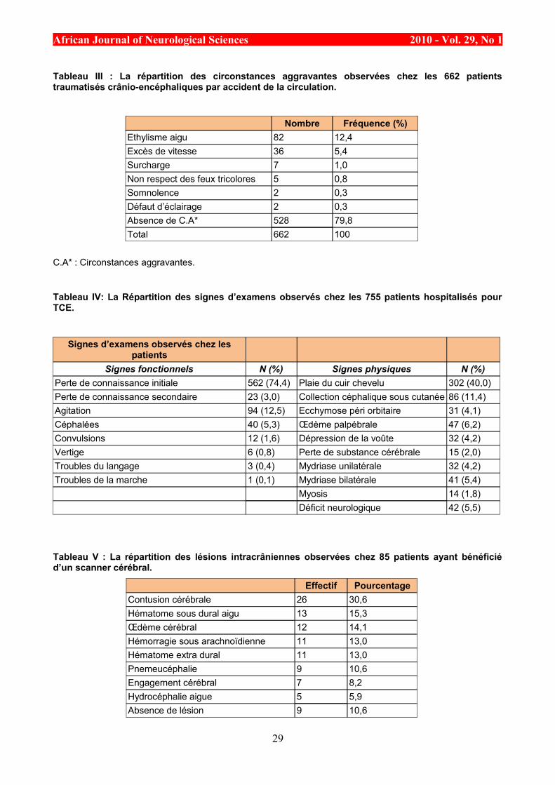
African Journal of Neurological Sciences 2010 - Vol. 29, No 1
Tableau III : La répartition des circonstances aggravantes observées chez les 662 patients traumatisés crânio-encéphaliques par accident de la circulation.
Nombre Fréquence (%)Ethylisme aigu 82 12,4Excès de vitesse 36 5,4Surcharge 7 1,0Non respect des feux tricolores 5 0,8Somnolence 2 0,3Défaut d’éclairage 2 0,3Absence de C.A* 528 79,8Total 662 100
C.A* : Circonstances aggravantes.
Tableau IV: La Répartition des signes d’examens observés chez les 755 patients hospitalisés pour TCE.
Signes d’examens observés chez les patients
Signes fonctionnels N (%) Signes physiques N (%)Perte de connaissance initiale 562 (74,4) Plaie du cuir chevelu 302 (40,0)Perte de connaissance secondaire 23 (3,0) Collection céphalique sous cutanée 86 (11,4)Agitation 94 (12,5) Ecchymose péri orbitaire 31 (4,1)Céphalées 40 (5,3) Œdème palpébrale 47 (6,2)Convulsions 12 (1,6) Dépression de la voûte 32 (4,2)Vertige 6 (0,8) Perte de substance cérébrale 15 (2,0)Troubles du langage 3 (0,4) Mydriase unilatérale 32 (4,2)Troubles de la marche 1 (0,1) Mydriase bilatérale 41 (5,4)
Myosis 14 (1,8)Déficit neurologique 42 (5,5)
Tableau V : La répartition des lésions intracrâniennes observées chez 85 patients ayant bénéficié d’un scanner cérébral.
Effectif PourcentageContusion cérébrale 26 30,6Hématome sous dural aigu 13 15,3Œdème cérébral 12 14,1Hémorragie sous arachnoïdienne 11 13,0Hématome extra dural 11 13,0Pnemeucéphalie 9 10,6Engagement cérébral 7 8,2Hydrocéphalie aigue 5 5,9Absence de lésion 9 10,6
29
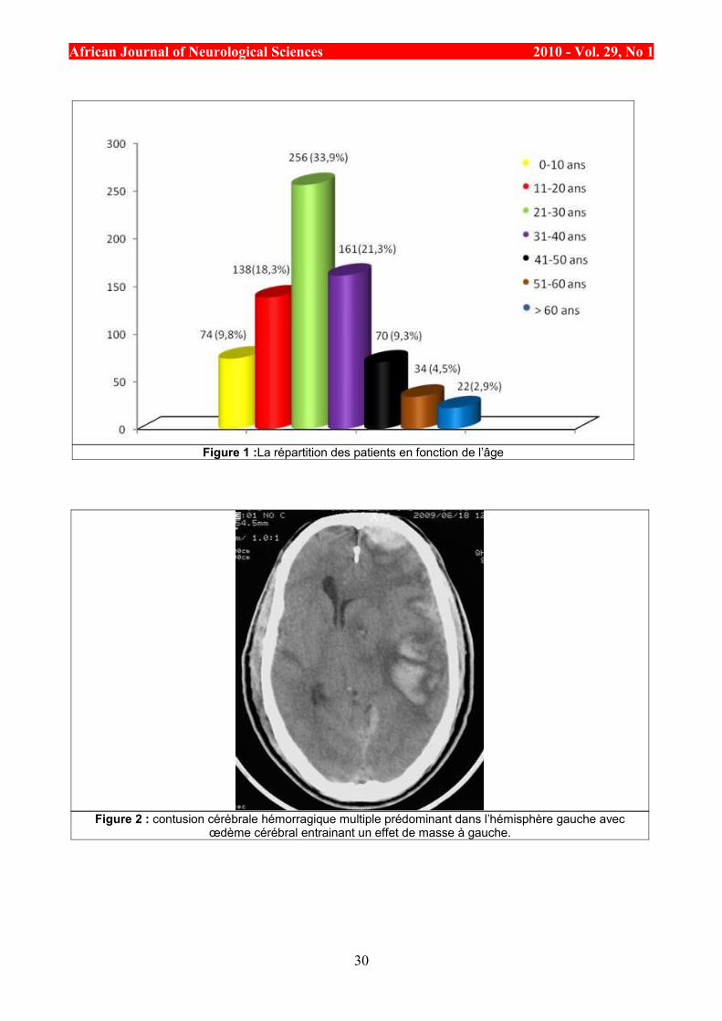
African Journal of Neurological Sciences 2010 - Vol. 29, No 1
Figure 1 :La répartition des patients en fonction de l’âge
Figure 2 : contusion cérébrale hémorragique multiple prédominant dans l’hémisphère gauche avec œdème cérébral entrainant un effet de masse à gauche.
30
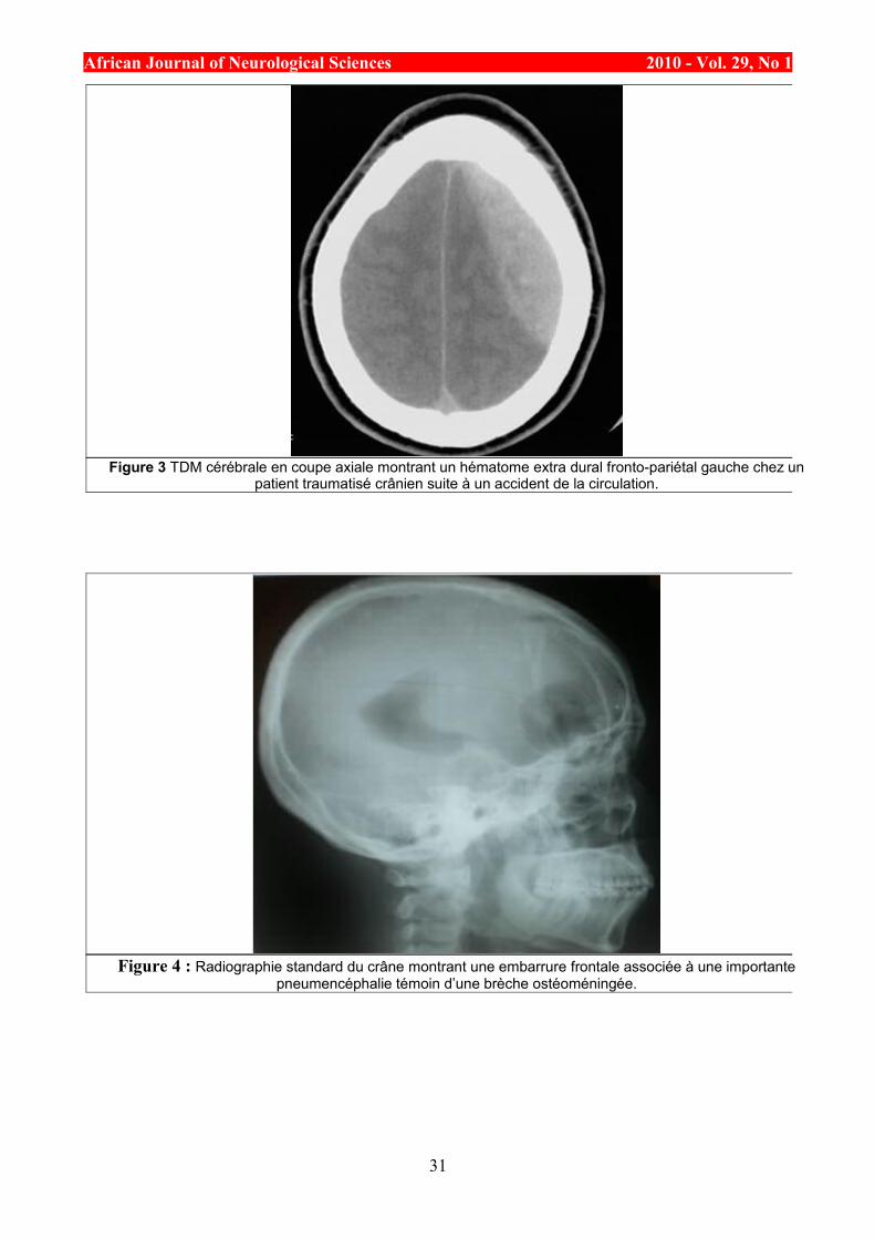
African Journal of Neurological Sciences 2010 - Vol. 29, No 1
Figure 3 TDM cérébrale en coupe axiale montrant un hématome extra dural fronto-pariétal gauche chez un patient traumatisé crânien suite à un accident de la circulation.
Figure 4 : Radiographie standard du crâne montrant une embarrure frontale associée à une importante pneumencéphalie témoin d’une brèche ostéoméningée.
31
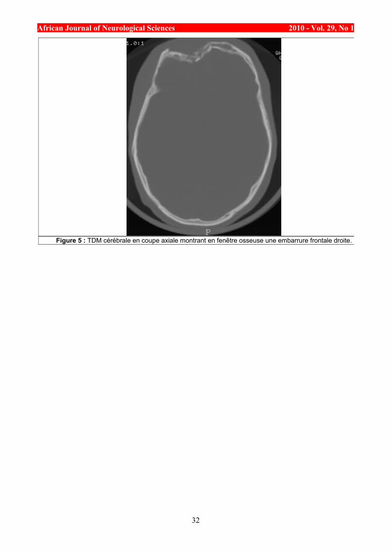
African Journal of Neurological Sciences 2010 - Vol. 29, No 1
Figure 5 : TDM cérébrale en coupe axiale montrant en fenêtre osseuse une embarrure frontale droite.
32
African Journal of Neurological Sciences 2010 - Vol. 29, No 1
REFERENCES1. BAHLOUL M, CHELLY H, BEN HAMIDA M et al. Prognosis of Traumatic Head Injury in South
Tunisia: A multivariate Analysis of 437 Cases. The Journal of Trauma Injury, Infection, and Critical Care. 57 •Number 2, 255-61.
2. BENER A, OMAR AO, AHMAD AE, AL-MULLA FH, ABDUL RAHMAN YS. The pattern of traumatic brain injuries: a country undergoing rapid development. Brain Inj. 2010 ;24(2):74-80.
3. CHIU WT, YEH KH, LI YC, GAN YH, CHEN HY, HUNG CC. Traumatic brain injury registry in Taiwan. Neurol Res. 1997; 19(3):261-4
4. CORRIGAN JD, SELASSIE AW, ORMAN JA. The epidemiology of traumatic brain injury. J Head Trauma Rehabil. 2010;25(2):72-80.
5. COULIBALY Y, MP E, DIALLO A et al. Le traumatisme crânien à L’hôpital du Point G : A propos de 80 cas. Mali Médical 2004 T XIX N° 3&4
6. CROCKER P, ZAD O, MILLING T, LAWSON KA. Alcohol, bicycling, and head and brain injury: a study of impaired cyclists’ riding patterns R1. Am J Emerg Med. 2010; 28(1):68-72.
7. EMEJULU JK. Epidemiological patterns of head injury in a newly established neurosurgical service: one-year prospective study. Afr J Med Med Sci. 2008; 37(4):383-8.
8. ENGBERG A. Severe traumatic brain injury. Epidemiology external causes, prevention and rehabilitation of mental and physical sequelae. Acta neurol Scand 1995; 92 suppl 164:1- 151.
9. HASSAN NA, KELANY RS, EMARA AM, AMER M. Pattern of craniofacial injuries in patients admitted to Tanta University Hospital—Egypt. J Forensic Leg Med. 2010 ;17(1):26-32.
10.LEVY F, MAKARAWO S. A Survey of 371 head injured patients admitted to the neurosurgical wards of the Parirewyatwa Hospital, Harare, Zimbabwe. Afr J Neur Sci 1997;16:2
11.MAAS AI. Standardisation of data collection in traumatic brain injury: key to the future? Crit Care. 2009; 13(6):1016. Epub 2009 Dec 16.
12.SIDIBE S, DIALLO A, TOURE M, TCHAMKO-DJEUTCHEU FR, TRAORE I. Apport de la tomodensitométrie dans la prise en charge des traumatismes crânio-encéphaliques à Bamako. Mali Médical 2005, T XX, N° 1&2.
13.SURIYAWONGPAISAL P, KANCHANUSUT S. Road traffic injuries in Thailand: Trends, selected underlying determinants and status of intervention. Injury control and safety promotion, 2003, 10:95 - 104.
14.TAGLIAFERRI F, COMPAGNONE C, KORSIC M, SERVADEI F, AND KRAUS J. A systematic review of brain injury epidemiology in Europe. Acta Neurochir (Wien) (2006) 148: 255-268
33
African Journal of Neurological Sciences 2010 - Vol. 29, No 1
CLINICAL STUDIES / ETUDES CLINIQUESEVALUATION DU TRAITEMENT DES EPILEPTIQUES ADULTES HOSPITALISES DANS LE SERVICE
DE NEUROLOGIE DU CHU DE YOPOUGON A ABIDJAN - COTE D’IVOIRE
EVALUATION OF THE TREATMENT OF ADULTS EPILEPTIC INPATIENTS IN THE DEPARTMENT OF NEUROLOGY AT TEACHING HOSPITAL OF YOPOUGON ABIDJAN - COTE D’IVOIRE
DOUMBIA-OUATTARA Mariam 1
BOA YAPO Félix 1
KOUASSI KOUAME Léonard 1
KOUAME-ASSOUAN Ange- Eric 2
AKA-ANGHUI DIARRA Evelyne 2
SONAN-DOUAYOUA Thérèse 1
1. Service de Neurologie, CHU de Yopougon 21 BP 632 Abidjan 21 Côte d’Ivoire2. Service de Neurologie, CHU de Cocody, BP V 13 Abidjan 01, Côte d’Ivoire.
E-Mail Contact - DOUMBIA-OUATTARA Mariam :
Mots-clés: Afrique, coût, épilepsie, prise en charge, traitement.Keywords:
RESUMEDescription Malgré l’efficacité reconnue des médicaments antiépileptiques (MAE) en Afrique, les structures sanitaires sont sous-utilisées pour des raisons culturelles et économiques.
Objectif Evaluer l’efficacité, la tolérance des MAE et le coût de la prise en charge de l’épilepsie de l’adulte hospitalisé dans un service de neurologie. Méthode: Une étude transversale descriptive fut réalisée sur une période de 2 ans allant de septembre 2002 à octobre 2004 dans l’unité d’hospitalisation du service de Neurologie du CHU de Yopougon. Elle portait sur les dossiers médicaux de 70 patients ayant présenté au moins 2 crises d’épilepsie.
Résultats Les MAE les plus utilisés étaient le phénobarbital (40%), la carbamazépine (24%) et l’acide valproique (14%). Le traitement avait entraîné un arrêt des crises chez 60% des patients. 82% des patients présentaient une bonne tolérance aux MAE. La durée moyenne d’hospitalisation par malade était d’environ 12 jours. Le coût estimatif direct de l’hospitalisation était de 148 715 FCFA (226,71 € ou 280,59 $ US), alors que 14% des patients étaient sans profession et 66% avaient un revenu mensuel inférieur à 50000 FCFA, (76,21 €) et que le Salaire Minimum Interprofessionnel Garanti est égal à 36000 FCFA (54,88 € ou 67,92 $ US). 22% des patients avaient dû quitter le service par manque de moyens financiers.
Conclusion Chez l’adulte épileptique, le traitement médical est efficace. Comme ailleurs en Afrique, les barbituriques restent la molécule de choix. Mais le coût global de la prise en charge, encore largement supérieur au SMIG contribue à rendre ce traitement difficilement accessible à la majorité des populations démunies.
34
African Journal of Neurological Sciences 2010 - Vol. 29, No 1
ABSTRACTObjective Evaluate the efficacy, the tolerance of AE drugs and the financial cost of the care of epilepsies in adult’s inpatients unit of the neurology department at Yopougon-Abidjan.
Methodology A transversal descriptive study took place during 2 years from September 2002 to October 2004 in the inpatients unit of the department of Neurology of the teaching hospital of Yopougon. The study included the charts of 70 patients which were admitted for at least 2 epileptic seizures.
Results The most used AE drugs were: phenobarbital (40%), carbamazepine (24%) and valproic acid (14%). The AE treatment stopped the occurrence of seizures in 60% of patients. 82% had no secondary effects of drugs. The average duration of hospitalisation in the unit was 12 days. Estimative cost of care was equal to 148 715 FCFA or 226.71 € or 280.59 $ US whereas 14% of patients had no occupation and 66% had a monthly salary lower than 50.000 FCFA or 76.21 € and SMIG is equal to 36000 FCFA (54.88 € or 67.92 $ US). 22% of patients had to leave the unit without a favourable evolution due to of lack of financial means.
Discussion As elsewhere in Africa, phenobarbital is the treatment of choice. The global cost of care is very high compared to the average salary (the minimal guaranteed salary which is equal to 36.000 FCFA or 54.88 € or 67.92 $ US) and this yields the problem of financial accessibility to the modern treatment of epilepsy.
Conclusion The treatment of epilepsy in adult inpatient is efficient but the high cost of global care contributes to the inaccessibility of MAE for populations with low socioeconomic level.
INTRODUCTIONEn Afrique Noire, la prise en charge de l’épilepsie est caractérisée par une sous-utilisation des structures sanitaires et des MAE pour des raisons essentiellement socioculturelles (11) mais également économiques. Notre travail a pour but d’évaluer l’efficacité, la tolérance des MAE et le coût de la prise en charge de l’épilepsie de l’adulte dans un service de neurologie en Côte d’Ivoire.
METHODOLOGIEIl s’agit d’une étude transversale de type descriptif qui s’est déroulée dans l’unité d’hospitalisation du service de Neurologie du Centre Hospitalier Universitaire (CHU) de Yopougon. Elle a été réalisée sur une période de 2 ans allant du mois de septembre 2002 au mois d’octobre 2004. Les 70 dossiers inclus dans l’étude concernaient les patients adultes reçus en hospitalisation, ayant présenté au moins 2 crises d’épilepsie, et ayant des dossiers médicaux complets.
Les données suivantes ont été recueillies à l’aide de fiches d’enquête: l’âge, le sexe, la profession, le revenu mensuel, la séméiologie des crises épileptiques, les résultats de la sérologie du Virus de l’Immunodéficience Humaine (VIH) et de la tomodensitométrie (TDM) crâniocérébrale, les médicaments anti-épileptiques (MAE) utilisés et leur tolérance, l’évolution de la maladie, la durée d’hospitalisation et le coût de l’hospitalisation. Ces données ont ensuite fait l’objet d’une analyse statistique à l’aide du logiciel SPSS.Vers.10. Résultats
Les âges extrêmes étaient 20 et 80 ans, avec une moyenne de 40 ans. On notait une légère prédominance masculine (52%) avec un sex-ratio de 1,12. 55% des patients exerçaient une activité professionnelle et 35% étaient sans activité (retraités et sans emploi). 66% de la population d’étude avait un revenu mensuel inférieur à 50.000 FCFA, soit 76,21 €.
Les crises étaient généralisées dans 58% des cas et partielles dans 36%. 6% des patients avaient présenté un état de mal épileptique. .La TDM crânio-cérébrale était anormale dans 54% des cas et les étiologies identifiées étaient l’abcès cérébral (25%), la méningo-encéphalite (12%) et les AVC (16%). La sérologie du VIH était positive dans 56% des cas.
Les MAE utilisés étaient le phénobarbital (40%), la carbamazépine (24%), le diazépam (20%) et l’acide valproique (14%). Dans 82% des cas, la tolérance aux MAE était bonne. Les effets indésirables observés dans 18% des cas étaient des nausées, des vomissements, une asthénie et des vertiges avec l’acide valproique (VPA) et la carbamazépine (CBZ). Sous traitement antiépileptique, on a noté un arrêt des crises chez 60% des patients.
La durée moyenne du séjour hospitalier par malade était de 12 jours avec des extrêmes de 1 et 29 jours. 60% des patients sont sortis du service au terme d’une évolution clinique favorable. 22% sont sortis à la
35
African Journal of Neurological Sciences 2010 - Vol. 29, No 1demande de leur famille, par manque de moyens financiers et 8% se sont évadés.
Le coût estimatif direct de l’hospitalisation était égal à 148.715 FCFA soit 226,71 € ou 280,59 $ US répartis entre les examens complémentaires (73.500 FCFA), l’hôtellerie (58.300 FCFA) et les MAE (16.915 FCFA), alors que 14% des patients étaient sans profession et que 66% avaient un revenu mensuel inférieur à 50000 FCFA (76,21 €) et que le SMIG est égal à 36.000 F CFA (54,88 € ou 67,92 $ US.)
DISCUSSIONComme dans la majorité des études africaines et européennes (3, 6, 9, 11, 12, 15), nous avons observé une prédominance masculine avec un sex-ratio de 1,12. Notre série comporte une majorité d’adultes jeunes. L’âge moyen était égal à 40 ans, et la tranche d’âge de 20 à 45 ans représentait 58% des patients. Giordano et al. (6) rapportent également une prédominance de l’épilepsie dans la tranche d’âge de 20 à 45 ans avec un taux de 59%. Pour Senanayaké et al. (13), près de 40% des épilepsies apparaissent entre 20 et 30 ans.
66% de nos patients avaient un revenu mensuel inférieur à 50.000 FCFA, soit 76,21 €, à peine le SMIG (égal à 36.000 FCFA, soit 54,88 € ou 67,92 $ US). Nous avons relevé également 14% de patients sans profession, ce qui suggère qu’un bon nombre de patients sont de véritables charges financières pour leurs familles, et qu’ils pourraient par ailleurs avoir une insertion socio-professionnelle difficile. Dans la série de Piraux (12), 30% des malades constituaient également de réels problèmes sociaux. Ces résultats posent le problème de l’aspect financier de la prise en charge des épileptiques et celui de leur insertion socioprofessionnelle. En Côte d’Ivoire, la mauvaise adaptation sociale de l’épileptique peut s’expliquer en partie par la connotation socioculturelle de l’épilepsie décrite par Hazera et Sieye cités par Giordano et al (6) qui ont montré une modification du jeu relationnel et une marginalisation de l’individu pour cause de contagiosité par la salive. Ailleurs en Afrique, la crise épileptique est considérée comme une maladie de Dieu, une fatalité ou une attaque en sorcellerie, souvent assimilée à une maladie des djinns ou de Satan (16). Dans un tel contexte économique et socioculturel, les thérapeutes traditionnels et les guérisseurs constituent le premier recours et ce n’est que le constat de plusieurs échecs qui motive une consultation à l’hôpital (11).
Sur le plan sémiologique, notre étude comme d’autres études africaines (3, 4, 6, 16) montre la prédominance des crises généralisées alors qu’en Occident, les crises partielles sont plus fréquentes que les crises généralisées (7). Il est possible qu’une analyse plus minutieuse des crises d’épilepsie identifiées comme généralisées en Afrique, révèle en son sein une grande proportion de crises partielles (6). 6% de nos patients ont présenté un état de mal épileptique. Ce chiffre est proche de ceux cités dans des travaux antérieurs en Côte d’Ivoire: 6,7% pour Giordano et al. (6) et 9% pour Sonan et al. (14).
Les étiologies identifiées par la TDM sont infectieuses (37%), vasculaires (16%) et tumorales (8%). Dans la série de Mbodj et al. (10), les causes infectieuses (67%) et les causes vasculaires (8%) étaient également les étiologies majeures. Dans notre étude le taux de séropositivité au VIH est élevé (56%), ce qui explique que la toxoplasmose soit l’infection la plus fréquente (25%) comme l’ont montré Bartolémei et al (1), Dumas et al (5) et Doumbia et al (4).
Parmi les antiépileptiques majeurs utilisés en monothérapie de première intention, nous avons observé une large prescription du phénobarbital (40%), suivi de la CBZ (24%) et du VPA (14%). Dans la série de Dongmo et al. (3) au Cameroun, les médicaments les plus utilisés étaient également le PB (75,2%) et la CBZ (15,2%). Heaney et al. (8) soulignent également l’utilisation du PB en première intention dans les pays en développement, en raison de son coût 15 à 30 fois moins élevé que celui de la CBZ et du VPA, et 100 fois plus faible que ceux des nouveaux antiépileptiques (Lamotrigine). Pour Collomb (2) et la majorité des auteurs africains, l’efficacité thérapeutique des barbituriques, doublée de leur accessibilité financière constituent un argument majeur pour leur prescription dans les populations à bas niveau socio-économique.
Les MAE ont entraîné l’arrêt des crises chez 60% des patients. Ce taux de rémission est voisin de celui observé par Dongmo et al. (69,6%) (3). Ces médicaments étaient bien tolérés dans 82% des cas. 18% des patients ont présenté des effets indésirables à type de nausées, de vomissements, d’asthénie et de vertiges. Ces effets ont été observés avec le VPA et la CBZ, et ont parfois occasionné le changement de l’antiépileptique. Dans la série de Dongmo et al., les effets indésirables des médicaments ont été notés chez 16% de patients, avec comme principal effet indésirable, le couple somnolence-ralentissement psychomoteur (3).
La durée moyenne du séjour hospitalier par malade était d’environ 12 jours avec des extrêmes de 1 et 29 jours. Le coût estimatif direct de l’hospitalisation était de 148.715 FCFA (226,67 €) ainsi réparti: examens complémentaires (73.500 FCFA), hôtellerie (58.300 FCFA) et MAE (16.915 FCFA). Le coût minimal de la TDM crâniocérébrale, examen-clé pour l’exploration étiologique des épilepsies, qui s’élève à 55.000 FCFA (83,83 €) accroit les difficultés de la prise en charge des patients sans profession ou issus d’un bas niveau socio-économique.
Le coût global de l’hospitalisation largement supérieur au SMIG, égal à 36 000 FCFA (54,88 €) pose le problème de l’accessibilité financière au traitement médical des épilepsies, et explique que 22% des patients soient sortis du service à la demande des familles épuisées financièrement.
36
African Journal of Neurological Sciences 2010 - Vol. 29, No 1
CONCLUSIONEn milieu hospitalier neurologique, le traitement de l’épilepsie de l’adulte jeune par les MAE est efficace et bien toléré, mais il reste encore peu accessible aux populations en majorité démunies en raison du coût onéreux de la prise en charge globale en hospitalisation.
REFERENCES
1. BARTOLEMEI F, PELLE GRINO P, DHIVER C, QUILICHINI R, GASTAUT JA, GASTAUT JL. Crise d’épilepsie au cours de l’infection par le VIH: 52 observations. Presse Méd. 1991; 20: 2135-38.
2. COLLOMB. Epidémiologie de l’épilepsie au Sénégal. Afr J of Med Sciences 1970;1: 125-48.3. DONGMO L, ECHOUFFO TJB, NJAMNSHI AK et al. Difficultés de la prise en charge de l’épilepsie
en milieu rural camerounais: le cas de la localité de Mbangassina. Afr J of Neurol Sci. 2003; 22(1): 18-26.
4. DOUMBIA M, DOUAYOUA-SONAN T, BOA YF. Aspects cliniques et étiologiques des épilepsies observés dans le service de Neurologie du CHU de Yopougon. Journées Scientifiques - CHU de Yopougon. Mars 2005.
5. DUMAS M, LEGER JM, PESTRE-ALEXANDRE M. Manifestations neurologiques et psychiatriques des parasitoses. Congrès de Psychiatrie et de Neurologie de langue française, session LXXXIV, 23-27 juin, le Mans (France). Masson 1986; 1: 332p.
6. GIORDANO C, HAZERA M, BADOUAL J, ASSI-ADOU J, ANDRE M, VIDAL H, et al. Aspects épidémiologiques, cliniques et électriques de l’épilepsie en Côte d’Ivoire (Abidjan). Méd Afr Noire. 1976; 23: 305-22.
7. GRANGER N, CONVERS P, BEAUCHET O, IMLER D, VIALLON A, LAURENT B, et al. Première crise d’épilepsie chez le sujet de plus de 60 ans: données électrocliniques et étiologiques, à propos d’une série de 341 cas. Rev Neurol. 2002; 158 (11): 1088-95.
8. HEANEY D, SANDER JWS. Antiepileptic drugs in developing countries. Lancet. 1998; 351: 1967.9. KABORE J, LENGANI A, DRABO YJ, MELAKU Z, PREUX PM, N’DIAYE IP. Clinical aspects of
seizures disorders at Ouagadougou-Burkina-Faso: retrospective study of 532 cases. Afr J Neurol Sci. 1995; 14: 24-6.
10.MBODJ, N’DIAYE M, SENE F, SALIF SOW P, SOW HP, DIAGANA M et al. Prise en charge de l’état de mal épileptique dans les conditions de pays en développement: les états de mal épileptiques périodiques. Congrès LA DA. 2000; 30(3): 165-9.
11.NUBUKPO P, PREUX PM, CLEMENT JP. Représentations socioculturelles de l’épilepsie en Afrique noire. Ann psychiatric. 2001; 16: 219-27.
12.PIRAUX A. Les épilepsies en Afrique centrale. World Neurol. 1960; 1: 510-22.13.SENANAYAKE N, ROMAN GC. Epidemiology of epilepsy in the tropics. J Trop Geogr Neurol. 1999;
2: 10-9.14.SONAN T, PIQUEMAL M, DECHAMBENOIT G, BOA Y, BEUGRE K, GIORDANO C. Epilepsies
partielles de l’adulte en milieu hospitalier: aspects cliniques et étiologiques. Soc Med CI: séance du 26 juin 1986. Rev Méd CI. 1986; 60 (abstract).
15.TEKLE-HAIMANOT R, FORSGEN L, ABÉDÉ M, GÉBRÉ-MARIAM A, HEIJBEL J, HOLMGREN G, et al. Clinical and electroencephalographic characteristics of epilepsy in rural ethiopians: a community-based study. Epilepsy Res. 1990; 7: 230-9.
16.TRAORE H, PREUX PM, DIAGANA M, DRUET-CABANAC M, DEBROCK C, DUMAS M. Aspects cliniques et étiologiques des épilepsies dans un service de neurologie à Nouakchott, Mauritanie. Afr J Neurol Sci. 2001; 1: 17-20
37
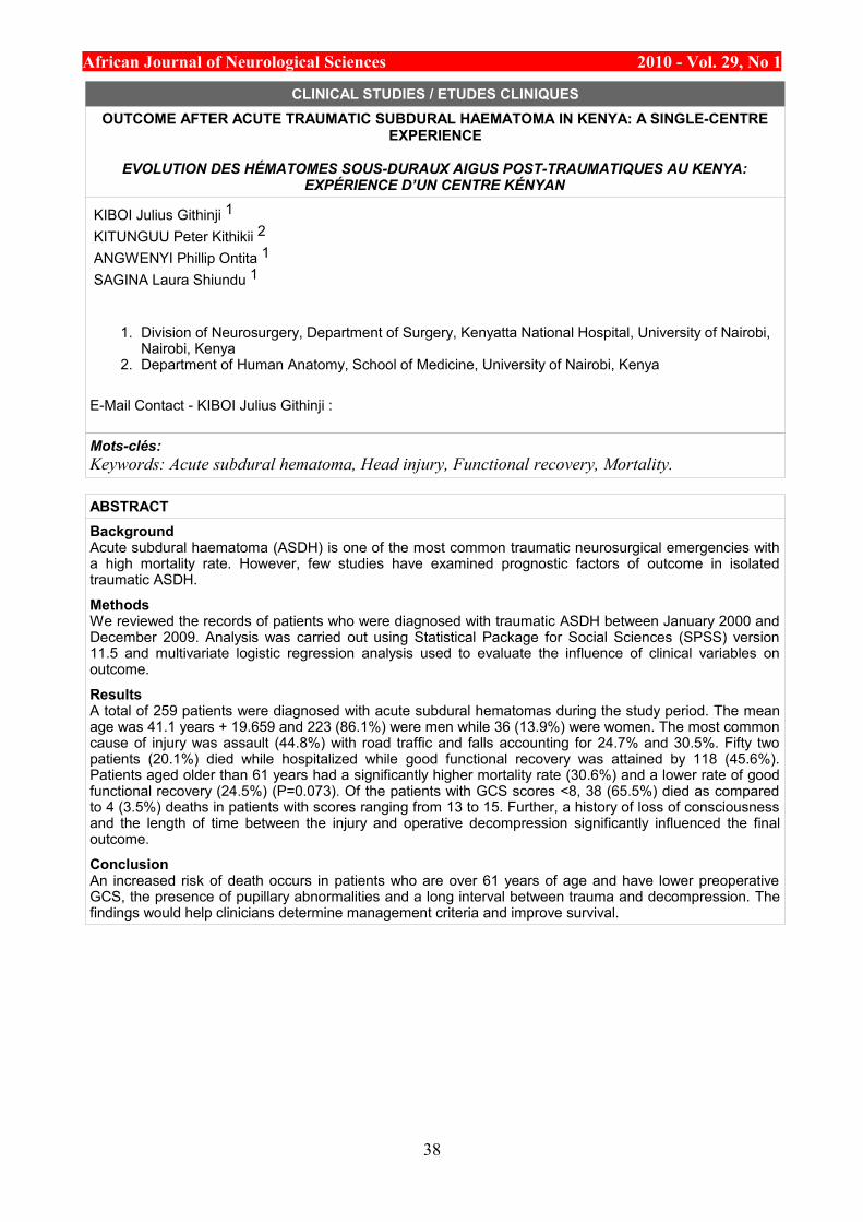
African Journal of Neurological Sciences 2010 - Vol. 29, No 1
CLINICAL STUDIES / ETUDES CLINIQUESOUTCOME AFTER ACUTE TRAUMATIC SUBDURAL HAEMATOMA IN KENYA: A SINGLE-CENTRE
EXPERIENCE
EVOLUTION DES HÉMATOMES SOUS-DURAUX AIGUS POST-TRAUMATIQUES AU KENYA: EXPÉRIENCE D’UN CENTRE KÉNYAN
KIBOI Julius Githinji 1
KITUNGUU Peter Kithikii 2
ANGWENYI Phillip Ontita 1
SAGINA Laura Shiundu 1
1. Division of Neurosurgery, Department of Surgery, Kenyatta National Hospital, University of Nairobi, Nairobi, Kenya
2. Department of Human Anatomy, School of Medicine, University of Nairobi, Kenya
E-Mail Contact - KIBOI Julius Githinji :
Mots-clés:Keywords: Acute subdural hematoma, Head injury, Functional recovery, Mortality.
ABSTRACTBackground Acute subdural haematoma (ASDH) is one of the most common traumatic neurosurgical emergencies with a high mortality rate. However, few studies have examined prognostic factors of outcome in isolated traumatic ASDH.
Methods We reviewed the records of patients who were diagnosed with traumatic ASDH between January 2000 and December 2009. Analysis was carried out using Statistical Package for Social Sciences (SPSS) version 11.5 and multivariate logistic regression analysis used to evaluate the influence of clinical variables on outcome.
Results A total of 259 patients were diagnosed with acute subdural hematomas during the study period. The mean age was 41.1 years + 19.659 and 223 (86.1%) were men while 36 (13.9%) were women. The most common cause of injury was assault (44.8%) with road traffic and falls accounting for 24.7% and 30.5%. Fifty two patients (20.1%) died while hospitalized while good functional recovery was attained by 118 (45.6%). Patients aged older than 61 years had a significantly higher mortality rate (30.6%) and a lower rate of good functional recovery (24.5%) (P=0.073). Of the patients with GCS scores <8, 38 (65.5%) died as compared to 4 (3.5%) deaths in patients with scores ranging from 13 to 15. Further, a history of loss of consciousness and the length of time between the injury and operative decompression significantly influenced the final outcome.
Conclusion An increased risk of death occurs in patients who are over 61 years of age and have lower preoperative GCS, the presence of pupillary abnormalities and a long interval between trauma and decompression. The findings would help clinicians determine management criteria and improve survival.
38
African Journal of Neurological Sciences 2010 - Vol. 29, No 1
RESUMEFond L’hématome sous dural aigu post-traumatique (HSDA) est une urgence neurochirurgicale avec un haut taux de mortalité. Peu d’études ont examiné les facteurs prognostiques lors d’ ASDH traumatisant isolé.
Méthodes Nous avons réexaminé les dossiers de patients avec ASDH post traumatique entre janvier 2000 et décembre 2009. L’analyse a été pratiqué en utilisant le Packet Statistique pour les Sciences Sociales (SPSS) la version 11,5.
Résultats Un total de 259 malades a été diagnostiqué avec les hématomes de subdural aigu pendant la période d’étude. L’âge moyen était 41,1 ans + 19,659 et 223 (86.1%) étaient des hommes pendant que 36 (13.9%) étaient des femmes. La cause la plus commune de blessure était l’assaut (44.8%) avec la circulation de route et les chutes représente 24,7% et 30,5%. Cinquante deux malades (20.1%) est mort pendant qu’a hospitalisé pendant que le bon rétablissement fonctionnel a été atteint par 118 (45.6%). Les malades plus vieux que 61 ans ont eu un significativement plus haut taux de mortalité (30.6%) et un taux plus bas de bon rétablissement fonctionnel (24.5%) (p=0.073). Des malades avec GCS scores <8, 38 (65.5%) est mort comme en comparaison de 4 (3.5%) les morts dans les malades avec scores étendant de 13 à 15. Plus, une histoire de perte de conscience et le temps qu’il faut entre la blessure et la décompression opérative a influencé significativement l’issue finale.
Conclusion Un risque augmenté de mort arrive dans les malades qui sont plus de 61 ans majeurs et a GCS préopératoire plus bas, la présence d’anomalies de pupillary et un intervalle long entre le traumatisme et la décompression. Les conclusions aideraient des practiciens déterminent les critères de direction et améliorent la survie.
INTRODUCTIONTraumatic acute subdural hematoma (ASDH) is one of the most common clinical entities encountered by any neurosurgical service and often requires surgical intervention (1, 2). ASDH occurs in 12% to 30% of patients with severe head injury (3) and reported mortality rates vary from 36% to 79% for patients who underwent surgery (4, 5). Although developments and improvements in emergency medical service systems, neuro-intensive monitoring and treatment, ASDH is a disorder with a very high mortality rate and extremely poor prognosis among traumatic brain injuries (5, 6, 7). Therefore, identifying reliable prognostic factors for ASDH to improve the surgical results in these patients is important. However relatively few studies have focused on the factors that affect the outcome of patients with traumatic ASDH. Kenyatta National Hospital, located in Nairobi Kenya, is a national teaching and referral hospital with a bed capacity of 2000 and the largest neurosurgical unit in the Great Lakes region. Through this center, the great majority of neurosurgical referrals are managed. Among these patients, we retrospectively reviewed patients who were diagnosed and treated for traumatic ASDH and sought to identify the factors related to functional recovery and mortality of this lethal disorder with a secondary aim to improve functional outcome of these patients hereafter.
METHODOLOGYFollowing ethical approval, the records of 259 patients who were admitted to the neurosurgical unit at the Kenyatta National Hospital and diagnosed with ASDH between January 2000 and December 2009 were reviewed. The records were analysed for demographic characteristics such as gender, age, GCS on admission, pupil abnormalities, mechanism of injury, and time elapsed from accident to surgery.
The GCS score is usually determined on admission and all patients were divided patients into three groups; those with GCS scores of 3 to 8, 9 to 12 and 13 to 15 for statistical analysis. For logistic regression analysis, patients were classified as having 0, 1, or 2 reactive pupils. Outcome was assessed according to the Glasgow Outcome Scale (GOS) at the time of discharge from hospital or mortality.
Data was collected in pre-formed questionnaires, coded and analysis carried out using Statistical Package for Social Sciences (SPSS) version 11.5. Frequencies and means were computed for description of the various variables, discrete variables compared using the Chi-square test and continuous variables compared using the Students’ t-test. A linear logistic multivariable regression model was run to determine which variables are independently associated with functional recovery and mortality. A P value <0.005 was considered statistically significant.
39
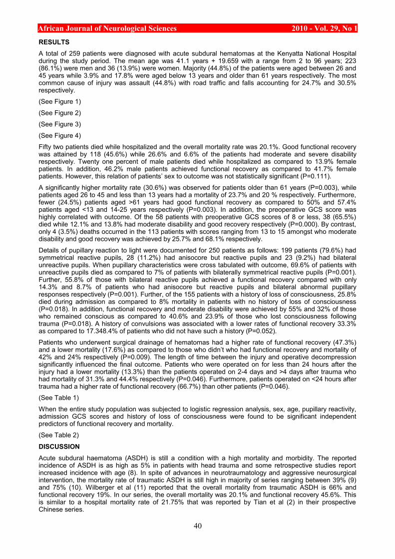
African Journal of Neurological Sciences 2010 - Vol. 29, No 1
RESULTSA total of 259 patients were diagnosed with acute subdural hematomas at the Kenyatta National Hospital during the study period. The mean age was 41.1 years + 19.659 with a range from 2 to 96 years; 223 (86.1%) were men and 36 (13.9%) were women. Majority (44.8%) of the patients were aged between 26 and 45 years while 3.9% and 17.8% were aged below 13 years and older than 61 years respectively. The most common cause of injury was assault (44.8%) with road traffic and falls accounting for 24.7% and 30.5% respectively.
(See Figure 1)
(See Figure 2)
(See Figure 3)
(See Figure 4)
Fifty two patients died while hospitalized and the overall mortality rate was 20.1%. Good functional recovery was attained by 118 (45.6%) while 26.6% and 6.6% of the patients had moderate and severe disability respectively. Twenty one percent of male patients died while hospitalized as compared to 13.9% female patients. In addition, 46.2% male patients achieved functional recovery as compared to 41.7% female patients. However, this relation of patients’ sex to outcome was not statistically significant (P=0.111).
A significantly higher mortality rate (30.6%) was observed for patients older than 61 years (P=0.003), while patients aged 26 to 45 and less than 13 years had a mortality of 23.7% and 20 % respectively. Furthermore, fewer (24.5%) patients aged >61 years had good functional recovery as compared to 50% and 57.4% patients aged <13 and 14-25 years respectively (P=0.003). In addition, the preoperative GCS score was highly correlated with outcome. Of the 58 patients with preoperative GCS scores of 8 or less, 38 (65.5%) died while 12.1% and 13.8% had moderate disability and good recovery respectively (P=0.000). By contrast, only 4 (3.5%) deaths occurred in the 113 patients with scores ranging from 13 to 15 amongst who moderate disability and good recovery was achieved by 25.7% and 68.1% respectively.
Details of pupillary reaction to light were documented for 250 patients as follows: 199 patients (79.6%) had symmetrical reactive pupils, 28 (11.2%) had anisocore but reactive pupils and 23 (9.2%) had bilateral unreactive pupils. When pupillary characteristics were cross tabulated with outcome, 69.6% of patients with unreactive pupils died as compared to 7% of patients with bilaterally symmetrical reactive pupils (P=0.001). Further, 55.8% of those with bilateral reactive pupils achieved a functional recovery compared with only 14.3% and 8.7% of patients who had anisocore but reactive pupils and bilateral abnormal pupillary responses respectively (P=0.001). Further, of the 155 patients with a history of loss of consciousness, 25.8% died during admission as compared to 8% mortality in patients with no history of loss of consciousness (P=0.018). In addition, functional recovery and moderate disability were achieved by 55% and 32% of those who remained conscious as compared to 40.6% and 23.9% of those who lost consciousness following trauma (P=0.018). A history of convulsions was associated with a lower rates of functional recovery 33.3% as compared to 17.348.4% of patients who did not have such a history (P=0.052).
Patients who underwent surgical drainage of hematomas had a higher rate of functional recovery (47.3%) and a lower mortality (17.6%) as compared to those who didn’t who had functional recovery and mortality of 42% and 24% respectively (P=0.009). The length of time between the injury and operative decompression significantly influenced the final outcome. Patients who were operated on for less than 24 hours after the injury had a lower mortality (13.3%) than the patients operated on 2-4 days and >4 days after trauma who had mortality of 31.3% and 44.4% respectively (P=0.046). Furthermore, patients operated on <24 hours after trauma had a higher rate of functional recovery (66.7%) than other patients (P=0.046).
(See Table 1)
When the entire study population was subjected to logistic regression analysis, sex, age, pupillary reactivity, admission GCS scores and history of loss of consciousness were found to be significant independent predictors of functional recovery and mortality.
(See Table 2)
DISCUSSIONAcute subdural haematoma (ASDH) is still a condition with a high mortality and morbidity. The reported incidence of ASDH is as high as 5% in patients with head trauma and some retrospective studies report increased incidence with age (8). In spite of advances in neurotraumatology and aggressive neurosurgical intervention, the mortality rate of traumatic ASDH is still high in majority of series ranging between 39% (9) and 75% (10). Wilberger et al (11) reported that the overall mortality from traumatic ASDH is 66% and functional recovery 19%. In our series, the overall mortality was 20.1% and functional recovery 45.6%. This is similar to a hospital mortality rate of 21.75% that was reported by Tian et al (2) in their prospective Chinese series.
40
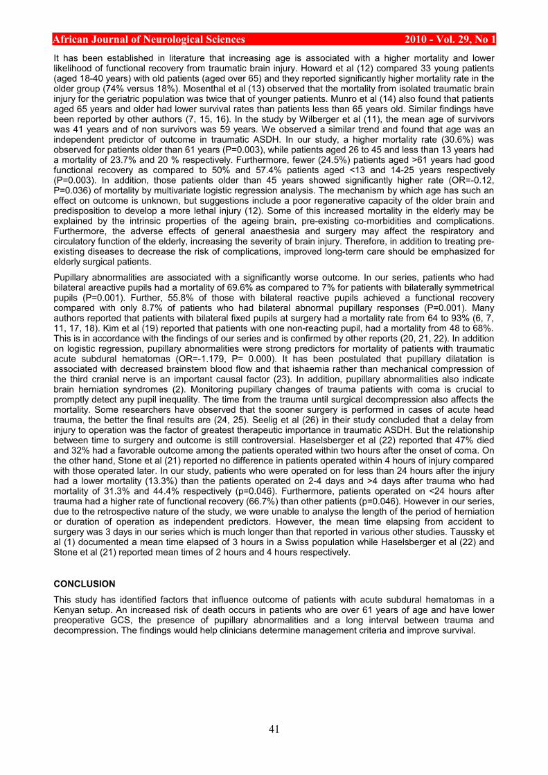
African Journal of Neurological Sciences 2010 - Vol. 29, No 1It has been established in literature that increasing age is associated with a higher mortality and lower likelihood of functional recovery from traumatic brain injury. Howard et al (12) compared 33 young patients (aged 18-40 years) with old patients (aged over 65) and they reported significantly higher mortality rate in the older group (74% versus 18%). Mosenthal et al (13) observed that the mortality from isolated traumatic brain injury for the geriatric population was twice that of younger patients. Munro et al (14) also found that patients aged 65 years and older had lower survival rates than patients less than 65 years old. Similar findings have been reported by other authors (7, 15, 16). In the study by Wilberger et al (11), the mean age of survivors was 41 years and of non survivors was 59 years. We observed a similar trend and found that age was an independent predictor of outcome in traumatic ASDH. In our study, a higher mortality rate (30.6%) was observed for patients older than 61 years (P=0.003), while patients aged 26 to 45 and less than 13 years had a mortality of 23.7% and 20 % respectively. Furthermore, fewer (24.5%) patients aged >61 years had good functional recovery as compared to 50% and 57.4% patients aged <13 and 14-25 years respectively (P=0.003). In addition, those patients older than 45 years showed significantly higher rate (OR=-0.12, P=0.036) of mortality by multivariate logistic regression analysis. The mechanism by which age has such an effect on outcome is unknown, but suggestions include a poor regenerative capacity of the older brain and predisposition to develop a more lethal injury (12). Some of this increased mortality in the elderly may be explained by the intrinsic properties of the ageing brain, pre-existing co-morbidities and complications. Furthermore, the adverse effects of general anaesthesia and surgery may affect the respiratory and circulatory function of the elderly, increasing the severity of brain injury. Therefore, in addition to treating pre-existing diseases to decrease the risk of complications, improved long-term care should be emphasized for elderly surgical patients.
Pupillary abnormalities are associated with a significantly worse outcome. In our series, patients who had bilateral areactive pupils had a mortality of 69.6% as compared to 7% for patients with bilaterally symmetrical pupils (P=0.001). Further, 55.8% of those with bilateral reactive pupils achieved a functional recovery compared with only 8.7% of patients who had bilateral abnormal pupillary responses (P=0.001). Many authors reported that patients with bilateral fixed pupils at surgery had a mortality rate from 64 to 93% (6, 7, 11, 17, 18). Kim et al (19) reported that patients with one non-reacting pupil, had a mortality from 48 to 68%. This is in accordance with the findings of our series and is confirmed by other reports (20, 21, 22). In addition on logistic regression, pupillary abnormalities were strong predictors for mortality of patients with traumatic acute subdural hematomas (OR=-1.179, P= 0.000). It has been postulated that pupillary dilatation is associated with decreased brainstem blood flow and that ishaemia rather than mechanical compression of the third cranial nerve is an important causal factor (23). In addition, pupillary abnormalities also indicate brain herniation syndromes (2). Monitoring pupillary changes of trauma patients with coma is crucial to promptly detect any pupil inequality. The time from the trauma until surgical decompression also affects the mortality. Some researchers have observed that the sooner surgery is performed in cases of acute head trauma, the better the final results are (24, 25). Seelig et al (26) in their study concluded that a delay from injury to operation was the factor of greatest therapeutic importance in traumatic ASDH. But the relationship between time to surgery and outcome is still controversial. Haselsberger et al (22) reported that 47% died and 32% had a favorable outcome among the patients operated within two hours after the onset of coma. On the other hand, Stone et al (21) reported no difference in patients operated within 4 hours of injury compared with those operated later. In our study, patients who were operated on for less than 24 hours after the injury had a lower mortality (13.3%) than the patients operated on 2-4 days and >4 days after trauma who had mortality of 31.3% and 44.4% respectively (p=0.046). Furthermore, patients operated on <24 hours after trauma had a higher rate of functional recovery (66.7%) than other patients (p=0.046). However in our series, due to the retrospective nature of the study, we were unable to analyse the length of the period of herniation or duration of operation as independent predictors. However, the mean time elapsing from accident to surgery was 3 days in our series which is much longer than that reported in various other studies. Taussky et al (1) documented a mean time elapsed of 3 hours in a Swiss population while Haselsberger et al (22) and Stone et al (21) reported mean times of 2 hours and 4 hours respectively.
CONCLUSIONThis study has identified factors that influence outcome of patients with acute subdural hematomas in a Kenyan setup. An increased risk of death occurs in patients who are over 61 years of age and have lower preoperative GCS, the presence of pupillary abnormalities and a long interval between trauma and decompression. The findings would help clinicians determine management criteria and improve survival.
41
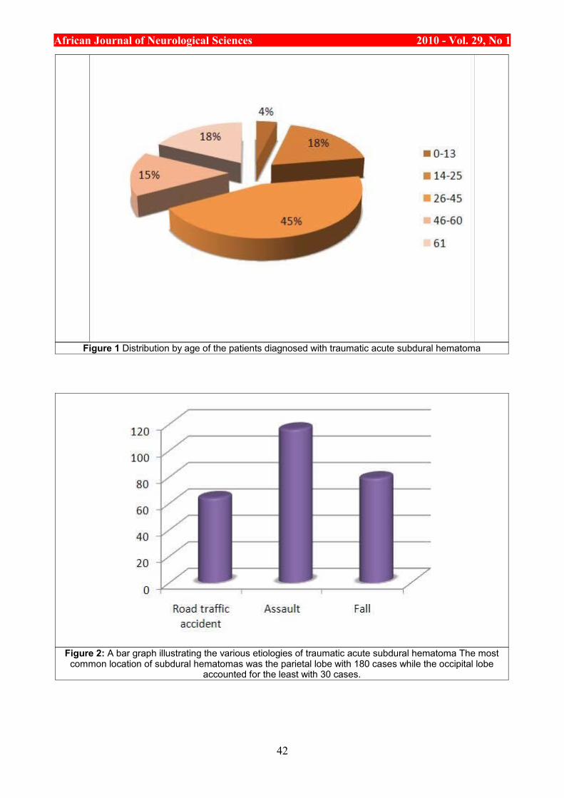
African Journal of Neurological Sciences 2010 - Vol. 29, No 1
Figure 1 Distribution by age of the patients diagnosed with traumatic acute subdural hematoma
Figure 2: A bar graph illustrating the various etiologies of traumatic acute subdural hematoma The most common location of subdural hematomas was the parietal lobe with 180 cases while the occipital lobe
accounted for the least with 30 cases.
42
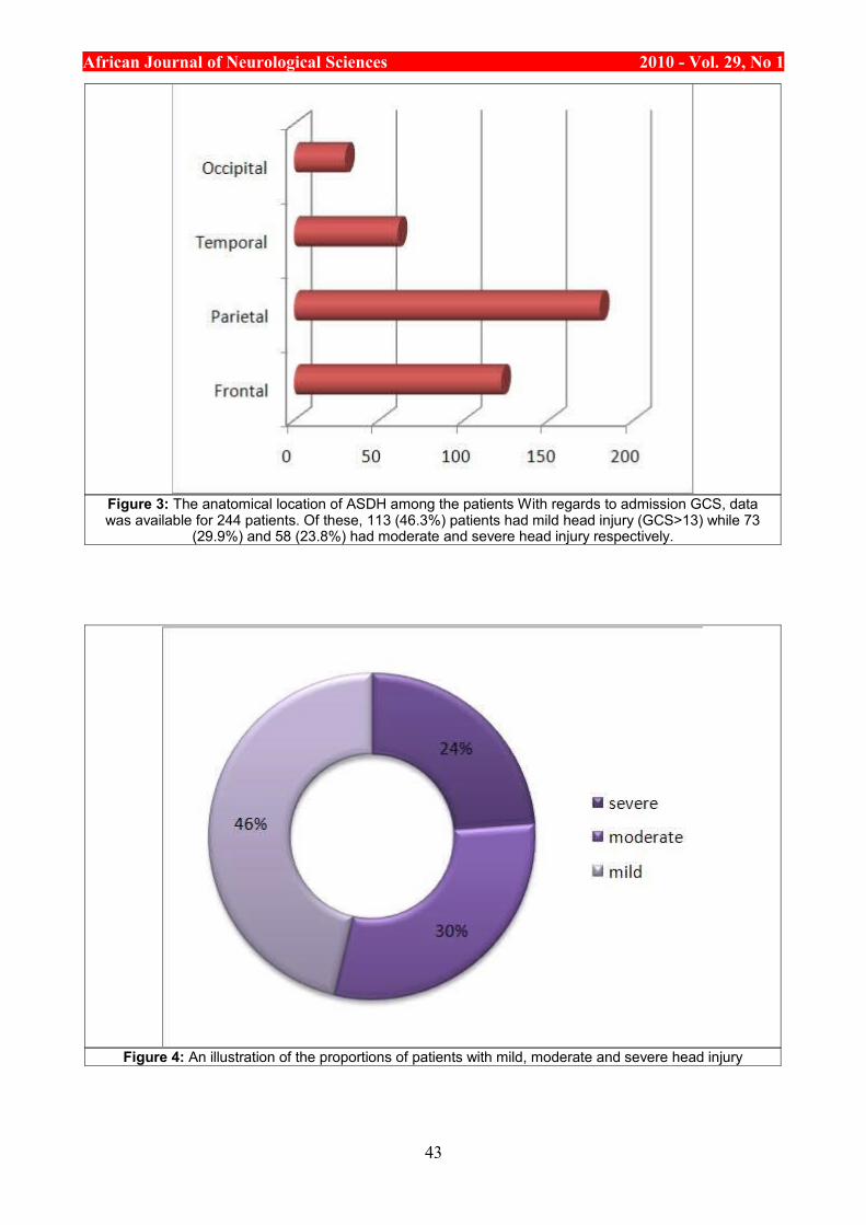
African Journal of Neurological Sciences 2010 - Vol. 29, No 1
Figure 3: The anatomical location of ASDH among the patients With regards to admission GCS, data was available for 244 patients. Of these, 113 (46.3%) patients had mild head injury (GCS>13) while 73
(29.9%) and 58 (23.8%) had moderate and severe head injury respectively.
Figure 4: An illustration of the proportions of patients with mild, moderate and severe head injury
43
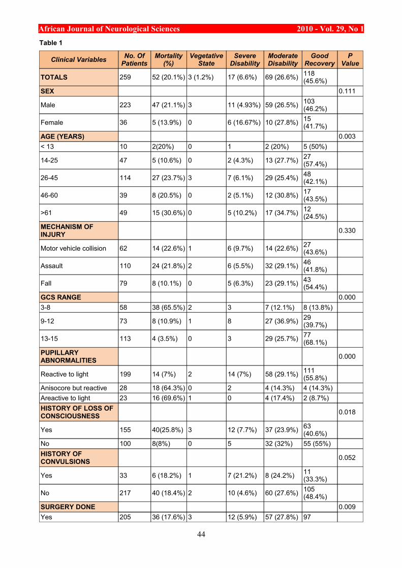
African Journal of Neurological Sciences 2010 - Vol. 29, No 1
Table 1
Clinical Variables No. Of Patients
Mortality (%)
Vegetative State
Severe Disability
Moderate Disability
Good Recovery
P Value
TOTALS 259 52 (20.1%) 3 (1.2%) 17 (6.6%) 69 (26.6%) 118 (45.6%)
SEX 0.111
Male 223 47 (21.1%) 3 11 (4.93%) 59 (26.5%) 103 (46.2%)
Female 36 5 (13.9%) 0 6 (16.67%) 10 (27.8%) 15 (41.7%)
AGE (YEARS) 0.003< 13 10 2(20%) 0 1 2 (20%) 5 (50%)
14-25 47 5 (10.6%) 0 2 (4.3%) 13 (27.7%) 27 (57.4%)
26-45 114 27 (23.7%) 3 7 (6.1%) 29 (25.4%) 48 (42.1%)
46-60 39 8 (20.5%) 0 2 (5.1%) 12 (30.8%) 17 (43.5%)
>61 49 15 (30.6%) 0 5 (10.2%) 17 (34.7%) 12 (24.5%)
MECHANISM OF INJURY 0.330
Motor vehicle collision 62 14 (22.6%) 1 6 (9.7%) 14 (22.6%) 27 (43.6%)
Assault 110 24 (21.8%) 2 6 (5.5%) 32 (29.1%) 46 (41.8%)
Fall 79 8 (10.1%) 0 5 (6.3%) 23 (29.1%) 43 (54.4%)
GCS RANGE 0.0003-8 58 38 (65.5%) 2 3 7 (12.1%) 8 (13.8%)
9-12 73 8 (10.9%) 1 8 27 (36.9%) 29 (39.7%)
13-15 113 4 (3.5%) 0 3 29 (25.7%) 77 (68.1%)
PUPILLARY ABNORMALITIES 0.000
Reactive to light 199 14 (7%) 2 14 (7%) 58 (29.1%) 111 (55.8%)
Anisocore but reactive 28 18 (64.3%) 0 2 4 (14.3%) 4 (14.3%)Areactive to light 23 16 (69.6%) 1 0 4 (17.4%) 2 (8.7%)HISTORY OF LOSS OF CONSCIOUSNESS 0.018
Yes 155 40(25.8%) 3 12 (7.7%) 37 (23.9%) 63 (40.6%)
No 100 8(8%) 0 5 32 (32%) 55 (55%)HISTORY OF CONVULSIONS 0.052
Yes 33 6 (18.2%) 1 7 (21.2%) 8 (24.2%) 11 (33.3%)
No 217 40 (18.4%) 2 10 (4.6%) 60 (27.6%) 105 (48.4%)
SURGERY DONE 0.009Yes 205 36 (17.6%) 3 12 (5.9%) 57 (27.8%) 97
44
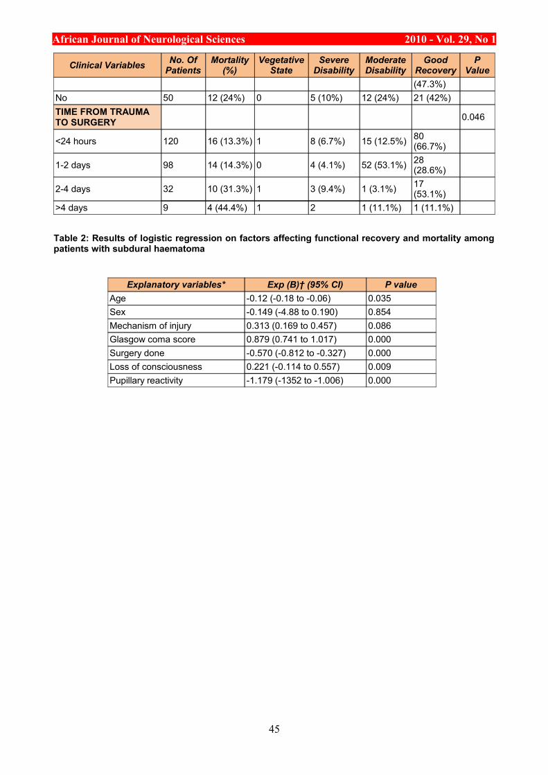
African Journal of Neurological Sciences 2010 - Vol. 29, No 1
Clinical Variables No. Of Patients
Mortality (%)
Vegetative State
Severe Disability
Moderate Disability
Good Recovery
P Value
(47.3%)No 50 12 (24%) 0 5 (10%) 12 (24%) 21 (42%)TIME FROM TRAUMA TO SURGERY 0.046
<24 hours 120 16 (13.3%) 1 8 (6.7%) 15 (12.5%) 80 (66.7%)
1-2 days 98 14 (14.3%) 0 4 (4.1%) 52 (53.1%) 28 (28.6%)
2-4 days 32 10 (31.3%) 1 3 (9.4%) 1 (3.1%) 17 (53.1%)
>4 days 9 4 (44.4%) 1 2 1 (11.1%) 1 (11.1%)
Table 2: Results of logistic regression on factors affecting functional recovery and mortality among patients with subdural haematoma
Explanatory variables* Exp (B)† (95% CI) P valueAge -0.12 (-0.18 to -0.06) 0.035Sex -0.149 (-4.88 to 0.190) 0.854Mechanism of injury 0.313 (0.169 to 0.457) 0.086Glasgow coma score 0.879 (0.741 to 1.017) 0.000Surgery done -0.570 (-0.812 to -0.327) 0.000Loss of consciousness 0.221 (-0.114 to 0.557) 0.009Pupillary reactivity -1.179 (-1352 to -1.006) 0.000
45
African Journal of Neurological Sciences 2010 - Vol. 29, No 1
REFERENCES1. BULLOCK MR, CHESNUT R, GHAJAR J, GORDON D, HARTL R, NEWELL DW. Surgical
management of acute subdural hematomas. Neurosurgery 2006; 58 (3): 16-24.2. FELL DA, FITZGERALD S, MOIEL RH, CARAM P. Acute subdural hematomas. Review of 144
cases. J Neurosurg 1975; 42: 37-42.3. HASELSBERGER K, PUCHER R, AUER LM. Prognosis after acute subdural or epidural
hemorrhage. Acta Neurochir (Wien) 1988; 90: 111-116.4. HATASHITA S, KOGA N, HOSAKA Y, TAKAGI S. Acute subdural hematoma: severity of injury,
surgical intervention, and mortality. Neurol Med Chir (Tokyo) 1993; 33: 13-18.5. HOWARD MA, GROSS AS, DACEY RG, WINN HR. Acute subdural hematomas: an agedependent
clinical entity. J Neurosurg 1989; 71: 85863.6. JANG HS, LEE YB, CHUNG C, LEE KC, PARK YS, MOK JH. Acute subdural hematoma: an
analysis of 244 operated cases. J Korean Neurosurg Soc 1996; 25: 111-118.7. KLUN B, FETTICH M. Factors influencing the outcome in acute subdural hematoma. A review of
330 cases. Acta Neurochir (Wien) 1984; 71 : 171-178.8. KOC RK, AKDEMIR H, OKTEM IS, MERAL M, MENKÜ A. Acute subdural hematoma : outcome
and outcome prediction. Neurosurg Rev 1997; 20: 239-244.9. KOTWICA Z, BRZEZINSKI J. Acute subdural hematoma in adults : an analysis of outcome in
comatose patients. Acta Neurochir (Wien) 1993; 121: 95-99.10.KYU-HONG KIM. Predictors for Functional Recovery and Mortality of Surgically Treated Traumatic
Acute Subdural Hematomas in 256 Patients. J Korean Neurosurg Soc 2009; 45 : 143-150.11.LEACH P, CHILDS C, EVANS J, JOHNSTON N, PROTHEROE R, KING A. Transfer times for
patients with extradural and subdural haematomas to neurosurgery in Greater Manchester. Br J Neurosurg 2007; 21: 11-15.
12.MARSHALL LF, GAUTILLE T, KLAUBER MR. The outcome of severe closed head injury. J Neurosurg 1991; 75: 528-536.
13.MOSENTHAL AC, LAVERY RF, ADDIS M, KAUL S, ROSS S, MARBURGER R. Isolated traumatic brain injury: age is an independent predictor of mortality and early outcome. J Trauma 2002; 52: 907-911.
14.MUNRO PT, SMITH RD, PARKE TR. Effect of patients’ age on management of acute intracranial haematoma: prospective national study. BMJ 2002; 325: 1001
15.PHUENPATHOM N, CHOOMUANG M, RATANALERT S. Outcome and outcome prediction in acute subdural hematoma. Surg Neurol 1993; 40: 22-25.
16.PIOTROWSKI WP, MÜHL BJ. Results of surgery in acute subdural hematoma. Unfauchirurg 1995; 98: 432-436.
17.RICHARDS T, HOFF J. Factors affecting survival from acute subdural hematoma. Surgery 1974; 75: 253-258.
18.RITTER AM, MUIZELAAR JP. Brainstem blood flow, pupillary response and outcome in patients with severe head injuries. Neurosurg 1999; 44: 941-948.
19.SAKAS DE, BULLOCK MR, TEASDALE GM. One-year outcome following craniotomy for traumatic hematoma in patients with fixed dilated pupils. J Neurosurg 1995; 82: 961-965.
20.SEELIG JM, BECKER DP, MILLER JD, GREENBERG RP, WARD JD, CHOI SC. Traumatic acute subdural hematoma: major mortality reduction in comatose patients treated within four hours. New Engl J Med 1981; 304:1511-1518.
21.SERVADEI F. Prognostic factors in severely head injured adult patients with acute subdural hematomas. Acta Neurochir (Wien) 1997; 139: 279-285.
22.STENING WA, BERRY G, DAN NG, KWOK B, MANDRYK JA, RING I. Experience with acute subdural hematomas in New South Wales. Aust N Z J Surg 1986; 56: 549-556.
23.STONE JL, RIFAI MH, SUGAR O, LANG RG, OLDERSHAW JB, MOODY RA. Subdural hematomas. I. Acute subdural hematoma: progress in definition, clinical pathology, and therapy. Surg Neurol 1983; 19 : 216-231.
24.TAUSSKY P, WIDMERA HR, TAKALAB J, FANDINOA J. Outcome after acute traumatic subdural and epidural haematoma in Switzerland: a single-centre experience. Swiss Med Wkly 2008; 138(19-20): 281-285.
25.TIAN H, CHEN S, XU T ET AL. Risk factors related to hospital mortality in patients with isolated traumatic acute subdural haematoma: analysis of 308 patients undergone surgery. Chin Med J 2008; 121(12):1080-1084.
26. WILBERGER JE JR, HARRIS M, DIAMOND DL. Acute subdural hematoma: morbidity, mortality, and operative timing. J Neurosurg 1991; 74: 212- 218.
46
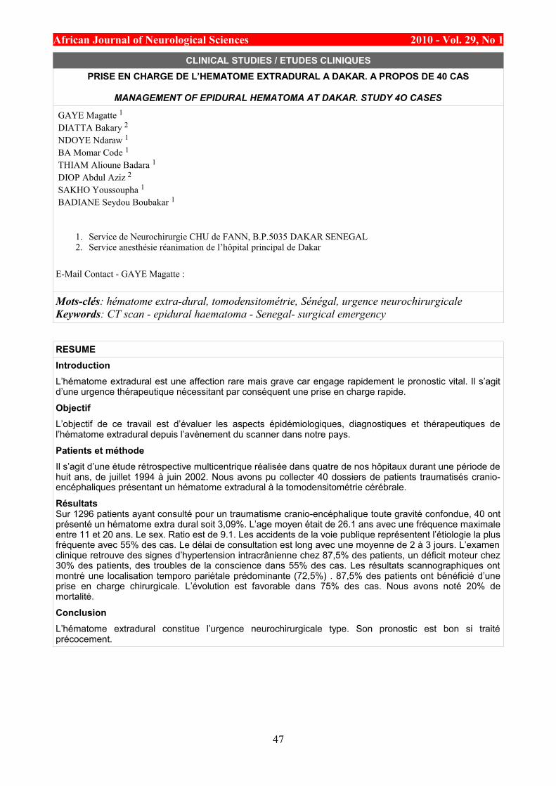
African Journal of Neurological Sciences 2010 - Vol. 29, No 1
CLINICAL STUDIES / ETUDES CLINIQUESPRISE EN CHARGE DE L’HEMATOME EXTRADURAL A DAKAR. A PROPOS DE 40 CAS
MANAGEMENT OF EPIDURAL HEMATOMA AT DAKAR. STUDY 4O CASES
GAYE Magatte 1 DIATTA Bakary 2 NDOYE Ndaraw 1 BA Momar Code 1 THIAM Alioune Badara 1 DIOP Abdul Aziz 2 SAKHO Youssoupha 1 BADIANE Seydou Boubakar 1
1. Service de Neurochirurgie CHU de FANN, B.P.5035 DAKAR SENEGAL 2. Service anesthésie réanimation de l’hôpital principal de Dakar
E-Mail Contact - GAYE Magatte :
Mots-clés: hématome extra-dural, tomodensitométrie, Sénégal, urgence neurochirurgicaleKeywords: CT scan - epidural haematoma - Senegal- surgical emergency
RESUMEIntroduction
L’hématome extradural est une affection rare mais grave car engage rapidement le pronostic vital. Il s’agit d’une urgence thérapeutique nécessitant par conséquent une prise en charge rapide.
Objectif L’objectif de ce travail est d’évaluer les aspects épidémiologiques, diagnostiques et thérapeutiques de l’hématome extradural depuis l’avènement du scanner dans notre pays.
Patients et méthode
Il s’agit d’une étude rétrospective multicentrique réalisée dans quatre de nos hôpitaux durant une période de huit ans, de juillet 1994 à juin 2002. Nous avons pu collecter 40 dossiers de patients traumatisés cranio-encéphaliques présentant un hématome extradural à la tomodensitométrie cérébrale.
Résultats Sur 1296 patients ayant consulté pour un traumatisme cranio-encéphalique toute gravité confondue, 40 ont présenté un hématome extra dural soit 3,09%. L’age moyen était de 26.1 ans avec une fréquence maximale entre 11 et 20 ans. Le sex. Ratio est de 9.1. Les accidents de la voie publique représentent l’étiologie la plus fréquente avec 55% des cas. Le délai de consultation est long avec une moyenne de 2 à 3 jours. L’examen clinique retrouve des signes d’hypertension intracrânienne chez 87,5% des patients, un déficit moteur chez 30% des patients, des troubles de la conscience dans 55% des cas. Les résultats scannographiques ont montré une localisation temporo pariétale prédominante (72,5%) . 87,5% des patients ont bénéficié d’une prise en charge chirurgicale. L’évolution est favorable dans 75% des cas. Nous avons noté 20% de mortalité.
Conclusion
L’hématome extradural constitue l’urgence neurochirurgicale type. Son pronostic est bon si traité précocement.
47
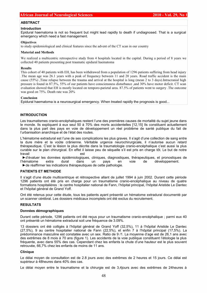
African Journal of Neurological Sciences 2010 - Vol. 29, No 1
ABSTRACTIntroduction Epidural haematoma is not so frequent but might lead rapidly to death if undiagnosed. That is a surgical emergency which need a fast management.
Objectives to study epidemiological and clinical features since the advent of the CT scan in our country
Material and Methods
We realized a multicentric retrospective study from 4 hospitals located in the capital. During a period of 8 years we collected 40 patients presenting post traumatic epidural haematoma
Results This cohort of 40 patients with EH, has been withdrawed from a population of 1296 patients suffering from head injury .The mean age was 26.1 years with a peak of frequency between 11 and 20 years. Road traffic accident is the main cause (55%) ,Time relapse between the trauma and arrival at the hospital is long (mean 2 to 3 days).Intracranial high pressure is found in 87.5%, 55% of our patients have consciensness disturbance ,and 30% have motor deficit. CT scan evaluation showed that EH is mostly located on temporo-parietal area. 87.5% of patients went to surgery .The outcome was good on 75%. Death rate was 20%
Conclusion Epidural haematoma is a neurosurgical emergency. When treated rapidly the prognosis is good...
INTRODUCTIONLes traumatismes cranio-encéphaliques restent l’une des premières causes de mortalité du sujet jeune dans le monde. Ils expliquent à eux seul 50 à 70% des morts accidentelles [12,15] Ils constituent actuellement dans la plus part des pays en voie de développement un réel problème de santé publique du fait de l’urbanisation anarchique et de l’état des routes.
L’hématome extradural est l’une de ses complications les plus graves. Il s’agit d’une collection de sang entre la dure mère et la voûte crânienne. Véritable urgence neurochirurgicale, il n’autorise aucun retard thérapeutique. C’est la lésion la plus décrite dans la traumatologie cranio-encéphalique c’est aussi la plus curable sur le plan chirurgical. En effet il laisse peu de séquelle s’il est pris en charge tôt. Le but de notre travail est : ►d’évaluer les données épidémiologiques, cliniques, diagnostiques, thérapeutiques, et pronostiques de l’hématome extra dural dans un pays en voie de développement. ►de réaffirmer les indications thérapeutiques de cette pathologie.
PATIENTS ET METHODEIl s’agit d’une étude multicentrique et rétrospective allant de juillet 1994 à juin 2002. Durant cette période 1296 patients ont été pris en charge pour un traumatisme cranio-encéphalique au niveau de quatre formations hospitalières : le centre hospitalier national de Fann, l’hôpital principal, l’hôpital Aristide Le Dantec et l’hôpital général de Grand Yoff.
Ont été retenus pour cette étude, tous les patients ayant présenté un hématome extradural documenté par un scanner cérébral. Les dossiers médicaux incomplets ont été exclus du recrutement.
RESULTATS
Données démographiquesDurant cette période, 1296 patients ont été reçus pour un traumatisme cranio-encéphalique ; parmi eux 40 ont présenté un hématome extradural soit une fréquence de 3.09%.
13 dossiers ont été colligés à l’hôpital général de Grand Yoff (32,5%), 11 à l’hôpital Aristide Le Dantec (27,5%), 9 au centre hospitalier national de Fann (22,5%), et enfin 7 à l’hôpital principal (17,5%). La prédominance masculine est constatée avec un sex. Ratio de 9 /1. La moyenne d’age est de 26,1 ans avec des extrêmes de 8 mois à 70 ans (figure 1). Les accidents de la voie publique constituent l’étiologie la plus fréquente, avec dans 55% des cas. Cependant chez les enfants la chute d’une hauteur est le plus souvent retrouvée, 66,7% chez les enfants de moins de 11 ans.
CliniqueLe délai moyen de consultation est de 2.8 jours avec des extrêmes de 2 heures et 15 jours. Ce délai est supérieur à 48heures dans 40% des cas.
Le délai moyen entre le traumatisme et la chirurgie est de 3,4jours avec des extrêmes de 24heures à
48
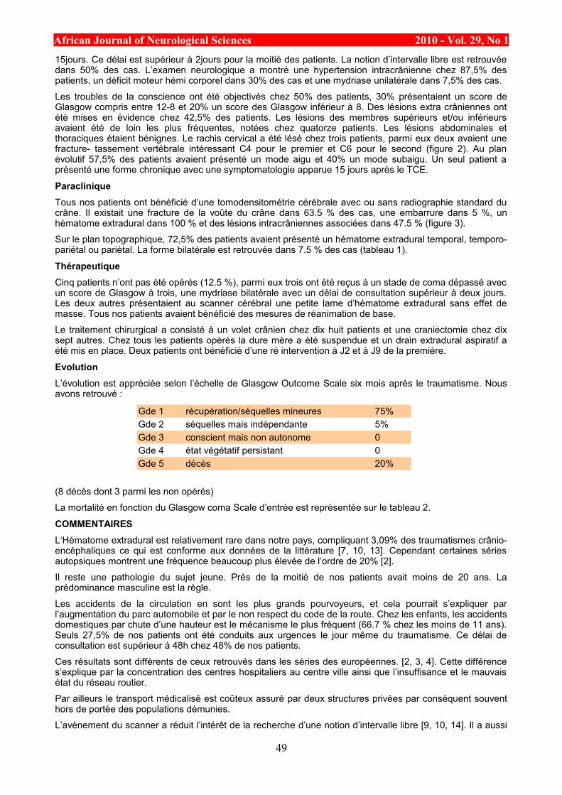
African Journal of Neurological Sciences 2010 - Vol. 29, No 115jours. Ce délai est supérieur à 2jours pour la moitié des patients. La notion d’intervalle libre est retrouvée dans 50% des cas. L’examen neurologique a montré une hypertension intracrânienne chez 87,5% des patients, un déficit moteur hémi corporel dans 30% des cas et une mydriase unilatérale dans 7,5% des cas.
Les troubles de la conscience ont été objectivés chez 50% des patients, 30% présentaient un score de Glasgow compris entre 12-8 et 20% un score des Glasgow inférieur à 8. Des lésions extra crâniennes ont été mises en évidence chez 42,5% des patients. Les lésions des membres supérieurs et/ou inférieurs avaient été de loin les plus fréquentes, notées chez quatorze patients. Les lésions abdominales et thoraciques étaient bénignes. Le rachis cervical a été lésé chez trois patients, parmi eux deux avaient une fracture- tassement vertébrale intéressant C4 pour le premier et C6 pour le second (figure 2). Au plan évolutif 57,5% des patients avaient présenté un mode aigu et 40% un mode subaigu. Un seul patient a présenté une forme chronique avec une symptomatologie apparue 15 jours après le TCE.
ParacliniqueTous nos patients ont bénéficié d’une tomodensitométrie cérébrale avec ou sans radiographie standard du crâne. Il existait une fracture de la voûte du crâne dans 63.5 % des cas, une embarrure dans 5 %, un hématome extradural dans 100 % et des lésions intracrâniennes associées dans 47.5 % (figure 3).
Sur le plan topographique, 72,5% des patients avaient présenté un hématome extradural temporal, temporo-pariétal ou pariétal. La forme bilatérale est retrouvée dans 7.5 % des cas (tableau 1).
ThérapeutiqueCinq patients n’ont pas été opérés (12.5 %), parmi eux trois ont été reçus à un stade de coma dépassé avec un score de Glasgow à trois, une mydriase bilatérale avec un délai de consultation supérieur à deux jours. Les deux autres présentaient au scanner cérébral une petite lame d’hématome extradural sans effet de masse. Tous nos patients avaient bénéficié des mesures de réanimation de base.
Le traitement chirurgical a consisté à un volet crânien chez dix huit patients et une craniectomie chez dix sept autres. Chez tous les patients opérés la dure mère a été suspendue et un drain extradural aspiratif a été mis en place. Deux patients ont bénéficié d’une ré intervention à J2 et à J9 de la première.
EvolutionL’évolution est appréciée selon l’échelle de Glasgow Outcome Scale six mois après le traumatisme. Nous avons retrouvé :
Gde 1 récupération/séquelles mineures 75% Gde 2 séquelles mais indépendante 5% Gde 3 conscient mais non autonome 0 Gde 4 état végétatif persistant 0 Gde 5 décès 20%
(8 décès dont 3 parmi les non opérés)
La mortalité en fonction du Glasgow coma Scale d’entrée est représentée sur le tableau 2.
COMMENTAIRESL‘Hématome extradural est relativement rare dans notre pays, compliquant 3,09% des traumatismes crânio-encéphaliques ce qui est conforme aux données de la littérature [7, 10, 13]. Cependant certaines séries autopsiques montrent une fréquence beaucoup plus élevée de l’ordre de 20% [2].
Il reste une pathologie du sujet jeune. Près de la moitié de nos patients avait moins de 20 ans. La prédominance masculine est la règle.
Les accidents de la circulation en sont les plus grands pourvoyeurs, et cela pourrait s’expliquer par l’augmentation du parc automobile et par le non respect du code de la route. Chez les enfants, les accidents domestiques par chute d’une hauteur est le mécanisme le plus fréquent (66.7 % chez les moins de 11 ans). Seuls 27,5% de nos patients ont été conduits aux urgences le jour même du traumatisme. Ce délai de consultation est supérieur à 48h chez 48% de nos patients.
Ces résultats sont différents de ceux retrouvés dans les séries des européennes. [2, 3, 4]. Cette différence s’explique par la concentration des centres hospitaliers au centre ville ainsi que l’insuffisance et le mauvais état du réseau routier.
Par ailleurs le transport médicalisé est coûteux assuré par deux structures privées par conséquent souvent hors de portée des populations démunies.
L’avènement du scanner a réduit l’intérêt de la recherche d’une notion d’intervalle libre [9, 10, 14]. Il a aussi
49
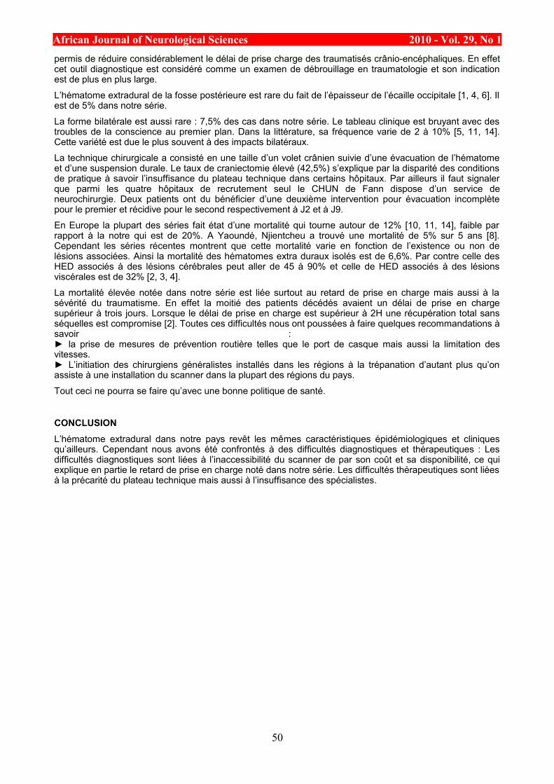
African Journal of Neurological Sciences 2010 - Vol. 29, No 1permis de réduire considérablement le délai de prise charge des traumatisés crânio-encéphaliques. En effet cet outil diagnostique est considéré comme un examen de débrouillage en traumatologie et son indication est de plus en plus large.
L’hématome extradural de la fosse postérieure est rare du fait de l’épaisseur de l’écaille occipitale [1, 4, 6]. Il est de 5% dans notre série.
La forme bilatérale est aussi rare : 7,5% des cas dans notre série. Le tableau clinique est bruyant avec des troubles de la conscience au premier plan. Dans la littérature, sa fréquence varie de 2 à 10% [5, 11, 14]. Cette variété est due le plus souvent à des impacts bilatéraux.
La technique chirurgicale a consisté en une taille d’un volet crânien suivie d’une évacuation de l’hématome et d’une suspension durale. Le taux de craniectomie élevé (42,5%) s’explique par la disparité des conditions de pratique à savoir l’insuffisance du plateau technique dans certains hôpitaux. Par ailleurs il faut signaler que parmi les quatre hôpitaux de recrutement seul le CHUN de Fann dispose d’un service de neurochirurgie. Deux patients ont du bénéficier d’une deuxième intervention pour évacuation incomplète pour le premier et récidive pour le second respectivement à J2 et à J9.
En Europe la plupart des séries fait état d’une mortalité qui tourne autour de 12% [10, 11, 14], faible par rapport à la notre qui est de 20%. A Yaoundé, Njientcheu a trouvé une mortalité de 5% sur 5 ans [8]. Cependant les séries récentes montrent que cette mortalité varie en fonction de l’existence ou non de lésions associées. Ainsi la mortalité des hématomes extra duraux isolés est de 6,6%. Par contre celle des HED associés à des lésions cérébrales peut aller de 45 à 90% et celle de HED associés à des lésions viscérales est de 32% [2, 3, 4].
La mortalité élevée notée dans notre série est liée surtout au retard de prise en charge mais aussi à la sévérité du traumatisme. En effet la moitié des patients décédés avaient un délai de prise en charge supérieur à trois jours. Lorsque le délai de prise en charge est supérieur à 2H une récupération total sans séquelles est compromise [2]. Toutes ces difficultés nous ont poussées à faire quelques recommandations à savoir : ► la prise de mesures de prévention routière telles que le port de casque mais aussi la limitation des vitesses. ► L’initiation des chirurgiens généralistes installés dans les régions à la trépanation d’autant plus qu’on assiste à une installation du scanner dans la plupart des régions du pays.
Tout ceci ne pourra se faire qu’avec une bonne politique de santé.
CONCLUSIONL’hématome extradural dans notre pays revêt les mêmes caractéristiques épidémiologiques et cliniques qu’ailleurs. Cependant nous avons été confrontés à des difficultés diagnostiques et thérapeutiques : Les difficultés diagnostiques sont liées à l’inaccessibilité du scanner de par son coût et sa disponibilité, ce qui explique en partie le retard de prise en charge noté dans notre série. Les difficultés thérapeutiques sont liées à la précarité du plateau technique mais aussi à l’insuffisance des spécialistes.
50

African Journal of Neurological Sciences 2010 - Vol. 29, No 1
Figure 1Répartition des cas selon l’age
Figure 2Lésions extracraniennes
51
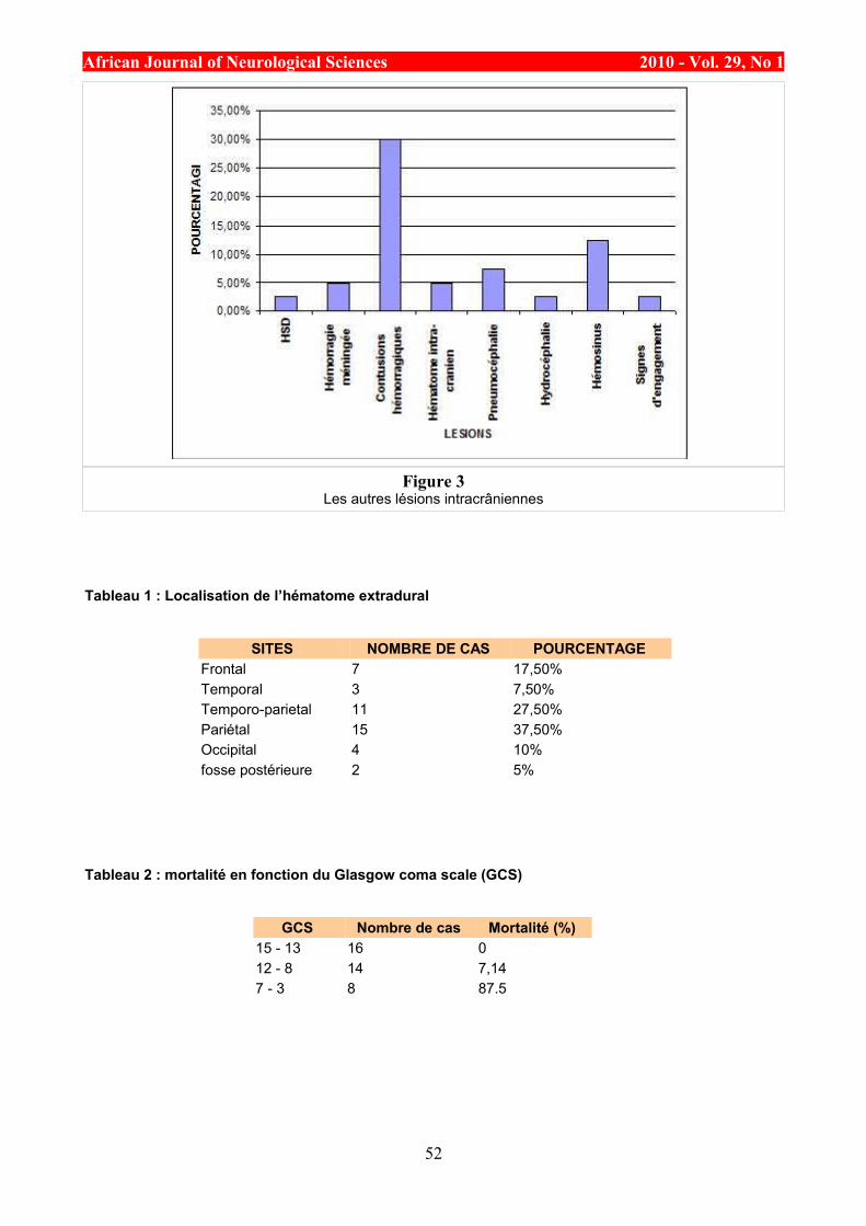
African Journal of Neurological Sciences 2010 - Vol. 29, No 1
Figure 3
Les autres lésions intracrâniennes
Tableau 1 : Localisation de l’hématome extradural
SITES NOMBRE DE CAS POURCENTAGE Frontal 7 17,50% Temporal 3 7,50% Temporo-parietal 11 27,50% Pariétal 15 37,50% Occipital 4 10% fosse postérieure 2 5%
Tableau 2 : mortalité en fonction du Glasgow coma scale (GCS)
GCS Nombre de cas Mortalité (%) 15 - 13 16 0 12 - 8 14 7,14 7 - 3 8 87.5
52
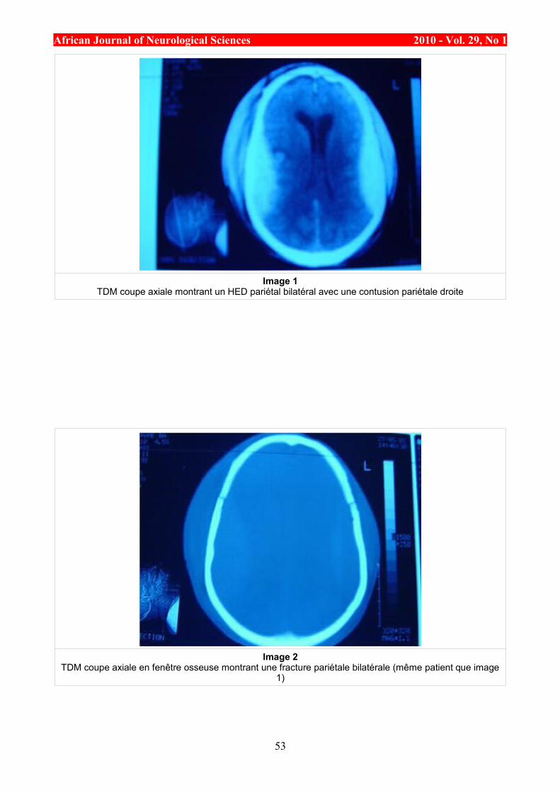
African Journal of Neurological Sciences 2010 - Vol. 29, No 1
Image 1TDM coupe axiale montrant un HED pariétal bilatéral avec une contusion pariétale droite
Image 2TDM coupe axiale en fenêtre osseuse montrant une fracture pariétale bilatérale (même patient que image
1)
53
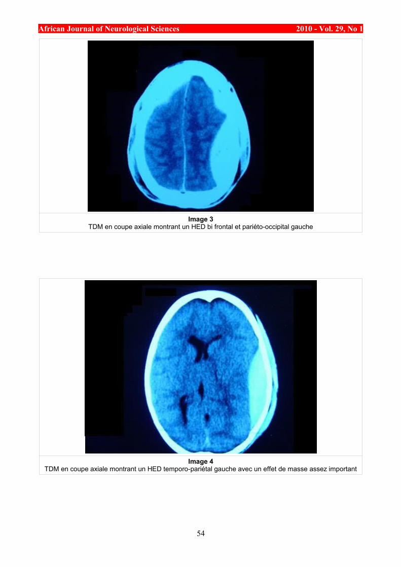
African Journal of Neurological Sciences 2010 - Vol. 29, No 1
Image 3TDM en coupe axiale montrant un HED bi frontal et pariéto-occipital gauche
Image 4TDM en coupe axiale montrant un HED temporo-pariétal gauche avec un effet de masse assez important
54
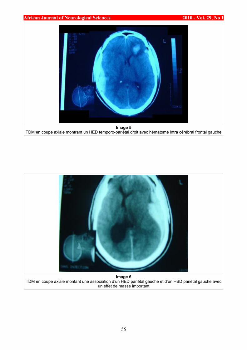
African Journal of Neurological Sciences 2010 - Vol. 29, No 1
Image 5TDM en coupe axiale montrant un HED temporo-pariétal droit avec hématome intra cérébral frontal gauche
Image 6TDM en coupe axiale montant une association d’un HED pariétal gauche et d’un HSD pariétal gauche avec
un effet de masse important
55
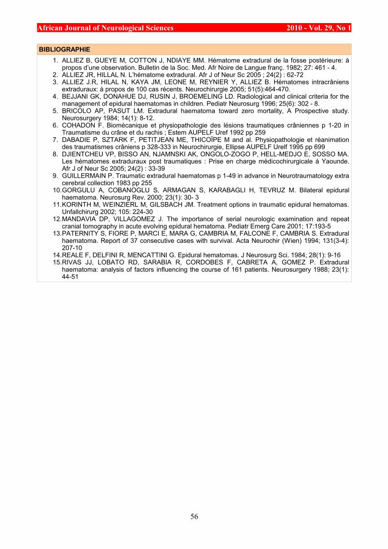
African Journal of Neurological Sciences 2010 - Vol. 29, No 1
BIBLIOGRAPHIE1. ALLIEZ B, GUEYE M, COTTON J, NDIAYE MM. Hématome extradural de la fosse postérieure: à
propos d’une observation. Bulletin de la Soc. Med. Afr Noire de Langue franç. 1982; 27: 461 - 4. 2. ALLIEZ JR, HILLAL N. L’hématome extradural. Afr J of Neur Sc 2005 ; 24(2) : 62-72 3. ALLIEZ J.R, HILAL N, KAYA JM, LEONE M, REYNIER Y, ALLIEZ B. Hématomes intracrâniens
extraduraux: à propos de 100 cas récents. Neurochirurgie 2005; 51(5):464-470. 4. BEJJANI GK, DONAHUE DJ, RUSIN J, BROEMELING LD. Radiological and clinical criteria for the
management of epidural haematomas in children. Pediatr Neurosurg 1996; 25(6): 302 - 8. 5. BRICOLO AP, PASUT LM. Extradural haematoma toward zero mortality, A Prospective study.
Neurosurgery 1984; 14(1): 8-12. 6. COHADON F. Biomécanique et physiopathologie des lésions traumatiques crâniennes p 1-20 in
Traumatisme du crâne et du rachis ; Estem AUPELF Uref 1992 pp 259 7. DABADIE P, SZTARK F, PETITJEAN ME, THICOÏPE M and al. Physiopathologie et réanimation
des traumatismes crâniens p 328-333 in Neurochirurgie, Ellipse AUPELF Urelf 1995 pp 699 8. DJIENTCHEU VP, BISSO AN, NJAMNSKI AK, ONGOLO-ZOGO P, HELL-MEDJO E, SOSSO MA.
Les hématomes extraduraux post traumatiques : Prise en charge médicochirurgicale à Yaounde. Afr J of Neur Sc 2005; 24(2) : 33-39
9. GUILLERMAIN P. Traumatic extradural haematomas p 1-49 in advance in Neurotraumatology extra cerebral collection 1983 pp 255
10.GORGULU A, COBANOGLU S, ARMAGAN S, KARABAGLI H, TEVRUZ M. Bilateral epidural haematoma. Neurosurg Rev. 2000; 23(1): 30- 3
11.KORINTH M, WEINZIERL M, GILSBACH JM. Treatment options in traumatic epidural hematomas. Unfallchirurg 2002; 105: 224-30
12.MANDAVIA DP, VILLAGOMEZ J. The importance of serial neurologic examination and repeat cranial tomography in acute evolving epidural hematoma. Pediatr Emerg Care 2001; 17:193-5
13.PATERNITY S, FIORE P, MARCI E, MARA G, CAMBRIA M, FALCONE F, CAMBRIA S. Extradural haematoma. Report of 37 consecutive cases with survival. Acta Neurochir (Wien) 1994; 131(3-4): 207-10
14.REALE F, DELFINI R, MENCATTINI G. Epidural hematomas. J Neurosurg Sci. 1984; 28(1): 9-16 15.RIVAS JJ, LOBATO RD, SARABIA R, CORDOBES F, CABRETA A, GOMEZ P. Extradural
haematoma: analysis of factors influencing the course of 161 patients. Neurosurgery 1988; 23(1): 44-51
56
African Journal of Neurological Sciences 2010 - Vol. 29, No 1
CASE REPORT / CAS CLINIQUEANOREXIE MENTALE ET MYELINOLYSE CENTROPONTINE ET EXTRAPONTINE
ANOREXIA NERVOSA ASSOCIATED WITH CENTRAL PONTINE MYELINOLYSIS AND EXTRAPONTINE MYELINOLYSIS
EL MIDAOUI Aouatef 1 MRANI Alia 1 FAIZ Fatima 1 EL HOUARI Nawfal 2 KHATOUF Mohammed 2 MESSOUAK Oufae 1 BELAHSEN Mohamed Faouzi 1
1. Service de Neurologie, CHU Hassan II, Fès, Maroc 2. Service de Réanimation polyvalente CHU Hassan II, Fès, Maroc
E-Mail Contact - EL MIDAOUI Aouatef :
Mots-clés: Anorexie mentale ; Hyponatrémie ; Myelinolyse centropontine; Myelinolyse extrapontine.Keywords: Anorexia nervosa; Centralpontine myelinolysis; Extrapontine myelinolysis; Hyponatremia.
RESUMEIntroduction La myélinolyse centropontine et extrapontine est une complication connue mais rare des hyponatrémies et de leur correction. Elle représente une entité pathologique définie par des arguments cliniques et radiologiques survenant dans un contexte étiologique susceptible d’orienter le diagnostic. L’anorexie mentale est rarement décrite comme responsable d’une hyponatrémie compliquée de myélinolyse centro et extrapontine.
Observation Nous rapportons l’observation d’une patiente de 19 ans ayant une histoire d’anorexie mentale depuis 4 ans admise pour trouble de conscience associée à une hyponatrémie. Malgré la correction adéquate des troubles hydro-électrolytiques, la patiente a présenté des crises convulsives tonico-cloniques généralisées et une tétraplégie. L’IRM cérébrale a confirmé le diagnostic de la myélinolyse centro et extrapontine. Malgré l’installation d’un tableau clinique particulièrement spectaculaire, l’évolution était marquée par l’amélioration progressive des troubles neurologiques.
Discussion La myélinolyse centropontine est une complication neurologique rattachée le plus souvent à la correction trop rapide d’une hyponatrémie. Dans le cas rapporté, la correction de l’hyponatrémie ne semble pas être l’agent déclenchant du syndrome, l’hypokaliémie et la dénutrition, en induisant une vulnérabilité particulière des cellules gliales aux variations osmotiques, ont probablement joué un rôle favorisant. Le pronostic de la myélinolyse est diversement apprécié mais dans le cadre de l’anorexie mentale, il semble être moins grave. Toutefois aucune corrélation n’existe entre la gravité du tableau clinique, l’aspect radiologique et l’évolution d’où l’importance d’une surveillance clinique et radiologique étroite surtout durant le premier mois.
57
African Journal of Neurological Sciences 2010 - Vol. 29, No 1
SUMMARYIntroduction Central pontine myelinolysis (CPM) and extrapontine myelinolysis (EPM) are demyelinating disorders of central nervous system, more commonly associated with rapid correction of hyponatremia. Several patients with CPM and EMP as a complication of anorexia nervosa have been reported.
Clinical case We report a case of a 19 year-old woman with a 4 year history of anorexia who presented a disturbance of consciousness, secondary to hyponatremia. She developed a quadriplegia associated with epileptic fits one week after a slow correction of hyponatremia. Central pontine myelinolysis and extrapontine myelinolysis were formally diagnosed by MRI findings. Despite of severe initial symptoms, the evolution was favourable with a quasi complete recovery.
Discussion Central pontine myelinolysis and extrapontine myelinolysis may occur despite a treatment of hyponatremia conducted according to published guidelines. In our case, the correction of hyponatraemiaI is not the only causal agent of this syndrome; hypokalaemia and denutrition seem to be predisposing factors. The prognosis has been varying in the last decades and it is highly heterogenous with complete recovery reported in some cases to progression and death in others. This prognosis does not seem to be fatal with anorexia nervosa. Thus, it has been observed that the size of the lesion in the CPM and EPM does not correlate with the seriousness of the neurological disorders or the clinical picture course.
INTRODUCTIONInitialement décrite par Adams, Victor et Mancall en 1959, la myélinolyse centropontine (MCP) encore appelée syndrome de démyélinisation osmotique, est une complication neurologique rare. Elle correspond à une démyélinisation caractérisée par la destruction des gaines de myéline de la partie centrale de la base de la protubérance anciennement appelée pont, épargnant les neurones. Cette myélinolyse peut s’étendre à d’autres structures cérébrales, définissant la myélinolyse extrapontine (MEP). (1, 27, 33).
Si la MCP peut s’accompagner d’une MEP dans environ 10 % des cas, l’atteinte extrapontine isolée est rare (5, 13, 30). Sa physiopathologie est complexe et demeure mal élucidée, elle est associée à la dénutrition, à l’alcoolisme chronique et elle est souvent la conséquence d’une correction trop rapide d’une hyponatrémie (5, 13, 33). Son traitement est mal codifié et repose essentiellement sur des mesures générales de réanimation, avec des essais thérapeutiques non spécifiques [corticothérapie, Thyrotropin releasing hormone (TRH)] et des résultats incertains (4, 7, 10).
Nous rapportons le cas d’une patiente de 19 ans ayant une histoire d’anorexie mentale depuis 4 ans avec des vomissements chroniques, qui a présenté une myélinolyse centropontine et extrapontique malgré une correction adéquate de l’hyponatrémie.
OBSERVATIONNous rapportons le cas d’une patiente âgée de 19 ans ayant comme antécédents une gastrite avec des vomissements chroniques depuis 4 ans qui s’est présenté aux urgences pour trouble de conscience sans signes de focalisation avec un score GSC (Glasgow Coma Scale) à 12 (Ouverture des yeux à 3, réponse verbale à 4 et réponse motrice á 5) et altération de l’état général . Une TDM et une ponction lombaire réalisée en urgence étaient normales. Son bilan biologique initial a révélé une hyponatrémie à 110 meq/l, une hypokaliémie à 1,7 meq/l et une hypocalcémie à 71 meq/l. La patiente a eu une correction adéquate de l’hyponatrémie : 7 meq/l le premier jour et 14 meq/l les 2 jours suivants (schéma 1). La kaliémie et la calcémie ont été corrigées en deux jours. Après la normalisation de l’ionogramme, les vomissements ont persisté et la fibroscopie a montré une œsophagite sévère et la patiente a présenté une hypokaliémie qui a persisté une semaine malgré une correction orale et parentérale. L’évolution était marquée par la résolution des troubles de la conscience.
Huit jours après son admission, la patiente a présenté une tétraplégie plus marquée à droite, un mutisme et des crises convulsives tonicocloniques généralisées.
L’IRM réalisée a montré un hypersignal centropontique (figure 1) et extrapontique au niveau mésencéphalique, cérébelleux, des noyaux gris centraux notamment le putamen, le noyau caudé et le thalamus et au niveau du cortex temporal; ce qui a permis de poser le diagnostic de la myélinolyse centropontine et extrapontique (figure 2-3-4).
L’interrogatoire avec la famille a révélé des troubles de la conduite alimentaire à type d’anorexie/boulimie,
58
African Journal of Neurological Sciences 2010 - Vol. 29, No 1des vomissements initialement provoqués devenus spontanés après chaque repas, un amaigrissement important (poids de la patiente : 27 kg), une aménorrhée secondaire ainsi qu’un trouble de la perception de l’image corporelle sur un fond de personnalité obsessionnelle. Ce tableau était compatible avec le diagnostic d’anorexie mentale.
L’évolution après 3 mois était marquée par une amélioration progressive et nette des signes cliniques: La patiente retrouvait son autonomie, elle marchait seule et son langage est devenu fluent. À l’examen clinique persistaient un syndrome tétrapyramidal non déficitaire et un syndrome cérébelleux. Une IRM cérébrale de contrôle réalisée après 3 mois montrait une évolution favorable des lésions avec un hypersignal T2 plus discret au niveau des noyaux lenticulaires (Figure 5). La patiente a bénéficié d’une prise en charge psychiatrique pour son anorexie mentale et l’évolution était marquée par la disparition des vomissements et une prise de poids de 18 kg.
DISCUSSIONLa myélinolyse centropontine (MCP) et extrapontine (MEP) faisant proposer le terme plus général de « osmotic demyelination syndrome » constitue une entité pathologique, définie par la présence de tableaux cliniques variés témoignant d’une démyélinisation du système nerveux central et de signes radiologiques (IRM) typiques faits d’une démyélinisation pontique et extra-pontique (4, 7, 8, 10, 30, 33).
La myélinolyse centropontine a été d’abord rapportée après autopsie chez des éthyliques dénutris puis avec l’avènement de l’IRM, les désordres des électrolytes et notamment la correction rapide de la natrémie dans le cadre d’une hyponatrémie chronique a été incriminée comme facteur causal (1). Actuellement, il est clair que des facteurs additionnels ont un rôle critique dans la prédisposition de la myéline pontique et extrapontique au stress osmotique: l’alcoolisme, les hépatopathies et la transplantation hépatique, et les déficits nutritionnels en particulier au cours d’une anorexie mentale récemment rapportée (3, 21). En effet, plusieurs cas de myélinolyse survenant comme complication de l’anorexie mentale ont été rapportés. Le mécanisme du développement de la myélinolyse est dans ce cas discuté par l’association des troubles électrolytiques et de leur correction rapide. Ces troubles sont attribuables à la malnutrition, aux vomissements, à l’intoxication à l’eau, l’abus des diurétiques et des laxatifs fréquent chez ces patients. (18, 33).
Les études expérimentales suggèrent que l’hyponatrémie chronique en elle-même n’induit pas de MCP et que celle-ci survient surtout en cas de correction trop rapide (14, 26). Ainsi les objectifs thérapeutiques recommandés dans les récents guidelines sont une élévation de la natrémie inférieure à 12 mmol/l dans les 24 premières heures et inférieure à 20 mmol/l dans les 48 heures suivantes (19). Des cas survenant, comme chez notre patient, lors d’un traitement conduit en accord avec les recommandations actuelles ont été décrits, témoignant sans doute d’une susceptibilité individuelle et l’intervention d’autres facteurs étiologiques (13).
Indépendamment de la profondeur et de la cause de l’hyponatrémie initiale, une durée d’évolution supérieure à 48 heures avant traitement semble être un facteur de risque de survenue d’une myélinolyse (13). Ce phénomène est plus fréquent quand le développement de l’hyponatrémie et sa correction surviennent en présence d’une hypokaliémie, de malnutrition ou en cas de situation de stress tel que l’hypoxie, le sepsis ou les brûlures (2). La durée de l’hyponatrémie était difficile à évaluer chez notre patiente puisque l’hyponatrémie peut être bien tolérée si elle s’installe lentement.
Les signes neurologiques révélateurs d’une myélinolyse extrapontine et/ ou centropontine sont généralement différents des signes neurologiques initiaux imputables à l’hyponatrémie et surviennent classiquement de façon retardée après un intervalle libre de 1 à 6 jours après correction de l’hyponatrémie (9, 13). Les symptômes de la myélinolyse peuvent cependant chevaucher ceux de l’hyponatrémie ou apparaître plusieurs mois après la correction de l’hyponatrémie (13, 25).
La myélinolyse centropontine a un large éventail de présentations cliniques et peut même rester silencieuse, de découverte fortuite lors de la réalisation d’une imagerie cérébrale. Classiquement, elle se manifeste par un syndrome pseudo-bulbaire, une tétraparésie spastique. Une myélinolyse étendue du pont peut se traduire par un locked-in syndrome (16, 23). Les lésions qui touchent la partie basse du pont peuvent être responsables d’une paralysie uni ou bilatérale du VI (16, 20).
Des variations de la vigilance peuvent être observées allant de l’obnubilation au coma profond traduisant le plus souvent une extension de la démyélinisation au tegmentum du pont (16).
En cas de MEP, la présentation clinique est dominée par des signes extrapyramidaux (syndrome parkinsonien, dystonie), trouble de comportement et convulsions. (6, 13, 15). Chez notre patiente, le seul signe clinique qui pouvait orienter vers une atteinte extrapontique était les crises convulsives.
La physiopathologie exacte de ce syndrome est mal connue. Norenberg (1983) dans une publication sur le mécanisme pathologique de la MCP, a suggéré que l’augmentation rapide de la natrémie produirait une perturbation osmotique de l’endothélium qui déclencherait la libération locale de facteurs myélinotoxiques dérivés des vaisseaux de la substance grise cérébrale (26). Ashrafian et Davey ont proposé une hypothèse
59
African Journal of Neurological Sciences 2010 - Vol. 29, No 1séduisante, faisant de l’apoptose des cellules gliales la cause principale de survenue des lésions de myélinolyse (3). L’apoptose serait due à un état de carence énergétique chronique chez certains patients comme les alcooliques, acutisée par le stress métabolique de l’adaptation du volume cellulaire aux variations de la tonicité plasmatique. La localisation préférentielle de ces phénomènes apoptotiques au niveau du pont peut être expliquée par l’organisation des axones et de la névroglie qui à cet endroit limite l’œdème cellulaire et le stockage du glucose, alors que la vascularisation régionale n’est pas optimisée pour répondre aux augmentations des besoins métaboliques. L’hypokaliémie dans ce contexte représente également un «challenge» métabolique puisque la pompe Na+/K+-ATPase, qui permet aux cellules gliales de transporter les osmolytes, doit fonctionner contre un gradient augmenté. Le scanner cérébral apparaît peu sensible pour la détection des lésions de myélinolyse (13).Une hypodensité médio-protubérantielle, non rehaussée par l’injection de produit de contraste iodé et sans effet de masse sur les structures avoisinantes est parfois trouvée (13, 15). L’IRM cérébrale plus sensible est l’examen de choix qui permet de mettre en évidence des lésions en hyposignal T1 et hypersignal T2 et FLAIR, non rehaussées par l’injection de Gadolinium (8). Dans la MCP ; cette anomalie de signal touche de manière symétrique le centre de la protubérance, épargnant les faisceaux corticospinaux et les régions ventrolatérales du pont (31).
Dans 10 % des cas, d’autres structures du système nerveux central peuvent être affectées par la myélinolyse définissant le terme de myélinolyse extrapontine touchant le cervelet, le thalamus, le putamen, le pallidum, le noyau sous thalamique, le cortex cérébral et la substance blanche sous-corticale (17). Ces anomalies peuvent avoir toutefois une apparition retardée et une IRM normale n’élimine pas le diagnostic (12, 17). Les sites de démyélinisation chez notre patiente étaient typiques : la région centrale de la protubérance, le mésencéphale, le putamen et le noyau caudé ; le thalamus ainsi que le cortex cérébral.
La taille des lésions radiologiques ne semble pas corrélée à la gravité de l’atteinte neurologique initiale, de même qu’il n’existe pas de corrélation entre l’évolution des anomalies radiologiques et la persistance des symptômes. L’étude du liquide céphalorachidien est habituellement normale, mais peut mettre en évidence une élévation de la protéinorachie ou des métabolites des monoamines, dosages non réalisés en pratique courante (13, 25). L’électro-encéphalogramme, non spécifique, montre le plus souvent un ralentissement diffus de l’activité cérébrale (13).
Le traitement est mal codifié et repose essentiellement sur des mesures symptomatiques, telle la correction douce et progressive des troubles hydroélectrolytiques, notamment une correction raisonnée de toute hyponatrémie, sans dépasser une vitesse de correction de 0,5 mmol/l par heure avec un contrôle pluriquotidien de la natrémie (7, 24). Une réinduction douce de l’hyponatrémie a été également proposée (10, 27, 28). Différents protocoles de corticothérapie ont été utilisés avec des résultats très contradictoires, allant d’une récupération quasi totale à un état stationnaire (7). Les tentatives de traitement par l’oxygène hyperbare ont été décevantes. Plus récemment, un traitement par TRH, à la dose de 2 mg/j en intraveineux, de durée variable, a été proposé avec des résultats encourageants (7, 28). Le mode d’action de la TRH sur la myélinolyse n’est pas connu ; des travaux cliniques expérimentaux suggèrent son rôle dans l’amélioration de la vigilance, du débit cérébral régional. Les plasmaphérèses et les immunoglobulines intraveineuses à la dose de 0,4 g/kg/j ont été essayées avec de bons résultats (11).
Le pronostic des syndromes de myélinolyse est diversement apprécié. L’évolution va de la récupération complète au décès en passant par des séquelles neurologiques. Les troubles bulbaires et la tétraparésie spastique sont des séquelles courantes en cas de MCP, alors que les séquelles habituelles de la MEP étant les troubles cognitifs, la dysarthrie et les troubles ataxiques (13, 22).
Cette évolution à long terme n’est corrélée ni à la sévérité initiale, ni à 1’évolution des signes radiologiques (persistance avec amélioration clinique, et vice versa). Dans les études les plus anciennes, des taux de mortalité de 50 à 90 % à six mois ont été rapportés, alors dans les études récentes, le pronostic de la myélinolyse n’est pas aussi sombre. La série de Menger portant sur 34 patients ne relève que deux décès, les autres ayant récupéré complètement pour un tiers, pour un autre tiers avec séquelles modérées et pour le dernier tiers avec séquelles lourdes (22). Cette récupération se fait au prix d’une longue période de rééducation comme en témoigne notre observation.
CONCLUSIONLa myélinolyse centropontine et extrapontine peut survenir chez les patients ayant des troubles de conduite alimentaire notamment dans l’anorexie mentale suite à des désordres métaboliques et hydroélectrolytiques secondaires aux vomissements et à la malnutrition Cette myélinolyse peut engager le pronostic vital et être la cause d’une lourde morbidité mais qui, à l’aide d’une rééducation, peut être réversible. Il faut redouter particulièrement la vitesse de correction des hyponatrémies, qui malgré une attention particulière, peut s’avérer trop rapide. Mais elle n’est pas le seul facteur prédisposant, et l’association à d’autres facteurs comme une hypokaliémie, une dénutrition ou un alcoolisme chronique, doivent inciter à une prudence encore accrue. Il faut alors mettre en balance le pronostic à court terme d’une hyponatrémie même profonde avec celui du stress métabolique induit par une correction encore trop rapide. L’absence d’un traitement spécifique souligne l’intérêt de l’essai de certaines thérapeutiques (corticothérapie, TRH, plasmaphérèse, immunoglobulines intraveineuses).
60
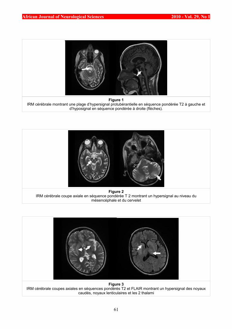
African Journal of Neurological Sciences 2010 - Vol. 29, No 1
Figure 1IRM cérébrale montrant une plage d’hypersignal protubérantielle en séquence pondérée T2 à gauche et
d’hyposignal en séquence pondérée à droite (flèches).
Figure 2IRM cérébrale coupe axiale en séquence pondérée T 2 montrant un hypersignal au niveau du
mésencéphale et du cervelet
Figure 3IRM cérébrale coupes axiales en séquences pondérés T2 et FLAIR montrant un hypersignal des noyaux
caudés, noyaux lenticulaires et les 2 thalami
61

African Journal of Neurological Sciences 2010 - Vol. 29, No 1
Figure 4IRM cérébrale coupe coronale en séquence pondérée FLAIR montrant un hypersignal des noyaux caudés,
noyaux lenticulaires et des lobes temporaux
Figure 5IRM cérébrale coupe axiale en séquence pondérée T2 montrant un hypersignal plus discret au niveau des
noyaux lenticulaires
62
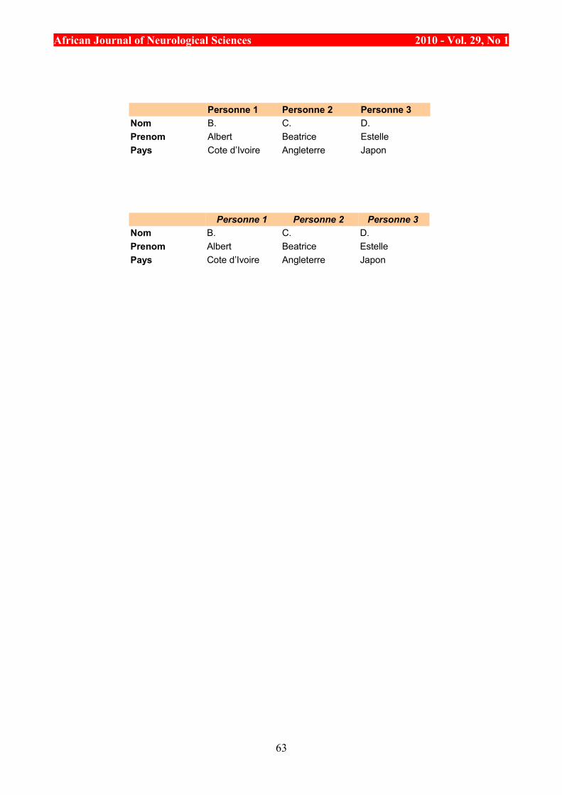
African Journal of Neurological Sciences 2010 - Vol. 29, No 1
Personne 1 Personne 2 Personne 3 Nom B. C. D. Prenom Albert Beatrice Estelle Pays Cote d’Ivoire Angleterre Japon
Personne 1 Personne 2 Personne 3 Nom B. C. D. Prenom Albert Beatrice Estelle Pays Cote d’Ivoire Angleterre Japon
63
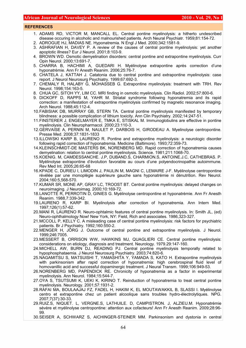
African Journal of Neurological Sciences 2010 - Vol. 29, No 1
REFERENCES1. ADAMS RD, VICTOR M, MANCALL EL. Central pontine myelinolysis: a hitherto undescribed
disease occuring in alcoholic and malnourished patients. Arch Neural Psychiatr. 1959;81:154-72. 2. ADROGUE HJ, MADIAS NE. Hyponatremia. N Engl J Med. 2000;342:1581-9. 3. ASHRAFIAN H, DAVEY P. A review of the causes of central pontine myelinolysis: yet another
apoptotic illness? Eur J Neurol. 2001;8:103-9. 4. BROWN WD. Osmotic demyelination disorders: central pontine and extrapontine myelinolysis. Curr
Opin Neurol. 2000;13:691-7. 5. CHARRA B, HACHIMI A, GUEDARI H. Myélinolyse extrapontine après correction d’une
hyponatrémie. Ann Fr Anesth Reanim. 2006;25:76-7. 6. CHATELA J, KATTAH J. Catatonia due to central pontine and extrapontine myelinolysis: case
report. J Neurol Neurosurg Psychiatry. 1999;67:692-3. 7. CHEMALY R, HALABY G, MOHASSEB G. Extrapontine myelinolysis: treatment with TRH. Rev
Neurol. 1998;154:163-5. 8. CHUA GC, SITOH YY, LIM CC. MRI finding in osmotic myelinolysis. Clin Radiol. 2002;57:800-6. 9. DICKOFF D, RAPPS M, YAHR M. Striatal syndrome following hyponatremia and its rapid
correction: a manifestation of extrapontine myelinolysis confirmed by magnetic resonance imaging. Arch Neurol. 1988;45:112-4.
10.FABISIAK DB, MURRAY GB, STERN TA. Central pontine myelinolysis manifested by temporary blindness: a possible complication of lithium toxicity. Ann Clin Psychiatry. 2002;14:247-51.
11.FINSTERER J, ENGELMAYER E, TMKA E, STISKAL M. Immunoglobulins are effective in pontine myelinolysis. Clin Neuropharmacol. 2000;23:110-3.
12.GERVAISE A, PERNIN M, NAULET P, DARBOIS H, GIRODEAU A. Myelinolyse centropontine. Presse Med. 2008;37:1831-1833
13.ILLOWSKI KARP B, LAURENO R. Pontine and extrapontine myelinolysis: a neurologic disorder following rapid correction of hyponatremia. Medicine (Baltimore). 1993;72:359-73.
14.KLEINSCHMIDT-DE MASTERS BK, NORENBERG MD. Rapid correction of hyponatremia causes demyelination: relation to central pontine myelinolysis. Science. 1981;211:1068-70.
15.KOENIG. M, CAMDESSANCHE. J.P, DUBAND.S, CHARMION.S, ANTOINE.J.C, CATHEBRAS. P. Myélinolyse extrapontine d’évolution favorable au cours d’une polyendocrinopathie autoimmune. Rev Med Int. 2005;26:65-68
16.KPADE C, DURIEU I, LIMODIN J, PAULIN M, MAGNI C, LEMAIRE J-F. Myélinolyse centropontine révélée par une monoplégie supérieure gauche sans hyponatrémie ni dénutrition. Rev Neurol. 2004;160:5,568-570.
17.KUMAR SR, MONE AP, GRAY LC, TROOST BT. Central pontine myelinolysis: delayed changes on neuroimaging. J Neuroimag. 2000;10:169-72.
18.LANOTTE R, PERROTIN D, GINIES G. Myélinolyse centropontine et hyponatrémie. Ann Fr Anesth Reanim. 1988,7:339-342.
19.LAURENO R, KARP BI. Myelinolysis after correction of hyponatremia. Ann Intern Med. 1997;126(1):57-62.
20.MANI R, LAURENO R. Neuro-ophtalmic features of central pontine myelinolysis. In: Smith JL, (ed) Neuro-ophthalmology Now! New York, NY: Field, Rich and associates. 1986,323-327.
21.MCCOLL P, KELLY C. A misleading case of central pontine myelinolysis: risk factors for psychiatric patients. Br J Psychiatry. 1992;160:550-2.
22.MENGER H, JÖRG J. Outcome of central pontine and extrapontine myelinolysis. J Neurol. 1999;246:7005.
23.MESSERT B, ORRISON WW, HAWKINS MJ, QUAGLIERI CE. Central pontine myelinolysis: considerations on etiology, diagnosis and treatment. Neurology. 1979.29:147-160.
24.MICHELL AW, BURN DJ, READING PJ. Central pontine myelinolysis temporally related to hypophosphataemia. J Neurol Neurosurg Psychiatry. 2003;74:820-6.
25.NAGAMITSU S, MATSUISHI T, YAMASHITA Y, YAMADA S, KATO H. Extrapontine myelinolysis with parkinsonism after rapid correction of hyponatremia: high cerebrospinal fluid level of homovanillic acid and successful dopaminergic treatment. J Neural Transm. 1999;106:949-53.
26.NORENBERG MD, PAPENDICK RE. Chronicity of hyponatremia as a factor in experimental myelinolysis. Ann Neurol. 1984;15:544-7.
27.OYA S, TSUTSUMI K, UEKI K, KIRINO T. Reinduction of hyponatremia to treat central pontine myelinolysis. Neurology. 2001;57:1931-2.
28.RAFAI MA, BOULAAJAJ FZ, FADEL H, HAKIM K, EL MOUTAWAKKIL B, SLASSI I. Myélinolyse centro et extrapontine chez un patient alcoolique sans troubles hydro-électrolytiques. NPG. 2007;7(37):30-33.
29.RUIZ.S, NIQUET. L, VERGNE.S, LATHUILE. D, CAMPISTRON. J, ALZIEU.M. Hyponatrémie sévère et myélinolyse centropontine: attention aux cofacteurs! Ann Fr Anesth Reanim. 2009;28:96-99.
30.SEISER A, SCHWARZ S, AICHINGER-STEINER MM. Parkinsonism and dystonia in central
64
African Journal of Neurological Sciences 2010 - Vol. 29, No 1
pontine and extrapontine myelinolysis. J Neurol Neurosurg Psychiatry. 1998;65:119-21. 31.SOTO-ARES G, DAÏDONE C, CLARISSE J, PRUVO JP. Imagerie des lésions encéphaliques liées
à l’alcool. Feuillets de Radiol. 1998;38:441-62. 32.STERNS RH, RIGGS JE, SCHOCHET JR. SS. Osmotic demyelination syndrome following
correction of hyponatremia. N Engl J Med. 1986;314:1535-42. 33.SUGIMOTO T, MURATA T, OMORI M. Central pontine myelinolysis associated with hypokalaemia
in anorexia nervosa. J Neurol Neurosurg Psychiatry. 2003;74:353-5.
65
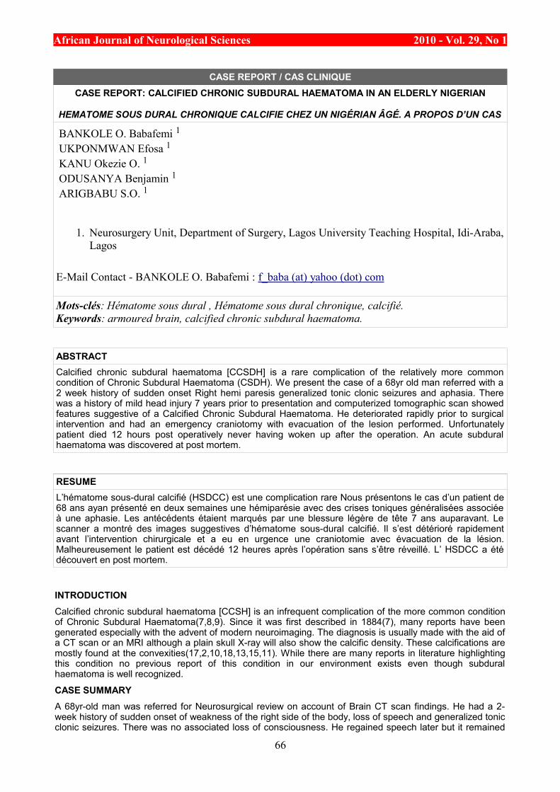
African Journal of Neurological Sciences 2010 - Vol. 29, No 1
CASE REPORT / CAS CLINIQUECASE REPORT: CALCIFIED CHRONIC SUBDURAL HAEMATOMA IN AN ELDERLY NIGERIAN
HEMATOME SOUS DURAL CHRONIQUE CALCIFIE CHEZ UN NIGÉRIAN ÂGÉ. A PROPOS D’UN CAS
BANKOLE O. Babafemi 1 UKPONMWAN Efosa 1 KANU Okezie O. 1 ODUSANYA Benjamin 1 ARIGBABU S.O. 1
1. Neurosurgery Unit, Department of Surgery, Lagos University Teaching Hospital, Idi-Araba, Lagos
E-Mail Contact - BANKOLE O. Babafemi : f_baba (at) yahoo (dot) com
Mots-clés: Hématome sous dural , Hématome sous dural chronique, calcifié.Keywords: armoured brain, calcified chronic subdural haematoma.
ABSTRACTCalcified chronic subdural haematoma [CCSDH] is a rare complication of the relatively more common condition of Chronic Subdural Haematoma (CSDH). We present the case of a 68yr old man referred with a 2 week history of sudden onset Right hemi paresis generalized tonic clonic seizures and aphasia. There was a history of mild head injury 7 years prior to presentation and computerized tomographic scan showed features suggestive of a Calcified Chronic Subdural Haematoma. He deteriorated rapidly prior to surgical intervention and had an emergency craniotomy with evacuation of the lesion performed. Unfortunately patient died 12 hours post operatively never having woken up after the operation. An acute subdural haematoma was discovered at post mortem.
RESUMEL’hématome sous-dural calcifié (HSDCC) est une complication rare Nous présentons le cas d’un patient de 68 ans ayan présenté en deux semaines une hémiparésie avec des crises toniques généralisées associée à une aphasie. Les antécédents étaient marqués par une blessure légère de tête 7 ans auparavant. Le scanner a montré des images suggestives d’hématome sous-dural calcifié. Il s’est détérioré rapidement avant l’intervention chirurgicale et a eu en urgence une craniotomie avec évacuation de la lésion. Malheureusement le patient est décédé 12 heures après l’opération sans s’être réveillé. L’ HSDCC a été découvert en post mortem.
INTRODUCTIONCalcified chronic subdural haematoma [CCSH] is an infrequent complication of the more common condition of Chronic Subdural Haematoma(7,8,9). Since it was first described in 1884(7), many reports have been generated especially with the advent of modern neuroimaging. The diagnosis is usually made with the aid of a CT scan or an MRI although a plain skull X-ray will also show the calcific density. These calcifications are mostly found at the convexities(17,2,10,18,13,15,11). While there are many reports in literature highlighting this condition no previous report of this condition in our environment exists even though subdural haematoma is well recognized.
CASE SUMMARYA 68yr-old man was referred for Neurosurgical review on account of Brain CT scan findings. He had a 2-week history of sudden onset of weakness of the right side of the body, loss of speech and generalized tonic clonic seizures. There was no associated loss of consciousness. He regained speech later but it remained
66
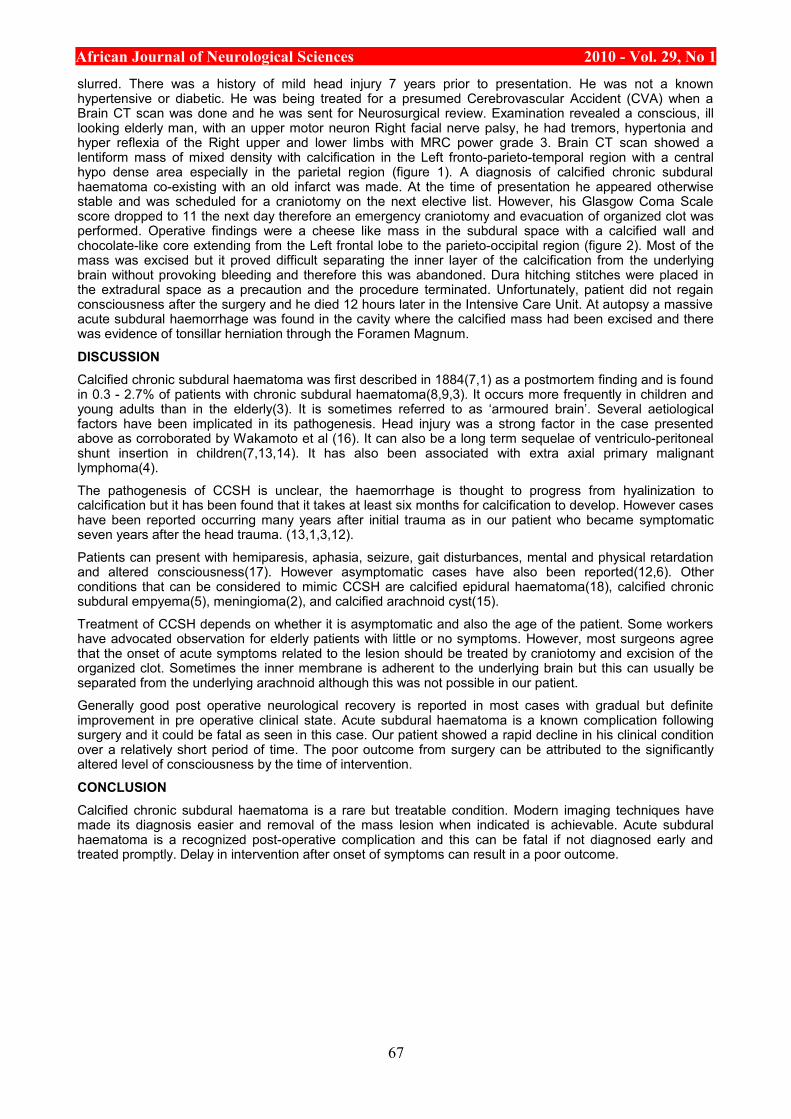
African Journal of Neurological Sciences 2010 - Vol. 29, No 1slurred. There was a history of mild head injury 7 years prior to presentation. He was not a known hypertensive or diabetic. He was being treated for a presumed Cerebrovascular Accident (CVA) when a Brain CT scan was done and he was sent for Neurosurgical review. Examination revealed a conscious, ill looking elderly man, with an upper motor neuron Right facial nerve palsy, he had tremors, hypertonia and hyper reflexia of the Right upper and lower limbs with MRC power grade 3. Brain CT scan showed a lentiform mass of mixed density with calcification in the Left fronto-parieto-temporal region with a central hypo dense area especially in the parietal region (figure 1). A diagnosis of calcified chronic subdural haematoma co-existing with an old infarct was made. At the time of presentation he appeared otherwise stable and was scheduled for a craniotomy on the next elective list. However, his Glasgow Coma Scale score dropped to 11 the next day therefore an emergency craniotomy and evacuation of organized clot was performed. Operative findings were a cheese like mass in the subdural space with a calcified wall and chocolate-like core extending from the Left frontal lobe to the parieto-occipital region (figure 2). Most of the mass was excised but it proved difficult separating the inner layer of the calcification from the underlying brain without provoking bleeding and therefore this was abandoned. Dura hitching stitches were placed in the extradural space as a precaution and the procedure terminated. Unfortunately, patient did not regain consciousness after the surgery and he died 12 hours later in the Intensive Care Unit. At autopsy a massive acute subdural haemorrhage was found in the cavity where the calcified mass had been excised and there was evidence of tonsillar herniation through the Foramen Magnum.
DISCUSSIONCalcified chronic subdural haematoma was first described in 1884(7,1) as a postmortem finding and is found in 0.3 - 2.7% of patients with chronic subdural haematoma(8,9,3). It occurs more frequently in children and young adults than in the elderly(3). It is sometimes referred to as ‘armoured brain’. Several aetiological factors have been implicated in its pathogenesis. Head injury was a strong factor in the case presented above as corroborated by Wakamoto et al (16). It can also be a long term sequelae of ventriculo-peritoneal shunt insertion in children(7,13,14). It has also been associated with extra axial primary malignant lymphoma(4).
The pathogenesis of CCSH is unclear, the haemorrhage is thought to progress from hyalinization to calcification but it has been found that it takes at least six months for calcification to develop. However cases have been reported occurring many years after initial trauma as in our patient who became symptomatic seven years after the head trauma. (13,1,3,12).
Patients can present with hemiparesis, aphasia, seizure, gait disturbances, mental and physical retardation and altered consciousness(17). However asymptomatic cases have also been reported(12,6). Other conditions that can be considered to mimic CCSH are calcified epidural haematoma(18), calcified chronic subdural empyema(5), meningioma(2), and calcified arachnoid cyst(15).
Treatment of CCSH depends on whether it is asymptomatic and also the age of the patient. Some workers have advocated observation for elderly patients with little or no symptoms. However, most surgeons agree that the onset of acute symptoms related to the lesion should be treated by craniotomy and excision of the organized clot. Sometimes the inner membrane is adherent to the underlying brain but this can usually be separated from the underlying arachnoid although this was not possible in our patient.
Generally good post operative neurological recovery is reported in most cases with gradual but definite improvement in pre operative clinical state. Acute subdural haematoma is a known complication following surgery and it could be fatal as seen in this case. Our patient showed a rapid decline in his clinical condition over a relatively short period of time. The poor outcome from surgery can be attributed to the significantly altered level of consciousness by the time of intervention.
CONCLUSIONCalcified chronic subdural haematoma is a rare but treatable condition. Modern imaging techniques have made its diagnosis easier and removal of the mass lesion when indicated is achievable. Acute subdural haematoma is a recognized post-operative complication and this can be fatal if not diagnosed early and treated promptly. Delay in intervention after onset of symptoms can result in a poor outcome.
67
African Journal of Neurological Sciences 2010 - Vol. 29, No 1
Figure 1Lentiform mass of mixed densities with calcification in the left fronto-parieto-temporal region with a central
hypo dense area (white arrow).
Figure 2Excised mass with a central core of chocolate coloured cheese-like material (black arrow) and the calcified
wall encasing the organized haematoma (white arrow).
68
African Journal of Neurological Sciences 2010 - Vol. 29, No 1
REFERENCES
1. AFRA D. Ossification of subdural hematoma. Report of two cases. J Neurosurg 1961; 18:393-397. 2. CUSICK JF, BAILEY OT. Association of ossified subdural hematoma and a meningioma. Case
report. J Neuro surg 1972; 37:731-734. 3. DAMMERS R, TER LAAK-POORT M, MAAS IR. Armoured brain: case report of a symptomatic
Calcified Chronic Subdural Haematoma. J Neurol Neurosurg Psychiatry 2007;78:542-543. 4. GOTOH M, TSUNO K, HANDA A, NISHIURA T, ISHIMITSU H, NISHIDA A. Extraaxial primary
malignant lymphoma associated with calcified chronic subdural haematoma: a case report. No Shinkei Geka 2001;29(3):259-64.
5. HIKAWA T,UCHIDA S,FUJIKI M,KOBAYASHI H, OOBA H. Calcified chronic subdural empyema: a case report. Neurosurgery Quarterly 2006;16(3):152-154.
6. IDE M, JIMBO M, YAMAMOTO M, UMEBARA Y, HAGIWARA S. Asymptomatic calcified chronic subdural haematoma-report of three cases. Neurol Med Chir (Tokyo) 1993; 33:559-563.
7. IMAIZUMI S, ONUMA T, KAMEYAMA M, NAGANUMA H. Organized chronic subdural hematoma requiring craniotomy. Five case reports. Neurol Med Chir (Tokyo) 2001;41:19-24.
8. JONG-SOO P, EUN-LK S, DONG-WON K,SANG-PYO K. Calcified Chronic Subdural Haematoma associated with intra cerebral haematoma. J Korean Neurosurg Soc 2003; 34:177-178.
9. Mc LAUREN RL, MC LAUREN KS. Calcified subdural haematoma in childhood. Journal of Neurosurgery 1966; 24(3): 648-655.
10.MIYAGI Y, MORIOKA T, KIMURA Y, FUKUI M. Calcified convexity dura mater and acute epidural hematoma mimicking calcified chronic subdural Haematoma. Neuroradiology 1995; 37:551-552.
11.MOON KS, LEE JK, KIM TS et al. contralateral acute subdural haematoma occurring after removal of calcified chronic subdural haematoma. J Clin Neuroscience 2007;14:283-286.
12.NIWA J, NAKAMURA T, FUJISHIGE M, HASHI K. Removal of a large asymptomatic calcified chronic subdural haematoma. Surg Neurol 1988; 30:135-139.
13.PAPANIKOLEOU PG, PALEOLOGOS TS,TRIANTOFYLLOU TM, CHATZIDAKIS EM. Shunt revision after 33years in a patient with bilateral Calcified Chronic Subdural Haematoma: case illustration. J Neurosurgery 2008; 108(2): 401.
14.SHARMA RR, MAHAPATRA A,PAWAR SJ,SOUSA J,ATHALE SD. Symptomatic calcified subdural haematomas. Paediatric Neurosurgery 1999 Sept;31(3):150-154.
15.SHUANGSHOTI S. Calcified congenital arachnoid cyst with heterotopic neuroglia in wall. J Neurol Neurosurg Psychiatry 1978; 41:88-94.
16.WAKAMOTO H, MIWA T,ORII M,MIYAZAKI H,ISHIYAMA N. Calcified chronic subdural haematoma complicated with subcortical haemorrhage:case report.No Shinkei Geka Mar 2oo3; 31(3):269-73.
17.YAN HJ, LIN KE, LEE ST, TZAAN WC. Calcified chronic subdural haematoma: Case report. Changgeng Yi Xue Za Zhi 1998; 21:521-525.
18.YUASA H, WATANABE H, UEMURA Y, TOKITO S. Intra osseous haematoma of the skull: case report. Neurosurgery: May 1992; 30(5):776-777
69
African Journal of Neurological Sciences 2010 - Vol. 29, No 1
CASE REPORT / CAS CLINIQUEIMAGERIE DE LA MALADIE DE CREUTZFELDT JACOB SPORADIQUE
IMAGING IN DIAGNOSIS OF SPORADIC CREUTZFELDT JACOB DISEASE
HASSANI Rachid 1 BENABDELJLIL Maria 1 AÏDI Sadia 1 EL ALAOUI Faris Mustapha 1 JIDDANE Mohammed 2
1. Service de Neurologie A et Neuropsychologie Hôpital des Spécialités, Rabat, Maroc 2. Service de neuroradiologie, Rabat, Royaume du Maroc
E-Mail Contact - HASSANI Rachid : rachidhassani1 (at) gmail (dot) com
Mots-clés: MALADIE DE CREUTZFELDT JACOB SPORADIQUE, IMAGERIE PAR RESONANCE MAGNETIQUE ENCEPHALIQUE, IRMKeywords: SPORADIC CREUTZFELDT JACOB DISEASE, MAGNETIC RESONANCE IMAGING, MRI
RESUMELes encéphalopathies spongiformes subaiguës transmissibles (ESST) sont des maladies infectieuses, neurodégénératives et génétiques. Elles sont caractérisées par la présence d’une substance protéique : le prion. L’imagerie par résonance magnétique (IRM) encéphalique peut actuellement contribuer au diagnostic des ESST.
Nous rapportons le cas d’une patiente de 70 ans qui présenta en Juin 2004 un syndrome démentiel avec myoclonies, d’aggravation rapide et d’évolution fatale. L’électroencéphalogramme (EEG) objectivait une activité de fond ralentie et symétrique sans activité périodique. L’IRM encéphalique montrait des hypersignaux au niveau des noyaux gris centraux surtout du striatum mais aussi au niveau cortical. L’étude du Liquide céphalorachidien (LCR), trouvait une cytochimie normale avec présence de la protéine 14-3-3. Les sérologies syphilitiques étaient négatives, la sérologie de l’hépatite C positive et le bilan thyroïdien normal. Le diagnostic de maladie de Creutzfeldt Jacob sporadique probable a été retenu.
Une preuve anatomopathologique est le seul moyen d’obtenir un diagnostic certain dans les ESST, l’apport de l’imagerie et surtout les nouvelles techniques d’IRM peuvent être d’une aide précieuse dans le diagnostic des maladies à prion.
Dans les formes sporadiques, l’IRM peut mettre en évidence des hypersignaux des NGC visibles sur les séquences T2, Flair ou de diffusion, ce qui peut être un argument pour le diagnostic de MCJ sporadique chez un patient présentant une démence d’évolution rapide.
SUMMARYThe subacute transmissible spongiform encephalopathies (TSE) are infectious diseases, and neurodegenerative genetic. They are characterized by the presence of a protein substance: a prion. The magnetic resonance imaging (MRI) brain can currently contribute to the diagnosis of TSE.
We report the case of a patient of 70 years who presented in June 2004 with myoclonus, syndrome dementia, rapid deterioration and fatalities. The electroencephalogram (EEG) showed basic activity slowed and symmetrical without periodic activity. MRI brain showed hypersignals at the basal ganglia (BG) especially in the striatum but also the cortical level.
The study of cerebrospinal fluid (CSF), cytochemistry was normal with presence of the protein 14-3-3. Syphilitic serology was negative, serology for hepatitis C positive and normal thyroid stock. The diagnosis of Creutzfeldt Jacob sporadic likely was incorporated.
Pathological evidence is the only way to get a certain diagnosis in the TSE, the contribution of imaging and especially new technologies MRI can be a valuable aid in the diagnosis of prion diseases.
In sporadic forms, MRI can highlight hypersignals of BG visible on the sequences T2, Flair or broadcast,
70
African Journal of Neurological Sciences 2010 - Vol. 29, No 1
which may be an argument for the diagnosis of sporadic Creutzfeldt Jacob in a patient with dementia of rapid change.
INTRODUCTIONA coté du tableau clinique, de l’électroencéphalographie, de la positivité de la protéine 14-3-3 dans le LCR, l’IRM encéphalique occupe actuellement une place importante dans le diagnostic des maladies à prions. Nous rapportons le cas d’une patiente qui a présenté une MCJ sporadique probable et chez qui l’IRM encéphalique a mis en évidence des anomalies évocatrices.
OBSERVATIONMme A.H, 70 ans, mariée, 10 enfants, antécédents d’hypertension artérielle modérée de découverte récente, le début des symptômes remonte à juin 2004 par des troubles de la posture, des hallucinations visuelles avec réactions de peur et de méfiance. En octobre 2004, installation de troubles de la mémoire et du langage d’aggravation progressive avec impotence fonctionnelle des membres inférieurs, mouvements anormaux des 4 membres. Ce tableau évolue rapidement vers un état grabataire avec troubles sphinctériens, suspension de la parole et détérioration intellectuelle. Hospitalisée en Neurologie en janvier 2005, l’examen trouve une patiente consciente mais présentant une désintégration phonétique avec détérioration intellectuelle sévère. La station debout est impossible sans aide avec rétropulsions, hypertonie axiale et des membres, roue dentée aux poignets. Il existe des mouvements choréiques avec stéréotypies gestuelles, les réflexes ostéotendineux étaient abolis aux 4 membres, il n’y avait pas d’atteinte des nerfs crâniens. L’examen somatique est normal. Le scanner cérébral (TDM) était normal. L’imagerie par résonance magnétique (IRM) encéphalique montre des hypersignaux en séquences pondérées T2 et FLAIR du striatum et du cortex temporal (fig. 1 et 2), sans anomalies bipallidales (absence « pulvinar sign »)
L’EEG met en évidence une activité de fond symétrique mais ralentie, sans autres anomalies. L’étude du liquide céphalorachidien : cytologie : 7 EB/mm3, protéinorachie : 0,49 g/l, glycorrachie : 0,92g/l. L’électrophorèse des protéines était normale. La protéine 14-3-3 positive. Le bilan biologique standard était normal. La sérologie TPHA - VDRL est négative, les anticorps anti-thyroglobuline et anti-thyroperoxydase négatifs et les hormones thyroïdiennes normales.
DISCUSSIONLe diagnostic de la maladie de Creutzfeldt Jacob sporadique probable a été retenu chez notre patiente devant la présentation clinique classique, la positivité de la protéine 14-3-3 dans le LCR et l’aspect évocateur à l’IRM encéphalique. La MCJ sporadique fait partie des ESST, dont l’agent responsable est la protéine prion, codé par le gène PRNP (prion protein (p27- 30) Le diagnostic se base sur un tableau clinique évocateur et les anomalies à l’EEG qui est toujours anormal et peut montrer un ralentissement du rythme de base comme chez notre patiente, des décharges d’ondes lentes delta, et dans 60% des cas des anomalies pseudo-périodiques.
L’examen du LCR est en général normal, en dehors d’une protéinorachie souvent modérément élevée, il permet la détection de la protéine 14-3-3. Cette protéine 14-3-3 est à ce jour, le test le plus sensible et le plus spécifique pour le diagnostic de cette forme de MCJ (1), elle peut cependant être retrouvée dans les encéphalites nécrosantes virales et les infarctus cérébraux. Le diagnostic de certitude reste neuropathologique (biopsie cérébrale ou autopsie).
Le scanner cérébral (TDM) peut être normal ou montrer une atrophie cérébrale ; cet examen était normal chez notre patiente. Dans la MCJ sporadique, l’IRM encéphalique peut être normale, mettre en évidence une atrophie, ou dans la moitié des cas révéler des hypersignaux dans les noyaux gris centraux (NGC), visibles sur les séquences T2, Flair ou de diffusion. Des hypersignaux dans les cortex cérébraux ou cérébelleux sont plus rarement observés (Schroter et al, 2000), (6).
Dans notre cas, des hypersignaux ont été visualisés sur les séquences T2 et FLAIR au niveau du striatum et du cortex temporal. Les hypersignaux dans les NGC, visibles sur les séquences T2 sont principalement localisés dans le putamen et le noyau caudé; et sont en général symétriques. Ce signe a une sensibilité diagnostic de 60 à 80% (8). La séquence FLAIR est plus sensible que la séquenceT2, elle sera préférée en cas de suspicion de MCJ, si les séquences conventionnelles sont normales (Vrancken, 2000). L’IRM encéphalique de diffusion montre des anomalies très précoces, dès la 3ème semaine (Shiga, 2004), (7). Elle permet également de visualiser des anomalies corticales (3). La Spectro IRM (4) peut également aider au diagnostic : une valeur abaissée du N- Acetyl-Aspartate témoigne d’une perte neuronale. Par ailleurs, selon Meissner, 2004 (2) : les cas avec anomalies à l’IRM sont corrélés à une démence plus précoce, et à une durée d’évolution plus courte ; les cas sans anomalies sont corrélés à une dépression et à une atteinte sensorielle. Il n’y a pas de corrélation entre les hypersignaux des ganglions de la base (GB) et les signes
71
African Journal of Neurological Sciences 2010 - Vol. 29, No 1extrapyramidaux.
Le mécanisme exact de ces lésions reste inconnu ; il pourrait s’agir de dépôts de protéine prion détectables sur le plan neuropathologique. Dans la nouvelle variante de la MCJ (nvMCJ), l’aspect IRM typique est le « pulvinar sign », cet hypersignal bilatéral du pulvinar en T2 et FLAIR est plus important que dans les autres ganglions de la base, par ailleurs cet aspect peut être retrouvé dans certains cas de MCJ sporadiques (5).
CONCLUSIONLe diagnostic de certitude des MCJ reste anatomopathologique, cependant les nouvelles techniques de l’IRM occupent actuellement une place importante et très prometteuse dans l’orientation et l’approche de ce diagnostic.
BIBLIOGRAPHIE
1. HSICH G, KENNEY K. The 14-3-3 brain protein in cerebrospinal fluid as a marker for transmissible spongiform encephalophathie. N Engl J Med. 1996 ;335(13):924-30
2. MEISSNER B, KÖRTNER K, BARTL M, JASTROW U, MOLLENHAUER B, et al. Sporadic Creutzfeldt-Jakob disease: magnetic resonance imaging and clinical findings. Neurology. 2004;10;63(3):450-6.
3. MITTAL S, FARMER P,KALINA P, KINGSLEY P. Correlation of diffusion-weighted magnetic resonance imaging with neuropathology in Creutzfeldt-Jakob disease. Arch Neurol. 2002;59:128-4.
4. OPPENHEIM C, ZUBER M, GALANAUD D, DETILLEUX M, BOLGERT F, MAS JL, CHIRAS J, MEDER JF.Spectroscopy and serial diffusion MR findings in hGH-Creutzfeldt-Jakob disease. J Neurol Neurosurg Psychiatry. 2004;75(7):1066-9.
5. PETZOLD GC, WESTNER I, BOHNER G, EINHÄUPL KM, KRETZSCHMAR HA, VALDUEZA JM. False-positive pulvinar sign on MRI in sporadic Creutzfeldt-Jakob disease.Neurology. 2004 ;62(7):1235-6. No abstract available.
6. SCHROTER A, ZERR I, HENKEL K. Magnetic resonance imaging in the clinical diagnostic of Creutzfeldt Jakob disease. Arch Neurol 2000;57:1751-1757.
7. SHIGA Y, MIYAZAWA K, SATO S, FUKUSHIMA R, SHIBUYA S, SATO Y et al. Diffusion-weighted MRI abnormalities as an early diagnostic marker for Creutzfeldt-Jakob disease. Neurology. 2004;63(3):443-9.
8. VRANKEN et al. Flair MRI in Sporadic Creutzfeldt Jakob disease. Neurology 2000; 12;55.147.
72
African Journal of Neurological Sciences 2010 - Vol. 29, No 1
CASE REPORT / CAS CLINIQUETRAUMATISME VERTEBRO-MEDULLAIRE COMPLIQUANT UNE SPONDYLARTHRITE
ANKYLOSANTE
SPINAL CORD INJURY COMPLICATING ANKYLOSING SPONDYLITIS
HARIFI Ghita 1 LAGHMARI Mehdi 2 LMEJJATI Mohammed 2 GHANNANE Houcine 2 AIT BENALI Said 2 EL HASSANI Selma 1
1. Service de Rhumatologie, CHU Mohammed VI, Université Cadi Ayyad, Marrakech, Maroc 2. Service de Neurochirurgie, CHU Mohammed VI, Université Cadi Ayyad, Marrakech,
Maroc
E-Mail Contact - HARIFI Ghita : ghitaharifi (at) yahoo (dot) fr Mots-clés: Spondylarthrite ankylosante ; Traumatisme vertébro-médullaire ; fracture vertébraleKeywords: Ankylosing spondylitis ; spinal cord injury ; vertebral fracture
RESUMELa survenue d’un traumatisme vertébral constitue un événement potentiellement grave dans l’évolution d’une spondylarthrite ankylosante (SPA). Le risque d’une atteinte médullaire est relativement élevé. Nous rapportons deux observations illustratives de la gravité du traumatisme vertébro-médullaire compliquant une SPA, avec une revue des différents facteurs de risque, et des aspects cliniques et thérapeutiques de la prise en charge dans cette situation.
ABSTRACTThe occurrence of spinal injury is a potentially serious event in ankylosing spondylitis (AS) patients. The risk of spinal cord injury (SCI) is greater in this situation. With this case report the authors outline the factors that increase the incidence of vertebral fractures and spinal cord injury in AS patients and discuss the management of SCI in these patients.
INTRODUCTIONLa spondylathrite ankylosante (SPA) est un rhumatisme inflammatoire chronique, lié à l’antigène HLA B27, touchant le plus souvent l’adulte jeune de sexe masculin et affectant essentiellement le squelette axial. Elle est caractérisée par une enthésopathie inflammatoire évoluant vers l’ossification et l’ankylose, conduisant progressivement à une raideur rachidienne avec altération des propriétés biomécaniques du rachis [3].
Les troubles de l’équilibre, la diminution des reflexes de protection lors de la chute et l’ossification des structures disco-ligamentaires et l’ostéopénie rachidienne, souvent associés, prédisposent au traumatisme vertèbro-médullaire mettant en jeu le pronostic vital et fonctionnel [1,14].
OBSERVATION 1Un homme de 52 ans, était suivi depuis une vingtaine d’années pour une spondylarthrite ankylosante (SPA). Le diagnostic de SPA avait été porté sur l’association d’un syndrome pelvi-rachidien d’horaire inflammatoire, une polyenthésopathie périphérique, une sacro-illite bilatérale stade 2 de forestier, et un terrain HLA B27 positif. Le patient a été mis sous indométacine et traitement physique, mais l’observance était mauvaise et le suivi irrégulier. L’évolution s’est faite progressivement vers une cyphose dorsale avec perte de l’horizontalité
73
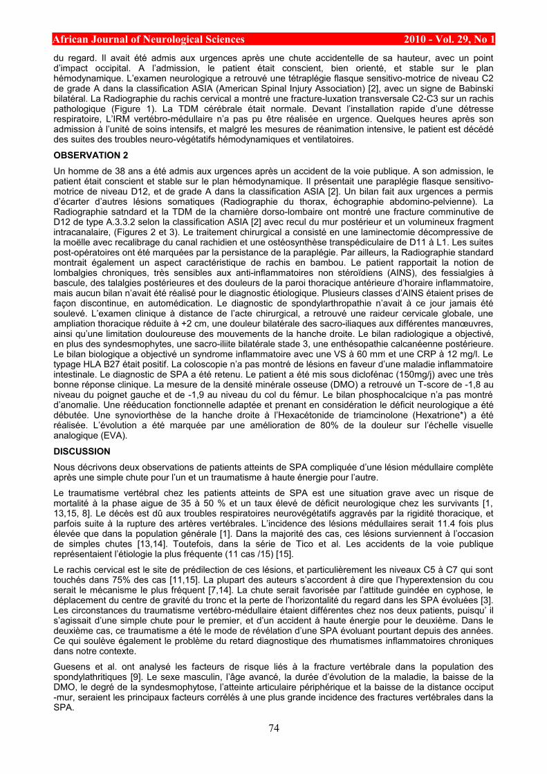
African Journal of Neurological Sciences 2010 - Vol. 29, No 1du regard. Il avait été admis aux urgences après une chute accidentelle de sa hauteur, avec un point d’impact occipital. A l’admission, le patient était conscient, bien orienté, et stable sur le plan hémodynamique. L’examen neurologique a retrouvé une tétraplégie flasque sensitivo-motrice de niveau C2 de grade A dans la classification ASIA (American Spinal Injury Association) [2], avec un signe de Babinski bilatéral. La Radiographie du rachis cervical a montré une fracture-luxation transversale C2-C3 sur un rachis pathologique (Figure 1). La TDM cérébrale était normale. Devant l’installation rapide d’une détresse respiratoire, L’IRM vertébro-médullaire n’a pas pu être réalisée en urgence. Quelques heures après son admission à l’unité de soins intensifs, et malgré les mesures de réanimation intensive, le patient est décédé des suites des troubles neuro-végétatifs hémodynamiques et ventilatoires.
OBSERVATION 2Un homme de 38 ans a été admis aux urgences après un accident de la voie publique. A son admission, le patient était conscient et stable sur le plan hémodynamique. Il présentait une paraplégie flasque sensitivo-motrice de niveau D12, et de grade A dans la classification ASIA [2]. Un bilan fait aux urgences a permis d’écarter d’autres lésions somatiques (Radiographie du thorax, échographie abdomino-pelvienne). La Radiographie satndard et la TDM de la charnière dorso-lombaire ont montré une fracture comminutive de D12 de type A.3.3.2 selon la classification ASIA [2] avec recul du mur postérieur et un volumineux fragment intracanalaire, (Figures 2 et 3). Le traitement chirurgical a consisté en une laminectomie décompressive de la moëlle avec recalibrage du canal rachidien et une ostéosynthèse transpédiculaire de D11 à L1. Les suites post-opératoires ont été marquées par la persistance de la paraplégie. Par ailleurs, la Radiographie standard montrait également un aspect caractéristique de rachis en bambou. Le patient rapportait la notion de lombalgies chroniques, très sensibles aux anti-inflammatoires non stéroïdiens (AINS), des fessialgies à bascule, des talalgies postérieures et des douleurs de la paroi thoracique antérieure d’horaire inflammatoire, mais aucun bilan n’avait été réalisé pour le diagnostic étiologique. Plusieurs classes d’AINS étaient prises de façon discontinue, en automédication. Le diagnostic de spondylarthropathie n’avait à ce jour jamais été soulevé. L’examen clinique à distance de l’acte chirurgical, a retrouvé une raideur cervicale globale, une ampliation thoracique réduite à +2 cm, une douleur bilatérale des sacro-iliaques aux différentes manœuvres, ainsi qu’une limitation douloureuse des mouvements de la hanche droite. Le bilan radiologique a objectivé, en plus des syndesmophytes, une sacro-iliite bilatérale stade 3, une enthésopathie calcanéenne postérieure. Le bilan biologique a objectivé un syndrome inflammatoire avec une VS à 60 mm et une CRP à 12 mg/l. Le typage HLA B27 était positif. La coloscopie n’a pas montré de lésions en faveur d’une maladie inflammatoire intestinale. Le diagnostic de SPA a été retenu. Le patient a été mis sous diclofénac (150mg/j) avec une très bonne réponse clinique. La mesure de la densité minérale osseuse (DMO) a retrouvé un T-score de -1,8 au niveau du poignet gauche et de -1,9 au niveau du col du fémur. Le bilan phosphocalcique n’a pas montré d’anomalie. Une rééducation fonctionnelle adaptée et prenant en considération le déficit neurologique a été débutée. Une synoviorthèse de la hanche droite à l’Hexacétonide de triamcinolone (Hexatrione*) a été réalisée. L’évolution a été marquée par une amélioration de 80% de la douleur sur l’échelle visuelle analogique (EVA).
DISCUSSIONNous décrivons deux observations de patients atteints de SPA compliquée d’une lésion médullaire complète après une simple chute pour l’un et un traumatisme à haute énergie pour l’autre.
Le traumatisme vertébral chez les patients atteints de SPA est une situation grave avec un risque de mortalité à la phase aigue de 35 à 50 % et un taux élevé de déficit neurologique chez les survivants [1, 13,15, 8]. Le décès est dû aux troubles respiratoires neurovégétatifs aggravés par la rigidité thoracique, et parfois suite à la rupture des artères vertébrales. L’incidence des lésions médullaires serait 11.4 fois plus élevée que dans la population générale [1]. Dans la majorité des cas, ces lésions surviennent à l’occasion de simples chutes [13,14]. Toutefois, dans la série de Tico et al. Les accidents de la voie publique représentaient l’étiologie la plus fréquente (11 cas /15) [15].
Le rachis cervical est le site de prédilection de ces lésions, et particulièrement les niveaux C5 à C7 qui sont touchés dans 75% des cas [11,15]. La plupart des auteurs s’accordent à dire que l’hyperextension du cou serait le mécanisme le plus fréquent [7,14]. La chute serait favorisée par l’attitude guindée en cyphose, le déplacement du centre de gravité du tronc et la perte de l’horizontalité du regard dans les SPA évoluées [3]. Les circonstances du traumatisme vertébro-médullaire étaient différentes chez nos deux patients, puisqu’ il s’agissait d’une simple chute pour le premier, et d’un accident à haute énergie pour le deuxième. Dans le deuxième cas, ce traumatisme a été le mode de révélation d’une SPA évoluant pourtant depuis des années. Ce qui soulève également le problème du retard diagnostique des rhumatismes inflammatoires chroniques dans notre contexte.
Guesens et al. ont analysé les facteurs de risque liés à la fracture vertébrale dans la population des spondylathritiques [9]. Le sexe masculin, l’âge avancé, la durée d’évolution de la maladie, la baisse de la DMO, le degré de la syndesmophytose, l’atteinte articulaire périphérique et la baisse de la distance occiput -mur, seraient les principaux facteurs corrélés à une plus grande incidence des fractures vertébrales dans la SPA.
74
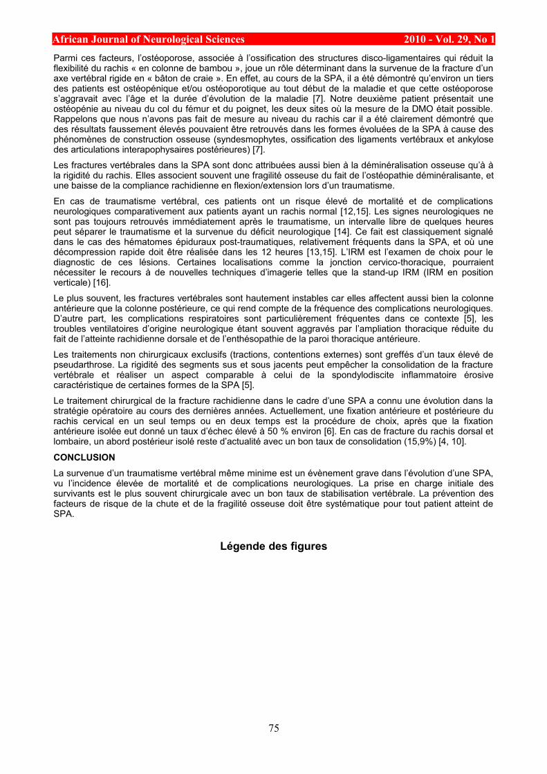
African Journal of Neurological Sciences 2010 - Vol. 29, No 1Parmi ces facteurs, l’ostéoporose, associée à l’ossification des structures disco-ligamentaires qui réduit la flexibilité du rachis « en colonne de bambou », joue un rôle déterminant dans la survenue de la fracture d’un axe vertébral rigide en « bâton de craie ». En effet, au cours de la SPA, il a été démontré qu’environ un tiers des patients est ostéopénique et/ou ostéoporotique au tout début de la maladie et que cette ostéoporose s’aggravait avec l’âge et la durée d’évolution de la maladie [7]. Notre deuxième patient présentait une ostéopénie au niveau du col du fémur et du poignet, les deux sites où la mesure de la DMO était possible. Rappelons que nous n’avons pas fait de mesure au niveau du rachis car il a été clairement démontré que des résultats faussement élevés pouvaient être retrouvés dans les formes évoluées de la SPA à cause des phénomènes de construction osseuse (syndesmophytes, ossification des ligaments vertébraux et ankylose des articulations interapophysaires postérieures) [7].
Les fractures vertébrales dans la SPA sont donc attribuées aussi bien à la déminéralisation osseuse qu’à à la rigidité du rachis. Elles associent souvent une fragilité osseuse du fait de l’ostéopathie déminéralisante, et une baisse de la compliance rachidienne en flexion/extension lors d’un traumatisme.
En cas de traumatisme vertébral, ces patients ont un risque élevé de mortalité et de complications neurologiques comparativement aux patients ayant un rachis normal [12,15]. Les signes neurologiques ne sont pas toujours retrouvés immédiatement après le traumatisme, un intervalle libre de quelques heures peut séparer le traumatisme et la survenue du déficit neurologique [14]. Ce fait est classiquement signalé dans le cas des hématomes épiduraux post-traumatiques, relativement fréquents dans la SPA, et où une décompression rapide doit être réalisée dans les 12 heures [13,15]. L’IRM est l’examen de choix pour le diagnostic de ces lésions. Certaines localisations comme la jonction cervico-thoracique, pourraient nécessiter le recours à de nouvelles techniques d’imagerie telles que la stand-up IRM (IRM en position verticale) [16].
Le plus souvent, les fractures vertébrales sont hautement instables car elles affectent aussi bien la colonne antérieure que la colonne postérieure, ce qui rend compte de la fréquence des complications neurologiques. D’autre part, les complications respiratoires sont particulièrement fréquentes dans ce contexte [5], les troubles ventilatoires d’origine neurologique étant souvent aggravés par l’ampliation thoracique réduite du fait de l’atteinte rachidienne dorsale et de l’enthésopathie de la paroi thoracique antérieure.
Les traitements non chirurgicaux exclusifs (tractions, contentions externes) sont greffés d’un taux élevé de pseudarthrose. La rigidité des segments sus et sous jacents peut empêcher la consolidation de la fracture vertébrale et réaliser un aspect comparable à celui de la spondylodiscite inflammatoire érosive caractéristique de certaines formes de la SPA [5].
Le traitement chirurgical de la fracture rachidienne dans le cadre d’une SPA a connu une évolution dans la stratégie opératoire au cours des dernières années. Actuellement, une fixation antérieure et postérieure du rachis cervical en un seul temps ou en deux temps est la procédure de choix, après que la fixation antérieure isolée eut donné un taux d’échec élevé à 50 % environ [6]. En cas de fracture du rachis dorsal et lombaire, un abord postérieur isolé reste d’actualité avec un bon taux de consolidation (15,9%) [4, 10].
CONCLUSIONLa survenue d’un traumatisme vertébral même minime est un évènement grave dans l’évolution d’une SPA, vu l’incidence élevée de mortalité et de complications neurologiques. La prise en charge initiale des survivants est le plus souvent chirurgicale avec un bon taux de stabilisation vertébrale. La prévention des facteurs de risque de la chute et de la fragilité osseuse doit être systématique pour tout patient atteint de SPA.
Légende des figures
75
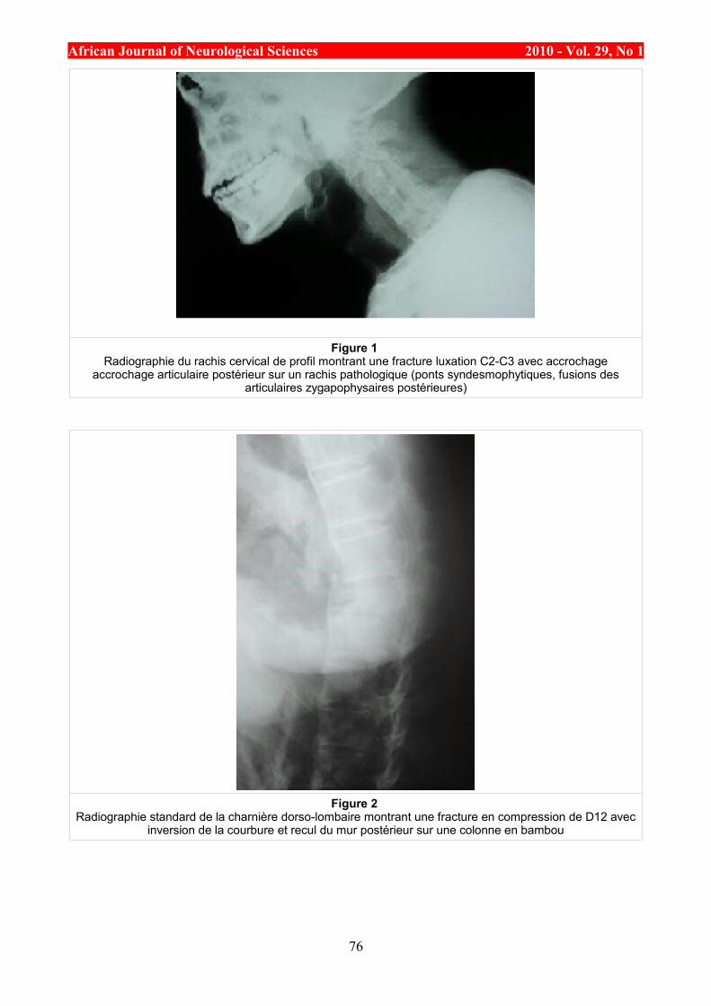
African Journal of Neurological Sciences 2010 - Vol. 29, No 1
Figure 1 Radiographie du rachis cervical de profil montrant une fracture luxation C2-C3 avec accrochage
accrochage articulaire postérieur sur un rachis pathologique (ponts syndesmophytiques, fusions des articulaires zygapophysaires postérieures)
Figure 2 Radiographie standard de la charnière dorso-lombaire montrant une fracture en compression de D12 avec
inversion de la courbure et recul du mur postérieur sur une colonne en bambou
76
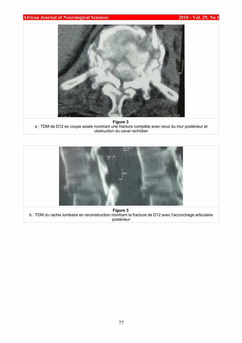
African Journal of Neurological Sciences 2010 - Vol. 29, No 1
Figure 3 a : TDM de D12 en coupe axiale montrant une fracture complète avec recul du mur postérieur et
obstruction du canal rachidien
Figure 3 b : TDM du rachis lombaire en reconstruction montrant la fracture de D12 avec l’accrochage articulaire
postérieur
77
African Journal of Neurological Sciences 2010 - Vol. 29, No 1
REFERENCES
1. ALARANTA H, LUOTO S, KONTTINEN YT. Traumatic spinal cord injury as a complication to ankylosing spondylitis. An extended report. Clin Exp Rheumatol. 2002;20(1):66-8
2. American Spinal Injury Association / International Medical Society Of Paraplegia: International standards for neurological and functional classification of spinal cord injury, revised 1996. Chocago, American Spinal Injury Association, 1996.
3. BOT SD, CASPERS M, VAN ROYEN BJ, TOUSSAINT HM, KINGMA I. Biomechanical analysis of posture in patients with spinal kyphosis due to ankylosing spondylitis: a pilot study. Rheumatology (Oxford) 1999;38:441-443.
4. BRON JL, DE VRIES MK, SNIEDERS MN, VAN DER HORST-BRUINSMA IE, VAN ROYEN BJ. Discovertebral (Andersson) lesions of the spine in ankylosing spondylitis revisited. Clin Rheumatol. 2009;28(8):883-92
5. DUHEM-TONNELLE V, DUHEM R, ALLAOUI M, CHASTANET P, ASSAKER R. Fracture luxation of the cervical spine in patients with ankylosing spondylitis : six cases. Neurochirurgie. 2008;54(1):46-52.
6. EINSIEDEL T, SCHMELZ A, ARAND M, WILKE HJ, GEBHARD F, HARTWIG E, KRAMER M, NEUGEBAUER R, KINZL L, SCHULTHEISS M. Injuries of the cervical spine in patients with ankylosing spondylitis: experience at two trauma centers. J Neurosurg Spine. 2006;5(1):33-45
7. EL MAGHRAOUI. Osteoporosis and ankylosing spondylitis. Joint Bone Spine. 2004 2004 ;71 (4): 291-5
8. FOO D, SARKARATI M, MARCELINO V. Cervical spinal cord injury complicating ankylosing spondylitis. Paraplegia 1985; 23: 358 ±633.
9. GEUSENS P, VOSSE D, VAN DER LINDEN S. Osteoporosis and vertebral fractures in ankylosing spondylitis. Curr Opin Rheumatol 2007;19:335-339.
10.HITCHON PW, FROM AM, BRENTON MD, GLASER JA, TORNER JC. Fractures of the thoracolumbar spine complicating ankylosing spondylitis. J Neurosurg 2002 ;97(2):218-22
11.MURRAY GC, PERSELLIN RH. Cervical fracture complicating ankylosing spondylitis. A report of eight cases and review of the literature. Am J Med 1981; 70: 1033 ± 1041.
12.OLERUD, C., FROST, A., BRING, J. Spinal fractures in patients with ankylosing spondylitis. Eur. Spine J. 1996 ; 5 : 51-55.
13.ROWED DW. Management of cervical spinal cord injury in ankylosing spondylitis: the intervertebral disc as a cause of cord compression. J Neurosurg 1992: 77: 241 ± 246.
14.THUMBIKAT P, HARIHARAN RP, RAVICHANDRAN G, MCCLELLAND MR, MATHEW KM. Spinal cord injury in patients with ankylosing spondylitis: a 10-year review. Spine 2007;32:2989-2995.
15.TICO N, RAMON S, GARCIA-ORTUN F ET AL. Traumatic spinal cord injury complicating ankylosing spondylitis. Spinal Cord 1998;36:349-352.
16.VIVES MJ, HARRIS C, REITER MF, DRZALA M. Use of stand-up magnetic resonance imaging for evaluation of a cervicothoracic injury in a patient with ankylosing spondylitis. Spine J. 2008;8(4):678-82.
78
African Journal of Neurological Sciences 2010 - Vol. 29, No 1
INFORMATIONS19th PAANS CONGRESS IN TRIPOLI
19eme CONGRES DE LA PAANS A TRIPOLI
Pan African Association of Neurological Sciences (PAANS) 1
E-Mail Contact - Pan African Association of Neurological Sciences (PAANS) :
The 19th PAANS congress in Tripoli holds between December 9th to 11th. The meeting will be at the Datelemad Convention Center.
Find more information at the congress web site www.paanstripoli.ly -
Highlights:
Congress Registration frees 200 LYD Teaching courses (per course) 50 LYD
Currency converter -
Fees for participants include: * Participation in scientific sessions * Visit of the exhibition * The printed material of the Congress * A certificate of attendance * Opening session and welcome reception
Payment:Payment of fees must be made in Dollars and should be paid using Bank Transfer.
Please make transfer to:
Bank name: Bank Of Commerce & Development Bank address: Tripoli/Libya - Burj El Fatah Agency Account number: 023.742993.001
Bank charges are the responsibility of the payer and should be paid in addition to the registration fees. Please indicate your name, address and reference number , chosen courses on the transaction draft and send the copy by fax to: +218-21-5650094 and email it to [email protected] -
The Conference Themes:
1 - Neurology : A - Main - Stroke - CNS Inflammatory Diseases B - miscellaneous - Epilepsy - Headaches
2- Neurosurgery : A - Brain Tumors B - Neuro traumatology
Abstracts for the Congress:* Participants who would like to actively participate in the Congress, are requested to submit an abstract for review by the Scientific Committee. * The Committee reserves the right to: 1) select the abstracts relevant to the sessions; 2) decide on the final form of presentation.
Abstracts will be published in the abstract book and JMJ (Jamahiriya Medical Journal ) in special edition only on receipt of payment in full for registration.
Before you begin, please prepare the following information: * Title of the abstract * Family names and initials of all authors * Details of affiliation of all author(s); institution, city, country * Contact details of the presenting author (email, tel, fax)
Please note: Abstracts cannot exceed 250 words.
About abstract submission:
79

African Journal of Neurological Sciences 2010 - Vol. 29, No 1Be sure that all changes, corrections and proofreading have been carried out before submitting.
Please note: once you click "Submit", you will not be able to edit the information provided. To make changes after you have clicked "Submit", please restart the process, or contact the Congress Secretariat at [email protected] -, mentioning your name and reference number.
80


![Journal of Neurological Sciences [Turkish] 22:(4)# 54; 359-373](https://img.pdfslide.net/doc/110x75/58679f401a28ab78408bd8f9/journal-of-neurological-sciences-turkish-224-54-359-373-.jpg)