Embed Size (px)
Citation preview
Agarose Gel Electrophoresis
What does gel electrophoresis do? employs electromotive force to move molecules through a porous gel separates molecules from each other on the basis of
size and/or charge and/or shape
basis of separation depends on how the sample and gel are prepared
Why perform electrophoresis on ds DNA?
To separate fragments from each other To determine the sizes of fragments To determine the presence or amount of DNA To analyze restriction digestion products
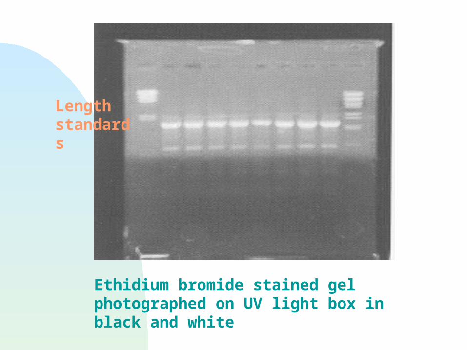
Ethidium bromide stained gel photographed on UV light box in black and white
Length standards
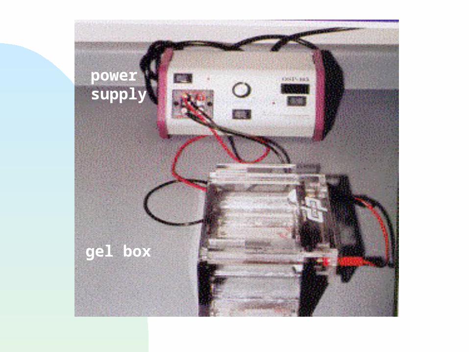
power supply
gel box
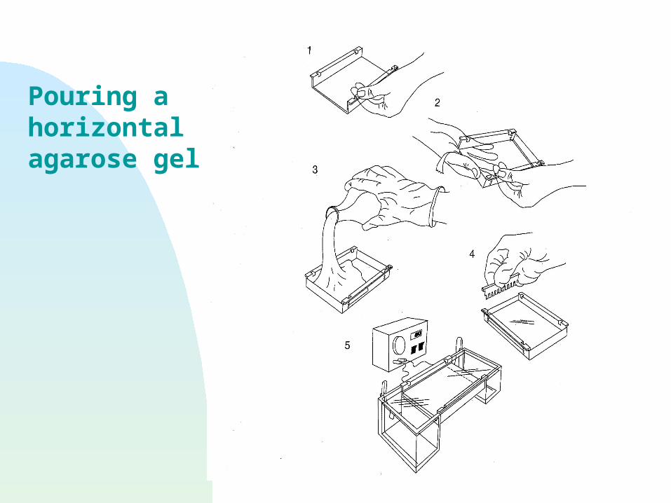
Pouring a horizontal agarose gel
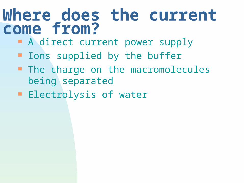
Where does the current come from? A direct current power supply Ions supplied by the buffer The charge on the macromolecules being
separated Electrolysis of water
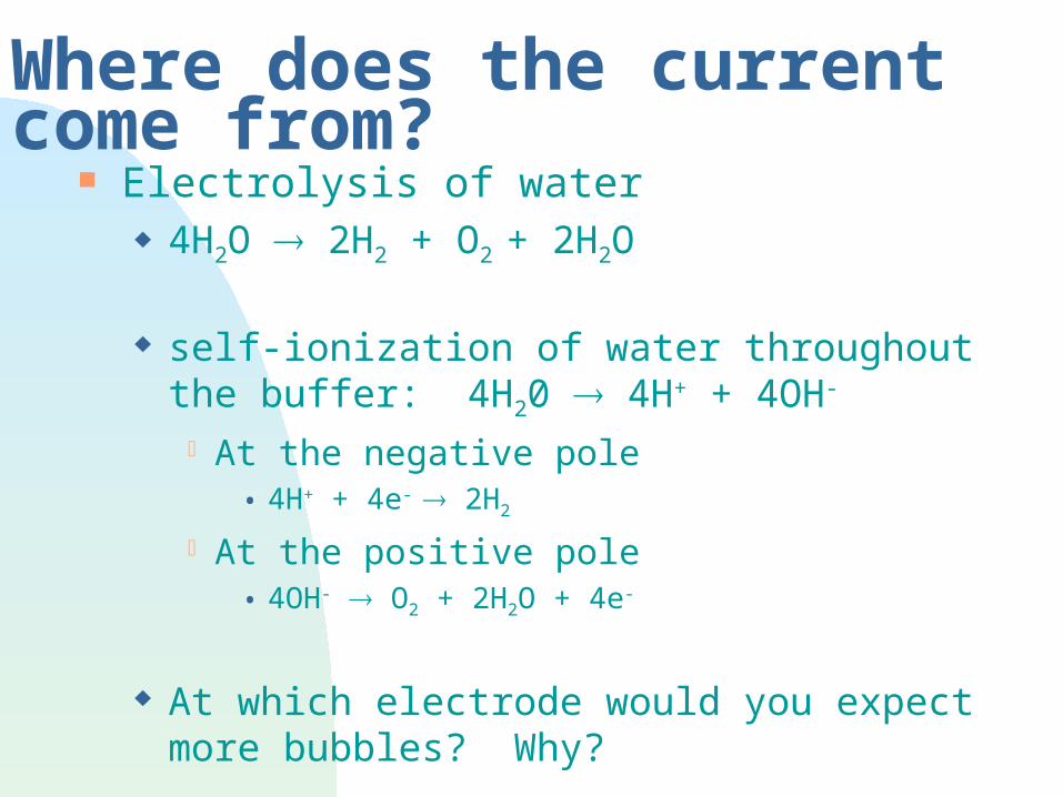
Where does the current come from? Electrolysis of water
4H2O 2H2 + O2 + 2H2O
self-ionization of water throughout the buffer: 4H20 4H+ + 4OH-
At the negative pole• 4H+ + 4e- 2H2
At the positive pole• 4OH- O2 + 2H2O + 4e-
At which electrode would you expect more bubbles? Why?
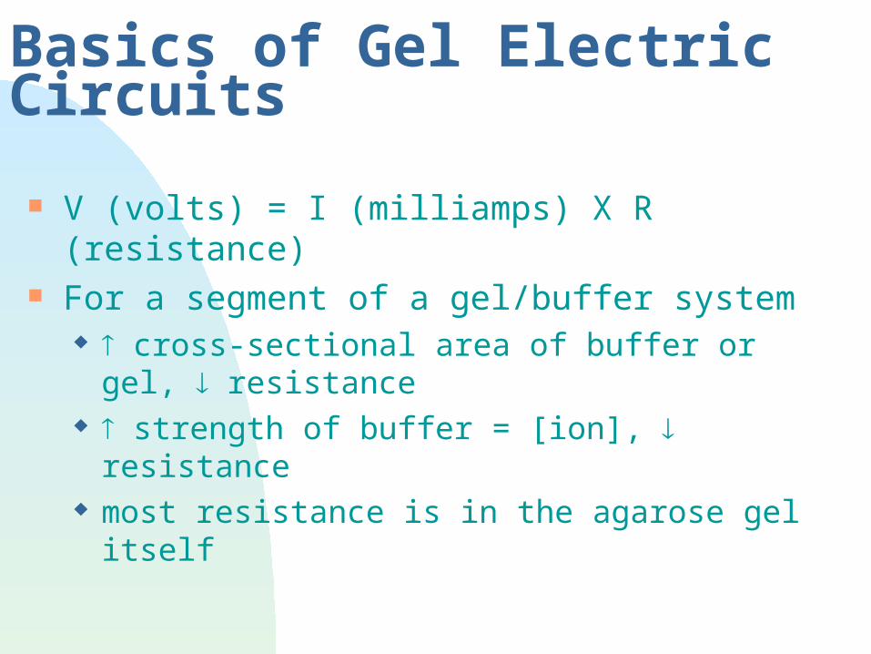
Basics of Gel Electric Circuits
V (volts) = I (milliamps) X R (resistance) For a segment of a gel/buffer system
cross-sectional area of buffer or gel, resistance strength of buffer = [ion], resistance most resistance is in the agarose gel itself
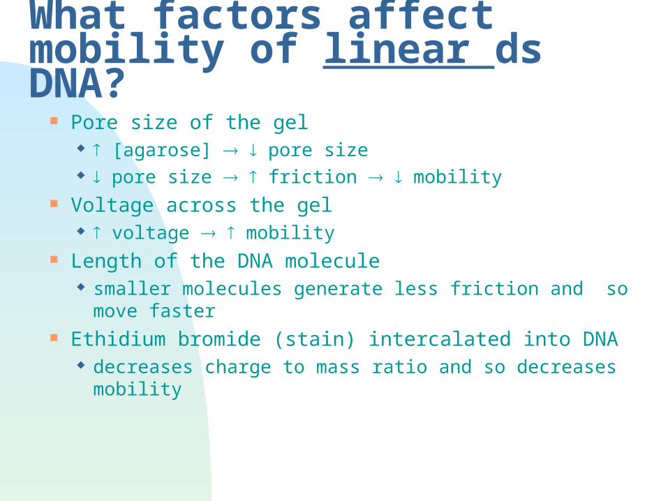
What factors affect mobility of linear ds DNA?
Pore size of the gel [agarose] pore size pore size friction mobility
Voltage across the gel voltage mobility
Length of the DNA molecule smaller molecules generate less friction and so move
faster Ethidium bromide (stain) intercalated into DNA
decreases charge to mass ratio and so decreases mobility
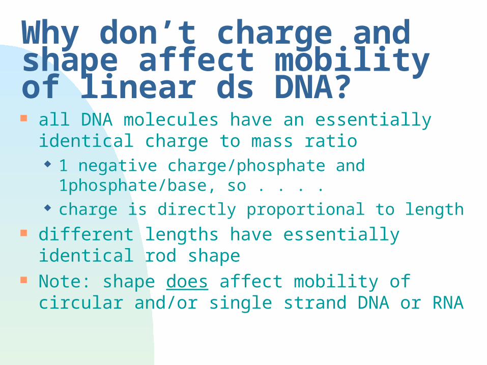
Why don’t charge and shape affect mobility of linear ds DNA? all DNA molecules have an essentially identical
charge to mass ratio 1 negative charge/phosphate and 1phosphate/base,
so . . . . charge is directly proportional to length
different lengths have essentially identical rod shape
Note: shape does affect mobility of circular and/or single strand DNA or RNA
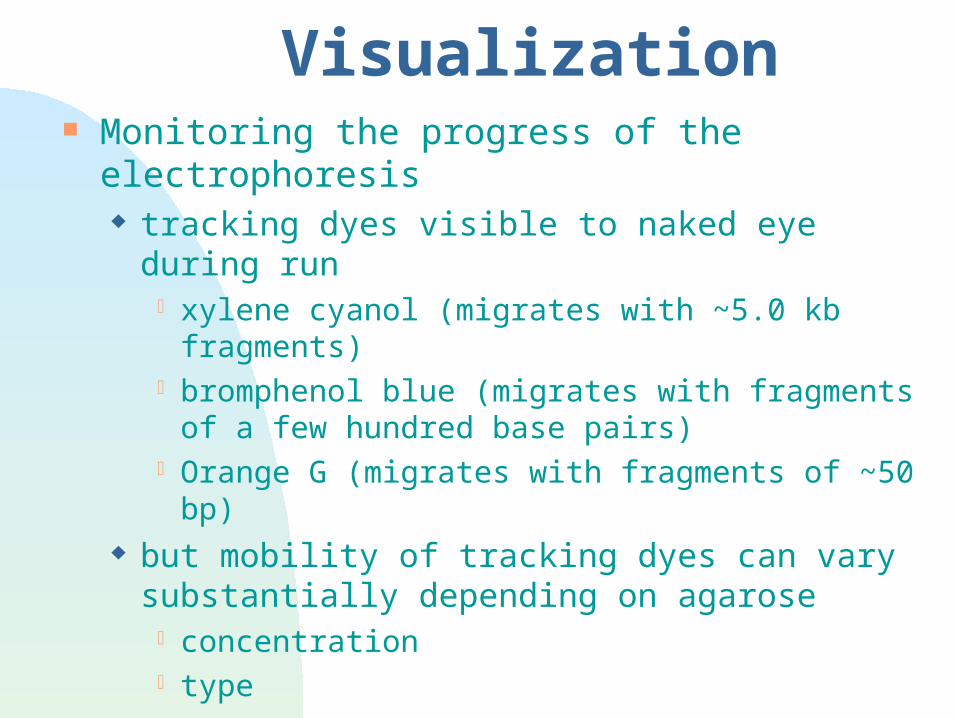
Visualization Monitoring the progress of the electrophoresis
tracking dyes visible to naked eye during run xylene cyanol (migrates with ~5.0 kb fragments) bromphenol blue (migrates with fragments of a few
hundred base pairs) Orange G (migrates with fragments of ~50 bp)
but mobility of tracking dyes can vary substantially depending on agarose concentration type

Visualization Locating the DNA fragments in the gel
ethidium bromide staining mutagen, wear gloves! visible under UV light wear UV opaque face or eye shield to observe!
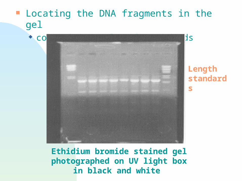
Locating the DNA fragments in the gel comparison to length standards
Ethidium bromide stained gel photographed on UV light box in black
and white
Length standards
Factors affecting resolution
Resolution = separation of fragments The “higher” the resolution, the more space between
fragments of similar, but different, lengths Resolution is affected by
agarose type agarose concentration salt concentration of buffer or sample amount of DNA loaded in the sample voltage
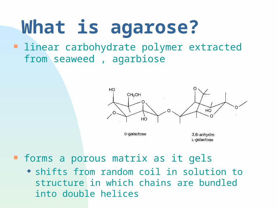
What is agarose? linear carbohydrate polymer extracted from
seaweed , agarbiose
forms a porous matrix as it gels shifts from random coil in solution to structure in
which chains are bundled into double helices
What is agarose? (cont’d) multiple types of agarose
Standard agarose - LE Gels at 35-38oC; Melts at 90-95oC Becomes opaque at high concentrations Makes a fairly sturdy gel
Low melting agarose (NuSieve) Gels at 35oC; Melts at 65oC
• Often used to isolate DNA fragments from gel Modified by hydroxyethylation to lower M.P. Is relatively translucent at high concentrations Makes a fragile gel
Intermediate forms or combinations of LE and NuSieve can provide sturdy, translucent gels at high agarose concentrations Good for resolving smaller fragments
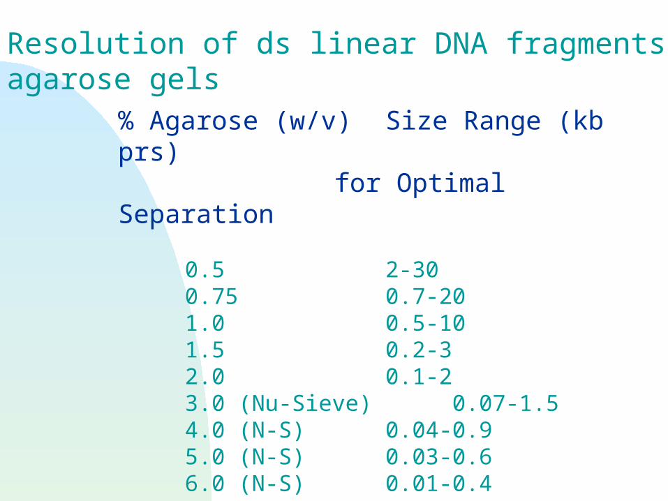
Resolution of ds linear DNA fragments in agarose gels
% Agarose (w/v) Size Range (kb prs) for Optimal Separation
0.5 2-300.75 0.7-201.0 0.5-101.5 0.2-32.0 0.1-23.0 (Nu-Sieve) 0.07-1.54.0 (N-S) 0.04-0.95.0 (N-S) 0.03-0.66.0 (N-S) 0.01-0.4
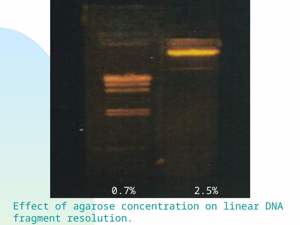
0.7% 2.5%
Effect of agarose concentration on linear DNA fragment resolution.The two lanes contain identical DNA samples.
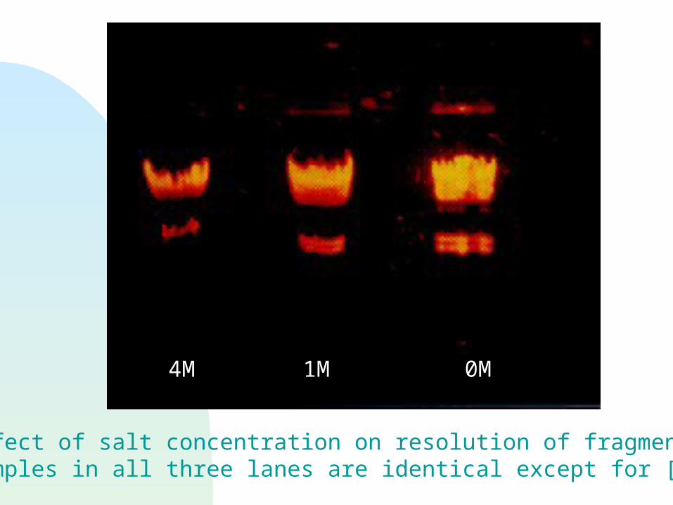
4M 1M 0M
Effect of salt concentration on resolution of fragments.Samples in all three lanes are identical except for [salt].
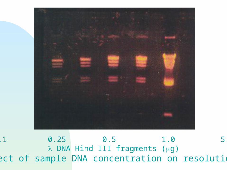
0.1 0.25 0.5 1.0 5.0 DNA Hind III fragments (g)
Effect of sample DNA concentration on resolution.
Voltage voltage, rate of migration to increase the voltage
increase the setting on the power supply increase the resistance
decrease the gel thickness decrease the ion concentration
if voltage is too high, gel melts as voltage is increased, large molecules migrate at a rate
proportionally faster than small molecules, so lower voltages are better for resolving large fragments but the larger ds DNA fragments are always slower than the smaller ones
Buffer Systems Remember, buffer systems include weak acids and/or bases
that do not dissociate completely. If ions resulting from dissociation are “removed,” more weak
acid and/or base will dissociate. Purposes of buffer
Keep solution at pH compatible with molecules being separated Generate ions consistently to
maintain current keep resistance low
Both gel and the solution in the gel box are buffered.
Buffer Systems (cont’d)
Two commonly used buffers for routine agarose gel electrophoresis TAE, pH 8.0, ~50 mM - Tris, Acetate, EDTA TBE, pH 8.0, ~50 mM - Tris, Borate, EDTA
Tris (T) is a weak base. Acetic (A) acid and boric (B) acid are weak acids.
Acetic acid is more completely ionized at pH 8.0 than is boric acid, so TBE has a high buffer capacity than TAE.
Buffer Systems (cont’d) TAE, pH 8.0, ~50 mM - Tris, Acetate, EDTA
loses buffer capacity during long or high voltage gel runs; • anode end of gel becomes acidic• gel may melt from the increased resistance that results from ion depletion
resolves high MW fragments better than TBE
TBE, pH 8.0, ~50 mM - Tris, Borate, EDTA higher buffer capacity somewhat more expensive resolves low MW fragments better than TAE may interfere with subsequent reactions
Non-denaturing agarose gel loading solutions
Composition tracking dyes
are used to follow progress of electrophoresis sometimes interfere with later visualization of DNA
a solute to increase density so that sample falls to bottom of loading well with minimal dilution solute examples: glycerol, Ficoll
Other gel types, with different purposes, use different loading solutions!
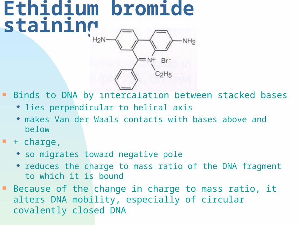
Ethidium bromide staining
Binds to DNA by intercalation between stacked bases lies perpendicular to helical axis makes Van der Waals contacts with bases above and below
+ charge, so migrates toward negative pole reduces the charge to mass ratio of the DNA fragment to which
it is bound Because of the change in charge to mass ratio, it alters DNA
mobility, especially of circular covalently closed DNA

Ethidium bromide staining Used to visualize DNA with UV light
uv 254 nm absorbed by DNA and transmitted to EtBr excitation at 302 or 366 nm fluorescence at 590 nm
560 nm = visible red/orange >/= 10ng/band required for visualization
Bound dye fluoresces 20-25X more than dye in solution because of fixed position of planar group proximity of dye to bases
UV light damages eyes and skin! Wear goggles and/or face shield.
Trouble shooting Smearing
torn sample wells voltage too high for large fragments too much DNA
Use </= 0.5 ug / fragment / 0.25cm2 migration area Gel melts
voltage too high ionic strength too low
Poor resolution wrong [agarose] small bands are fuzzy – the gel run may have been too long at too low
a voltage, allowing diffusion of the DNA and broadening of the band