Embed Size (px)
Citation preview
A manual for age determination of
southern bluefin tuna Thunnus maccoyii
Otolith sampling, preparation and interpretation
The Direct Age Estimation Workshop of the CCSBT 11-14 June, 2002
Queenscliff, Australia

Contents
CONTENTS .........................................................................................................................2
1 INTRODUCTION.........................................................................................................3 1.1 BACKGROUND TO THIS DOCUMENT............................................................................3 1.2 REFERENCE COLLECTION .........................................................................................3 1.3 CURRENT OTOLITH COLLECTIONS AND ARCHIVED OTOLITHS.....................................3
2 BACKGROUND ON SBT.............................................................................................4 2.1 LIFE HISTORY AND BIOLOGY .....................................................................................4 2.2 MAP INCLUDING RANGE AND SPAWNING GROUNDS ...................................................5
3 RESULTS FROM PREVIOUS OTOLITH AGE ESTIMATION STUDIES .................5
4 SAMPLING OF OTOLITHS ........................................................................................6 4.1 DESCRIPTION OF SBT OTOLITH MORPHOLOGY AND POSITION ...................................6 4.2 TECHNIQUES FOR EXTRACTION OF OTOLITHS ............................................................7 4.3 CLEANING, HANDLING AND STORAGE......................................................................12 4.4 SAMPLING STRATEGIES...........................................................................................13
5 AGE ESTIMATES USING WHOLE OTOLITHS FROM JUVENILE SBT............... 13 5.1 PREPARATION .........................................................................................................13 5.2 INTERPRETATION OF ANNUAL GROWTH INCREMENTS IN WHOLE OTOLITHS .............13 5.3 SCALES OF READABILITY FOR WHOLE OTOLITH READINGS.......................................14
6 PREPARING OTOLITH SECTIONS FOR AGE ESTIMATES ................................. 15 6.1 SECTIONS APPROPRIATE FOR SBT AGE ESTIMATION ...............................................15 6.2 HOW TO SECTION ....................................................................................................15 6.3 ADVANTAGES AND DISADVANTAGES OF PREPARATION TECHNIQUES: MULTIPLE SECTIONS VERSUS SINGLE SECTIONS ..................................................................................16
7 INTERPRETATION AND MEASUREMENT OF STRUCTURES IN OTOLITH SECTIONS......................................................................................................................... 19
7.1 INTERPRETATION OF ANNUAL GROWTH INCREMENTS IN OTOLITH SECTIONS ...........19 7.2 SCALES OF READABILITY AND CONFIDENCE FOR OTOLITH SECTIONS .......................22 7.3 CONVERTING ZONE COUNTS TO AGE ESTIMATES....................................................22 7.4 USE OF DIGITAL IMAGES IN AGE ESTIMATION ..........................................................23
8 LEVELS OF EXPECTED PRECISION FROM AGE ESTIMATION STUDIES ....... 24 8.1 PRECISION AND ACCURACY .....................................................................................24 8.2 MEASURES OF PRECISION TO DETERMINE INTRA- AND INTER-READER DIFFERENCES .. ...............................................................................................................................24
9 PROTOCOLS FOR ESTIMATING AGES USING OTOLITHS................................. 25
APPENDIX A. GLOSSARY ............................................................................................... 27
APPENDIX B. IMAGES OF OTOLITH SECTIONS FROM JUVENILE SBT AND FINAL AGE ESTIMATES ............................................................................................................. 30
APPENDIX C. WORKSHOP PARTICIPANTS ................................................................. 33
APPENDIX D. REFERENCES.......................................................................................... 36
___________________________________________________________________________
3
1 Introduction
1.1 Background to this document The 6th Scientific Committee Meeting of the Commission for the Conservation of Southern Bluefin Tuna (CCSBT) agreed that a dedicated technical Age Estimation Workshop should be held during 2002 to develop standard protocols for estimating age of southern bluefin tuna (SBT) from otoliths. This workshop was held at the Central Ageing Facility of the Marine and Freshwater Resources Institute (MAFRI) in Queenscliff, Victoria, Australia on 11-14 June 2002. Scientists from Australia, Japan, Korea, New Zealand and Taiwan attended the workshop (Appendix C). The workshop had two main aims. The first was to facilitate skills exchange between the member scientists for the collection, preparation and reading of otoliths from SBT. The second aim was to develop a common standard in estimating age of southern bluefin tuna from otoliths and consequently to produce a working manual for use by members’ laboratories. This document is an overall outcome of collaborative effort through the Workshop and includes the standards for sampling and preparation of otoliths and criteria for identifying annual marks in SBT otoliths. This manual is expected to guide laboratories undertaking this work, to maintain common procedures, and to increase the precision and accuracy of age estimates contributing to stock assessment. Preparation of the manual was a collaborative effort by all participants listed in Appendix C. Naomi Clear led a primary drafting group which also included Tomoyuki Itoh, Malcolm Francis, Doo Hae An and Wann-Nian Tzeng. Individuals’ contributions to particular sections are acknowledged where possible. The manual also reflects previous work undertaken in the laboratories of members and where appropriate this is referenced in the manual.
1.2 Reference collection The Workshop agreed to store a reference set of SBT otolith sections comprising 60 otoliths with agreed final age estimates at the CCSBT Secretariat. Images of the same set of otoliths on which annual increments are marked will be available on CD-ROM and at the CCSBT website. This reference set of otolith will be available for loan to CCSBT members.
1.3 Current otolith collections and archived otoliths The CCSBT agreed at the 1997 SC to establish an otolith collection from all fishing sectors. The followings are the current status of the otolith sampling system and historically archived otoliths. Other Members are expected to establish similar otolith collection systems at the earliest possible time. Australia: Australia started collecting SBT otoliths in the 1960s. In recent years Australia has routinely collected otoliths from several hundred fish per year in their surface and longline fisheries for SBT. The total number of otoliths currently archived in Australia is about 15,000. In addition, some 500 otoliths are collected per year from the Indonesian fishery under a collaborative research programme, and age estimates of these are made routinely at the Central Aging Facility.
___________________________________________________________________________
4
Japan: Japan started collecting otoliths using observers placed on Japanese longline vessels in the early 1990’s. The level of co-operation and the success rate for retrieval of otoliths has improved steadily since the programme’s inception. In recent years, some 200–300 otoliths have been collected per year, and sampling is stratified in 10 cm size classes to ensure sampling of larger fish. New Zealand: New Zealand initiated SBT otolith collection in 1998 using observers on longline vessels. Otoliths from about 2100 fish have now been collected. 2 Background on SBT
2.1 Life history and biology The content in this section has been adapted from information from the CCSBT web site. Further references on the biology of SBT are available from the website (http://www.ccsbt.org). Southern bluefin tuna (SBT), Thunnus maccoyii, is a large, oceanic migratory fish. It is found throughout the southern hemisphere mainly in waters between 30 and 50 degrees south but only rarely in the eastern Pacific. The only known spawning area is in the Indian Ocean southeast of Java, Indonesia. SBT can live for up to forty years, reach a weight of over 200 kilograms, and grow to more than 2 metres in length. There is some uncertainty about the size and age when on average they become mature. This is the subject of current research by Commission members. The available data suggests that it is around 1.5 metres and no younger than 8 years. Mature females can produce in excess of several million eggs in a single spawning period. Breeding takes place from September to April in warm waters southeast of Java. The juveniles migrate south down the west coast of Australia. During the summer/autumn months (December-April), they tend to congregate in the coastal waters off the southern coast of Australia and spend their winters in deeper, temperate oceanic waters. After age 5, they are seldom found in near-shore surface-waters. Some other known facts about SBT are: -
• they swim at an average speed of 2-3 km/hr;
• average growth rate for a three-year old is 1.5 cm per month (fish have been growing faster since about 1980 than previously);
• they can tolerate a wide range of seawater temperatures because of their advanced circulatory system which tends to keep the temperature of their body warmer than the surrounding water;
• they are known to dive to at least 500 metres.

___________________________________________________________________________
5
2.2 Map including range and spawning grounds
Figure 2.1. Distribution of southern bluefin tuna (hatched) and the spawning ground (shaded). Reproduced with permission from Caton (1991). 3 Results from previous otolith age estimation studies Daily growth increments: Jenkins and Davis (1990) examined the otolith microstructure of SBT larvae under light and scanning electron microscopes. They verified daily formation of increments using marginal increment analysis and daily progression of increment number in otoliths sampled from a single cohort. Itoh and Tsuji (1996) examined growth increments in sagittal otoliths collected from juvenile SBT during 1990-1993. By viewing etched otoliths under SEM they found that increments were deposited daily in otoliths of juvenile fish. Counts of daily increments estimated that mean size-at-age 1 was 50.8 cm and size-at-age 2 was 78.6 cm. Comparison among various hard parts for direct age estimate: Gunn et al. (1996) examined three hard parts collected from SBT — scales, caudal vertebrae and sagittal otoliths —to determine which structure produced the most accurate age estimates in SBT. Scales proved useful for estimating ages of SBT only up to 4 years. Marginal increment analysis verified that the zones of dense circuli visible on the scales were deposited annually up to this age. In larger fish, “piling up” of circuli on the margin of scales and high incidence of regenerated scales made estimation of age very difficult. Obvious increments were present on both the vertebrae and otoliths as opaque and translucent zones. Age estimates from the two structures matched closely for the first 10 years of life. However, in larger fish the counts diverged, otoliths consistently providing higher age estimates. Using
___________________________________________________________________________
6
results from the concomitant validation study (see below) it was determined that annual increments are formed on sagittal otoliths throughout life but that this is not the case in vertebrae, where bands are formed less frequently. Validation with annual deposition of increment: Clear et al. (2000) validated annual deposition of otolith increments using strontium-marking experiments. During a large-scale tag-and-release programme conducted by CSIRO in the Great Australian Bight, 20,204 SBT were injected with strontium chloride (SrCl2). The strontium was deposited in the otoliths of injected fish, marking them with a “time-stamp” that was visible under SEM. Using marked otoliths the periodicity of increment formation was examined and it was established that in SBT with 1 to 6 increments in their otoliths, one increment is deposited per year at liberty. The otoliths recovered from the smallest tagged fish had one translucent zone. Evidence that this first increment was deposited during the first year of life came from the studies of daily growth increments (Itoh and Tsuji, 1996; Rees et al. 1996), both of whom determined that size-at-age 1 is 50 cm. In the study of annual increments, 50 cm fish had one increment on their otoliths and were estimated to be aged 1+. Evidence that increments visible in otoliths of larger SBT are deposited annually came from close agreement between increment counts on otoliths and the sum of age at tagging and time at liberty for two fish tagged in the 1980s and recaptured in the 1990s. These data indicated that increment formation continues to be annual in fish up to at least 13 years old. Kalish et al. (1996) provided further evidence that increments in SBT sagittae are formed annually throughout life by a comparison of increment counts with age estimates derived from levels of bomb-radiocarbon in the early growth zones of sagittae. That study reported close agreement between the two methods of estimating age for fish between 23 and 34 years old. In summary, three sources of data, those from the strontium-marking experiment, the increment counts for two fish at liberty for over a decade, and the bomb radiocarbon dataprovided strong evidence that increments identifiable in the sagittal otoliths of SBT are deposited annually and can be used to estimate the age of individual fish. The data developed using these techniques has been used to develop growth models and in stock assessments by members of the CCSBT. 4 Sampling of otoliths
4.1 Description of SBT otolith morphology and position The otoliths (or ear bones) of fish are small structures located in the semi-circular canals at the base of the brain. They are formed by the daily accretion of a layer of calcium carbonate bound within a protein matrix. In tunas, as in other teleost fish, there are 3 pairs of otoliths. The sagittal otoliths are the largest of the 3 pairs in SBT and are oblong with a pointed rostrum and a deep sulcus.
___________________________________________________________________________
7
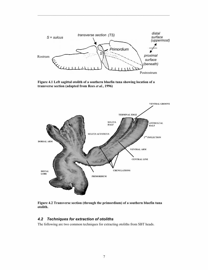
Figure 4.1 Left sagittal otolith of a southern bluefin tuna showing location of a transverse section (adapted from Rees et al., 1996)
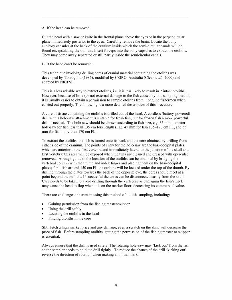
Figure 4.2 Transverse section (through the primordium) of a southern bluefin tuna otolith.
4.2 Techniques for extraction of otoliths The following are two common techniques for extracting otoliths from SBT heads.
Postrostrum
Rostrum
distal surface
(uppermost)
proximal surface
(beneath)
transverse section (TS) S = sulcus
S Primordium
PRIMORDIUM
SULCUS ACUSTICUS
TERMINAL EDGE
SULCUL HALF
ANTISULCAL HALF
CRENULATIONS
2ND INFLECTION
VENTRAL ARM
DORSAL ARM
VENTRAL GROOVE
CENTRAL LINE
DISTAL LOBE
___________________________________________________________________________
8
A. If the head can be removed: Cut the head with a saw or knife in the frontal plane above the eyes or in the perpendicular plane immediately posterior to the eyes. Carefully remove the brain. Locate the bony auditory capsules at the back of the cranium inside which the semi-circular canals will be found encapsulating the otoliths. Insert forceps into the bony capsules to extract the otoliths. They may come away separated or still partly inside the semicircular canals. B. If the head can’t be removed: This technique involving drilling cores of cranial material containing the otoliths was developed by Thorogood (1986), modified by CSIRO, Australia (Clear et al., 2000) and adapted by NRIFSF. This is a less reliable way to extract otoliths, i.e. it is less likely to result in 2 intact otoliths. However, because of little (or no) external damage to the fish caused by this sampling method, it is usually easier to obtain a permission to sample otoliths from longline fishermen when carried out properly. The following is a more detailed description of this procedure: A core of tissue containing the otoliths is drilled out of the head. A cordless (battery-powered) drill with a hole-saw attachment is suitable for fresh fish, but for frozen fish a more powerful drill is needed. The hole-saw should be chosen according to fish size, e.g. 35 mm diameter hole-saw for fish less than 135 cm fork length (FL), 45 mm for fish 135–170 cm FL, and 55 mm for fish more than 170 cm FL. To extract the otoliths, the fish is turned onto its back and the core obtained by drilling from either side of the cranium. The points of entry for the hole-saw are the basi-occipital plates, which are anterior to the first vertebra and immediately lateral to the junction of the skull and first vertebra; this area will be exposed when the tuna are cleaned and dressed with operculae removed. A rough guide to the location of the otoliths can be obtained by bridging the vertebral column with the thumb and index finger and placing them on the basi-occipital plates; for a fish around 150 cm FL the otoliths will be located under the top of the thumb. By drilling through the plates towards the back of the opposite eye, the cores should meet at a point beyond the otoliths. If successful the cores can be disconnected easily from the skull. Care needs to be taken to avoid drilling through the vertebrae as damaging the fish’s neck may cause the head to flop when it is on the market floor, decreasing its commercial value. There are challenges inherent in using this method of otolith sampling, including: • Gaining permission from the fishing master/skipper • Using the drill safely • Locating the otoliths in the head • Finding otoliths in the core SBT fetch a high market price and any damage, even a scratch on the skin, will decrease the price of fish. Before sampling otoliths, getting the permission of the fishing master or skipper is essential. Always ensure that the drill is used safely. The rotating hole-saw may ‘kick out’ from the fish so the sampler needs to hold the drill tightly. To reduce the chance of the drill ‘kicking out’ reverse the direction of rotation when making an initial mark.
___________________________________________________________________________
9
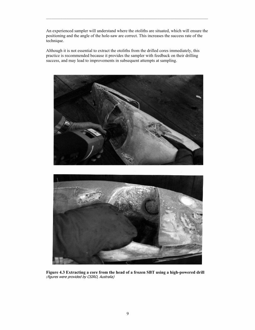
An experienced sampler will understand where the otoliths are situated, which will ensure the positioning and the angle of the hole-saw are correct. This increases the success rate of the technique. Although it is not essential to extract the otoliths from the drilled cores immediately, this practice is recommended because it provides the sampler with feedback on their drilling success, and may lead to improvements in subsequent attempts at sampling.
Figure 4.3 Extracting a core from the head of a frozen SBT using a high-powered drill (figures were provided by CSIRO, Australia)

___________________________________________________________________________
10
Figure 4.4 Battery-powered drills can be used for extracting cores from un-frozen fish (figures were provided by CSIRO, Australia and NRIFSF, Japan).

___________________________________________________________________________
11
Figure 4.5 To drill a core that contains the otoliths, the holesaw is positioned over the basi-occipital plates (indicated) and aimed towards the opposite eye (figures were provided by NRIFSF, Japan and CSIRO, Australia).
___________________________________________________________________________
12
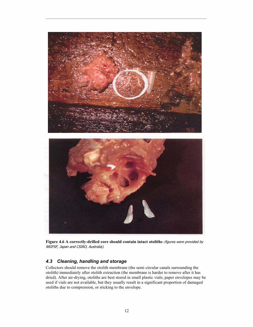
Figure 4.6 A correctly-drilled core should contain intact otoliths (figures were provided by NRIFSF, Japan and CSIRO, Australia).
4.3 Cleaning, handling and storage Collectors should remove the otolith membrane (the semi circular canals surrounding the otolith) immediately after otolith extraction (the membrane is harder to remove after it has dried). After air-drying, otoliths are best stored in small plastic vials; paper envelopes may be used if vials are not available, but they usually result in a significant proportion of damaged otoliths due to compression, or sticking to the envelope.
___________________________________________________________________________
13
Cleaning otoliths immediately after collection reduces the amount of time required to prepare them for sectioning.
4.4 Sampling strategies Two different sampling strategies have been used by members when collecting otoliths: • Length-stratified sampling in which a pre-determined number of otoliths are collected
from each length class. • Random sampling in which otoliths are collected from randomly selected fish. Length-stratified sampling provides good samples of otoliths across the full length range, and is particularly useful for the development of growth curves, age-length keys and the estimation of longevity. Population age structures can be derived by applying age-length keys to a length-frequency distribution of the catch that has been scaled to represent the population. Random sampling provides a direct estimate of the age structure of the population, but may not be suitable for the development of growth curves if all age classes are not fully represented. For both sampling methods, it is difficult to obtain adequate samples of otoliths to represent the whole stock that is fished by a number of fleets throughout the year. The appropriate sampling strategy depends on how to use of direct age information in stock assessment and this mater is currently considered by the CCSBT Scientific Committee. 5 Age estimates using whole otoliths from juvenile SBT This section describes methods developed by CSIRO. Further details can be found in Clear et al. (2000) and Gunn et al.(in press). Otoliths from fish less than 135 cm FL can generally be read whole. Increments comprise two zones: an opaque zone (assumed to be fast growth), and a narrower, translucent zone (assumed to be slow growth).
5.1 Preparation • Burn otoliths on a hot plate until they turn golden brown; the translucent zones will
become more obvious than the opaque zones • View the burnt otoliths under a light microscope, with reflected light and a black
background.
5.2 Interpretation of annual growth increments in whole otoliths • The first one or two increments deposited on the otolith are often broader and less
distinct than subsequent increments. • Increments are most obvious on the rostral and post-rostral axes (see figures 5.1 and
5.2); however, the translucent zones can usually be seen on the otolith surface between these axes, parallel to the ventral margin.
• Count increments on whole sagittae from the primordium to the margin along the rostral and post-rostral axes.

___________________________________________________________________________
14
• Measure the length of the rostral and post-rostral axes and the distance between the primordium and the inside of each translucent zone (the outer margin of the zone is often difficult to identify in otoliths from small fish) along the rostral and post-rostral axes.
• Measure the marginal increment along the rostral axis, from the beginning of the most recently deposited translucent zone to the rostrum.
• See figures 5.1, 5.2 and Appendix B for examples of age estimates from whole otoliths.
Figure 5.1. A whole, burnt sagittal otolith from a 98 cm FL SBT, caught in January. Three increments were counted on this right-hand sagitta: the darker, translucent zones can be followed along the otolith surface from the rostral axis to the post-rostral axis, parallel to the ventral margin. Scale Bar: 1 mm [Reproduced from Clear et al. (2000).]
Figure 5.2. Sagitta (specimen OB 764) from a fish tagged and injected in March 1994 at 57 cm FL and recaptured in February 1997 at 111 cm FL. Four increments were counted on the whole, burnt otolith and measured from the primordium (P) to the beginning of each translucent zone apparent on the distal surface along the post-rostral axis at 2.2 mm, 3.6 mm, 4.4 mm and 4.9 mm. Scale Bar: 1 mm [Source: Naomi Clear, CSIRO, Australia]
5.3 Scales of readability for whole otolith readings • Assign each otolith a “readability” score as follows:
1. An exceptional otolith – no doubt about the number of increments counted. 2. Some doubt is present but only 1 interpretation is considered. 3. 2 interpretations are possible – the best estimate is given. 4. More than 2 interpretations are possible – the best estimate is given.
P
12
3 4
P
12
3 4
P
12
3 4

___________________________________________________________________________
15
5. Unreadable (no pattern obvious) 6. Broken or missing
6 Preparing otolith sections for age estimates
6.1 Sections appropriate for SBT age estimation In southern bluefin tuna, the otolith thickens laterally mainly because material is deposited along two growth axes: dorso-medial and ventro-medial. We refer to these as the dorsal and ventral arms respectively (Figure 4.2). This results in a very deep sulcus. It has been found that a transverse section approximately 300-350 µm thick produced the greatest clarity of growth increments.
6.2 How to section Currently, three laboratories are involved in the preparation of SBT otoliths: the Central Ageing Facility, Victoria, Australia (CAF), the National Institute of Water and Atmospheric Research, Wellington, New Zealand (NIWA), and Marino Research, Mie, Japan. Each laboratory uses different methods to produce thin transverse sections as outlined below. CAF, Australia: Clean dry otoliths are arranged in two columns of 5 otoliths and embedded in clear casting polyester resin (methyl-ethyl ketone peroxide is used as hardening agent), ensuring that the primordia of the otoliths are aligned. A minimum of 4 transverse sections approximately 250-400 µm thick are cut from the centres of the otoliths using a modified Gemasta lapidary diamond cutting saw fitted with a 250 µm wide diamond impregnated blade. The sections are then cleaned in water, rinsed with alcohol and dried. Sections are then mounted on numbered microscope slides using polyester resin and covered with glass cover-slips. Generally the otoliths are not baked in this process although this can be done if desired. Approximately 250 otoliths can be prepared per day. (See section 6.3 for photographs of the preparation process). Marino Research Inc., Japan: Large otoliths (major axis > 10 mm) The otolith primordium is marked, and cut with a small hand-held cutting device (Minitor, diamond cutting disc). The cut surface is polished with a grinder-polisher (2000 grit), and then fixed to a glass slide with a strong adhesive. When the otolith has been firmly mounted onto the glass slide, the other end is removed with the small cutting device, until the remainder is approximately 1 mm thick. It is then ground further to about 300 µm with the grinder-polisher. The surface is coated by dripping epoxy embedding resin (Euparal) over it to produce a smooth surface. Small otoliths (major axis < 10 mm) A rectangular block is formed by placing the otolith in a silicone mould, and pouring in epoxy embedding resin. Next, the block is secured, and part of the otolith is removed with a cutting device (micro cutter). The blade of the cutting device is repositioned, and adjusted so that it cuts a slice approximately 300 µm thick. This section is attached to a slide with epoxy embedding resin, and coated with Euparal to produce a smooth surface. NIWA, New Zealand: Otoliths are cleaned of tissue using an ultra-sonic cleaner. Each otolith is dried, embedded individually in a clear epoxy resin block, and left for 24 hours while the resin cures. The core is identified under reflected light using a binocular microscope. A tungsten tipped pen is then

___________________________________________________________________________
16
used to scribe a transverse line across the surface of the resin block. This allows the core to be located easily during sectioning. A thin transverse section (approx. 650 µm thick) is cut from the resin block using a Struers (Accutom-2) saw fitted with two diamond-edged blades separated by a spacer.
The thin section is examined under a binocular microscope (using reflected light) to determine the clearest side, i.e. the side closest to the primordium. This side is ground manually (with 1200 grit, followed by 4000 grit carborundum) to enhance the primordium. Once this has been achieved, 1 µm alumina paste (Linde) is used to remove any remaining scratches. The section is then glued to a microscope slide (polished surface face down) using fast curing clear epoxy resin. The upper surface of the section is polished using 1200 grit carborundum followed by 1 µm linde to a thickness of approx. 350 µm.
6.3 Advantages and disadvantages of preparation techniques: multiple sections versus single sections
While the CAF prepares multiple sections of multiple otoliths at one time, Marino Research Inc. and NIWA prepare one section of each otolith. Advantage and disadvantages in preparing multiple sections are discussed below. Preparing four sections provides a greater chance that at least one of these sections will be clear. The level of cutting precision is also less critical for multiple sections as it is not necessary for each section to pass through the primordium. This means that preparation time is reduced as the primordium does not have to be marked. However, this technique requires a specialised high-speed machine with a thin blade (approximately 250 µm thick). Commonly available machines with a thick blade generally lose too much otolith material, which increases the risk of losing the primordium. Even with a special machine with a thin blade, the primordium may occasionally be missed completely in all four sections. The disadvantage with preparing single section is that locating and marking the primordium is critical, as each section must be as clear as possible. This is often a very time consuming process. The techniques of multiple sectioning used at the CAF are shown in following series of photos:
___________________________________________________________________________
17
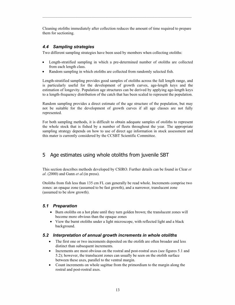
1. Silicone moulds used to embed otoliths in polyester resin. Source: Kyne Krusic-Golub, Central Ageing Facility, Australia.
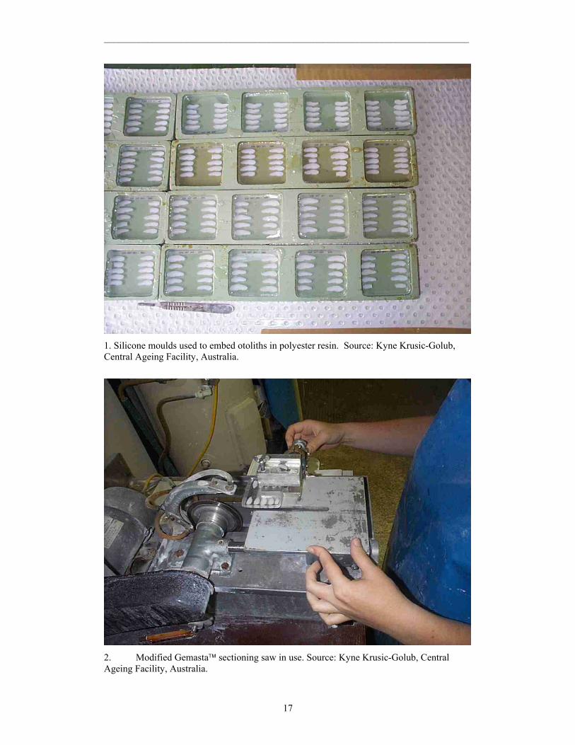
2. Modified Gemasta sectioning saw in use. Source: Kyne Krusic-Golub, Central Ageing Facility, Australia.
___________________________________________________________________________
18
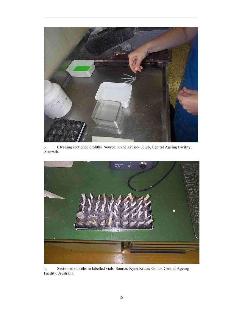
3. Cleaning sectioned otoliths. Source: Kyne Krusic-Golub, Central Ageing Facility, Australia.
4. Sectioned otoliths in labelled vials. Source: Kyne Krusic-Golub, Central Ageing Facility, Australia.
___________________________________________________________________________
19
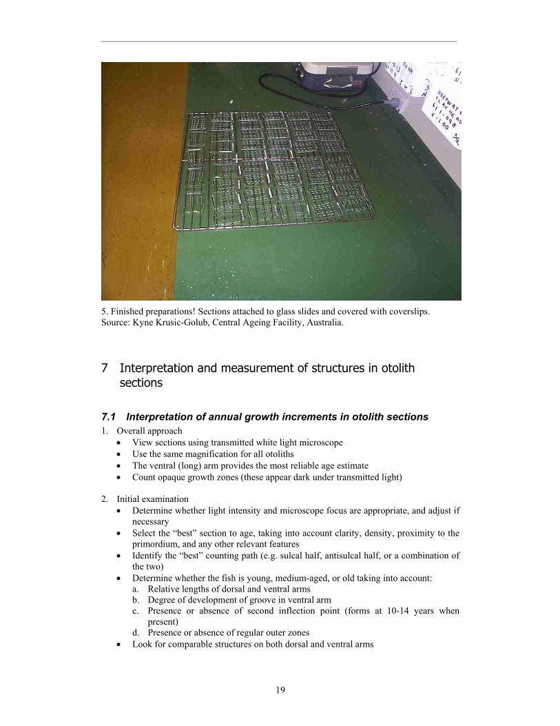
5. Finished preparations! Sections attached to glass slides and covered with coverslips. Source: Kyne Krusic-Golub, Central Ageing Facility, Australia. 7 Interpretation and measurement of structures in otolith
sections
7.1 Interpretation of annual growth increments in otolith sections 1. Overall approach
• View sections using transmitted white light microscope • Use the same magnification for all otoliths • The ventral (long) arm provides the most reliable age estimate • Count opaque growth zones (these appear dark under transmitted light)
2. Initial examination
• Determine whether light intensity and microscope focus are appropriate, and adjust if necessary
• Select the “best” section to age, taking into account clarity, density, proximity to the primordium, and any other relevant features
• Identify the “best” counting path (e.g. sulcal half, antisulcal half, or a combination of the two)
• Determine whether the fish is young, medium-aged, or old taking into account: a. Relative lengths of dorsal and ventral arms b. Degree of development of groove in ventral arm c. Presence or absence of second inflection point (forms at 10-14 years when
present) d. Presence or absence of regular outer zones
• Look for comparable structures on both dorsal and ventral arms
___________________________________________________________________________
20
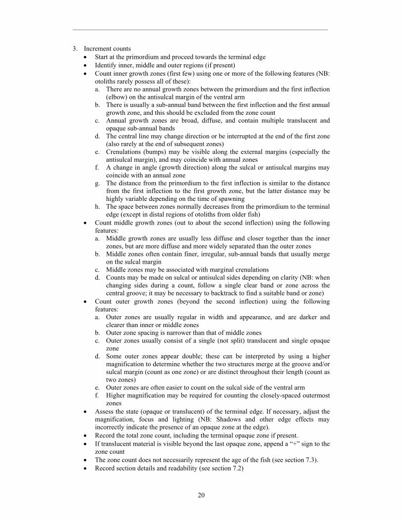
3. Increment counts • Start at the primordium and proceed towards the terminal edge • Identify inner, middle and outer regions (if present) • Count inner growth zones (first few) using one or more of the following features (NB:
otoliths rarely possess all of these): a. There are no annual growth zones between the primordium and the first inflection
(elbow) on the antisulcal margin of the ventral arm b. There is usually a sub-annual band between the first inflection and the first annual
growth zone, and this should be excluded from the zone count c. Annual growth zones are broad, diffuse, and contain multiple translucent and
opaque sub-annual bands d. The central line may change direction or be interrupted at the end of the first zone
(also rarely at the end of subsequent zones) e. Crenulations (bumps) may be visible along the external margins (especially the
antisulcal margin), and may coincide with annual zones f. A change in angle (growth direction) along the sulcal or antisulcal margins may
coincide with an annual zone g. The distance from the primordium to the first inflection is similar to the distance
from the first inflection to the first growth zone, but the latter distance may be highly variable depending on the time of spawning
h. The space between zones normally decreases from the primordium to the terminal edge (except in distal regions of otoliths from older fish)
• Count middle growth zones (out to about the second inflection) using the following features: a. Middle growth zones are usually less diffuse and closer together than the inner
zones, but are more diffuse and more widely separated than the outer zones b. Middle zones often contain finer, irregular, sub-annual bands that usually merge
on the sulcal margin c. Middle zones may be associated with marginal crenulations d. Counts may be made on sulcal or antisulcal sides depending on clarity (NB: when
changing sides during a count, follow a single clear band or zone across the central groove; it may be necessary to backtrack to find a suitable band or zone)
• Count outer growth zones (beyond the second inflection) using the following features: a. Outer zones are usually regular in width and appearance, and are darker and
clearer than inner or middle zones b. Outer zone spacing is narrower than that of middle zones c. Outer zones usually consist of a single (not split) translucent and single opaque
zone d. Some outer zones appear double; these can be interpreted by using a higher
magnification to determine whether the two structures merge at the groove and/or sulcal margin (count as one zone) or are distinct throughout their length (count as two zones)
e. Outer zones are often easier to count on the sulcal side of the ventral arm f. Higher magnification may be required for counting the closely-spaced outermost
zones • Assess the state (opaque or translucent) of the terminal edge. If necessary, adjust the
magnification, focus and lighting (NB: Shadows and other edge effects may incorrectly indicate the presence of an opaque zone at the edge).
• Record the total zone count, including the terminal opaque zone if present. • If translucent material is visible beyond the last opaque zone, append a “+” sign to the
zone count • The zone count does not necessarily represent the age of the fish (see section 7.3). • Record section details and readability (see section 7.2)
___________________________________________________________________________
21
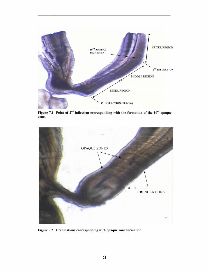
Figure 7.1 Point of 2nd inflection corresponding with the formation of the 10th opaque zone.
Figure 7.2 Crenulations corresponding with opaque zone formation
1st INFLECTION (ELBOW)
2ND INFLECTION
10TH ANNUAL INCREMENT
INNER REGION
MIDDLE REGION
OUTER REGION
CRENULATIONS
OPAQUE ZONES
___________________________________________________________________________
22
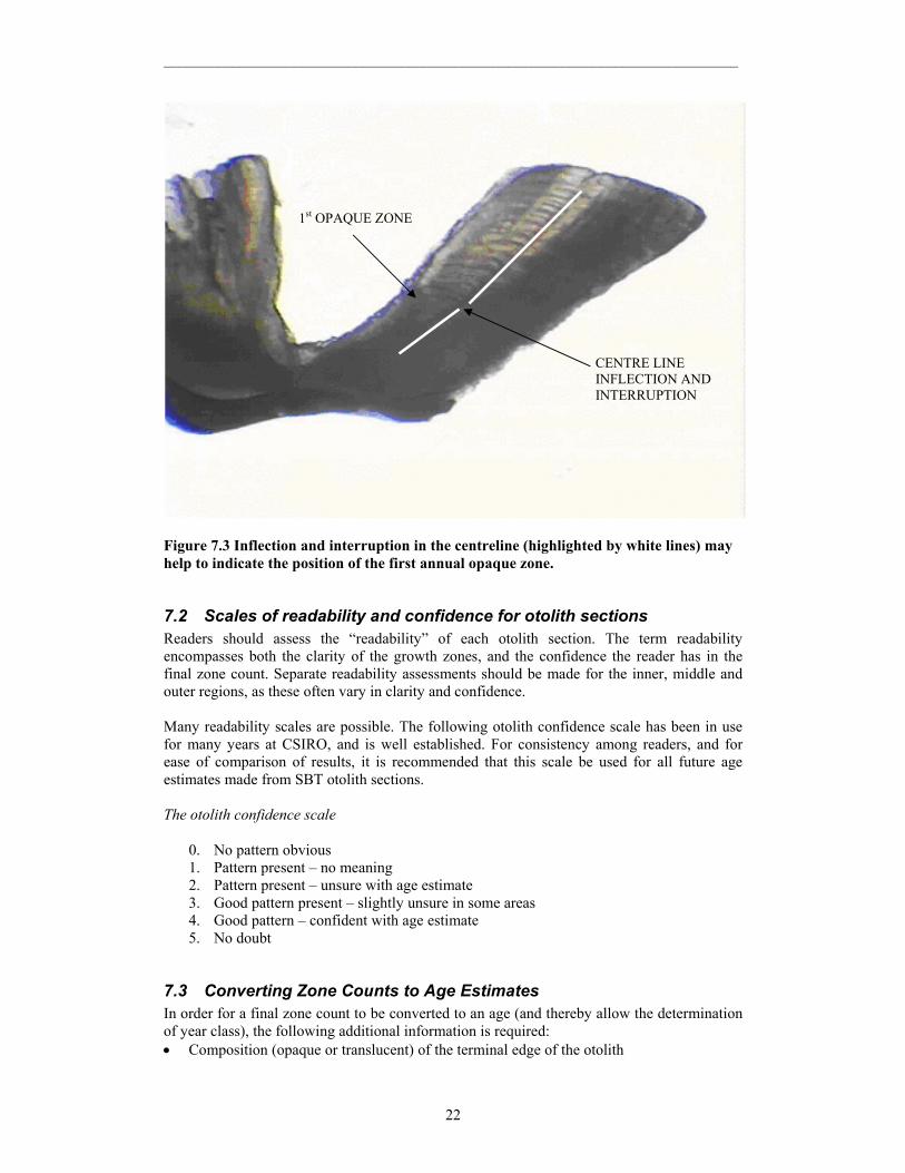
Figure 7.3 Inflection and interruption in the centreline (highlighted by white lines) may help to indicate the position of the first annual opaque zone.
7.2 Scales of readability and confidence for otolith sections Readers should assess the “readability” of each otolith section. The term readability encompasses both the clarity of the growth zones, and the confidence the reader has in the final zone count. Separate readability assessments should be made for the inner, middle and outer regions, as these often vary in clarity and confidence. Many readability scales are possible. The following otolith confidence scale has been in use for many years at CSIRO, and is well established. For consistency among readers, and for ease of comparison of results, it is recommended that this scale be used for all future age estimates made from SBT otolith sections. The otolith confidence scale
0. No pattern obvious 1. Pattern present – no meaning 2. Pattern present – unsure with age estimate 3. Good pattern present – slightly unsure in some areas 4. Good pattern – confident with age estimate 5. No doubt
7.3 Converting Zone Counts to Age Estimates In order for a final zone count to be converted to an age (and thereby allow the determination of year class), the following additional information is required: • Composition (opaque or translucent) of the terminal edge of the otolith
1st OPAQUE ZONE
CENTRE LINE INFLECTION AND INTERRUPTION
___________________________________________________________________________
23
• Theoretical birth date • Time of year during which opaque zones are formed • Date of capture The marginal composition is determined during the otolith interpretation stage. Note, however, that deposition of a zone may begin some time before it becomes visible at the terminal edge of the otolith. The theoretical birth date of SBT has been defined as 1 January. However, spawning lasts from September to March, with peaks occurring near the beginning and end of that period. So individual fish may have been spawned up to 3 months before or after the theoretical birth date. In whole otoliths, the first translucent zone is formed in May-September (“winter”) and the subsequent opaque zone during October-April (“summer”) (Clear et al. in press). Translucent zones are narrower than opaque zones. However, the relationship between the zones seen in whole otoliths and those seen in thin sections has not been determined. In the inner region of otolith sections, the opaque zones appear broader than the translucent zones (though it is very difficult to determine the start and finish points of each zone). This interpretation is consistent with that in whole otoliths. However, in the outer region of otolith sections from old fish, the opaque zones may be narrower than the translucent zones. More research is required to determine whether the opaque zones that are being counted are always deposited in summer across the entire otolith. This could be determined by: • examining the composition of the terminal edge in different months • examining the composition of the material deposited after tagging
7.4 Use of digital images in age estimation Digital images are often used for counting growth zones, and for measuring inter-zone distances and otolith radii. Digital images have advantages and disadvantages over microscope examination, and readers should consider these when deciding which method to use. Advantages of direct microscopic examination:
• Greater ability to perceive variations in intensity, density and colour of otolith structures
• Greater ability to interpret three-dimensional structure by altering the focal plane • Greater resolution • Faster for a simple zone count (but slower if measurements or many hard copy
images are required) Advantages of digital images:
• Ability to mark, count and display growth zones on the image for later examination, discussion, and dissemination
• Reduced chance of counting same zone twice, or missing zones (if zones are marked on-screen while counting)
• Ability to enhance images digitally to improve clarity and discrimination of growth zones
• Facilitate and speed up measurement of distances • Automatic data acquisition reduces likelihood of transcription errors
___________________________________________________________________________
24
Automated zone counting: In theory, automated counting of otolith zones is possible using software developed for that purpose, or adapted from another application. However, to date, automated counts have only been possible for species having clear, discrete growth zones. The participants at the workshop felt that automated age determination for southern bluefin tuna is unlikely to be possible in the near future. 8 Levels of expected precision from age estimation studies
8.1 Precision and accuracy In studies of age determination, accuracy refers to the closeness of an age estimate to the true age whereas precision is a measure of the variability between individual readings, either within or between readers. The aim in age determination studies is to increase precision and accuracy. However, age estimates are still useful where precision is low but the mean age reflects the true age. Problems arise when there is bias in age estimates (often obvious in age-bias plots – see section 8.2) and estimates are inaccurate. Accuracy of age estimates can only be tested by validation, using an independent and absolute method, such as the strontium-chloride marking of SBT hard parts. Indices of precision can also be a useful tool for comparing between readers and between methodologies (e.g. otoliths and vertebrae). They can also indicate samples that are “difficult”, i.e. produce unreliable estimates of age.
8.2 Measures of precision to determine intra- and inter-reader differences
Intra- and inter-reader variability (consistency) between otolith readings can be quantified using several methods. Average Percentage Error (e.g. Beamish and Fournier 1981):
,X
XXR1*100APE
R
1i j
jijj ∑
=
−=
where Xij is the ith age estimate of the jth fish, Xj the mean of readings for the jth fish and R is the number of times each fish is aged. Using the standard deviation rather than the absolute deviation from the mean age produces a coefficient of variation (CV) (Campana et al. 1995):
( )
j
R
1i
2jij
j X1R
XX
*100CV∑
= −−
=
Experienced readers of SBT have achieved average percentage error (APE) levels of around 3% for intra- and inter-reader precision. We recommend a maximum level of 10%, beyond which resultant age estimates should be treated with a caution.
___________________________________________________________________________
25
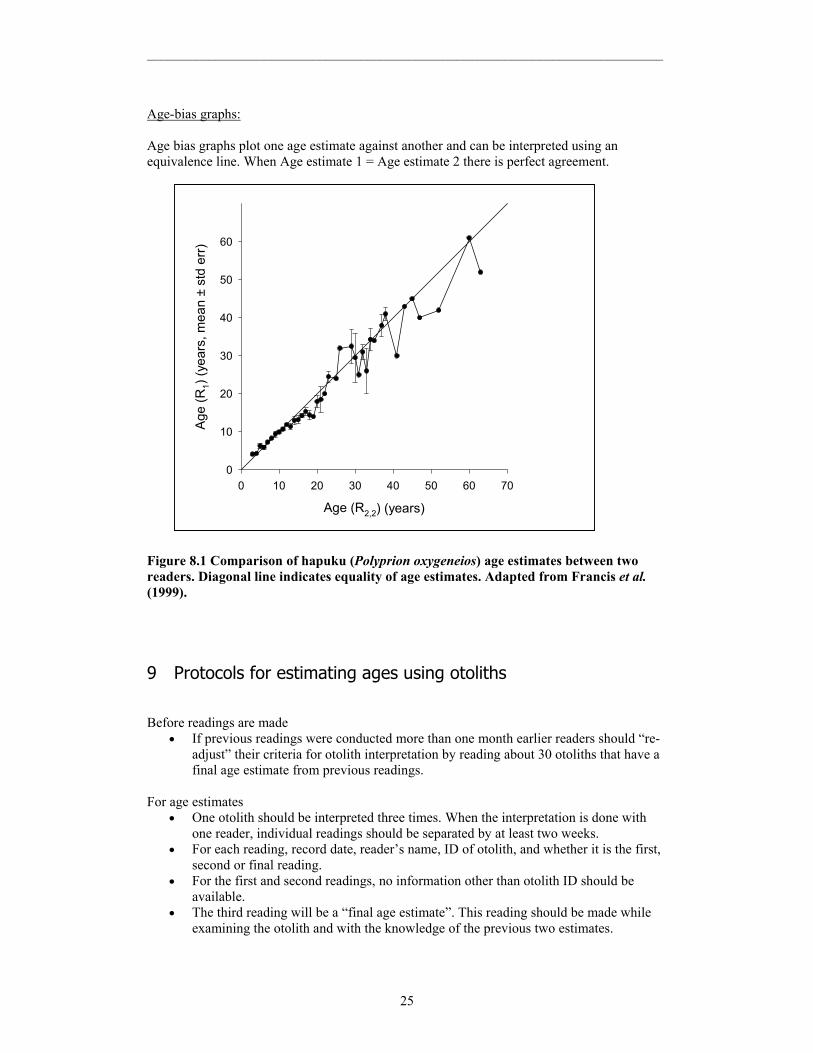
Age-bias graphs: Age bias graphs plot one age estimate against another and can be interpreted using an equivalence line. When Age estimate 1 = Age estimate 2 there is perfect agreement.
Age (R2,2) (years)
0 10 20 30 40 50 60 70
Age
(R1)
(yea
rs, m
ean
± st
d er
r)
0
10
20
30
40
50
60
Figure 8.1 Comparison of hapuku (Polyprion oxygeneios) age estimates between two readers. Diagonal line indicates equality of age estimates. Adapted from Francis et al. (1999).
9 Protocols for estimating ages using otoliths Before readings are made
• If previous readings were conducted more than one month earlier readers should “re-adjust” their criteria for otolith interpretation by reading about 30 otoliths that have a final age estimate from previous readings.
For age estimates
• One otolith should be interpreted three times. When the interpretation is done with one reader, individual readings should be separated by at least two weeks.
• For each reading, record date, reader’s name, ID of otolith, and whether it is the first, second or final reading.
• For the first and second readings, no information other than otolith ID should be available.
• The third reading will be a “final age estimate”. This reading should be made while examining the otolith and with the knowledge of the previous two estimates.
___________________________________________________________________________
26
• Read otoliths following the criteria described in 7.1 of this manual. If the reader is working with an electronic image they should mark the zones identified as annual.
• Assign readability level using ‘the scale of readability’ as described in 7.2 of this manual.
• Record estimated age and a level of readability. If reader is not confident with their interpretation, they should record a plausible age range in addition to the best estimate.
Age estimates by more than one reader
• When more than one reader is involved in otolith interpretation, readers should interpret some otoliths as a group in order to ensure that the interpretation is consistent among readers as well as over time. The group should collectively interpret at least 30 otoliths that already have final age estimates assigned.
• The group of readers should make a final age estimate. The estimate should be made while examining the otolith with the knowledge of each reader’s previous estimates.

___________________________________________________________________________
27
Appendix A. Glossary
This glossary has been developed to reduce confusion and aid interpretation of SBT otolith structure. It is not intended to describe or define all parts of the otolith, rather, it provides a standardized terminology for convenient communication between otolith readers. The definitions have been adapted from the following sources:
Lee DW, Prince ED, Crow ME (1982). Interpretation of growth bands on vertebrae and otoliths of Atlantic Bluefin tuna, Thunnus thynnus. In: Eric D. Prince and Lynn M. Pulos (editors). Proceedings of the International Workshop on Age Determination of Oceanic Pelagic Fishes: Tunas, Billfishes, and Sharks. NOAA Technical Report. NMFS 8, P61-69.
Secor DH, Dean JM, Campana SE (1995) Recent Developments in Fish Otolith Research. The Belle W. Baruch Library in Marine Science Number 19.
Smale MJ, Watson G, Hecht T (1995) Otolith Atlas of Southern African Marine Fishes. J.L.B. Smith Institute of Ichthyology, Grahamstown, South Africa. Ichthyologic Monographs Number 1.
Summerfelt RC, Hall GE (1987) Age and Growth of Fish. IOWA State University Press. Accuracy - The closeness of a measured or computed value to its true value. Age estimation, age determination - These terms are preferred when discussing the process of assigning ages to fish. The term aging (ageing) should not be used as it refers to time-related processes and the alteration of an organism’s composition, structure, and function over time. Age group - The cohort of fish that have a given age (e.g., the 5-year-old age-group). The term is not synonymous with year-class or day-class. Antirostrum - The antero-dorsal corner or projection of the otolith. Annulus (pl. annuli) - One of a series of concentric zones on a structure that may be interpreted in terms of age. The annulus is defined as either a continuous translucent or opaque zone that can be seen along the entire structure or as a ridge or a groove in or on the structure. In some cases, an annulus may not be continuous or obviously concentric. The optical appearance of these marks depends on the otolith structure. Band – A sub-unit of a growth increment (See Zone) Basi-occipital plates – The bony plates at either side of the base of the cranium situated anterior to the first vertebra and immediately lateral to the junction of the cranium and first vertebra. Check - A discontinuity (e.g., a stress-induced mark) in a zone, or in a pattern of opaque and translucent zones. Core - The area surrounding the primordium and bounded by the first prominent growth zone. Cranium – Skull or brain case.
___________________________________________________________________________
28
Crenulation – Wave, bump or indentation on the margin (sulcal or antisulcal) of the ventral arm. May be adjacent to an opaque growth zone. Crenulations typically have a “scalloped” appearance and maybe useful annuli indicators. Increment - The region between similar zones on a structure used for age estimation. The term refers to a structure, but it may be qualified to refer to portions of the otolith formed over a specified time interval (e.g. sub-daily, daily or annual). Depending on the portion of the otolith considered, the dimensions, chemistry, and period of formation can vary widely. An annual increment comprises an opaque zone and a translucent zone. Increments can be complex structures, comprising multiple opaque and translucent zones. Inflection - Change in the direction of the growth axis. Marginal increment - The region beyond the last identifiable zone at the margin of a structure used for age estimation. Quantitatively, this increment is usually expressed in relative terms, that is, as a fraction or proportion of the last complete annual or daily increment. Microincrement - Increments that are typically less than 50 µm in width; the prefix “micro” serves to indicate that the object denoted is of relatively small size and that it may be observed only with a microscope. Often used to describe daily and sub-daily increments. See increment. Nucleus - Originally used to indicate the primordium and core of the otolith but is now considered ambiguous and should not be used. The preferred terms are primordium and core (see definitions). Opaque zone - A zone that restricts the passage of light when compared with a translucent zone. The term is relative, because a zone is determined to be opaque on the basis of the appearance of adjacent zones in the otolith (see Translucent zone). In transmitted light, the opaque zone appears dark and the translucent zone appears light. Under reflected light the opaque zone appears light and the translucent zone appears dark if viewed against a black background. Precision - A measure of the variability between individual age estimates. Primordium (pl. primordia) - The first-formed part of an otolith. It consists of granular or fibrillar material surrounding one or more optically dense nuclei from 0.5 µm to 1.0 µm in diameter. Rostrum - The anterior extension of the otolith. Sagitta (pl. sagittae) - One of the three otolith pairs found in the membranous labyrinth of osteichthyan fishes. Sulcus acusticus (commonly shortened to sulcus) - A groove along the medial surface of the sagitta. Translucent zone - A zone that allows the passage of greater quantities of light than an opaque zone. The term is a relative one because a zone is determined to be translucent on the basis of the appearance of adjacent zones in the otolith (see opaque zone). Under transmitted light, the translucent zone appears light and the opaque zone appears dark. Under reflected light with a dark background, the translucent zone appears dark and the opaque zone appears light. The term hyaline has been used, but translucent is the preferred term.

___________________________________________________________________________
29
Validation - The process of demonstrating that an age estimation method is accurate, i.e. confirming the temporal meaning of the structures being counted. Zone - Region of similar structure or optical density. Synonymous with ring. The term zone is preferred. A band is a sub-unit of a zone.
___________________________________________________________________________
30
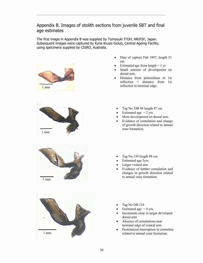
Appendix B. Images of otolith sections from juvenile SBT and final age estimates The first image in Appendix B was supplied by Tomoyuki ITOH, NRIFSF, Japan. Subsequent images were captured by Kyne Krusic-Golub, Central Ageing Facility, using specimens supplied by CSIRO, Australia.
1 mm
1 mm
1 mm
• Tag No. OB 94 length 87 cm • Estimated age = 2 yrs • More development on dorsal arm. • Evidence of crenulation and change
of growth direction related to annual zone formation.
• Tag No.139 length 98 cm. • Estimated age 3yrs. • Larger ventral arm • Evidence of further crenulation and
changes in growth direction related to annual zone formation.
• Tag No OB 214 • Estimated age = 4 yrs. • Increments clear in larger developed
dorsal arm • Absence of crenulations near
terminal edge of ventral arm. • Pronounced interruption in centreline
related to annual zone formation.
• Date of capture Feb 1997, length 51 cm
• Estimated age from length = 1 yr • Small amount of development on
dorsal arm. • Distance from primordium to 1st
inflection = distance from 1st inflection to terminal edge. 1 mm
___________________________________________________________________________
31
1 mm
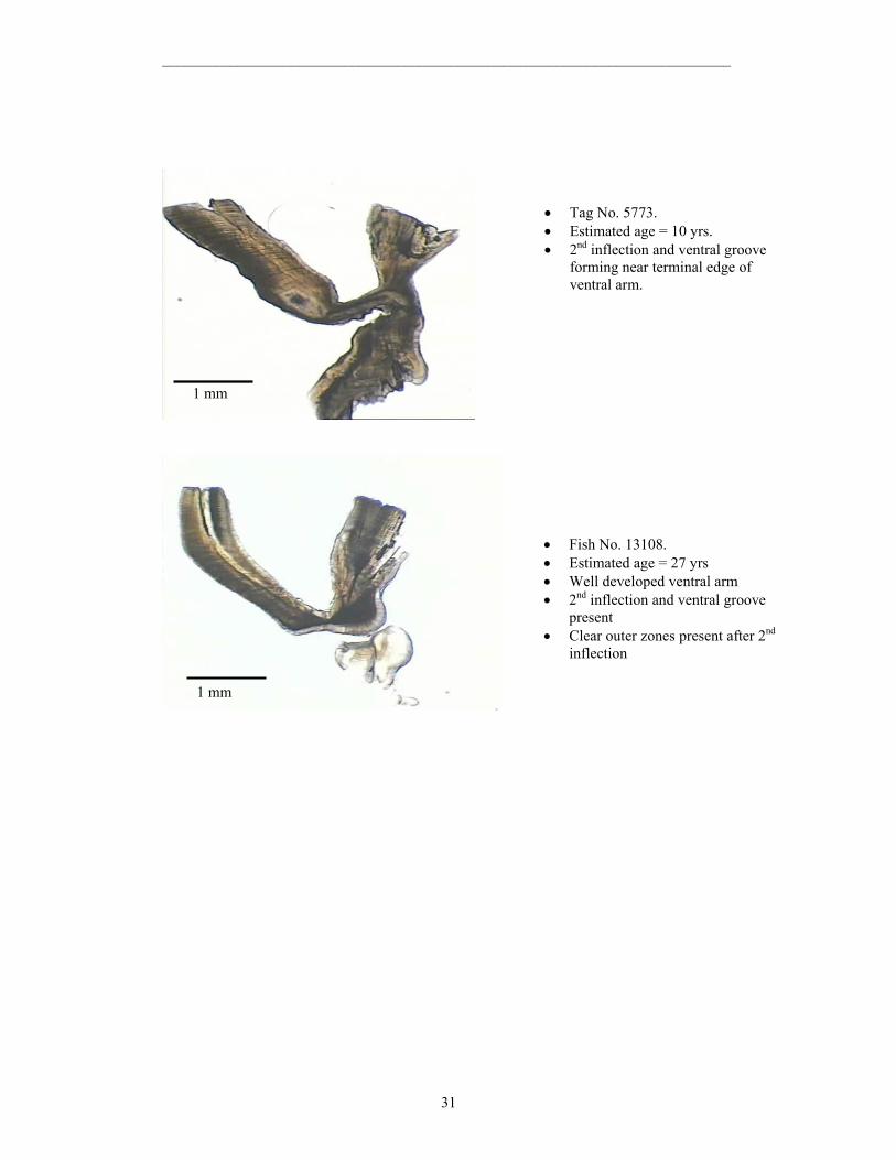
• Tag No. 5773. • Estimated age = 10 yrs. • 2nd inflection and ventral groove
forming near terminal edge of ventral arm.
1 mm
• Fish No. 13108. • Estimated age = 27 yrs • Well developed ventral arm • 2nd inflection and ventral groove
present • Clear outer zones present after 2nd
inflection

___________________________________________________________________________
32
Figure. Burnt whole otoliths; translucent zones have burnt more than opaque zones and appear brown in these images. Ages estimated from these otoliths are, from top of the page, 1 year, 2 years, 3 years and 4 years. Images were supplied by CSIRO, Australia.

___________________________________________________________________________
33
Appendix C. Workshop Participants CHAIR Dr James FINDLAY Senior Research Scientist Fisheries and Marine Sciences Bureau of Rural Sciences PO Box E11 Kingston ACT 2604 Phone: +61 2 6272 5534 Fax: +61 2 6272 3882 Email: [email protected] AUSTRALIA Ms Naomi CLEAR CSIRO P.O. Box 1538 Hobart, Tasmania, 7001 Phone: +61 3 6232 5336 Fax: +61 3 6232 5012 Email: [email protected] Mr Kyne KRUSIC-GOLUB Marine and Freshwater Resources Institute PO Box 114 Queenscliff, VIC, 3225 Phone: +61 3 5258 0111 Fax: +61 3 5258 0270 Email: [email protected] JAPAN Dr Sachiko TSUJI Section Chief Temperate Tuna Section National Research Institute of Far Seas Fisheries 5-7-1 Shimizu-Orido, Shizuoka 424-8633 Phone: +81 543 36 6042 Fax: +81 543 35 9642 Email: [email protected]

___________________________________________________________________________
34
Mr Tomoyuki ITOH Temperate Tuna Section National Research Institute of Far Seas Fisheries 5-7-1 Shimizu-Orido, Shizuoka 424-8633 Phone: +81 543 36 6043 Fax: +81 543 35 9642 Email: [email protected] Dr Akio HIRAI Marino Research 60 Nakayama-cho, Kuwana Mie, 511-0914 Phone: +81 594 32 9901 Fax: +81 594 32 9902 Email: [email protected] Mr Kenichiro OMOTE Marino Research 60 Nakayama-cho, Kuwana Mie, 511-0914 Phone: +81 594 32 9901 Fax: +81 594 32 9902 Email: [email protected] NEW ZEALAND Dr Malcolm FRANCIS National Institute of Water & Atmospheric Research Ltd PO Box 14901, Kilbirnie, Wellington Phone: +64 4 386 0377 Fax: +64 4 386 0574 Email: [email protected] Mr Colin SUTTON National Institute of Water & Atmospheric Research Ltd. PO Box 14901, Kilbirnie, Wellington Phone: +64 4 386 0300 Fax: +64 4 386 0574 Email: [email protected] REPUBLIC OF KOREA Dr Doo Hae AN Distant Water Fisheries Resources Division National Fisheries Research & Development Institute 408-1, Sirang-Ri, Gijang-Up, Gijang-Gun, Busan, 619-900 Phone: +82 51 720 2325 Fax: +82 51 720 2337 E-mail: [email protected]

___________________________________________________________________________
35
Dr Yang Jae IM Fisheries & Environment Division West Sea Fisheries Research Institute 98-36, BukSeong-Dong IGa, Chung-Gu, Inchon, 400-201 Phone: +82 32 773 7090 Fax: +82 32 761 0467 E-mail: [email protected] TAIWAN Dr Wann-Nian TZENG Department of Zoology College of Science National Taiwan University No.1 Sec.4, Roosevelt Road Taipei, 106 Phone: +886 2 2363 9570 Fax: +886 2 2363 6837 Email: [email protected] Dr Jay Jen-Chieh SHIAO Department of Zoology College of Science National Taiwan University No.1 Sec.4, Roosevelt Road Taipei, 106 Phone: +886 2 2363 9570 Fax: +886 2 2363 6837 Email: [email protected] CCSBT SECRETARIAT PO Box 37, Deakin West ACT 2600 AUSTRALIA Phone: +61 2 6282 8396 Fax: +61 2 6282 8407 Mr Morio KANEKO Deputy Executive Secretary Email: [email protected] INTERPRETER Ms Kumi KOIKE
___________________________________________________________________________
36
Appendix D. References Beamish, R. J. and Fournier, D. A. (1981). A method for comparing the precision of a set of
age determinations. Can. J. of Fish. Aquat. Sci. 38:982-983. Campana S. E., Annand, M. C. and McMillan, J. I. (1995). Graphical and statistical methods
for determining the consistency of age determinations. Trans. Amer. Fish. Soc. 124:131-138.
Caton, A. E. (ed.) 1991. Review of aspects of southern bluefin tuna biology, population and
fisheries. In R. B. Deriso and W. H. Bayliff (eds). World meeting on stock assessment of bluefin tunas: strengths and weaknesses. Inter–Amer. Trop. Tuna Comm. Special Report No. 7:181–350.
Clear, N. P., Gunn, J., and Rees, A. J. (2000). Direct validation of annual increments in the
otoliths of juvenile southern bluefin tuna, Thunnus maccoyii, by means of a large-scale mark-recapture experiment with strontium chloride. Fishery Bulletin 98: 25-40.
Clear, N. P., Eveson, J.P. and Polacheck, T. (in press). Investigating the timing of annual
growth zones in otoliths of southern bluefin tuna (Thunnus maccoyii). Appendix 11. In, Polacheck, T., L., Laslett, G. M. and Eveson, J. P. An integrated analysis of growth rates of southern bluefin tuna for use in estimating catch at age in stock assessments. FRDC Final Report Project Number 99/104.
Francis, M. P.; Mulligan, K. P.; Davies and N. M.; Beentjes, M. P. (1999). Age and growth
estimates for New Zealand hapuku, Polyprion oxygeneios. Fishery bulletin 97(2): 227-242.
Gunn, J. S., Clear, N. P., Carter, T. I., Rees, A. J., Stanley, C. A., Farley, J. H., and Kalish, J.
M. (in press). The direct estimation of age and growth in SBT, Thunnus maccoyii, using otolith, scales and vertebrae. Fishery Bulletin.
Itoh, T. and Tsuji, S. (1996). Age and growth of juvenile southern bluefin tuna Thunnus
maccoyii based on otolith microstructure. Fisheries Science 62: 892-896. Jenkins, G. P. and Davis, T. L. O. (1990). Age, growth rate, and growth trajectory determined
from otolith microstructure of southern bluefin tuna Thunnus maccoyii larvae. Marine Ecology Progress Series 63: 93-104.
Kalish, J. M., Johnston, J. M., Gunn, J. S., and Clear, N. P. (1996). Use of the bomb
radiocarbon chronometer to determine age of southern bluefin tuna (Thunnus maccoyii). Marine Ecology Progress Series 143: 1-8.
Rees, A. J., Gunn, J. S. and N. P. Clear. (1996). Age determination of juvenile southern
bluefin tuna, Thunnus maccoyii, based on scanning electron microscopy of otolith microincrements. CCSBT/SC/96/8 (Appendix). Second Meeting of the Commission for the Conservation of Southern Bluefin Tuna (CCSBT), 26 Aug-5 Sep 1996, CSIRO Marine Laboratories, Hobart, Tasmania. 122 pp.
Thorogood, J. (1986), New technique for sampling otoliths of sashimi-grade scombrid fishes.
Trans. Amer. Fish. Soc. 115:913–914.