Embed Size (px)
Citation preview
Journal of the American College of Cardiology Vol. 60, No. 5, 2012© 2012 by the American College of Cardiology Foundation ISSN 0735-1097/$36.00
Vascular Disease
Aggressive Cardiovascular Phenotypeof Aneurysms-Osteoarthritis SyndromeCaused by Pathogenic SMAD3 VariantsDenise van der Linde, MSC,* Ingrid M. B. H. van de Laar, MD,† Aida M. Bertoli-Avella, MD, PHD,†Rogier A. Oldenburg, MD, PHD,† Jos A. Bekkers, MD,‡ Francesco U. S. Mattace-Raso, MD, PHD,§Anton H. van den Meiracker, MD, PHD,§ Adriaan Moelker, MD, PHD,� Fop van Kooten, MD, PHD,¶Ingrid M. E. Frohn-Mulder, MD,# Janneke Timmermans, MD,** Els Moltzer, PHD,§Jan M. Cobben, MD, PHD,†† Lut van Laer, PHD,‡‡ Bart Loeys, MD, PHD,‡‡Julie De Backer, MD, PHD,§§ Paul J. Coucke, PHD,§§ Anne De Paepe, MD, PHD,§§Yvonne Hilhorst-Hofstee, MD,�� Marja W. Wessels, MD, PHD,†Jolien W. Roos-Hesselink, MD, PHD*
Rotterdam, Nijmegen, Amsterdam, and Leiden, the Netherlands; and Antwerp and Ghent, Belgium
Objectives The purpose of this study was describe the cardiovascular phenotype of the aneurysms-osteoarthritis syndrome(AOS) and to provide clinical recommendations.
Background AOS, caused by pathogenic SMAD3 variants, is a recently described autosomal dominant syndrome character-ized by aneurysms and arterial tortuosity in combination with osteoarthritis.
Methods AOS patients in participating centers underwent extensive cardiovascular evaluation, including imaging, arterialstiffness measurements, and biochemical studies.
Results We included 44 AOS patients from 7 families with pathogenic SMAD3 variants (mean age: 42 � 17 years). In71%, an aortic root aneurysm was found. In 33%, aneurysms in other arteries in the thorax and abdomen werediagnosed, and in 48%, arterial tortuosity was diagnosed. In 16 patients, cerebrovascular imaging was per-formed, and cerebrovascular abnormalities were detected in 56% of them. Fifteen deaths occurred at a meanage of 54 � 15 years. The main cause of death was aortic dissection (9 of 15; 60%), which occurred at mildlyincreased aortic diameters (range: 40 to 63 mm). Furthermore, cardiac abnormalities were diagnosed, such as con-genital heart defects (6%), mitral valve abnormalities (51%), left ventricular hypertrophy (19%), and atrial fibrillation(22%). N-terminal brain natriuretic peptide (NT-proBNP) was significantly higher in AOS patients compared withmatched controls (p � 0.001). Aortic pulse wave velocity was high-normal (9.2 � 2.2 m/s), indicating increased aor-tic stiffness, which strongly correlated with NT-proBNP (r � 0.731, p � 0.005).
Conclusions AOS predisposes patients to aggressive and widespread cardiovascular disease and is associated with high mor-tality. Dissections can occur at relatively mildly increased aortic diameters; therefore, early elective repair of theascending aorta should be considered. Moreover, cerebrovascular abnormalities were encountered in mostpatients. (J Am Coll Cardiol 2012;60:397–403) © 2012 by the American College of Cardiology Foundation
Published by Elsevier Inc. http://dx.doi.org/10.1016/j.jacc.2011.12.052
ment of Clinical Genetics, Ghent University Hospital, Ghent, Belgium; and the� �Department of Clinical Genetics, Leiden University Medical Center, Leiden, theNetherlands. This work was funded partially by an Erasmus Fellowship to Dr.Bertoli-Avella; a Research Foundation Flanders grant to Dr. Loeys; the FlandersFund for Scientific Research to Dr. De Backer as a Senior Clinical Researcher; andMethusalem grant number BOF08/01M01108 from Ghent University to Dr. DePaepe. All other authors have reported that they have no relationships relevant to thecontents of this paper to disclose. The first two authors contributed equally to thiswork.
From the *Department of Cardiology, Erasmus Medical Center, Rotterdam, theNetherlands; †Department of Clinical Genetics, Erasmus Medical Center, Rotterdam,the Netherlands; ‡Department of Cardio-Thoracic Surgery, Erasmus Medical Center,Rotterdam, the Netherlands; §Department of Internal Medicine, Erasmus Medical Center,Rotterdam, the Netherlands; �Department of Radiology, Erasmus Medical Center, Rotter-dam, the Netherlands; ¶Department of Neurology, Erasmus Medical Center, Rotterdam, theNetherlands; #Department of Pediatrics, Erasmus Medical Center, Rotterdam, the Nether-lands; **Department of Cardiology, Radboud University Nijmegen Medical Center,Nijmegen, the Netherlands; ††Department of Pediatric Genetics, Academic Medical
Center, Amsterdam, the Netherlands; ‡‡Center for Medical Genetics, AntwerpUniversity Hospital, University of Antwerp, Antwerp-Edegem, Belgium; §§Depart-Manuscript received September 6, 2011; revised manuscript received December 5,2011, accepted December 18, 2011.
sa
v(a(aap(amsto
M
FSthcs1poessawe
ci1thosIEcDCp1udadrtawwotS
R
WpOdTph
398 van der Linde et al. JACC Vol. 60, No. 5, 2012Aneurysms-Osteoarthritis Syndrome July 31, 2012:397–403
Aortic aneurysms and dissectionswere ranked as the nineteenthmost common cause of death inthe United States in 2007 (1). Thetrue incidence is probably muchhigher, because many aortic aneu-rysms are silent. Thoracic aortic an-eurysms and dissections (TAADs)
See page 404
often are found in the context ofgenetic syndromes, such as Marfansyndrome (MFS) and Loeys-Dietzsyndrome (LDS), but also are asso-ciated with bicuspid aortic valves(2–4). MFS is one of the mostcommon hereditable connective tis-sue disorders, with abnormalitiespredominantly in the skeletal,ocular, pulmonary, and cardiovas-cular systems (2). LDS shows some
imilarities with MFS, but exhibits widespread arterialneurysms and tortuosity (3).
Recently, our group found that pathogenic SMAD3ariants cause aneurysms-osteoarthritis syndrome (AOS)5). AOS is inherited as an autosomal dominant disordernd is found to be responsible for 2% of familial TAADs5,6). Aneurysms, dissections, and tortuosity throughout therterial tree are the main cardiovascular features (5). Inddition, early-onset osteoarthritis is present in almost allatients and often is the first reason to seek medical advice5). Mild craniofacial abnormalities, such as hypertelorismnd bifid uvula, also are associated with AOS (5). Further-ore, umbilical or inguinal hernias, or both; varices; velvety
kin; and striae are common findings (5). The purpose ofhis study was to describe the cardiovascular consequencesf AOS and to provide clinical recommendations.
ethods
rom 2009 onward, all AOS patients with a pathogenicMAD3 variant in participating centers were included inhis ongoing cohort study. Genetic identification methodsave been described previously (5). Patients underwentomprehensive clinical evaluation, including risk factor as-essment, physical examination, biochemical measurements,2-lead electrocardiography, transthoracic echocardiogra-hy (TTE), and computed tomography angiography (CTA)f the thorax and abdomen. For logistical reasons, not allxaminations could be performed in every patient. In aubset of patients, CTA of the cerebral vessels and arterialtiffness measurements also were performed. These methodsre described extensively in the Online Appendix. Patientsere monitored for occurrence of cardiovascular events,
Abbreviationsand Acronyms
AF � atrial fibrillation
AOS � aneurysms-osteoarthritis syndrome
aPWV � aortic pulsewave velocity
CTA � computedtomography angiography
LDS � Loeys-Dietzsyndrome
MFS � Marfan syndrome
NT-proBNP � N-terminalbrain natriuretic peptide
TAAD � thoracic aorticaneurysms and dissections
TGF � transforming growthfactor
TTE � transthoracicechocardiography
specially dissection or mortality. Autopsy was requested in
ase of death and was performed when possible. Biochem-cal and arterial stiffness measurements were compared-to-1 with age-, sex-, and smoking status-matched con-rols. Apparently healthy controls were recruited amongospital personnel and their acquaintances and underwentnly biochemical and arterial stiffness measurements andmoking status assessment. The study was approved by thenstitutional Review Board and Ethical Committee of therasmus Medical Center in Rotterdam. Written informed
onsent was obtained from each patient.ata analysis. SPSS software version 15.0 (SPSS, Inc.,hicago, Illinois) was used for the statistical analyses. Avalue of �0.05 was considered statistically significant. The-sample Kolmogorov-Smirnov test and histograms weresed to check normality. Normally distributed continuousata are presented as mean � SD, and categorical variablesre presented as frequency (n) and percentages. Non-normalistributed data are presented as median with interquartileange (25th and 75th percentiles). For comparison betweenhe control and patient groups, a Student t test taking intoccount the 1-to-1 pairing or the signed-rank Wilcoxon testas used. Biochemical measurements also were comparedith reference values from the clinical chemical laboratoryf the Erasmus Medical Center in Rotterdam. For correla-ion analysis, the Pearson r correlation coefficient andpearman correlation test were used.
esults
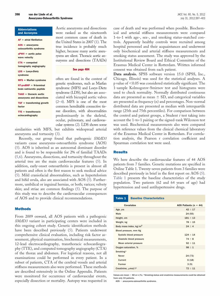
e here describe the cardiovascular features of 44 AOSatients from 7 families. Genetic mutations are specified innline Table 1. Twenty-seven patients from 3 families were
escribed previously in brief in the first report on AOS (5).able 1 presents the baseline characteristics of the studyopulation. Two patients (62 and 64 years of age) hadypertension and used antihypertensive drugs.
Baseline CharacteristicsTable 1 Baseline Characteristics
Covariates AOS Patients (n � 44)
Age, yrs 42 � 17
Male 24 (55)
Height, cm 181 � 13
Weight, kg 78 � 15
Body mass index, kg/m2 24 � 4
Blood pressure, mm Hg
Systolic blood pressure 124 � 14
Diastolic blood pressure 74 � 8
Mean arterial pressure 92 � 11
Oxygen saturation, % 98 � 1
Smoking*
Never 24 (73)
Current 6 (18)
Former 3 (9)
Creatinine, �mol/l* 72 � 11
Values are mean � SD or n (%). *Smoking status and creatinine measurements could be obtained
from only 33 patients.AOS � aneurysms-osteoarthritis syndrome.
p
Ob
399JACC Vol. 60, No. 5, 2012 van der Linde et al.July 31, 2012:397–403 Aneurysms-Osteoarthritis Syndrome
Survival. Fifteen deaths in AOS patients with confirmedathogenic SMAD3 variants occurred at a mean age of 54 �
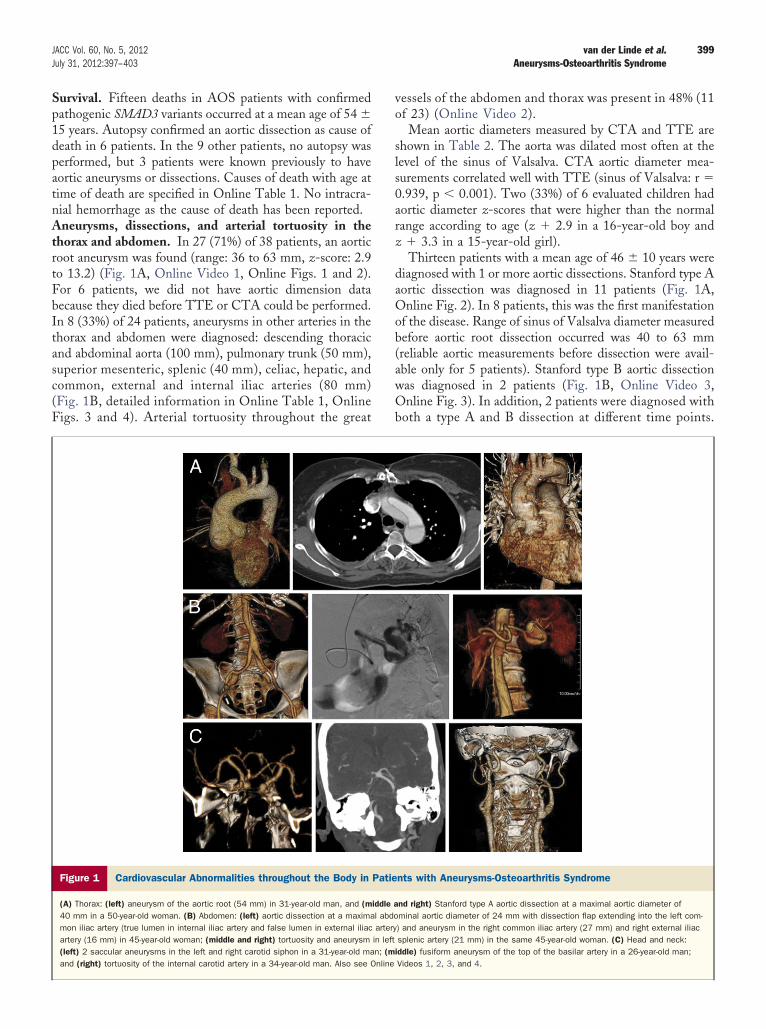
15 years. Autopsy confirmed an aortic dissection as cause ofdeath in 6 patients. In the 9 other patients, no autopsy wasperformed, but 3 patients were known previously to haveaortic aneurysms or dissections. Causes of death with age attime of death are specified in Online Table 1. No intracra-nial hemorrhage as the cause of death has been reported.Aneurysms, dissections, and arterial tortuosity in thethorax and abdomen. In 27 (71%) of 38 patients, an aorticroot aneurysm was found (range: 36 to 63 mm, z-score: 2.9to 13.2) (Fig. 1A, Online Video 1, Online Figs. 1 and 2).For 6 patients, we did not have aortic dimension databecause they died before TTE or CTA could be performed.In 8 (33%) of 24 patients, aneurysms in other arteries in thethorax and abdomen were diagnosed: descending thoracicand abdominal aorta (100 mm), pulmonary trunk (50 mm),superior mesenteric, splenic (40 mm), celiac, hepatic, andcommon, external and internal iliac arteries (80 mm)(Fig. 1B, detailed information in Online Table 1, OnlineFigs. 3 and 4). Arterial tortuosity throughout the great
Figure 1 Cardiovascular Abnormalities throughout the Body in
(A) Thorax: (left) aneurysm of the aortic root (54 mm) in 31-year-old man, and (m40 mm in a 50-year-old woman. (B) Abdomen: (left) aortic dissection at a maximamon iliac artery (true lumen in internal iliac artery and false lumen in external iliacartery (16 mm) in 45-year-old woman; (middle and right) tortuosity and aneurysm(left) 2 saccular aneurysms in the left and right carotid siphon in a 31-year-old maand (right) tortuosity of the internal carotid artery in a 34-year-old man. Also see O
vessels of the abdomen and thorax was present in 48% (11of 23) (Online Video 2).
Mean aortic diameters measured by CTA and TTE areshown in Table 2. The aorta was dilated most often at thelevel of the sinus of Valsalva. CTA aortic diameter mea-surements correlated well with TTE (sinus of Valsalva: r �0.939, p � 0.001). Two (33%) of 6 evaluated children hadaortic diameter z-scores that were higher than the normalrange according to age (z � 2.9 in a 16-year-old boy andz � 3.3 in a 15-year-old girl).
Thirteen patients with a mean age of 46 � 10 years werediagnosed with 1 or more aortic dissections. Stanford type Aaortic dissection was diagnosed in 11 patients (Fig. 1A,Online Fig. 2). In 8 patients, this was the first manifestationof the disease. Range of sinus of Valsalva diameter measuredbefore aortic root dissection occurred was 40 to 63 mm(reliable aortic measurements before dissection were avail-able only for 5 patients). Stanford type B aortic dissectionwas diagnosed in 2 patients (Fig. 1B, Online Video 3,
nline Fig. 3). In addition, 2 patients were diagnosed withoth a type A and B dissection at different time points.
nts with Aneurysms-Osteoarthritis Syndrome
nd right) Stanford type A aortic dissection at a maximal aortic diameter ofminal aortic diameter of 24 mm with dissection flap extending into the left com-) and aneurysm in the right common iliac artery (27 mm) and right external iliacsplenic artery (21 mm) in the same 45-year-old woman. (C) Head and neck:iddle) fusiform aneurysm of the top of the basilar artery in a 26-year-old man;Videos 1, 2, 3, and 4.
Patie
iddle al abdoarteryin leftn; (mnline
ssraosaviActmc
OmTfV1aibrahsCm5midh1fipf
thivla
400 van der Linde et al. JACC Vol. 60, No. 5, 2012Aneurysms-Osteoarthritis Syndrome July 31, 2012:397–403
None of these aortic dissections occurred during the 23pregnancies and deliveries in our AOS cohort. In 1patient, a dissection in a nondilated proximal left anteriordescending coronary artery was found.Elective cardiovascular operations and interventions.Fifteen patients underwent 1 or more elective cardiovascularinterventions at a mean age of 41 � 11 years: 12 valve-paring aortic root replacements, 1 Bentall procedure, 2plenic artery coiling procedures, and 1 abdominal aneurysmepair; 1 patient underwent aortic repair surgeries in thoraxnd abdomen and mitral valve repair. In 2 patients, post-perative complications occurred: 1 patient had painfulplenic ischemia for which reoperation was necessary andnother patient had a total atrioventricular block afteralve-sparing aortic root replacement, for which pacemakermplantation was necessary.
neurysms and tortuosity of brachiocephalic and intra-ranial vasculature. CTA of the brachiocephalic and in-racranial vasculature was performed in 16 patients with aean age of 37 � 14 years. In 56% (9 of 16), we found
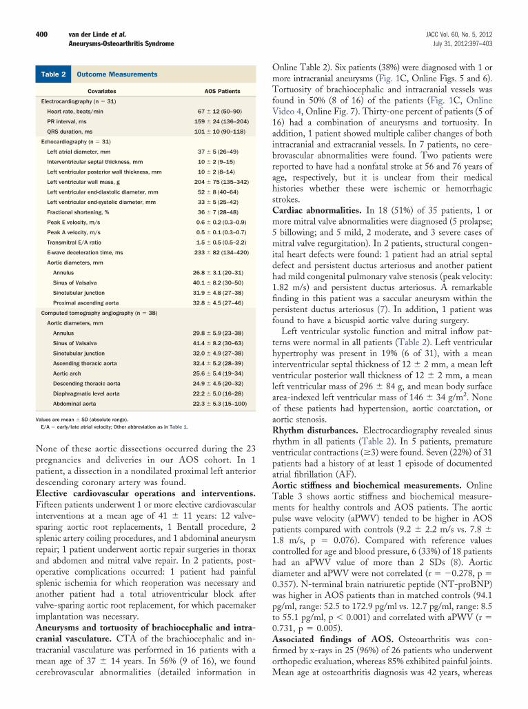
Outcome MeasurementsTable 2 Outcome Measurements
Covariates AOS Patients
Electrocardiography (n � 31)
Heart rate, beats/min 67 � 12 (50–90)
PR interval, ms 159 � 24 (136–204)
QRS duration, ms 101 � 10 (90–118)
Echocardiography (n � 31)
Left atrial diameter, mm 37 � 5 (26–49)
Interventricular septal thickness, mm 10 � 2 (9–15)
Left ventricular posterior wall thickness, mm 10 � 2 (8–14)
Left ventricular wall mass, g 204 � 75 (135–342)
Left ventricular end-diastolic diameter, mm 52 � 8 (40–64)
Left ventricular end-systolic diameter, mm 33 � 5 (25–42)
Fractional shortening, % 36 � 7 (28–48)
Peak E velocity, m/s 0.6 � 0.2 (0.3–0.9)
Peak A velocity, m/s 0.5 � 0.1 (0.3–0.7)
Transmitral E/A ratio 1.5 � 0.5 (0.5–2.2)
E-wave deceleration time, ms 233 � 82 (134–420)
Aortic diameters, mm
Annulus 26.8 � 3.1 (20–31)
Sinus of Valsalva 40.1 � 8.2 (30–50)
Sinotubular junction 31.9 � 4.8 (27–38)
Proximal ascending aorta 32.8 � 4.5 (27–46)
Computed tomography angiography (n � 38)
Aortic diameters, mm
Annulus 29.8 � 5.9 (23–38)
Sinus of Valsalva 41.4 � 8.2 (30–63)
Sinotubular junction 32.0 � 4.9 (27–38)
Ascending thoracic aorta 32.4 � 5.2 (28–39)
Aortic arch 25.6 � 5.4 (19–34)
Descending thoracic aorta 24.9 � 4.5 (20–32)
Diaphragmatic level aorta 22.2 � 5.0 (16–28)
Abdominal aorta 22.3 � 5.3 (15–100)
Values are mean � SD (absolute range).E/A � early/late atrial velocity; Other abbreviation as in Table 1.
erebrovascular abnormalities (detailed information in
nline Table 2). Six patients (38%) were diagnosed with 1 orore intracranial aneurysms (Fig. 1C, Online Figs. 5 and 6).ortuosity of brachiocephalic and intracranial vessels was
ound in 50% (8 of 16) of the patients (Fig. 1C, Onlineideo 4, Online Fig. 7). Thirty-one percent of patients (5 of6) had a combination of aneurysms and tortuosity. Inddition, 1 patient showed multiple caliber changes of bothntracranial and extracranial vessels. In 7 patients, no cere-rovascular abnormalities were found. Two patients wereeported to have had a nonfatal stroke at 56 and 76 years ofge, respectively, but it is unclear from their medicalistories whether these were ischemic or hemorrhagictrokes.ardiac abnormalities. In 18 (51%) of 35 patients, 1 orore mitral valve abnormalities were diagnosed (5 prolapse;billowing; and 5 mild, 2 moderate, and 3 severe cases ofitral valve regurgitation). In 2 patients, structural congen-
tal heart defects were found: 1 patient had an atrial septalefect and persistent ductus arteriosus and another patientad mild congenital pulmonary valve stenosis (peak velocity:.82 m/s) and persistent ductus arteriosus. A remarkablending in this patient was a saccular aneurysm within theersistent ductus arteriosus (7). In addition, 1 patient wasound to have a bicuspid aortic valve during surgery.
Left ventricular systolic function and mitral inflow pat-erns were normal in all patients (Table 2). Left ventricularypertrophy was present in 19% (6 of 31), with a mean
nterventricular septal thickness of 12 � 2 mm, a mean leftentricular posterior wall thickness of 12 � 2 mm, a meaneft ventricular mass of 296 � 84 g, and mean body surfacerea-indexed left ventricular mass of 146 � 34 g/m2. None
of these patients had hypertension, aortic coarctation, oraortic stenosis.Rhythm disturbances. Electrocardiography revealed sinusrhythm in all patients (Table 2). In 5 patients, prematureventricular contractions (�3) were found. Seven (22%) of 31patients had a history of at least 1 episode of documentedatrial fibrillation (AF).Aortic stiffness and biochemical measurements. OnlineTable 3 shows aortic stiffness and biochemical measure-ments for healthy controls and AOS patients. The aorticpulse wave velocity (aPWV) tended to be higher in AOSpatients compared with controls (9.2 � 2.2 m/s vs. 7.8 �1.8 m/s, p � 0.076). Compared with reference valuescontrolled for age and blood pressure, 6 (33%) of 18 patientshad an aPWV value of more than 2 SDs (8). Aorticdiameter and aPWV were not correlated (r � �0.278, p �0.357). N-terminal brain natriuretic peptide (NT-proBNP)was higher in AOS patients than in matched controls (94.1pg/ml, range: 52.5 to 172.9 pg/ml vs. 12.7 pg/ml, range: 8.5to 55.1 pg/ml, p � 0.001) and correlated with aPWV (r �0.731, p � 0.005).Associated findings of AOS. Osteoarthritis was con-firmed by x-rays in 25 (96%) of 26 patients who underwentorthopedic evaluation, whereas 85% exhibited painful joints.
Mean age at osteoarthritis diagnosis was 42 years, whereas
cppl
hw
pdStlssss
ardditd
401JACC Vol. 60, No. 5, 2012 van der Linde et al.July 31, 2012:397–403 Aneurysms-Osteoarthritis Syndrome
the youngest patient was 12 years of age. Spine, hands orwrists, and knees most often were affected (detailed infor-mation in Online Table 1). Pes planus was present in 91%of patients and scoliosis was present in 61%. Other associ-ated anomalies included hypertelorism (31%); abnormalpalate (54%); abnormal uvula (52%); hernia inguinalis orumbilicalis (43%); and uterus, bladder, or bowel prolapse(41%). More detailed information about these associatedfindings will be reported separately (9).
Discussion
AOS is a recently described autosomal dominant connectivetissue disorder characterized by aneurysms, dissections, andtortuosity throughout the arterial tree in combination withosteoarthritis and mild craniofacial features. The AOSphenotype may resemble that of other connective tissuedisorders such as MFS and LDS (Online Table 4). Themain site of aortic aneurysms in AOS is the sinus ofValsalva. Similar to LDS, AOS is an aggressive disease withsubstantial mortality and a high risk of aortic rupture anddissection in mildly dilated aortas (10). AOS and LDS bothare associated with widespread arterial tortuosity and aneu-rysms in the thorax and abdomen (10). In contrast to MFS,cerebrovascular abnormalities frequently occur in AOS andLDS (11). Identification of the underlying genetic defect inTAAD patients is crucial, considering the variability inprognosis, treatment strategy, and risk assessment in familymembers.Cardiac abnormalities in AOS. In addition to the aneu-rysms and tortuosity of the arterial tree, we also foundcardiac abnormalities. A remarkable finding in approxi-mately one fifth of the patients was left ventricular hyper-trophy in the absence of hypertension or aortic stenosis.Primary cardiomyopathy is reported in one quarter of MFSpatients showing mainly a reduced left ventricular ejectionfraction, but only in a minority (2.9%) was LV massincreased (12). Mice studies have determined that TGF-�induces proliferation of cardiac fibroblasts and hypertrophicgrowth of cardiomyocytes (13). Furthermore, TGF-� neu-tralizing antibodies were able to attenuate LV hypertrophy,and losartan reduced nonmyocyte proliferation, implyingpossible therapeutic implications in humans as well (14).
Similar to MFS, mitral valve abnormalities were commonin AOS patients, and 22% of AOS patients had a history ofAF. Mice studies have shown that TGF-�1–induced myo-ardial fibrosis in the atria plays an important role inredisposing individuals to AF (15). Atrial fibrogenesis inatients with AF occurs in 2 phases: an early increase, but
ater loss of responsiveness to TGF-�1, while the fibrosisprogresses (16).
Furthermore, evidence from mouse studies suggests thatTGF-� signaling is essential in the embryogenesis of the
eart, valvular pathogenesis, and organization of the aorticall (17,18). In many mouse models with disrupted TGF-�
signaling activities, congenital heart defects are present (17). a
In the future, SMAD3 knockdown mice will help to explorethe mechanism behind the cardiac abnormalities in AOS.Aortic stiffness and NT-proBNP in AOS. NT-proBNPin AOS patients was elevated compared with that incontrols, although none of the patients had extremely highNT-proBNP levels of more than 250 pg/ml. In vivo and invitro studies have shown that treatment with brain natri-uretic peptide can attenuate cardiac hypertrophy via theTGF-�1 pathway (19). One may hypothesize that theelevated NT-proBNP levels in AOS patients in fact are aprotective mechanism against the emergence of LV hypertro-phy. Because (mildly to moderately) elevated NT-proBNPlevels in other patient groups are reported to predictcardiovascular outcome and AF recurrence, evaluation ofthe prognostic value of NT-proBNP in AOS patients withrespect to clinical outcome may be important (20,21).
The aPWV as a measure of aortic stiffness was high-normal in AOS patients, as was described previously in, forinstance, patients with MFS and bicuspid aortic valve(22,23). Ascending aortic diameter and aPWV were notcorrelated, suggesting that arterial stiffness occurs indepen-dently of aneurysm formation. In MFS patients, an aug-mentation index of more than 11% has been reported topredict progression of aortic diameters, so further research iswarranted to test whether this also holds true for AOSpatients (24).Clinical suggestions for cardiologists treating AOSpatients. Although AOS is a recently discovered aneurysmsyndrome and the full spectrum of the disease and its progres-sion need to be clarified, some preliminary suggestions may bederived from the current findings. Because multisystem in-volvement frequently is observed, cooperation in a multidisci-plinary team with clinical geneticists, cardiologists, orthopedicsurgeons, radiologists, neurologists, and, when necessary, (vas-cular or cardiothoracic) surgeons is important.
MONITORING AND SCREENING. Cardiologists should sus-ect AOS in every TAAD patient without moleculariagnosis or known cause and should test these patients forMAD3 mutations. Furthermore, we suggest that cliniciansreating patients with arterial aneurysmal disease in anyarge artery (intracranial, iliac, splenic artery, and so on)hould at least ask whether these patients report jointymptoms. In the physical examination, one must paypecial attention to presence of AOS-associated findings,uch as joint anomalies and abnormal uvula.
Extensive cardiovascular evaluation using echocardiographynd CTA or magnetic resonance imaging (head to pelvis) isecommended in every adult AOS patient. Initially, theseiagnostic investigations should be performed annually toetermine rate of progression. Thereafter, frequency of imag-
ng should be guided by the findings, for instance, annually ifhe aortic diameter is more than 35 mm or if the aorticiameter shows significant growth (�5 mm/year).The phenotype seems to be age-dependent, because
neurysms mainly and dissections only occurred in adult-
AmtAbilcm
ttas(ppl
aitaiSibhbtri
C
ActdmAis
wr
1
1
1
1
402 van der Linde et al. JACC Vol. 60, No. 5, 2012Aneurysms-Osteoarthritis Syndrome July 31, 2012:397–403
hood; however, our series included only 6 children withAOS. Concerning screening in childhood, clear suggestionsare difficult to formulate at this time. We suggest thatfrequency of cardiologic evaluation with TTE, magneticresonance imaging, or both must be guided by the aorticroot z-score and presence of other cardiac abnormalities.
Although in our cohort no dissections occurred duringpregnancy or delivery, pregnancy should be considered highrisk in AOS patients with aneurysms, as in those with MFSand LDS (25).
TREATMENT. The implication of TGF-� signaling in thepathogenesis of aortic aneurysm syndromes suggests aTGF-� antagonist as a specific pharmaceutical target (26).
lthough losartan showed promising results in MFS mouseodels, we have to await the results of randomized clinical
rials in MFS, SMAD3 knockdown mice, and consequentlyOS clinical trials (26). At the moment, attention shoulde focused on genetic counseling, screening of relatives, andnterventional or surgical treatment. Medical treatment withosartan, beta-blockade, or both may be beneficial. Stringentontrol of hypertension to limit aortic wall stress is recom-ended (27).Because dissections in AOS patients can occur at rela-
ively small aortic diameters, early elective surgical interven-ion is indicated to reduce the risk of mortality. Because datare limited and the rate of progression is unknown, weuggest applying the surgical recommendations for LDS27). Valve-sparing aortic root replacement using the reim-lantation technique is the intervention of choice (28). Foreripheral aneurysms, individual size or rate of growth and
ocation must determine the treatment strategy.Currently, the risk of rupture of intracranial aneurysms
ssociated with AOS is unknown. No deaths resulting fromntracranial hemorrhage occurred in our series. Life expec-ancy and size, location, and rate of growth of the aneurysmre the most important determinants to decide whetherntervention is needed.tudy limitations. First, the number of subjects included
n the present study is relatively small, because AOS haseen discovered only recently. Second, the population is quiteeterogeneous, particularly in disease severity and age, andecause of logistical reasons and mortality, it was not possibleo perform every examination in all 44 patients. Furtheresearch is necessary to confirm our findings and to gain morensight in the disease mechanism and progression.
onclusions
OS is an aggressive, inherited, connective tissue disorderharacterized by arterial tortuosity, aneurysms, and osteoar-hritis. Aortic root enlargement is the most common car-iovascular finding in our series, but cerebrovascular abnor-alities were also present in more than 50% of patients.ortic dissections occur at smaller diameters than observed
n, for instance, MFS, and as such need early elective
urgical treatment. Larger prospective follow-up studies are1
arranted to determine progression over time and clinicalelevancy of the cardiac and intracranial abnormalities.
AcknowledgmentsThe authors thank the participating patients, their families,and their referring physicians. The authors also thank allcontrol subjects and technician assistants from the partici-pating centers.
Reprints requests and correspondence: Dr. Jolien W. Roos-Hesselink, Department of Cardiology, Ba-583a, Erasmus MedicalCenter, P. O. Box 2040, 3000 CA, Rotterdam, the Netherlands.E-mail: [email protected].
REFERENCES
1. National Center for Injury Prevention and Control. WISQARS LeadingCauses of Death Reports 2007. Available at: http://webappa.cdc.gov/sasweb/ncipc/leadcaus10.html. Accessed January 17, 2011.
2. Judge DP, Dietz HC. Marfan’s syndrome. Lancet 2005;366:1965–76.3. Loeys BL, Chen J, Neptune ER, et al. A syndrome of altered
cardiovascular, craniofacial, neurocognitive and skeletal developmentcaused by mutations in TGFBR1 or TGFBR2. Nat Genet 2005;37:275–81.
4. Tadros TM, Klein MD, Shapira OM. Ascending aortic dilatationassociated with bicuspid aortic valve: pathophysiology, molecularbiology, and clinical implications. Circulation 2009;119:880–90.
5. van de Laar IM, Oldenburg RA, Pals G, et al. Mutations in SMAD3cause a syndromic form of aortic aneurysms and dissections withearly-onset osteoarthritis. Nat Genet 2011;43:121–6.
6. Regalado ES, Guo DC, Villamizar C, et al. Exome sequencingidentifies SMAD3 mutations as a cause of familial thoracic aorticaneurysm and dissection with intracranial and other arterial aneurysms.Circ Res 2011;109:680–6.
7. Van der Linde D, Witsenburg M, van de Laar I, Moelker A,Roos-Hesselink J. Saccular aneurysm within a persistent ductusarteriosus. Lancet 2012;379:e33.
8. Reference Values for Arterial Stiffness’ Collaboration. Determinants ofpulse wave velocity in healthy people and in the presence of cardio-vascular risk factors: ‘establishing normal and reference values.’ EurHeart J 2010;31:2338–50.
9. Van de Laar IM, Van der Linde D, Oei EH, et al. Phenotypicspectrum of the SMAD3-related aneurysms-osteoarthritis syndrome.J Med Genet 2012;49:47–57.
10. Loeys BL, Schwarze U, Holm T, et al. Aneurysm syndromes caused bymutations in the TGF-beta receptor. N Engl J Med 2006;355:788–98.
11. Rodrigues VJ, Elsayed S, Loeys BL, Dietz HC, Yousem DM.Neuroradiologic manifestations of Loeys-Dietz syndrome type 1.AJNR Am J Neuroradiol 2009;30:1614–9.
12. Alpendurada F, Wong J, Kiotsekoglou A, et al. Evidence for Marfancardiomyopathy. Eur J Heart Fail 2010;12:1085–91.
13. Rosenkranz S. TGF-beta1 and angiotensin networking in cardiacremodeling. Cardiovasc Res 2004;63:423–32.
14. Teekakirikul P, Eminaga S, Toka O, et al. Cardiac fibrosis in micewith hypertrophic cardiomyopathy is mediated by non-myocyte pro-liferation and requires TGF-�. J Clin Invest 2010;120:3520–9.
5. Khan R, Sheppard R. Fibrosis in heart disease: understanding the roleof transforming growth factor-beta in cardiomyopathy, valvular diseaseand arrhythmia. Immunology 2006;118:10–24.
6. Gramley F, Lorenzen J, Koellensperger E, Kettering K, Weiss C,Munzel T. Atrial fibrosis and atrial fibrillation: the role of the TGF-�1signaling pathway. Int J Cardiol 2010;143:405–13.
7. Arthur HM, Bamforth SD. TGF� signaling and congenital heartdisease: insights from mouse studies. Birth Defects Res A Clin MolTeratol 2011;91:423–34.
8. Armstrong EJ, Bischoff J. Heart valve development: endothelial cellsignaling and differentiation. Circ Res 2004;95:459–70.
9. He JG, Chen YL, Chen BL, et al. B-type natriuretic peptide
attenuates cardiac hypertrophy via the transforming growth factor-�1/
2
2
2
2
2
2
2
2
2
403JACC Vol. 60, No. 5, 2012 van der Linde et al.July 31, 2012:397–403 Aneurysms-Osteoarthritis Syndrome
smad7 pathway in vivo and in vitro. Clin Exp Pharmacol Physiol2010;37:283–9.
0. den Uijl DW, Delgado V, Tops LF, et al. Natriuretic peptide levelspredict recurrence of atrial fibrillation after radiofrequency catheterablation. Am Heart J 2011;161:197–203.
1. Goei D, van Kuijk JP, Flu WJ, et al. Usefulness of repeated N-terminalpro-B-type natriuretic peptide measurements as incremental predictorfor long-term cardiovascular outcome after vascular surgery. Am JCardiol 2011;107:609–14.
2. Kiotsekoglou A, Moggridge JC, Saha SK, et al. Assessment of aorticstiffness in Marfan syndrome using two-dimensional and Dopplerechocardiography. Echocardiography 2011;28:29–37.
3. Tzemos N, Lyseggen E, Silversides C, et al. Endothelial function,carotid-femoral stiffness, and plasma matrix metalloproteinase-2 inmen with bicuspid aortic valve and dilated aorta. J Am Coll Cardiol2010;55:660–8.
4. Mortensen K, Baulmann J, Rybczynski M, et al. Augmentation indexand the evolution of aortic disease in Marfan-like syndromes. Am JHypertens 2010;23:716–24.
5. Regitz-Zagrosek V, Lundqvist CB, Borghi C, et al. ESC guidelines onthe management of cardiovascular diseases during pregnancy: the TaskForce on the Management of Cardiovascular Diseases during Preg-
nancy of the European Society of Cardiology (ESC). Eur Heart J2011;32:3147–97.6. Brooke BS, Habashi JP, Judge DP, Patel N, Loeys B, Dietz HC 3rd.Angiotensin II blockade and aortic-root dilation in Marfan’s syn-drome. N Engl J Med 2008;358:2787–95.
7. Hiratzka LF, Bakris GL, Beckman JA, et al. 2010 ACCF/AHA/AATS/ACR/ASA/SCA/SCAI/SIR/STS/SVM guidelines for the di-agnosis and management of patients with thoracic aortic disease: areport of the American College of Cardiology Foundation/AmericanHeart Association Task Force on Practice Guidelines, AmericanAssociation for Thoracic Surgery, American College of Radiology,American Stroke Association, Society of Cardiovascular Anesthesiol-ogists, Society for Cardiovascular Angiography and Interventions,Society of Interventional Radiology, Society of Thoracic Surgeons, andSociety for Vascular Medicine. J Am Coll Cardiol 2010;55:e27–130.
8. David TE, Feindel CM, Webb GD, Colman JM, Armstrong S,Maganti M. Long-term results of aortic valve-sparing operations foraortic root aneurysm. J Thorac Cardiovasc Surg 2006;132:347–54.
Key Words: aneurysm y aorta y cerebrovascular disorders y genetics ySMAD3.
APPENDIX
For an expanded Methods section and for supplementary figures,
videos, and tables, please see the online version of this article.