Embed Size (px)
Citation preview
5.1
Airway/Breathing
Chapter 5
Airway/Breathing
IntroductionSkillful, rapid, assessment and management of airway andventilation are critical to preventing morbidity and mortality.Airway compromise can occur rapidly or slowly and may recur.Frequent reassessment is necessary. Preventable causes of deathfrom airway problems in trauma include the following:� Failure to recognize the need for an airway.� Inability to establish an airway.� Failure to recognize the incorrect placement of an airway.� Displacement of a previously established airway.� Failure to recognize the need for ventilation.� Aspiration of the gastric contents.Initial airway management at any level, but especially outsideof medical treatment facilities (MTFs).Immediate goal: Move tongue, pharyngeal soft tissues, andsecretions out of airway. Until a formal airway is established,place patients in the lateral or prone position (rescue position).� Chin-lift and head tilt: Place fingers under the tip of the
mandible to lift the chin outward from face.� Two-Handed Jaw Thrust: Place both hands behind the angles
of the mandible and displace forward. This method can beused on the patient with cervical injury.
� Oropharyngeal airway:ο Insert oral airway upright if a tongue depressor is used
(preferred method).ο Keep the airway inverted past the tongue then rotate 180°.ο Too small an airway will not alleviate the obstruction.
Too long an airway may fold the epiglottis caudally,worsening the obstruction.
ο Estimate airway size by distance from corner of mouthto ear lobe.
ο Oral airways are not used in conscious patients.
5.2
Emergency War Surgery
� Nasopharyngeal airway.ο Pass lubricated nasal airway gently through one nostril.ο Not used in suspected facial or basal skull injuries.ο Is tolerated by conscious patients.
� Field expedient.ο Pull tongue forward and safety pin or suture it to corner
of mouth.� Cricothyrotomy.
Ventilation� Ventilate patient with bag valve mask (BVM).ο Bring the face into the mask rather than pushing the
mask onto the face.ο The chin-lift and head tilt are also employed during mask
ventilation unless they are contraindicated due tocervical spine precautions.
Assess air movement during mask ventilation by observingrise and fall of the chest, auscultation, absence of a maskleak, compliant feel of self-inflating bag, and stable oxygensaturation.
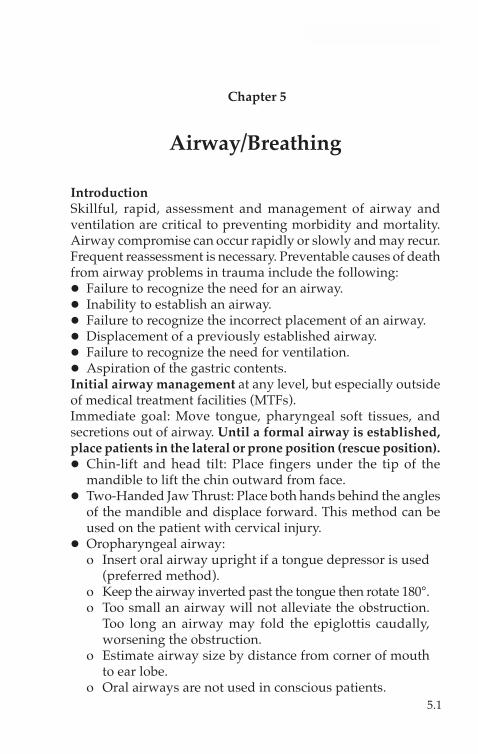
ο If air movement is not achieved, use two-person maskventilation (Fig. 5-1).♦ One person lifts the jaw aggressively at the angles of
the mandible; the other holds the mask and ventilates.♦ If air movement is still not present, obtain a definitive
airway.
Fig. 5-1. Two-person mask ventilation.
5.3
Airway/Breathing
ο Unsuccessful and aggressive attempts at ventilation mayresult in inflation of the stomach, placing the patient atincreased risk for vomiting and aspiration.
Positive pressure ventilation can convert a simplepneumothorax into a tension pneumothorax. Performfrequent assessment and have equipment available forneedle chest decompression.
Orotracheal Intubation
Rapid Sequence Intubation (RSI)—7 steps.1. Preoxygenate with 100% oxygen by mask.2. Consider fentanyl—titrate to maintain adequate blood
pressure and effect (2.0–2.5 µg/kg).3. Cricoid Pressure—Selleck maneuver until endotracheal
tube (ETT) placement is confirmed and balloon is inflated.4. Induction Agent: etomidate 0.1–0.4 mg/kg IV push.5. Muscle Relaxant: succinylcholine 1.0–1.5 mg/kg IV push.6. Laryngoscopy and orotracheal intubation.7. Verify tube placement.
� Direct laryngoscopy technique.ο Ensure optimal “sniffing” position is achieved unless
contraindicated by cervical spine injury.ο Open the mouth by scissoring the right thumb and
middle finger.ο Hold the laryngoscope in the left hand and insert the
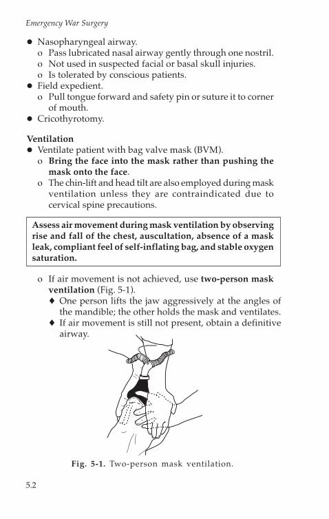
blade along the right side of the mouth, slightlydisplacing the tongue to the left.♦ Macintosh (curved) blade: Advance the tip of the blade
into the space between the base of the tongue and theepiglottis (valecula). Apply force at a 30°–45° angle,lifting the entire laryngoscope/blade, without rockingit backward (Fig. 5-2).
♦ Miller (straight) blade: Advance the tip of the blade intothe posterior oropharynx, picking up the epiglottis andtongue base anteriorly and laterally, and apply a forcevector like that of the Macintosh blade. Avoid rockingthe laryngoscope backward (Fig. 5-3).
5.4
Emergency War Surgery
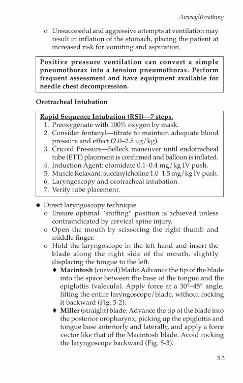
ο Visualize the vocal cords.ο Consider the “BURP” maneuver when the laryngoscopic
view is poor (Fig. 5-4).♦ “Backward-Upward-Rightward-Pressure” of the larynx,
also referred to as external laryngeal manipulation.♦ Place the fingers of an assistant onto the larynx with
your right hand and manipulate the glottic opening intothe field of view.
♦ Assistant then holds the position for intubation.
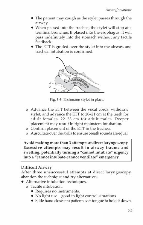
Eschmann stylet or Gum Elastic Bougie (GEB) (Fig. 5-5).♦ Blindly guide the tip of the stylet beneath the epiglottis,
then anteriorly through the vocal cords.♦ Advance the bougie deeply. Placement into the trachea
results in the sensation of tracheal ring “clicks”, andturning of the stylet as it passes airway bifurcations.
Fig. 5-3. Use of straight bladelaryngoscope.
Fig. 5-2. Use of curved bladelaryngoscope.
Force Vector
Fig. 5-4. BURP maneuver.
5.5
Airway/Breathing
♦ The patient may cough as the stylet passes through theairway.
♦ When passed into the trachea, the stylet will stop at aterminal bronchus. If placed into the esophagus, it willpass indefinitely into the stomach without any tactilefeedback.
♦ The ETT is guided over the stylet into the airway, andtracheal intubation is confirmed.
ο Advance the ETT between the vocal cords, withdrawstylet, and advance the ETT to 20–21 cm at the teeth foradult females, 22–23 cm for adult males. Deeperplacement may result in right mainstem intubation.
ο Confirm placement of the ETT in the trachea.ο Auscultate over the axilla to ensure breath sounds are equal.
Avoid making more than 3 attempts at direct laryngoscopy.Excessive attempts may result in airway trauma andswelling, potentially turning a “cannot intubate” urgencyinto a “cannot intubate-cannot ventilate” emergency.
Difficult AirwayAfter three unsuccessful attempts at direct laryngoscopy,abandon the technique and try alternatives.� Alternative intubation techniques.ο Tactile intubation.♦ Requires no instruments.♦ No light use—good in light control situations.♦ Slide hand closest to patient over tongue to hold it down.
Fig. 5-5. Eschmann stylet in place.
5.6
Emergency War Surgery
♦ Lift epiglottis with first two fingers.♦ Slide ETT along the “v” between the two fingers into
the airway.ο Lighted stylet or “light wand” intubation.♦ Flexible wand, lighted at the tip, is placed through the
ETT.♦ Wand is advanced by tactile guidance into the
trachea.♦ Position in trachea is verified by transillumination.♦ The ETT is advanced over the wand.
ο Flexible fiberoptic oral or nasal intubation.ο Retrograde wire intubation.ο Rigid fiberoptic intubation (Bullard laryngoscope).ο Alternative Airways.♦ May NOT be definitive airways.♦ Allow for oxygenation and ventilation when standard
airways cannot be placed.♦ “Fastrach” model laryngeal mask airway (LMA).♦ Esophageal-tracheal combitube (ETC).
� Perform a surgical airway.� Wake the patient up and attempt an awake technique if
possible.
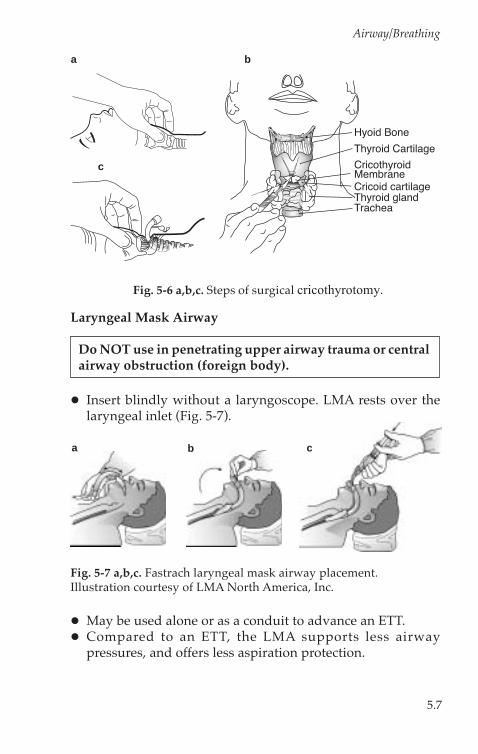
Surgical Cricothyrotomy� Identify cricothyroid membrane (between cricoid ring and
thyroid cartilage [Fig. 5-6a]).� Prep skin widely.� Grasp and hold trachea until airway is completely in place.� Make a vertical SKIN incision down to the cricothyroid
membrane (a No. 10 or 11 blade).� Bluntly dissect the tissues to expose the membrane.� Make a horizontal MEMBRANE incision (Fig. 5-6b).� Open the membrane with forceps or the scalpel handle.� Insert a small, cuffed ETT, 6.0–7.0 inner diameter (ID), to just
above the balloon (Fig. 5-6c).� Confirm tracheal intubation.� Suture the ETT in place, and secure it with ties that pass
around the neck.
5.7
Airway/Breathing
Laryngeal Mask Airway
Do NOT use in penetrating upper airway trauma or centralairway obstruction (foreign body).
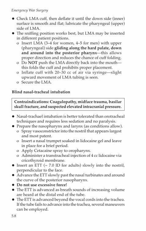
� Insert blindly without a laryngoscope. LMA rests over thelaryngeal inlet (Fig. 5-7).
� May be used alone or as a conduit to advance an ETT.� Compared to an ETT, the LMA supports less airway
pressures, and offers less aspiration protection.
a b
c
Fig. 5-6 a,b,c. Steps of surgical cricothyrotomy.
a b c
Fig. 5-7 a,b,c. Fastrach laryngeal mask airway placement.Illustration courtesy of LMA North America, Inc.
Hyoid Bone
Thyroid Cartilage
Thyroid glandCricoid cartilage
Trachea
CricothyroidMembrane
5.8
Emergency War Surgery
� Check LMA cuff, then deflate it until the down side (inner)surface is smooth and flat; lubricate the pharyngeal (upper)side of LMA.
� The sniffing position works best, but LMA may be insertedin different patient positions.ο Insert LMA (3–4 for women, 4–5 for men) with upper
(pharyngeal) side gliding along the hard palate, downand around into the posterior pharynx—this allowsproper direction and reduces the chance of cuff folding.
ο Do NOT push the LMA directly back into the mouth—this folds the cuff and prohibits proper placement.
ο Inflate cuff with 20–30 cc of air via syringe—slightupward movement of LMA tubing is seen.
ο Secure the LMA.
Blind nasal-tracheal intubation
Contraindications: Coagulopathy, midface trauma, basilarskull fracture, and suspected elevated intracranial pressure.
� Nasal-trachael intubation is better tolerated than orotrachealtechniques and requires less sedation and no paralysis.
� Prepare the nasopharynx and larynx (as conditions allow).ο Spray vasoconstrictor into the nostril that appears largest
and most patent.ο Insert a nasal trumpet soaked in lidocaine gel and leave
in place for a brief period.ο Apply Cetacaine spray to oropharynx.ο Administer a transtracheal injection of 4 cc lidocaine via
cricothyroid membrane.� Insert an ETT (~ 7.0 ID for adults) slowly into the nostril,
perpendicular to the face.� Advance the ETT slowly past the nasal turbinates and around
the curve of the posterior nasopharynx.� Do not use excessive force!� The ETT is advanced as breath sounds of increasing volume
are heard at the distal end of the tube.� The ETT is advanced beyond the vocal cords into the trachea.
If the tube fails to advance into the trachea, several maneuverscan be employed.
5.9
Airway/Breathing
ο Tilt the head.ο Apply external, downward pressure to the larynx.ο Inflate the ETT balloon to help center the tube, then deflate
and advance it once it is engaged in the glottic opening.


![Airway & Breathing Management [Compatibility Mode]](https://img.pdfslide.net/doc/110x75/563db829550346aa9a911da5/airway-breathing-management-compatibility-mode.jpg)